doi: 10.1098/rspb.2010.0647
, 3381-3389 first published online 16 June 2010
277
2010
Proc. R. Soc. B
Thomas Butts, Peter W. H. Holland and David E. K. Ferrier
gene expression
brain and pharynx development: deductions from amphioxus
Ancient homeobox gene loss and the evolution of chordate
References
http://rspb.royalsocietypublishing.org/content/277/1699/3381.full.html#related-urls Article cited in:
http://rspb.royalsocietypublishing.org/content/277/1699/3381.full.html#ref-list-1 This article cites 73 articles, 13 of which can be accessed free
This article is free to access
Subject collections
(1159 articles) evolution
(87 articles) developmental biology
Articles on similar topics can be found in the following collections
Email alerting service
right-hand corner of the article or click Receive free email alerts when new articles cite this article - sign up in the box at the topherehttp://rspb.royalsocietypublishing.org/subscriptions go to:
Ancient homeobox gene loss and the
evolution of chordate brain and pharynx
development: deductions from amphioxus
gene expression
Thomas Butts
1,†, Peter W. H. Holland
1and David E. K. Ferrier
2,*
1
Department of Zoology, University of Oxford, South Parks Road, Oxford OX1 3PS, UK
2
Scottish Oceans Institute, University of St Andrews, East Sands, St Andrews, Fife KY16 8LB, UK
Homeobox genes encode a large superclass of transcription factors with widespread roles in animal development. Within chordates there are over 100 homeobox genes in the invertebrate cephalochordate amphioxus and over 200 in humans. Set against this general trend of increasing gene number in vertebrate evolution, some ancient homeobox genes that were present in the last common ancestor of chordates have been lost from vertebrates. Here, we describe the embryonic expression of four amphioxus descendants of these genes—AmphiNedxa, AmphiNedxb, AmphiMsxlxandAmphiNKx7. All four genes are expressed with a striking asymmetry about the left – right axis in the pharyngeal region of neurula embryos, mirroring the pronounced asymmetry of amphioxus embryogenesis.AmphiMsxlxandAmphiNKx7are also transiently expressed in an anterior neural tube region destined to become the cerebral vesicle. These findings suggest significant rewiring of developmental gene regulatory networks occurred during chordate evol-ution, coincident with homeobox gene loss. We propose that loss of otherwise widely conserved genes is possible when these genes function in a confined role in development that is subsequently lost or sig-nificantly modified during evolution. In the case of these homeobox genes, we propose that this has occurred in relation to the evolution of the chordate pharynx and brain.
Keywords: cephalochordate; homeobox; gene loss; pharynx evolution; brain evolution
1. INTRODUCTION
Homeobox genes encode a large group of transcription factors that expanded very early in metazoan evolution (Ryanet al. 2006), and the diversification of these genes has been implicated in the evolution of the diversity of animal body plans. Frequently, homeobox genes have major directive roles in developmental gene-regulatory networks and many, such as theHoxgenes in the context of the anterior – posterior axis (Deschamps 2007; Duboule 2007) andPax6in the case of eye specification (Gehring 2002), function in aspects of development that are highly conserved across bilaterian taxa. Our under-standing of the evolution of the homeobox genes, and of animal development more generally, has in recent years been greatly impacted by the progressive advance of genome-sequencing projects across the Metazoa. In this regard, the genome of the chordate amphioxus (Branchiostoma floridae; Putnam et al. 2008) has been particularly revealing.
The lancelets, or amphioxus, were long thought to be the closest invertebrate relatives of the vertebrates, but recent molecular evidence has led to their relocation to the most basal lineage within the chordates (Bourlat et al. 2006; Delsuc et al. 2006; Putnam et al. 2008). Notwithstanding this repositioning, amphioxus remains
a most informative extant outgroup taxon for studying the early evolution of the vertebrates (Holland & Chen 2001). In large part, this importance is a result of the derived nature of development in the other invertebrate chordate lineage, the urochordates. In contrast, amphioxus in many ways resembles a typical vertebrate: gastrulation is coordinated by an organizer (Yuet al. 2007), and is fol-lowed by neurulation, producing an animal with a subepidermal dorsal hollow nerve cord (Holland & Holland 1999) that is enlarged, albeit not very much, at its anterior end, where the various sense organs are situ-ated, and that is surrounded laterally and ventrally by mesoderm that develops from somites (Beaster-Jones et al. 2008). A notochord sits atop a through gut that at the anterior end includes a muscular pharynx. Following a larval stage, amphioxus undergoes a metamorphic tran-sition that is homologous to that seen in amphibians (Pariset al. 2008a,b), and as an adult even resembles a small, poorly cephalized fish.
Classical descriptive morphological studies (Hatschek 1893; Conklin 1932) underlined the differences as well as the similarities between the lancelets and their ver-tebrate cousins, and the embryological and larval development of amphioxus differs from that of vertebrates in a number of important ways. Amphioxus possesses unpaired sense organs: a single photoreceptive eyespot (though this is not the only photoreceptive structure), and a single putative balance organ located in the cerebral vesicle (Lacalli & Kelly 2000) but no clear olfactory organ (Lacalli 2004). The notochord of amphioxus, unlike its
*Author for correspondence ([email protected]). †
Present address: MRC Center for Developmental Neurobiology, King’s College London, 4th floor, New Hunt’s House, Guy’s Hospital Campus, London SE1 1UL, UK.
Proc. R. Soc. B(2010)277, 3381–3389 doi:10.1098/rspb.2010.0647
Published online16 June 2010
Received25 March 2010
vertebrate counterparts, is muscular (Flood et al. 1969) and extends the full length of the body, acting as an antag-onist to the lateral muscles during swimming or burrowing (Guthrie & Banks 1970). The formation of the mesodermal somites also differs between amphioxus and vertebrates, with the first eight somites inB. floridae
(Holland et al. 1997) forming by enterocoely rather than schizocoely, which instead occurs during the addition of more posterior somites in amphioxus and in vertebrate development. Interestingly, the somites are generally asymmetric with the right series offset by half a somite length to the posterior relative to the left series, a result of their alternating production from the posterior growth zone (Schubert et al. 2001). Perhaps the most striking difference is the asymmetry that accom-panies the development of the pharyngeal region of the amphioxus neurula. The mouth opens on the left-hand side, just behind a pre-oral pit, a product of the fusion of the left anterior head cavity with the ectoderm. Both these structures have no equivalent on the right-hand side, where the enigmatic club-shaped gland develops just below the endostyle, the presumed thyroid homo-logue (Ogasawara 2000). Posterior to these, the first few gill slits form ventrally and migrate to the right side, where they break through the body wall (Whittaker 1997). Thus, despite the extensive similarity and homology, amphioxus embryogenesis and larval develop-ment differs from that of vertebrates in several important aspects, not least of which is profound asymmetry in early development.
In genomic terms, amphioxus has retained an unprece-dented number of ancestral features, both in terms of gene organization at the microsyntenic (e.g. Ferrier et al. 2005;Mazetet al. 2006;Buttset al. 2008;Horton et al. 2008) and macrosyntenic (Castro & Holland 2003; Putnam et al. 2008) scales, and in terms of gene retention. Indeed, following the completion of the genome project, several genomic surveys of different gene families have underscored the conclusion that amphioxus generally has undergone comparatively little gene loss in its evolutionary history (D’Aniello et al. 2008; Holland et al. 2008; Paris et al. 2008b; Schubert et al. 2008;Shimeld 2008;Yuet al. 2008;Daiet al. 2009). Interestingly, in regard to the homeobox gene comp-lement, amphioxus has retained members of seven gene families (Abox, Bari, Msxlx, Nedx, NK7, Repo and Rough) that date back to at least the last common ances-tor of protostomes and deuterostomes, but have been lost from the vertebrate lineage (Hollandet al. 2008;Takatori et al. 2008). The early evolution of vertebrates was characterized by whole-genome duplications (Dehal & Boore 2005; Putnam et al. 2008), and the recruitment of duplicated genes and gene-regulatory networks to developmental innovations is one of the key themes of vertebrate developmental evolution. Given that homeo-box genes frequently function in developmental cascades in determinitive roles, specifying embryonic ter-ritories and fates, the loss of ancient homeobox genes presents a particularly interesting contrast to the genetic expansion widely described for early vertebrate evolution. In order to shed light on this transition, we have examined the developmental expression of the amphioxus ortholo-gues of the pan-bilaterian homeobox gene families lost from vertebrates. We find that four genes are detectably
expressed during amphioxus embryogenesis, and that intriguingly, all these four are expressed in territories that, in vertebrates, have undergone significant modifi-cation since the last common ancestor of the chordates: the anterior central nervous system and the pharynx.
2. MATERIAL AND METHODS
(a)Animal collection
Adult amphioxus, Branchiostoma floridae, were collected in July and August 2006 from Old Tampa Bay, FL, USA and fertilized in vitro as described by Holland & Holland (1993). Developing embryos were fixed for in situ hybridiz-ation at regular intervals by incubhybridiz-ation for 60 min at room temperature or overnight at 48C in 4 per cent PFA in MOPS buffer (0.1 M MOPS, 0.5 M NaCl, pH 7.0). After fixation, embryos were washed twice in 70 per cent ethanol and stored at2208C in 70 per cent ethanol.
(b)Probes
All aqueous solutions for subsequent stages were treated for 2 h with 0.5 per cent diethyl pyrocarbonate (DEPC) and autoclaved as a precaution against RNAse contamination. Probes were obtained by polymerase chain reaction on amphioxus genomic DNA obtained by phenol – chloroform extraction from fixed adult specimens. Primers to exonic sequences were designed using the v. 1.0 genome assembly of Branchiostoma floridae (http://genomeportal.jgi-psf.org/ Brafl1/Brafl1.home.html). Primers used to clone exons of
AmphiNedxa, AmphiNedxb, AmphiMsxlx and AmphiNKx7
were as follows: BfNedxaex1f, 50 -ATGTCGGGGTCTGA-TAACC-30, BfNedxaex1r, 50-CTTCTGTTCGCAAGT TGTTGA-30; BfNedxbex1f, 50
-ACGTGCAGGAGAGG-GAGAG-30; BfNedxbex1r, 50-TCGCTTTCATCTTCT
TGCTG-30; BfMsxlxex1f, 50 -GGCACTCCTATCC-CACTTGT-30; BfMsxlxex1r, 50-GAGTTTTGGCGGT TTGTACC-30; BfMsxlxex2f, 50
-ACGAAAAGATGGGAG-CAAGA-30; BfMsxlxex2r, 50-GATTTTCGGACAGGTTG
AGC-30; BfMsxlxex3f, 50- CGAGCCCGAGAGAGACGA-30; BfMsxlxex3r, 50- TCAATAGGTGAACGACACAG-GAG-30; BfNk7ex1f, 50-GGCGATGCAGCAGGAGTC-30;
BfNk7ex1r, 50- CTCGGAGTCAGAGTCTTCTCGC-30;
BfNk7ex3f, 50-TCTGGTTCCAAAATCGGCG-30; BfNk7ex3r, 50-TAGTCCGTGTGGCACGTTTG30. The probe for AmphiNedxa consisted of 373 bp in exon 1, for
AmphiNedxb363 bp of exon 1, forAmphiMsxlxan equimolar mixture of probes of length 514, 161 and 210 bp from exons 1, 2 and 3, respectively, and for AmphiNKx7 an equimolar mixture of probes of length 298 and 353 bp from exons 1 and 3, respectively.
Gene fragments were cloned into pGEMT-easy (Pro-mega). Plasmid DNA was isolated using QIAPREP Spin
Mini Kit (Qiagen) according to the manufacturer’s instruc-tions. Fragments were re-amplified from minipreps using M13 primers. This re-amplification product was run on a gel, excised and purified using the GFX gel extraction kit (Amersham).
Antisense and sense (control) probes were transcribedin vitro using DIG-RNA labelling (Roche) according to the manufacturer’s instructions. The reaction was incubated at 378C for at least 2 h. One microlitre of probe was run on an agarose gel to check for complete transcription and the probes were subsequently cleaned using Mini Quick Spin RNA columns (Roche) and then precipitated with 70 per
3382 T. Buttset al. Chordate homeobox evolution
cent ethanol and 100 mM LiCl. The probes were then washed with 70 per cent ethanol, dried, resuspended in DEPC-treated H2O and stored at2208C.
(c)In situhybridization
In situhybridizations were performed essentially as described elsewhere (Hollandet al. 1996;Osborneet al. 2009) with the following modifications. After rehydration, embryos were digested with 7.5mg ml21 Proteinase K for 5 – 30 min depending on the size and stage of development. Mid-neurula stages (9/10 somites; 15 h development) were digested for 7 min. Late neurula stages (12 somites; 18 h development) were digested for 10 min. Prehybridization was conducted at 50 – 658C with gentle shaking for at least 3 h. Hybridization was undertaken overnight at the same temperature with 50 – 200 ng of labelled probe. The embryos were blocked in 10 per cent sheep serum (in phosphate-buffered saline – Tween buffer) for at least 3 h at room temperature and then incubated in preabsorbed 1 : 1500 anti-DIG-alkaline phosphatase (Roche) at 48C overnight.
Following staining, embryos mounted in 80 or 100 per cent glycerol were visualized and photographed under a Zeiss Axioskop 2 microscope. All digital images were pro-cessed with Adobe PHOTOSHOP, with adjustments to
brightness, colour balance and contrast being made uniformly across the entirety of each image.
3. RESULTS
The ancient homeobox genes that are the subject of this study have been identified previously and classified based upon phylogenetic reconstruction (Holland et al. 2008;Takatoriet al. 2008). As with most ancient homeo-box gene families, phylogenetic classification at the family level is robust and reflects the conservation of diagno-stic residue combinations within the homeodomain (figure 1). The four genes studied here belong to three families: Nedx, Msxlx and NK7, and are all expressed in specific spatio-temporal patterns during amphioxus embryogenesis, only within the first day of amphioxus development.
(a)AmphiNedxexpression
The amphioxus genome possesses two lineage-specific duplicates of Nedx: AmphiNedxa and AmphiNedxb
(Takatori et al. 2008). We find that both are expressed in an overlapping pattern solely in the neurula stage of amphioxus embryogenesis, although AmphiNedxb is expressed in a broader territory and at higher levels than AmphiNedxa. Expression is observed in the 9-somite neurula in a region of ventrolateral epidermis on the left-hand side of the anterior portion of the embryo (figure 2). At the 12-somite stage, approximately 3 h later, expression is no longer detectable (data not shown). At this stage in embryogenesis, many of the mor-phological features of the ventral pharyngeal region are yet to develop and the transient nature of the expression coupled with the lack of lineage tracing studies in amphioxus makes it hard to identify whether Nedx expression is confined to a particular organ-specific terri-tory. In this regard, comparison with the previously reported expression patterns of AmphiPax3/7 (Holland et al. 1999), AmphiPax2/5/8 (Kozmik et al. 1999) and
AmphiSix4/5(Kozmiket al. 2007) is informative. Amphi-Pax2/5/8 is expressed in a number of tissues including asymmetrical structures. The strongest pharyngeal expression at neurula stages is endodermal and is observed in a region that at the early larval stage will mark the position of the mouth. In the early larva, as the mouth breaks through the body wall, the expression marks both the ectoderm and endoderm surrounding the mouth, which are in the process of fusing (Kozmik et al. 1999). In contrast, the expression of AmphiPax3/7
[image:4.595.119.493.53.193.2]in the neurula stage is much broader throughout the mesendoderm in the anterior third of the embryo. In the mouth region of the early larva, it specifically marks the endoderm both dorsal and ventral of the mouth open-ing (Holland et al. 1999). AmphiSix4/5 is also widely expressed in the developing pharyngeal endoderm at neurula stages, but in and around the opening larval mouth its expression is confined to the endoderm located ventral to the mouth opening (Kozmiket al. 2007). The expression of the Nedx paralogues is comparable with the ventral half of the pharyngealAmphiPax2/5/8territory in the neurula stage, though it is epidermal. Potentially these ectodermal cells then proceed to locate to the region ventral of the mouth opening in a position adjacent to theAmphiSix4/5-expressing cells, though this is specu-lative as by the larval stages of development Nedx expression is not detectable byin situhybridization.
(b)AmphiMsxlxexpression
AmphiMsxlx is expressed in two territories during amphioxus embryogenesis: the anterior central nervous system and the left anterior gut diverticulum of the nine-somite neurula (figure 3). The neural expression is in the ventral half of the neural tube and is situated at the same anteroposterior level as the neuropore, within the territory that will develop into the cerebral vesicle. Expression does not extend to the anterior tip of the cere-bral vesicle but is within the anterior vesicle, which is putatively homologous to the vertebrate diencephalon (Lacalli 2008). A second site of AmphiMsxlxexpression is in the anterior endoderm. AmphiMsxlx is expressed here solely on the left-hand side in Hatschek’s left diverti-culum, which will go on to fuse with the ectoderm at the left-hand body wall and form the larval pre-oral pit, the amphioxus homologue of the vertebrate adenohypophysis (Candianiet al. 2008).
(c)AmphiNKx7expression
AmphiNKx7, the amphioxus orthologue of the NK7.1
gene ofDrosophila, is also expressed in a left-sided terri-tory in the anterior endoderm and an anterior neural territory in the region that will develop into the cerebral vesicle (figure 4) in the neurula stage. The expression of
AmphiNKx7 is slightly less transient than the other genes discussed here and its expression is detectable at very low levels in the neural ectoderm just after hatching (roughly 12 h post-fertilization) and persists in the 12-somite stage (figure 4), though is not detectable at the time of mouth opening at 24 h post-fertilization (data not shown). The endodermal expression of
AmphiNKx7 is qualitatively weaker than the neural
expression and diffusely covers a relatively extended area, especially at the 12-somite stage (figure 4).
4. DISCUSSION
(a)Gene loss and the rewiring of gene-regulatory networks
The expression of AmphiNedxa, AmphiNedxb, Amphi Msxlx and AmphiNKx7 has important implications for the evolution of the structures in which they are expressed: the anterior central nervous system and the pharynx. The expression ofAmphiMsxlxin Hatschek’s left anterior diver-ticulum, and its inferred role in the early development of the pre-oral pit along with POU1F1/Pit-1, is an instance where expression resides within a structure that has ver-tebrate homologues, in this case the adenohypophysis (Candiani et al. 2008). A scenario can be envisaged whereby amphioxus represents the ancestral condition and the loss ofMsxlxexpression from this territory in ver-tebrates was associated with a change in the function of the ancestral adenohypophysis-like organ from external secretion to internal secretion of peptide hormones. An alternative possibility is that the function of Msxlxin the homologue of the adenohypophysis is a derived feature of amphioxus embryogenesis. WhetherMsxlxexists and per-forms a similar function in hemichordates, perhaps in the protocoel and proboscis pore, will be of considerable importance in choosing between these hypotheses and in clarifying the evolution of the coelomopore complex and an adenohypophysis-like organ.
A whole suite of genes have been found to be expressed in the pre-oral pit including AmphiEomes/Tbr1 (Satoh et al. 2002), AmphihairyA, AmphihairyD (Minguillo´n
(a) (b)
[image:5.595.90.489.49.218.2](c) (d)
Figure 2.Nedxexpression. (a,b)AmphiNedxaand (c,d)AmphiNedxbare both expressed at the mid-neurula stage in ventrolat-eral pharyngeal epidermis on the left-hand side, though the expression ofAmphiNedxbis considerably stronger and covers a much broader area. (a,c) Lateral views, (b,d) ventral views. Anterior is to the left in all panels; scale bar, 50mm.
(a) (b) (c)
Figure 3.AmphiMsxlxexpression.AmphiMsxlxis expressed at the mid-neurula stage in the left anterior gut diverticulum (aster-isks) and in bilateral spots in the ventral anterior neural plate (arrowheads). (a) Lateral view; (b) dorsal view; (c) is rotated slightly laterally, off dorsal, to reveal these bilateral spots more clearly. Anterior is to the left in all panels; scale bar, 50mm.
3384 T. Buttset al. Chordate homeobox evolution
[image:5.595.61.516.278.341.2]et al. 2003), AmphiVent (Kozmik et al. 2001), Amphi-POU1F1/Pit-1 (Candianiet al. 2008),Amphilhx3(Wang et al. 2002) and AmphiPax-6 (Glardon et al. 1998). Importantly, the expression of AmphiPOU1F1/Pit-1, which is one of the earliest specific markers of the ver-tebrate adenophypophysis and amphioxus pre-oral pit (Candianiet al. 2008) is first detected at the same stage as the AmphiMsxlx expression reported here, implying that AmphiMsxlx is one of the earliest genes active in the initial phase of the development of this organ. Two of the genes mentioned above, namely Amphilhx3 and
AmphiPax-6 are expressed in the same territories as
AmphiMsxlx in both the neuroectoderm and the endo-derm. Thus, AmphiMsxlx may act as part of a regulatory circuit of transcription factors in concert with these genes during the development of both the pre-oral pit and the brain of amphioxus.
In common with the AmphiMsxlx gene, AmphiNKx7
and the two Nedxparalogues—AmphiNedxaand Amphi-Nedxb—are expressed asymmetrically on the left-hand side during early pharyngeal development. However, the asymmetrical expression of the latter three genes is more caudal than that of AmphiMsxlx, in the regions where the mouth and first gill slit will later break through the body wall. The endodermal expression of
AmphiNKx7 is at the same anteroposterior position within the embryo as the ectodermal Nedx expression and accords well with the pharyngeal expression of
AmphiPax2/5/8(Kozmiket al. 1999). In addition, the pat-tern is almost identical to that of AmphiNK2-2 in this region, though this gene is expressed in the mid- and hindgut also (Holland et al. 1998). It is tempting to speculate therefore that the function ofAmphiNKx7, the
AmphiNedx genes, AmphiPax2/5/8 and AmphiNK2-2 are intertwined in specifying aspects of the asymmetric early development of pharyngeal structures in the amphioxus embryo, and thatNKx7 andNedx have been lost in the vertebrate and ascidian lineages because of modulations in the developmental programme controlling pharyngeal development.
The expression of Nedx has also been examined in
Drosophila (Nedx is CG13424 in D. melanogaster) where it is specific to developing larval muscle, initially in the thoracic segments but later in all segments (Tomancak et al. 2007). It has recently been found to be a direct target of Twist, the central transcription factor in the early development of Drosophila mesoderm (Sandmann et al. 2007). The amphioxus Twist gene is likewise expressed during early mesodermal differentiation
(Yasui et al. 1998) and its expression does not overlap with Nedx, implying that Nedx possesses distinct, non-homologous developmental roles in Drosophila and amphioxus.
The implication of changes in transcription factor net-works is mirrored in the brain. The anterior end of the neural tube in amphioxus has been homologized with the vertebrate anterior central nervous system based upon both neuroanatomical and developmental genetic studies (Williams & Holland 1996;Lacalli 2004;Holland 2005). In both taxa, the anterior border of the Hox gene-expressing hindbrain is caudal to a domain gene-expressing Otx family genes (Williams & Holland 1996). The expression of AmphiMsxlx and AmphiNKx7 within the AmphiOtx -positive region of the cerebral vesicle suggests that these genes act to regionalize this structure in a manner that is accomplished in a different way in its putative ver-tebrate homologue, the diencephalon. The expression of
AmphiNKx7 is located posterior to that of AmphiMsxlx
and possibly the two genes act to regionalize the
AmphiOtx-expressing cerebral vesicle. This makes the loss of these genes in vertebrates particularly striking as it implies profound regulatory change during the evol-ution of a well-conserved part of the ancestral chordate brain, the diencephalon (Williams & Holland 1996; Holland & Holland 1999).
(b)The gain(or loss)of asymmetry
The profound asymmetry of the amphioxus embryo has to date been largely ignored in molecular terms owing to the understandable concentration upon those many features that are conserved between amphioxus and other bilaterians, particularly the vertebrates. Traditional embryological study has led to the hypothesis that the mouth and the club-shaped gland represent modified gill slits (Whittaker 1997), a theory that is supported by the presence of Hatschek’s nephron above the mouth, coupled with the actual gill slit pairs each being accompanied by a pair of nephrons, and the fact that the club-shaped gland has no known homologue in other chordate phyla. The corollary is that the original primordial mouth has been lost, with the implication that the asymmetric nature of amphioxus is secondarily derived.
Data from developmental genetic studies over the past decade have further refined the question of the origin of amphioxus pharyngeal asymmetry with many genes pos-sessing sites of expression on one side of the pharyngeal
(a) (b) (c)
[image:6.595.70.543.51.173.2](d) (e) (f)
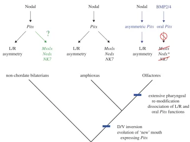
endoderm in addition to sites in other developmental contexts that are conserved with vertebrates. Crucially though, the ectodermal invagination that will form the mouth, the stomodeum, has been homologized across chordates (Christiaen et al. 2007) using the expression ofPitxdownstream of conserved ventralizing BMP signal-ling (Yuet al. 2007). This pattern of mouth development, and a loss ofBrachyuryand Goosecoidexpression, is dis-tinct from non-chordate deuterostomes and protostomes (Arendt et al. 2001; Christiaen et al. 2007) and has led to the suggestion that a ‘new mouth’ is a chordate inno-vation (Christiaen et al. 2007; Hejnol & Martindale 2008).
In addition to its role in chordate oral ectoderm spe-cification, Pitx involvement in left – right asymmetry downstream of Nodal signalling is conserved across deuterostomes (Duboc & Lepage 2008) and probably Bila-teria (Grande & Patel 2009). It has thus been hypothesized that the evolution of the ‘new’ chordate mouth was the result of the evolution of a stomodealPitxexpression domain that was under the control ofBMPsignalling and distinct from the asymmetric domain ofPitxexpression that was down-stream ofNodal(Christiaenet al. 2007). Evidence for the distinct regulation of these domains of expression has been uncovered in both mouse (Goodyeret al. 2003;Shiratori et al. 2006) andCiona(Christiaenet al. 2005).
Against this background, the correspondence between the expression ofAmphiMsxlx,AmphiNedxa,AmphiNedxb
and AmphiNKx7and the subsequent asymmetric devel-opment of the pharyngeal apparatus in amphioxus is striking, and the expression is likely, from its spatial and
temporal profile, to be downstream of the conserved
Nodal–Pitx symmetry-breaking pathway (Yasui et al. 2000;Boorman & Shimeld 2002;Yuet al. 2002). We pro-pose that the morphogenetic asymmetry of the amphioxus embryo/larva is dependent upon these ancient homeobox genes that are integrated downstream of the ancestral symmetry-breaking pathway. Under this hypothesis, amphioxus would represent an ancestral developmental gene-regulatory programme, where Pitx is expressed asymmetrically and is responsible for directing sub-sequent asymmetric pharyngeal (including oral and pre-oral) development, withMsxlx,NedxandNKx7being part of the ancestral, asymmetric pharyngeal patterning system.
In Olfactores (vertebratesþurochordates), the stomo-deal function of Pitx (which is under the control of BMP signalling) has dissociated from the asymmetric function (under the control of Nodal signalling), and this correlates with the loss of asymmetry in pharyngeal development (figure 5). The loss of the genes examined here from vertebrates, would thus be correlated with a loss of ancestral pharyngeal developmental programmes that paved the way for the evolution of the neural crest-dependent pharyngeal development characteristic of vertebrates (Gans & Northcutt 1983;Graham 2001;Ota et al. 2007). In light of this suggestion, the expression of the Oikopleura dioica Nedx paralogues, which represent the only one of the ANTP-class homeobox families dis-cussed here that is retained in a member of Olfactores, will be of considerable interest. A direct prediction of the model presented here would be that the Oikopleura
Nodal
Pitx
L/R asymmetry
non-chordate bilaterians amphioxus Olfactores
extensive pharyngeal re-modification dissociation of L/R and oral Pitx functions
D/V inversion
evolution of ‘new’ mouth expressing Pitx
L/R asymmetry
L/R asymmetry
Msxlx Nedx NK7
?
Msxlx Nedx NK7
Msxlx Nedx* NK7 Pitx
Nodal Nodal BMP2/4
[image:7.595.95.481.49.336.2]asymmetric Pitx oral Pitx
Figure 5. A hypothesis for the evolution of pharynx development. The evolution of chordates was marked by the relocation of the mouth to aPitx-expressing territory, the expression of which may have been controlled by BMP signalling (Olfactores) or Nodal signalling (amphioxus). In the Olfactores lineage, the pharynx has undergone significant modification with the oral function of Pitx being controlled independently of the asymmetry function (blue). Msxlx, Nedx and NK7, which in amphioxus are putatively downstream ofPitxhave been lost from Olfactores (red), except two Nedx paralogues in the appen-dicularianOikopleura(*). The expression ofMsxlx,NedxandNK7in non-chordate bilaterians is unexplored (green).
3386 T. Buttset al. Chordate homeobox evolution
genes are expressed in a novel and species-specific fashion that is distinct from BMP-dependent oralPitxexpression. Of course, it remains possible that amphioxus pharyn-geal development is derived relative to the chordate ancestor, with the asymmetric role of the genes examined here representing a co-option of ancient genes for a novel function. To resolve this uncertainty, comparative devel-opmental data from non-chordate phyla will be necessary, especially from enteropneusts, which pattern their anteroposterior and dorsoventral axes in a compar-able way to chordates (Lowe et al. 2003, 2006) and represent a crucial reference for reconstructing the ances-tral deuterostome (Gerhart 2006), assuming that the genes discussed here are present in enteropneust gen-omes. Interestingly, an expression domain ofPitx in the model hemichordateSaccoglossus kowalevskii (Loweet al. 2006) is reminiscent of the position of the chordate mouth in the conserved ectodermal expression map, and corresponds to the site where the proboscis pore later forms, the structure that indicates left – right asymmetry in this animal (Loweet al. 2003).
5. CONCLUSION
We have described the expression of four genes from three ancient ANTP-class homeobox families in the basal chor-date lineage of amphioxus. These three ancient gene families were all lost during vertebrate evolution. It is striking that the expression of these ‘lost’ genes is both transient and spatially restricted, and the structures that the genes are expressed in in amphioxus have been exten-sively modified during chordate evolution, namely the pharynx and the anterior CNS. This raises the distinct possibility that these genes have been ‘sidetracked’ into restricted developmental roles that were dispensed with during vertebrate evolution. The role of gene loss in the evolution of development has not been widely considered (reviewed inDe Robertis 2008), but loss of ancient genes is clearly widespread (e.g. Kortschak et al. 2003;Wyder et al. 2007; Kuraku et al. 2008; Takahashi et al. 2009). A more complete appreciation of the role of gene loss in evolutionary developmental biology will come from extending the type of comparative genomic and embryo-logical work highlighted here.
Irrespective of the ancestral role of Msxlx, Nedx and
NKx7, it is clear that the evolution of the vertebrates was accompanied not just by the addition of developmen-tal networks and structures (like the telencephalon and neural crest) onto a pre-existing amphioxus-like body plan, but that some ancestral gene-regulatory networks were dismantled leading ultimately to the loss of some of the constituent genes.
The authors would like to thank Seb Shimeld for advice and critical reading of the manuscript, John Lawrence for providing lab space at the University of Florida during the amphioxus spawning season and Nick and Linda Holland for help during amphioxus fieldwork. Work in the authors’ laboratoriess is supported by the BBSRC, Wellcome Trust (081233/Z/06/Z) and the Royal Society.
REFERENCES
Arendt, D., Technau, U. & Wittbrodt, J. 2001 Evolution of the bilaterian foregut.Nature409, 81 – 85. (doi:10.1038/ 35051075)
Beaster-Jones, L., Kaltenbach, S. L., Koop, D., Yuan, S., Chastain, R. & Holland, L. Z. 2008 Expression of somite segmentation genes in amphioxus: a clock without a wavefront? Dev. Genes Evol. 218, 599 – 611. (doi:10. 1007/s00427-008-0257-5)
Boorman, C. J. & Shimeld, S. M. 2002 Pitx homeobox genes inCiona and amphioxus show left – right asymmetry is a conserved chordate character and define the ascidian ade-nohypophysis. Evol. Dev. 4, 354 – 365. (doi:10.1046/j. 1525-142X.2002.02021.x)
Bourlat, S. J. et al. 2006 Deuterostome phylogeny reveals monophyletic chordates and the new phylum Xenotur-bellida.Nature444, 85 – 88. (doi:10.1038/nature05241) Butts, T., Holland, P. W. H. & Ferrier, D. E. K. 2008
The urbilaterian Super-Hox cluster. Trends Genet. 24, 259 – 262. (doi:10.1016/j.tig.2007.09.006)
Candiani, S., Holland, N. D., Oliveri, D., Parodi, M. & Pestarino, M. 2008 Expression of the amphioxus Pit-1
gene (AmphiPOU1F1/Pit-1) exclusively in the developing preoral organ, a putative homologue of the vertebrate ade-nohypophysis. Brain Res. Bull. 75, 324 – 330. (doi:10. 1016/j.brainresbull.2007.10.023)
Castro, L. F. C. & Holland, P. W. H. 2003 Chromosomal mapping of ANTP class homeobox genes in amphioxus: piecing together ancestral genomes. Evol. Dev. 5, 459 – 465. (doi:10.1046/j.1525-142X.2003.03052.x) Christiaen, L., Bourrat, F. & Joly, J. S. 2005 A modular
cis-regulatory system controls isoform-specific Pitx
expression in ascidian stomodeum. Dev. Biol. 277, 557 – 566. (doi:10.1016/j.ydbio.2004.10.008)
Christiaen, L., Jaszczyszyn, Y., Kerfant, M., Kano, S., Thermes, V. & Joly, J. S. 2007 Evolutionary modification of mouth position in deuterostomes.Semin. Cell Dev. Biol.
18, 502 – 511. (doi:10.1016/j.semcdb.2007.06.002) Conklin, E. G. 1932 The embryology of amphioxus.
J. Morphol 54, 69 – 151. (doi:10.1002/jmor.10505 40103)
Dai, Z., Chen, Z., Ye, H., Zhou, L., Cao, L. & Arendt, D. 2009 Characterisation of microRNAs in cephalo-chordates reveals a correlation between microRNA repertoire homology and morphological similarity in chordate evolution.Evol. Dev.11, 41 – 49. (doi:10.1111/ j.1525-142X.2008.00301.x)
D’Aniello, S., Irimia, M., Maeso, I., Pascual-Anaya, J., Jime´nez-Delgado, S., Bertrand, S. & Garcia-Ferna`ndez, J. 2008 Gene expansion and retention leads to a diverse tyrosine kinase superfamily in amphioxus. Mol. Biol. Evol.25, 1841 – 1854. (doi:10.1093/molbev/msn132) Dehal, P. & Boore, J. L. 2005 Two rounds of whole genome
duplication in the ancestral vertebrate.PLoS Biol.3, e314. (doi:10.1371/journal.pbio.0030314)
Delsuc, F., Brinkmann, H., Chorrout, D. & Philippe, H. 2006 Tunicates and not cephalochordates are the closest living relatives of vertebrates. Nature 439, 965–968. (doi:10. 1038/nature04336)
De Robertis, E. M. 2008 Evo-Devo: variations on ancestral themes.Cell132, 185 – 195.
Deschamps, J. 2007 Ancestral and recently recruited global control of the Hox genes in development. Curr. Opin. Genet. Dev.17, 422 –427. (doi:10.1016/j.gde.2007.07.008) Duboc, V. & Lepage, T. 2008 A conserved role for the nodal signalling pathway in the establishment of dorso-ventral and left-right axes in deuterostomes. J. Exp. Zool. B
(Mol. Dev. Evol.)310, 41 – 53.
Duboule, D. 2007 The rise and fall of Hox gene clusters.
Flood, P. R., Guthrie, D. M. & Banks, J. R. 1969 Paramyosin muscle in the notochord of amphioxus. Nature 222, 87 – 88. (doi:10.1038/222087a0)
Gans, C. & Northcutt, R. G. 1983 Neural crest and the origin of vertebrates: a new head.Science220, 268 – 274. (doi:10.1126/science.220.4594.268)
Gehring, W. J. 2002 The genetic control of eye development and its implications for the evolution of the various eye-types.Int. J. Dev. Biol.46, 65 – 73.
Gerhart, J. 2006 The deuterostome ancestor.J. Cell Physiol.
209, 677 – 685. (doi:10.1002/jcp.20803)
Glardon, S., Holland, L. Z., Gehring, W. J. & Holland, N. D. 1998 Isolation and developmental expression of the amphioxus Pax-6 gene (AmphiPax-6): insights into eye and photoreceptor evolution. Development 125, 2701 – 2710.
Goodyer, C. G., Tremblay, J. J., Paradis, F. W., Marcil, A., Gauthier, C., Lanctoˆt, Y. & Drouin, J. 2003 Pitx1 in vivopromoter activity and mechanisms of positive autore-gulation.Neuroendocrinology78, 129 – 137. (doi:10.1159/ 000072794)
Graham, A. 2001 The development and evolution of the pharyngeal arches.J. Anat.199, 133 – 141.
Grande, C. & Patel, N. H. 2009 Nodal signalling is involved in left – right asymmetry in snails.Nature457, 1007 – 1011. (doi:10.1038/nature07603)
Guthrie, D. M. & Banks, J. R. 1970 Observations on the function and physiological properties of a fast paramyosin muscle—the notochord of amphioxus (Branchiostoma lanceolatum).J. Exp. Biol.52, 125 – 138.
Hatschek, B. 1893The amphioxus and its development. Swan Sonnenschein & Co. Edited and translated by James Tuckey.
Hejnol, A. & Martindale, M. Q. 2008 Acoel development indicates the independent evolution of the bilaterian mouth and anus. Nature 456, 382 – 386. (doi:10.1038/ nature07309)
Holland, L. Z. 2005 Non-neural ectoderm is really neural: evolution of developmental patterning mechanisms in the non-neural ectoderm of chordates and the problem of sensory cell homologies. J. Exp. Zool. B (Mol. Dev. Evol.)304, 304 – 323.
Holland, L. Z. & Holland, N. D. 1993 Embryos and larvae of invertebrate deuterostomes. InEssential developmental biology: a practical approach(eds C. D. Stern & P. W. H. Holland), pp. 21–32. Oxford, UK: Oxford University Press.
Holland, L. Z. & Holland, N. D. 1999 Chordate origins of the vertebrate central nervous system. Curr. Opin. Neurobiol. 9, 596 – 602. (doi:10.1016/S0959-4388 (99)00003-3)
Holland, L. Z., Holland, P. W. H. & Holland, N. D. 1996 Revealing homologies between body parts of distantly related animals byin situhybridization to developmental genes: amphioxus vs vertebrates. In Molecular zoology: advances, strategies and protocols (eds J. D. Ferraris & S. R. Palumbi), pp. 267 – 282. New York, NY: John Wiley & Sons.
Holland, L. Z., Kene, M., Williams, N. A. & Holland, N. D. 1997 Sequence and embryonic expression of the amphioxus engrailed gene (AmphiEn): the metameric pat-tern of transcription resembles that of its segment polarity homologue inDrosophila.Development124, 1723 – 1732. Holland, L. Z., Venkatesh, T. V., Gorlin, A., Bodmer, R. &
Holland, N. D. 1998 Characterization and developmental expression ofAmphiNk2-2, an NK2 class homeobox gene from amphioxus (Phylum Chordata; Subphylum Cepha-lochordata). Dev. Genes Evol. 208, 100 – 105. (doi:10. 1007/s004270050159)
Holland, L. Z., Schubert, M., Kozmik, Z. & Holland, N. D. 1999 AmphiPax3/7, an amphioxus paired box gene:
insights into chordate myogenesis, neurogenesis, and the possible evolutionary precursor of definitive vertebrate neural crest. Evol. Dev. 1, 153 – 165. (doi:10.1046/j. 1525-142x.1999.99019.x)
Holland, L. Z. et al. 2008 The amphioxus genome illuminates vertebrate origins and cephalochordate biology. Genome Res. 18, 1100 – 1111. (doi:10.1101/gr. 073676.107)
Holland, N. D. & Chen, J. 2001 Origin and early evolution of the vertebrates: new insights from advances in molecular biology, anatomy, and palaeontology. Bioessays 23, 142 – 151. (doi:10.1002/1521-1878(200102)23:2,142:: AID-BIES1021.3.0.CO;2-5)
Horton, A. C.et al.2008 Conservation of linkage and evol-ution of developmental function within the Tbx2/3/4/5
subfamily of T-box genes: implications for the origin of vertebrate limbs. Dev. Genes Evol. 218, 613 – 628. (doi:10.1007/s00427-008-0249-5)
Kortschak, R. D., Samuel, G., Saint, R. & Miller, D. J. 2003 EST analysis of the cnidarian Acropora milleporareveals extensive gene loss and rapid sequence divergence in the model invertebrates.Curr. Biol.13, 2190 – 2195. (doi:10. 1016/j.cub.2003.11.030)
Kozmik, Z., Holland, N. D., Kalousova, A., Paces, J., Schubert, M. & Holland, L. Z. 1999 Characterisation of an amphioxus paired box gene,AmphiPax2/5/8: devel-opmental expression patterns in optic support cells, nephridium, thyroid-like structures and pharyngeal gill slits, but not in the midbrain-hindbrain boundary region.Development126, 1295 – 1304.
Kozmik, Z., Holland, L. Z., Schubert, M., Lacalli, T. C., Kreslova, J., Vlcek, C. & Holland, N. D. 2001 Character-isation of amphioxus AmphiVent, an evolutionarily conserved marker for chordate ventral mesoderm.Genesis
29, 172 – 179. (doi:10.1002/gene.1021)
Kozmik, Z.et al.2007Pax – Six – Eya – Dachnetwork during amphioxus development: conservationin vitrobut context specificityin vivo.Dev. Biol.306, 143 – 159. (doi:10.1016/ j.ydbio.2007.03.009)
Kuraku, S., Takio, Y., Tamura, K., Aono, H., Meyer, A. & Kuratani, S. 2008 Noncanonical role of Hox14 revealed by its expression patterns in lamprey and shark. Proc. Natl Acad. Sci. USA 105, 6679 – 6683. (doi:10.1073/ pnas.0710947105)
Lacalli, T. C. 2004 Sensory systems in amphioxus: a window on the ancestral chordate condition. Brain Behav. Evol.
64, 148 – 162. (doi:10.1159/000079744)
Lacalli, T. C. 2008 Basic features of the ancestral chordate brain: a protochordate perspective. Brain Res. Bull. 75, 319 – 323. (doi:10.1016/j.brainresbull.2007.10.038) Lacalli, T. C. & Kelly, S. J. 2000 The infundibular
bal-ance organ in amphioxus larvae and related aspects of cerebral vesicle organisation.Acta Zool.81, 37–47. (doi:10. 1046/j.1463-6395.2000.00036.x)
Lowe, C. J., Wu, M., Salic, A., Evans, L., Lander, E., Stange-Thomann, N., Gruber, C. E., Gerhart, J. & Kirschner, M. 2003 Anteroposterior patterning in hemi-chordates and the origins of the chordate nervous system. Cell 113, 853 – 865. ( doi:10.1016/S0092-8674(03)00469-0)
Lowe, C. J.et al.2006 Dorsoventral patterning in hemichor-dates: insights into early chordate evolution.PloS Biol.4, e291. (doi:10.1371/journal.pbio.0040291)
Mazet, F., Amemiya, C. T. & Shimeld, S. M. 2006 An ancient Fox gene cluster in bilaterian animals.Curr. Biol.
16, R314 – R316. (doi:10.1016/j.cub.2006.03.088) Minguillo´n, C., Jime´nez-Delgado, S., Panopoulou, G. &
Garcia-Ferna`ndez, J. 2003 The amphioxus Hairy family: differential fate after duplication. Development 130, 5903 – 5914. (doi:10.1242/dev.00811)
3388 T. Buttset al. Chordate homeobox evolution
Ogasawara, M. 2000 Overlapping expression of amphioxus homologues of the thyroid transcription factor-1 gene and thyroid peroxidase gene in the endostyle: insight into the evolution of the thyroid gland. Dev. Genes Evol.
210, 231 – 242. (doi:10.1007/s004270050309)
Osborne, P. W., Benoit, G., Laudet, V., Schubert, M. & Ferrier, D. E. K. 2009 Differential regulation of ParaHox genes by retinoic acid in the invertebrate chordate amphioxus (Branchiostoma floridae). Dev. Biol. 327, 252 – 262.
Ota, K. G., Kuraku, S. & Kuratani, S. 2007 Hagfish embryology with reference to the evolution of the neural crest.Nature446, 672 – 675. (doi:10.1038/nature05633) Paris, M.et al.2008aAmphioxus postembryonic development
reveals homology of chordate metamorphosis. Curr. Biol.
18, 825 – 830. (doi:10.1016/j.cub.2008.04.078)
Paris, M., Brunet, F., Markov, G. V., Schubert, M. & Laudet, V. 2008bThe amphioxus genome enlightens the evolution of the thyroid hormone signalling pathway.
Dev. Genes Evol. 218, 667 – 680. ( doi:10.1007/s00427-008-0255-7)
Putnam, N. H. et al. 2008 The amphioxus genome and the evolution of the chordate karyotype. Nature 453, 1064 – 1071. (doi:10.1038/nature06967)
Ryan, J. F., Burton, P. M., Mazza, M. E., Kwong, G. K., Mullikin, J. C. & Finnerty, J. R. 2006 The cnidarian-bilaterian ancestor possessed at least 56 homeoboxes: evidence from the starlet sea anemone, Nematostella vectensis. Genome Biol. 7, R64. ( doi:10.1186/gb-2006-7-7-r64)
Sandmann, T., Girardot, C., Brehme, M., Tongprasit, W., Stolc, V. & Furlong, E. E. M. 2007 A core transcriptional network for early mesoderm development in Drosophila melanogaster.Genes Dev.21, 436 – 449. (doi:10.1101/gad. 1509007)
Satoh, G., Takeuchi, J. K., Yasui, K., Tagawa, K., Saiga, H., Zhang, P. & Satoh, N. 2002 Amphi-Eomes/Tbr1: an amphioxus cognate of vertebrate Eomesodermin and
T-Brain1genes whose expression reveals an evolutionarily distinct domain in amphioxus development.J. Exp. Zool. B
(Mol. Dev. Evol.) 294, 136 – 145. (doi:10.1002/jez. 10149)
Schubert, M., Holland, L. Z., Stokes, M. D. & Holland, N. D. 2001 Three amphioxus Wnt genes (AmphiWnt3,
AmphiWnt5, and AmphiWnt6) associated with the tail bud: the evolution of somitogenesis in chordates. Dev. Biol.240, 262 – 273. (doi:10.1006/dbio.2001.0460) Schubert, M., Brunet, F., Paris, M., Bertrand, S., Benoit, G. &
Laudet, V. 2008 Nuclear hormone receptor signalling in amphioxus. Dev. Genes Evol. 218, 651 – 665. (doi:10. 1007/s00427-008-0251-y)
Shimeld, S. M. 2008 C2H2 zinc finger genes of theGli,Zic,
KLF, SP, Wilms’ tumour, Huckebein, Snail, Ovo, Spalt,
Odd, Blimp-1, Fez and related gene families from
Branchiostoma floridae. Dev. Genes Evol. 218, 639 – 649. (doi:10.1007/s00427-008-0248-6)
Shiratori, H., Yashiro, K., Shen, M. M. & Hamada, H. 2006 Conserved regulation and role of Pitx2 in situ-specific
morphogenesis of visceral organs. Development 133, 3015 – 3025. (doi:10.1242/dev.02470)
Takahashi, T., McDougall, C., Troscianko, J., Chen, W.-C., Jayaraman-Nagarajan, A., Shimeld, S. M. & Ferrier, D. E. K. 2009 An EST screen from the annelid
Pomatoceros lamarckii reveals patterns of gene loss and gain in animals. BMC Evol. Biol 9, 240. (doi:10.1186/ 1471-2148-9-240)
Takatori, N., Butts, T., Candiani, S., Pestarino, M., Ferrier, D. E. K., Saiga, H. & Holland, P. W. H. 2008 Com-prehensive survey and classification of homeobox genes in the genome of amphioxus, Branchiostoma floridae.
Dev. Genes Evol. 218, 579 – 590. ( doi:10.1007/s00427-008-0245-9)
Tomancak, P., Berman, B. P., Beaton, A., Kwan, R. W. E., Hartenstein, V., Celniker, S. E. & Rubin, G. M. 2007 Global analysis of gene expression during Drosophila
embryogenesis. Genome Biol. 8, R145. ( doi:10.1186/gb-2007-8-7-r145)
Wang, Y., Zhang, P.-J., Yasui, K. & Saiga, H. 2002 Expression of Bblhx3, a LIM homeobox gene, in the development of amphioxus Branchiostoma belcheri tsingtauense. Mech. Dev. 117, 315 – 319. (doi:10.1016/ S0925-4773(02)00197-1)
Whittaker, J. R. 1997 Cephalochordates, the lancelets. In
Embryology: constructing the organism(eds S. F. Gilbert & A. M. Raunio), pp. 365 – 381. Sunderland, MA: Sinauer Associates Inc.
Williams, N. A. & Holland, P. W. H. 1996 Old head on young shoulders. Nature 383, 490. (doi:10.1038/ 383490a0)
Wyder, S. et al. 2007 Quantification of ortholog losses in insects and vertebrates. Genome Biol. 8, R242. (doi:10. 1186/gb-2007-8-11-r242)
Yasui, Zhang, S.-C., Uemura, M., Aizawa, S. & Ueki, T. 1998 Expression of a twist-related gene,Bbtwist, during the development of a lancelet species and its relation to cephalochordate anterior structures.Dev. Biol.195, 49 – 59. (doi:10.1006/dbio.1997.8834)
Yasui, K., Zhang, S., Uemura, M. & Saiga, H. 2000 Left – right asymmetric expression of BbPtx, a Ptx-related gene, in a lancelet species and the developmental left-sidedness in deuterostomes.Development127, 187 – 195. Yu, J. K., Holland, L. Z. & Holland, N. D. 2002 An
amphioxus nodal gene (AmphiNodal) with early sym-metrical expression in the organiser and mesoderm and later asymmetrical expression associated with left-right axis formation. Evol. Dev. 4, 418 – 425. (doi:10.1046/j. 1525-142X.2002.02030.x)
Yu, J. K., Satou, Y., Holland, N. D., Shin-I, T., Kohara, Y., Satoh, N., Bronner-Fraser, M. & Holland, L. Z. 2007 Axial patterning in cephalochordates and the evolution of the organizer. Nature 445, 613 – 617. (doi:10.1038/ nature05472)