EVALUATION OF CARDIOVASCULAR
FUNCTION IN OVERT AND SUBLINICAL
HYPOTHYROIDISM
Dissertation Submitted to
THE TAMIL NADU DR. M.G.R MEDICAL UNIVERSITY
In partial fulfillment of the regulations For the award of the degree of
M.D. BRANCH – I GENERAL MEDICINE
INSTITUTE OF INTERNAL MEDICINE
M
ADRASM
EDICALC
OLLEGE&
R
AJIVG
ANDHIGOVERNMENT
GENERAL
H
OSPITAL,
CERTIFICATE
This is to certify that this dissertation entitled “EVALUATION OF CARDIOVASCULAR FUNCTION IN OVERT AND SUBLINICAL
HYPOTHYROIDISM” submitted by Dr. J.JACINTH PREETHI, appearing for
Part II M.D.Branch I General Medicine Degree examination in April 2012 is a bonafide record of work done by her under my direct audience and supervision in partial fulfillment of regulations of the Tamil Nadu Dr. M.G.R. Medical University, Chennai. I forward this to the Tamil Nadu Dr.M.G.R. Medical University, Chennai, Tamil Nadu, India.
Prof.A.RADHAKRISHNAN, M.D., Prof.C.RAJENDIRAN, M.D., Professor of Medicine, Guide & Supervisor, Director and Professor,
Institute of internal medicine, Institute of Internal Medicine,
MMC & RGGGH, MMC & RGGGH,
Chennai – 600 003 Chennai – 600 003.
DR.V.KANAGASABAI,M.D.,
D E A N
Madras Medical College and
DECLARATION
I solemnly declare that this dissertation entitled “EVALUATION OF CARDIOVASCULAR FUNCTION IN OVERT AND SUBLINICAL
HYPOTHYROIDISM” was done by me at Madras Medical College and
Government General Hospital, during the academic year 2009-2012 under the guidance and supervision of Prof.A.Radhakrishnan M.D. This dissertation is submitted to the Tamil Nadu Dr.M.G.R. Medical University towards the partial fulfillment of requirements for the award of M.D. Degree in General Medicine (Branch-I).
ACKNOWLEDGEMENT
I would like to thank Prof.V.Kanagasabai, M.D., Dean, Madras Medical College for his kind permission to use the hospital resources for this study.
I would like to thank Prof.V.Palani, M.S., Medical Superintendent, Madras Medical College and Rajiv Gandhi Government General Hospital for his kind permission to use the hospital resources for this study.
I would like to express my sincere gratitude to Prof. C.Rajendiran,M.D Professor and Director, Institute of Internal Medicine for his guidance and encouragement.
I am extremely grateful to my guide Prof .A Radhakrishnan M.D., Professor, Institute of internal medicine for his guidance and help.
I am extremely thankful to Prof. Geetha Subramanian M.D D.M, former Professor and Head of the Department of cardiology, Madras Medical College & Rajiv Gandhi Government General Hospital for permitting me to use the facilities of the department.
I thank the Department of Biochemistry for their extreme cooperation extended to me without whom the study would not have been possible.
I am also thankful to my colleagues and house officers for their full cooperation and help in this study. I am immensely grateful to the generosity shown by the patients who participated in this study.
S.NO
TITLE
PAGE NO
1 INTRODUCTION 1
2 AIMS AND OBJECTIVES 7
3 REVIEW OF LITERATURE 9
4 MATERIALS AND METHODS 29
5 RESULTS AND OBSERVATIONS 38
6 DISCUSSION 57
7 CONCLUSIONS 65
8 LIMITATIONS 67
9 BIBLIOGRAPHY 69
10 PROFORMA
11 INSTITUTIONAL ETHICAL COMMITTEE APPROVAL
12 MASTER CHART
ABBREVIATIONS
ASE- American society of echocardiography BMI – Body mass index
DM- Diabetes mellitus EDV- End diastolic volume EF- Ejection fraction
ESV- End systolic volume FS-Fractional shortening
HDL- High density lipoprotein IVSW- Inter ventricular septal wall LDL-Low density lipoprotein LV- Left ventricle
LVEF- Left ventricular ejection fraction
LVID (D)- Left ventricular internal diameter (diastolic) LVID(S)- Left ventricular internal diameter (systolic) LVPW- Left ventricular posterior wall
T3- triiodothyronine T4- thyroxine
INTRODUCTION
The thyroid gland maintains the level of metabolism in the tissues that is optimal for their normal function. The function of the thyroid is to generate the quantity of thyroid hormone necessary to meet the demands of the peripheral tissues. The principal hormones secreted by the thyroid are thyroxine (T4) and triiodothyronine (T3). T3 is also formed in the peripheral tissues by deiodination
of T4. Both hormones are iodine-containing amino acids. Small amounts of reverse
triiodothyronine (3, 3´, 5´-triiodothyronine, RT3) and other compounds are also
found in thyroid venous blood. T3 is more active than T4, whereas RT3 is inactive. Anatomy
The thyroid is one of the largest of the endocrine organs, weighing approximately 15 to 20 g. The normal thyroid is made up of two lobes joined by a thin band of tissue, the isthmus. The gland is composed of closely packed spherical units termed follicles, which are invested with a rich capillary network. The
Thyroid Hormone Synthesis
The thyroid cell membranes facing the capillaries contain a symporter, or iodide pump, that transports Na+ and I- into the cells against the electrochemical gradient for I-. This Na+/I- symporter (NIS) is capable of producing intracellular I -concentrations that are 20-40 times as great as the concentration in plasma.
In the thyroid gland, iodide is oxidized to iodine .In the process of hormone synthesis; the first product is monoiodotyrosine (MIT). MIT is next iodinated in the 5 position to form diiodotyrosine (DIT). Two DIT molecules then undergo an oxidative condensation to form T4 with the elimination of the alanine side chain
from the molecule that forms the outer ring. Thyroid peroxidase is probably involved in coupling as well as iodination. T3 is probably formed by condensation
of MIT with DIT. A small amount of RT3 is also formed, probably by condensation
of DIT with MIT and bound to the 3 position of tyrosine residues that are part of the thyroglobulin molecule in the colloid.(2)
Secretion
The human thyroid secretes about 80 ug (103 nmol) of T4, 4 ug (7 nmol) of T3, and
2 ug (3.5 nmol) of RT3 per day .However, MIT and DIT are not secreted. The
Transport & Metabolism
The plasma proteins that bind thyroid hormones are albumin; a prealbumin formerly called thyroxine-binding prealbumin (TBPA) and now called
transthyretin; and a globulin with an electrophoretic mobility between those of
α1- and α2-globulin, thyroxine-binding globulin (TBG). Of the three proteins, albumin has the largest capacity to bind T4—ie, it can bind the most T4 before
becoming saturated—and TBG has the smallest capacity.
Mechanism of Action
Thyroid hormones enter cells, and T3 binds to thyroid receptors (TR) in the
nuclei. T4 can also bind, but not as avidly. The hormone-receptor complex then
Effects on the Cardiovascular System
Thyroid hormone transcriptionally regulates many cardiac proteins the thyroid hormone exerts most of its direct effects on cardiac contractility by regulating calcium cycling through the SERCA-phospholamban system.
AIM OF THE STUDY
The aim of this study is to assess
i. The cardiovascular functions in primary overt and subclinical hypothyroid patients.
REVIEW OF LITERATURE
Thyroid diseases are among the commonest endocrine disorders worldwide. India, is no exception. There are various causes of primary hypothyroidism, but autoimmune disease (Hashimoto's thyroiditis) and thyroid failure following I 131 or surgical treatment account for over 90% of cases in those parts of the world which are not significantly iodine-deficient. Iodine deficiency as a cause of hypothyroidism is more common in developing countries.
The terminology 'subclinical hypothyroidism' was probably first used by Evered and colleagues to describe a group of individuals in whom 'conventional tests of thyroid function showed nothing abnormal ... but they were all found to have a raised serum TSH concentration' (Evered et al., 1973). Subclinical
hypothyroidism is defined today by normal serum free T4 levels and serum TSH levels above the upper limit of the reference range.
dyslipidaemia and a host of other problems, as well as progression to overt hypothyroidism, it is important to screen the general population and treat appropriate patients.
According to a survey by N. Kochupillai the countrywide prevalence of thyroid disorders is 42 millions. (5) Jayarama, K. S. et al the prevalence of hypothyroidism in India is 0.071 %.( 6). In a population-based study done in Cochin on 971 adult subjects, the prevalence of hypothyroidism was 3.9%. The prevalence of subclinical hypothyroidism was also high in this study, the value being 9.4%. In women, the prevalence was higher, at 11.4%, when compared with men, in whom the prevalence was 6.2%. (7) The worldwide prevalence of subclinical hypothyroidism ranges from 1% to 10%; the highest age and sex specific rates are in woman over 60 years, approaching to 20% (8, 9).
According to a study by Jangid DR et al thyroid surgery and decompensated iodine deficiency were the major known causes of hypothyroidism (19.5% and 15.3% respectively), while in the majority of patients (45.14%) the aetiology was obscure.(10)
groups having some form of hypothyroidism. The Framingham study found hypothyroidism (TSH >10 mIU/L) in 5.9% of women and 2.4% of men older than 60 years (11).
Causes of hypothyroidism
Primary Hypothyroidism
Acquired
Hashimoto's thyroiditis
Iodine deficiency (endemic goiter)
Drugs blocking synthesis or release of T4 (e.g., lithium, ethionamide, sulfonamides,
iodide)
Goitrogens in foodstuffs or as endemic substances or pollutants Cytokines (interferon-α, interleukin-2)
Thyroid infiltration (amyloidosis, hemochromatosis, sarcoidosis, Riedel's struma, cystinosis, scleroderma)
Congenital
Iodide transport or utilization defect (NIS or pendrin mutations) Iodotyrosine dehalogenase deficiency
Organification disorders (TPO* deficiency or dysfunction) Defects in thyroglobulin synthesis or processing
Thyroid agenesis or dysplasia TSH receptor* defects
Thyroidal Gs protein abnormalities (pseudohypoparathyroidism type 1a) Idiopathic TSH unresponsiveness
Transient (Post-thyroiditis) Hypothyroidism
Following subacute, painless, or postpartum thyroiditis
Consumptive Hypothyroidism
Rapid destruction of thyroid hormone due to D3 expression in large hemangiomas or hemangioendotheliomas
Defects of Thyroxine to Triiodothyronine Conversion
Drug-Induced Thyroid Destruction
Tyrosine kinase inhibitor (sunitinib)
Central Hypothyroidism
Acquired
Pituitary origin (secondary) Hypothalamic disorders (tertiary)
Bexarotene (retinaid X receptor agonist) Dopamine and/or severe illness
Congenital
TSH deficiency or structural abnormality TSH receptor defect
Resistance to Thyroid Hormone Generalized
Signs and Symptoms of Hypothyroidism (Descending Order of Frequency)
Symptoms
Tiredness, weakness Dry skin
Feeling cold Hair loss
Difficulty concentrating and poor memory Constipation
Weight gain with poor appetite Dyspnea
Hoarse voice
Menorrhagia (later oligomenorrhea or amenorrhea) Paresthesia
Signs
Dry coarse skin; cool peripheral extremities Puffy face, hands, and feet (myxedema) Diffuse alopecia
Bradycardia Peripheral edema
Delayed tendon reflex relaxation Carpal tunnel syndrome
Serous cavity effusions
Cardiac involvement in myxedema was first described by Zondek in 1918(12).The cardiovascular findings of hypothyroidism are more subtle. The clinical presentation of overt hypothyroidism is not obvious and most patients have few symptoms and signs (13).The CVS manifestations of hypothyroidism include the following:
c) Reduced heart rate (All leading to decreased cardiac output)
d) Raised systemic vascular resistance (leading to increased diastolic blood pressure)
e) Raised capillary permeability (leading to pericardial effusion)
Bradycardia and systemic hypertension, with narrow pulse pressure and slightly increased mean arterial pressure, and some degree of exercise impairment are the most-common findings in patients with overt hypothyroidism (13, 14).
No specific pathophysiological changes can be identified that characterize the myxedema heart. The cardiac silhouette is enlarged; however, heart weight is usually normal. Cardiac papillary muscle shows a depression of the force velocity curve and reduced rate of tension development, indicating significant contractile abnormalities. Myofibril swelling with loss of striation and some degree of interstitial fibrosis occurs on histological examination of hypothyroid hearts. In addition, accumulation of mucopolysaccharides can be demonstrated.
INTERACTION OF THYROID HORMONES WITH THE SYMPATHETIC NERVOUS
SYSTEM
concentrations are normal or decreased in hyperthyroidism and elevated in hypothyroidism.(15-17) The latter finding is not related to decreased norepinephrine clearance but rather to increased norepinephrine release from sympathetic nerves.(17). This interpretation is strengthened by the observation that thyrotropin releasing hormone (TRH),which is elevated in primary hypothyroidism, directly stimulates sympathetic outflow within the central nervous system and may be taken up by nerve endings to serve as a neurotransmitter. This apparent paradox of clinical signs evoking decreased sympathetic tone in the presence of elevated norepinephrine release from sympathetic nerve endings is consistent with the hypothesis of desensitization to the effects of catecholamines in hypothyroidism.
(18).
HYPERTENSION
Ikuo Saito et al in his study showed hypertension is more often associated with hypothyroidism than euthyroidism in patients over 50 years old, and that blood pressure is often reduced in response to adequate thyroid hormone replacement therapy alone (22)
LV FUNCTION
The most-consistent cardiac abnormality recognized in patients with overt hypothyroidism is impairment of LV diastolic function, which is characterized by slowed myocardial relaxation and impaired early ventricular filling (20, 23). This is completely reversed by replacement therapy.
ventricular morphology. Assessment of diastolic function showed significant shortening of isovolumic relaxation time, reduction of A wave, and increase of early diastolic mitral flow velocity/late diastolic mitral flow velocity ratio. These findings indicate that subclinical hypothyroidism affects diastolic function and that this abnormality may be reversed by L-T4 substitutive therapy. (24)
LV systolic function usually is only marginally subnormal, as demonstrated by slightly reduced values of ejection fraction and stroke volume (20, 21). Systolic function has been evaluated in overt and subclinical hypothyroidism by different techniques. All showed the abnormalities reverting on replacement therapy.
PERICARDIAL EFFUSION
cholesterol and protein content and is sometimes described as resembling 'gold paint’.
Kerber and Sherman (25) found echocardiographic evidence of pericardial effusion in 10 of 33 patients with hypothyroidism. Hardisty CA, Naik DR, Munro DS. studied 39 patients with untreated hypothyroidism using echocardiography for the presence of a pericardial effusion. Effusions were present in twelve patients who tended to be more severely hypothyroid. 9 were reinvestigated during thyroxine replacement therapy and the effusions did not disappear until thyroid function tests had returned to normal. (26)
LV MASS
Overt hypothyroidism may be associated with some increase in LV mass. In
both hypothyroidism and myxedema, reversible diastolic abnormalities have been
ANGINA
The increase in peripheral vascular resistance and arterial stiffness in overt hypothyroidism contributes to increased cardiac after load. This causes a disproportionate increase in myocardial oxygen uptake with respect to the level of cardiac performance explains in part why overt hypothyroidism may precipitate or worsen angina in patients with suspected or known ischemic heart disease (28) and why some of these patients have an improvement in anginal symptoms after thyroid hormone replacement is initiated.
Raised serum cholesterol level and other lipid abnormalities are known to occur in patients with hypothyroidism. Agdeppa er al.(29) found that high-density lipoprotein cholesterol (HDL-C) levels are reduced and low density lipoprotein cholesterol (LDL-C) levels are increased in hypothyroidism. The ratio between LDL-C and HDL-C is high in hypothyroid patients. According to Steinberg (30) ischemic heart disease is more prevalent among hypothyroid patients when hypertension is present.
HEART FAILURE
ventricular filling may be impaired by the simultaneous presence of left ventricular hypertrophy, abnormal relaxation, and bradycardia. Heart failure may occur
(32-34) when peripheral metabolic demand cannot be matched by an adequate cardiac
output. In addition, cardiac tamponade may be a rare cause of cardiac failure in myxedematous patients.(35) Heart failure is, however, more common in patients with underlying coronary artery disease Heart failure may be the result of patchy myocardial necrosis or ongoing ischemia, which may worsen at the time of thyroid replacement therapy, due to increased myocardial oxygen demand in the face of decreased coronary reserve.
ECG
ECG in hypothyroidism is characterized by sinus bradycardia, low voltage, right bundle branch block (RBBB), flat or inverted T wave and prolongation of the action potential duration and the QT interval. The latter in turn predisposes the patients to ventricular arrhythmias, and cases of patients with acquired torsades de pointes that have improved or completely resolved with thyroid hormone replacement have been reported (36)
LIPID ABNORMALITIES
effect of accumulation of LDL and triglycerides (37).HDL concentrations are reduced. Plasma free fatty acid levels are decreased, and the mobilization of free fatty acids in response to fasting, catecholamines, and growth hormone is impaired. All of these abnormalities are relieved by treatment.
Patrícia De Fátima et al performed, a cross-sectional study with 226 participants [subclinical hypothyroidism (SH) = 133 participants, manifest hypothyroidism (MH) = 23 participants, and euthyroidism (EU) = 70 participants]. The mean levels of atherogenic lipid variables were greater in MH than in SH and were greater in SH than in EU, although the differences between SH and EU did not reach statistical significance. The SH subgroup with greater serum thyrotropin (TSH) levels and that with positive antithyroperoxidase antibodies (TPO-Ab) had greater levels of triglycerides and of the atherogenic index Apo B/Apo A. A positive correlation exists between serum TSH and total cholesterol (rs = 0.167; P = 0.006), triglycerides (rs = 0.219; P < 0.001), and ApoB levels (rs =
0.205; P < 0.001). (38)
Ganotakis ES at al studied the frequency and type of lipid disorders associated with subclinical hypothyroidism (SH) in older women .Fasting serum lipid profiles and thyroid function tests were measured in 333 apparently healthy women (mean age: 71.8 +/- 7 years). Low-density lipoprotein cholesterol (LDL-C) concentrations were higher in the women with SH (p = 0.037). The mean values of total cholesterol (TC), high-density lipoprotein cholesterol (HDL-C), TC/HDL-C ratio, lipoprotein (a) (Lp[a]), apolipoprotein A-I (apo AI) apolipoprotein B100 (apo B) and apo B/apo A ratio were higher and triglycerides (TG) were lower, compared with those with normal levels of thyrotropin. Restoration of euthyroid status (thyroxine: 50-100 microg/day) in 17 SH women significantly improved TC (p = 0.017), LDL-C (p = 0.014), TC/HDL-C (p = 0.05), LDL-C/HDL-C (p = 0.03), apo B (p = 0.013), and Lp(a) (p = 0.0005) values. Thyroid hormone replacement therapy significantly improved serum lipids. In particular, the reduction in LDL-C and Lp(a) concentrations may be of clinical benefit(39)
SUBCLINICAL HYPOTHYROIDISM
compensation to maintain euthyroidism but mild tissue hypothyroidism sensu strictu. Subclinical hypothyroidism alters lipid metabolism, atherosclerosis, cardiac
contractility, and systemic vascular resistance.
A large study of women in Rotterdam showed that atherosclerosis and myocardial infarction increased with odds ratios of 1.7 and 2.3 in subclinical hypothyroid women, respectively. Restoration of serum TSH to normal after thyroid hormone replacement improved lipid levels, lowered systemic vascular resistance, and improved cardiac contractility.[44] Patients with subclinical hypothyroidism have prolonged isovolumic relaxation times, whereas systolic contractile function does not change.
Subclinical hypothyroidism is associated with an atherogenic lipid profile, characterized by increased circulating levels of total and low-density cholesterol and increased levels of oxidized low-density lipoproteins (45,46). Flow-mediated vasodilatation, a marker of endothelial function, is significantly impaired in subclinical hypothyroidism (47)
most-dysfunction, characterized by slowed myocardial relaxation and impaired early ventricular filling, both at rest and with exercise (48-51). In terms of systolic function,
(48) documented improvement from pretreatment values, although no difference
when compared with the control group. Among measures, parameters of LV morphology were shown to be significantly higher in patients with subclinical hypothyroidism compared with controls (52). In contrast, Biondi et al. (48) reported no abnormalities of LV morphology seen in the patient group.
TREATMENT
absorption from the gut. The response to treatment of hypothyroidism is predictable, especially from a cardiovascular perspective (53)
Subclinical Hypothyroidism
MATERIALS AND METHODS
The study was conducted in the Out Patient Department, Department of Endocrinology, Institute of Internal Medicine, Madras Medical College & Rajiv Gandhi Government General Hospital, Chennai.
Ethical committee Approval- Obtained Design of study- Descriptive Study
Sample size - In the study period patients attending the Endocrinology OPD, Institute of Internal Medicine after applying inclusion and exclusion criteria, 80 patients were included in the study. Patients thus selected were divided into three categories according to the level of thyroid stimulating hormone (TSH) as follows:
(i) Mild hypothyroidism (< 20 mIU/ml)
(ii) Moderate hypothyroidism (20 – 50 mIU/ml) (iii) Severe hypothyroidism (> 50 mIU/ml)
SUBJECTS
Inclusion criterita
1. Female patients older than 18yrs of age
Exclusion criteria
1. Patients already on treatment with Thyroxine. 2. Patients less than 18 years of age
3. Patients with known primary cardiac disease, DM,Hypertension.
4. Patients who were taking drugs that alter the cardiovascular functions like amiadarone, Beta blockers and calcium channels blockers etc.
Patients attending the Endocrinology outpatient who satisfied the inclusion criteria were registered for the study after obtaining their consent. A detail questionnaire was used to elicit symptoms of hypothyroidism. The patient was examined to look for signs of hypothyroidism. Special attention was given to examination of the cardiovascular system.
All the patients were evaluated for following parameters: a. Pulse rate
b. Blood pressure - measured thrice and the average was taken (As per recommendations of Joint National Committee – 7)
c. Body mass index (< 25 is normal)
e. Serum TSH. (Normal Range – 0.34–4.25 mIU/L)(54) Measurement of total T3 and T4 were done by radioimmunoassay (RIA) technique and TSH levels were measured by Immuno-radiometric assay (IRMA). f. Chest x ray-For cardiomegaly
g. ECG
h. Total cholesterol (As per recommendations of American National Cholesterol Eradication Programe III)
i. USG neck j. FNAC thyroid
Echocardiography was done in all the patients and the following parameters were looked for
1. Chamber dimensions:
Women Men
Reference
range abnormal Mildly Moderatelyabnormal abnormalSeverely ReferenceRange abnormal Mildly Moderatelyabnormal abnormalSeverely
Septal thickness, cm
0.6-0.9 1.0-1.2 1.3-1.5 >1.6 0.6-1 1.1-1.3 1.4-1.6 >1.7
Posterior wall thickness, cm
0.6-0.9 1.0-1.2 1.3-1.5 >1.6 0.6-1 1.1-1.3 1.4-1.6 >1.7
LV diastolic diameter
3.9–5.3 5.4–5.7 5.8–6.1 ≥6.2 4.2–5.9 6.0–6.3 6.4–6.8 ≥6.9
2.
Diastolic function:
The E/A ratio is the ratio between early (E) and late (atrial - A) ventricular
filling velocity (mitral inflow velocity).The early (E) diastolic one is caused by
accumulation of blood in the atria during previous systole, and second, a late one created by atrial contraction (A).
Grading of ventricular diastolic dysfunction
Grade II-Pseudo normal filling dynamics. This is considered moderate diastolic dysfunction and is associated with elevated left atrial filling pressures. These patients more commonly have symptoms of heart failure and many have left atrial enlargement due to the elevated pressures in the left heart.
Grade III-Reversible restrictive diastolic dysfunction. There is reversal of their diastolic abnormalities on echocardiogram when they perform the Valsalva maneuver
Grade IV – Fixed restrictive diastolic dysfunction. No reversibility of their echocardiogram abnormalities.
Grade III and IV are both severe forms of diastolic dysfunction and patients tend to have advanced heart failure symptoms.
3. Systolic function:
Linear method Women Men Refer ence range Mildly abnormal Moderately abnormal Severely abnormal Referen ce Range Mildly abnormal Modera tely abnormal Severely abnormal Reference range Endocardial fractional shortening, %
27–45 22–26 17–21 ≤16 25–43 20–24 15–19 ≤14
Endocardial fractional shortening, % Midwall fractional shortening, %
15–23 13–14 11–12 ≤10 14–22 12–13 10–11 ≤10
Midwall fractional shortening, % 2D Method Ejection fraction, %
≥55 45–54 30–44 <30 ≥55 45–54 30–44 <30
2D Method
Ejection fraction, %
LV Ejection Fraction = EDV-ESV
--- X 100 EDV
Grading of systolic dysfunction: i) Mild - EF 45 to 55 %
Fractional Shortening = LVID (D) – LVID(S)
--- X 100 LVID (D)
This systolic function parameter is now rarely used for diagnosis or clinical decision making
4. Wall Motion abnormalities:
For purposes of regional wall motion analysis, the ASE has recommended a 16-segment model or, optionally, a 17-segment model with an addition of the apical cap. The following numerical score is assigned to each wall segment on the basis of its contractile function as assessed visually: 1 = normal (>40% thickening with systole); 2 = hypokinesis (10% to 40% thickening); 3 = severe hypokinesis to akinesis (<10% thickening); 4 = dyskinesis; and 5 = aneurysm. On the basis of this wall motion analysis scheme, a wall motion score index (WMSI) is calculated to semiquantitate the extent of regional wall motion abnormalities
5. Pericardial Effusion:
Posterior atrio-ventricular groove shows echo free space, this is seen in systolic phase only. It represents normal pericardial fluid.
Mild pericardial effusion: Echo free space < 1cm.
Moderate pericardial effusion: Echo free space 1 – 2 cm. Large pericardial effusion: Echo free space > 2cm
Statistical analysis
SPSS12 and Excel were used for data analysis.
Consent
All participants gave written informed consent.
Ethical Committee Approval
8 group o years o patients - 21 to
Ag 21 31 41t 51 0 patients of 21 to 30.
f age.10 ( s were in th
59 Yrs) w
TABL
ge in years
to 30 to 40 to 50 to 60 were incl . Majority 13%)patie he age of 5
ith SEM (S
F
21
LE: 1 AG
s Total
luded in ou , 32(40%) ents were i 51 to 60.Th Standard e
40% 13%
1
Fig
1:Age
1 to 30 31 t
GE DISTR
l (n=80) 30 32 10 8ur study o of patient in the age
he mean a rror of me
37
% 10%
e
Distrib
to 40 41to
RIBUTIO
Percentag 37 40 13 10 of which 3 ts were in t group of age was 35 an 1.093)7%
bution
50 51 to 60
ON
ge %
30(37%) w the age gro 41 to 50. 5.21 +/- 9.
0
were in the oup of 31 t About 8(1 74 Yrs (Ra
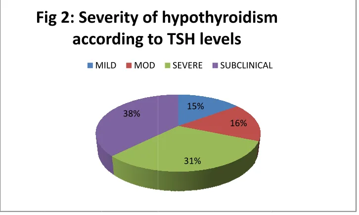
T TSH Sub Mild Mod Sev A patients 12 (15 hypothy (31%).T among t group h TABLE: 2 H mIU/l bclinical d (0.5-20) derate (20-5 ere( >50) Among the s were maj
%) patien yroidism. The mean
the modera had a mean
Fi
2 SEVER
No 30 12 0) 13 25
e 80 patie ority with nts had m
Severe hy TSH in t ate hypoth n TSH of 10
ig
2:
Sev
acco
M
RITY OF HY L
o of patients
ents inclu 30 (38%) mild hypo ypothyroid the mild h hyroid grou
07.58 mIU
38%
verity
of
ording
to
MILD MOD YPOTHYRO LEVELS s Percenta 38 15 16 31
ded in ou in number othyroidism d constitut
hypothyroi up was 33. U /L.
15%
31%
hypothy
o
TSH
lev
SEVERE
OIDISM ACC
age % M
18 11
33 10
ur study, r. Among t m while
ted the re id group w 43 mIU/L. 16%
yroidism
vels
SUBCLINICAL CORDING Tean S
8.75 8 .32 3
.43 10
07.58 40
subclinica the hypoth 13(16%) est with 2
was 11.32 . The sever
m
L
TO TSH
TABLE: 3 METABOLIC PARAMETERS IN HYPOTHYROIDISM
3A. BMI (BODY MASS INDEX)
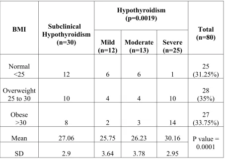
The mean BMI in the patients with subclinical hypothyroid was 27.06, while among the mildly hypothyroid it was 25.75. the moderately hypothyroid patients had a mean BMI of 26.23 and the severely hypothyroid was 30.16.The two-tailed P value by the one sample t test is 0.0001, considered extremely significant.
BMI Subclinical Hypothyroidism (n=30) Hypothyroidism (p=0.0019) Total (n=80) Mild (n=12) Moderate (n=13) Severe (n=25) Normal
<25 12 6 6 1
25 (31.25%) Overweight
25 to 30 10 4 4 10
28 (35%) Obese
>30 8 2 3 14
27 (33.75%) Mean 27.06 25.75 26.23 30.16 P value = 0.0001
3B. WAIST CICUMFERENCE
Waist (cm)
Subclinical
Hypothyroidism Hypothyroidism
Mild Moderate Severe
Female P= 0.0972
<88 19(63.33%) 8(66.67%) 7(53.84%) 8(32%) >88
11(36.67%) 4(33.33%) 6(46.15%) 17(68%)
Mean 83.06 84.91 87.46 89.48
SD 13.44 5.91 6.47 6.00
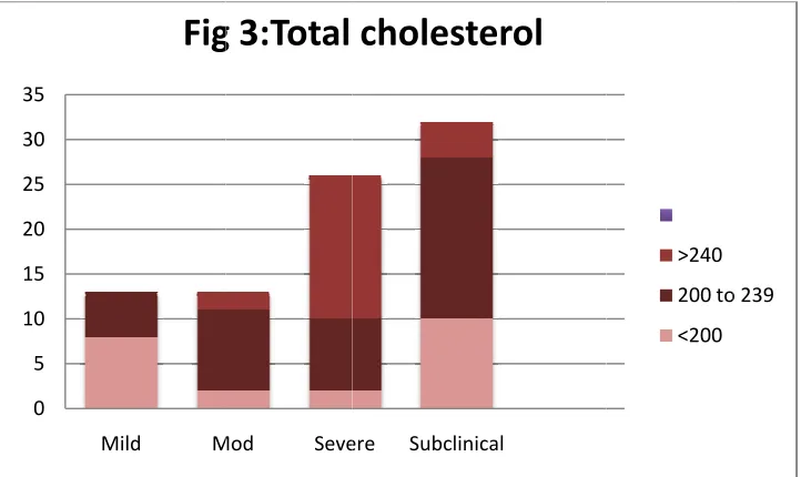
B T while th hypothy mean of statistic Total Cho (mg/ Normal Borderline - Elevated Mea SD
The Mean T he mean fo yroid had a
f 217.6mg/ cally signif 0 5 10 15 20 25 30 35 3C olesterol /dl) - <200
200 to 239
d- >240
an
D
Total Chol for modera
a mean of 2 /dl.The tw ficant. Mild M
Fig
C .TOTAL Sub Hypot 2 2 lesterol for ate hypothy 253.32 wh o-tailed P Mod Seveg
3:Total
L CHOLE bclinical hyroidism 10 18 4 217.6 29.93
r mild hyp yroid patie hile the sub value by o
ere Subclini
cholest
STEROL H Mild 8 5 0 203.16 15.52 pothyroid p ents is 220 bclinical hy one sample calerol
Hypothyroi Moderat 2 9 2 220.76 30.42 patients is 0.76 mg/dl ypothyroid e t test is 0
>240
200 to <200 idism te Sever 2 8 16 253.3 39.63 203.16 mg . The seve d patients h .0002 whic
o 239
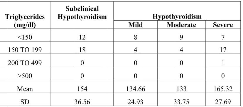
[image:50.612.127.488.481.696.2]T while t hypoth a mean which T The Mean the mean hyroid had n of 154 mg
is statistica 0 5 10 15 20 25 30 35 Triglyceride (mg/dl) <150 150 TO 199
200 TO 499
>500
Mean
SD
Triglyceri for moder a mean of g/dl. The P ally signifi
Fig
3
es HypotSub
9
9
3D.TRIG
ides for m rate hypot f 165.32 w P value by
icant.
3:Triglyc
bclinical thyroidism 12 18 0 0 154 36.56 GLYCER mild hypot thyroid pa while the suChi-squar
ceride
Le
Mild 8 4 0 0 134.6 24.93 RIDES thyroid pa atients is 1 ubclinical h
ed Test for
evels
Hypoth
d Mo
6
3 3
atients is 1 133 mg/dl.
hypothyroi r Independ
200 TO 499 150 TO 199 <150 yroidism oderate 9 4 0 0 133 33.75 134.66 mg . The seve id patients dence is 0.0
[image:51.612.102.516.122.304.2]L prevalen blocks a and 5 %
Abn Sinu BBB Rhy Low Low voltag nce. Next and rhythm %( 4) respec
0 5 10 15 20 25 sin T normalitie us Bradyca B ythm chang w voltage ge complex was Sinu m changes ctively.
[image:52.612.125.490.65.513.2]us bradycardia
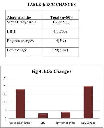
TABLE 4:
es
ardia
ges
xes was th us bradyca in the form
a BBB
Fig
4:
E
ECG CHA Total (n 18(22. 3(3.75 4(5% 20(25 he common ardia foun m of atrial
Rhyth
ECG
Chan
ANGES n=80) .5%) 5%) %) 5%) nest ECG nd in 22.5
fibrillation
hm changes
nges
finding w % (18). B n was foun
Low voltage
with 25 %( Bundle bra nd in 3.75%
e
TABLE:5
MYOCARDIAL
WALLTHICKNESSES
(
MM)
IN
HYPOTHYROID
PATIENTS.
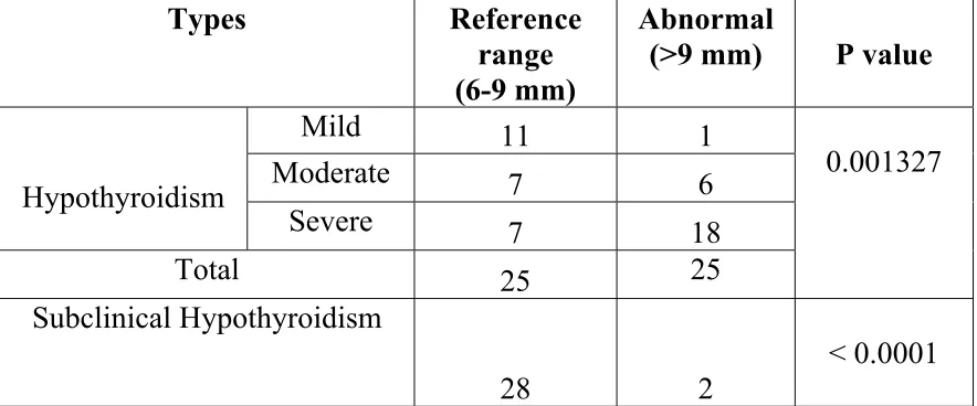
5A. LVPW THICKNESS
Types Reference range
(6-9 mm)
Abnormal
(>9 mm) P value
Hypothyroidism
Mild 11 1
0.001327
Moderate 7 6
Severe 7 18
Total 25 25
Subclinical Hypothyroidism
28 2
< 0.0001
5B.IVS THICKNESS
Types Reference range
(6-9 mm)
Abnormal
(>9 mm) P value
Hypothyroidism Mild 11 1 0.0007 Moderate
4 9 Severe
7 18 Total
22 28 Subclinical Hypothyroidism
25 5
0.0009
Among the mildly hypothyroid patients abnormal septal wall thickness was found in 1 (8.3%) and in the moderately hypothyroid it was noted in 9 (69.23%).18 (72%) out of the severely hypothyroid patients had increased septal wall thickness, while this finding was noted in only 5(16.66%) of the subclinical hypothyroid patients. On comparing the occurrence of increased septal wall thickness with increasing severity of disease by the Chi-squared Test for Independence the P value is 0.0007and on comparing the hypothyroid with the subclinical hypothyroid patients by the Fisher's Exact Test the two-sided P value is 0.0009 with Relative risk = 0.5280.
0 2 4 6 8 10 12 14
0 50 100 150 200
IV Septal wall thickness TSH
Fig
5:
IV
Septal
wall
thickness
Echo‐septal wall
thickness (mm) Linear (Echo‐septal
wall thickness (mm))
0 2 4 6 8 10 12 14
0 50 100 150 200
LV Posterior wall thickness (mm) TSH
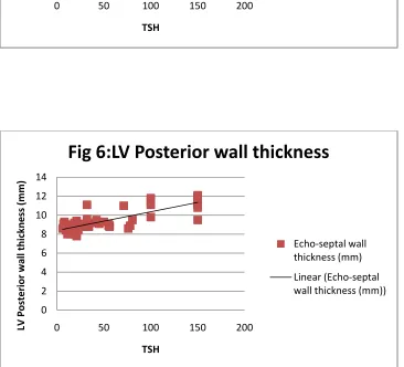
Fig
6:LV
Posterior
wall
thickness
Echo‐septal wall
thickness (mm) Linear (Echo‐septal
[image:55.612.124.488.102.321.2] [image:55.612.126.492.276.608.2]TABLE:6CARDIAC CHAMBER SIZE (CM) IN HYPOTHYROID PATIENTS
LVID(D) cm
(p=0.6250)
Subclinical Hypothyroidism
Hypothyroidism
Mild Moderate Severe
<5.4 12 13 24 30
>5.3 0 0 1 0
[image:56.612.92.518.128.272.2]TABLE
:7
LV
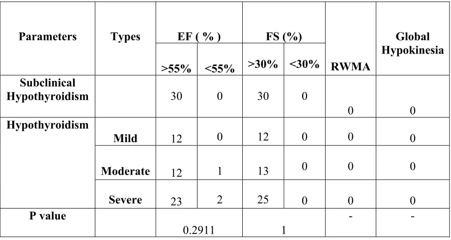
SYSTOLIC FUNCTIONS IN HYPOTHYROID PATIENTS
Parameters Types EF ( % ) FS (%)
RWMA
Global Hypokinesia >55% <55% >30% <30%
Subclinical
Hypothyroidism 30 0 30 0
0 0
Hypothyroidism
Mild 12 0 12 0 0 0
Moderate 12 1 13 0 0 0
Severe 23 2 25 0 0 0
P value
0.2911 1
- -
[image:57.612.78.537.127.371.2]
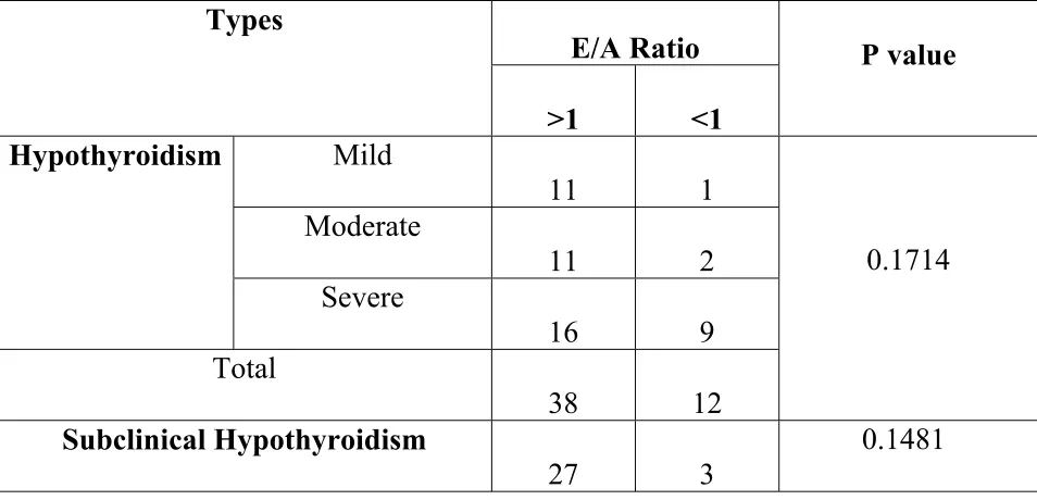
TABLE 8: DIASTOLIC DYSFUNCTION IN HYPOTHYROIDISM
Diastolic dysfunction was found in 1(8.33%) patient in the mild hypothyroid group and in 2 (15.38%) of the moderately hypothyroid group.9 (36%) patients of the severely hypothyroid group had diastolic dysfunction. In the subclinical hypothyroid group the same was noted in 3(10%) patients.
Types
E/A Ratio P value
>1 <1
Hypothyroidism Mild
11 1
0.1714 Moderate
11 2 Severe
16 9 Total
38 12
Subclinical Hypothyroidism
27 3
TABLE: 9 ANALYSES OF THE MEANS OF ECHO
PARAMETERS
Mean
values
Subclinical Hypothyroidism
Hypothyroidism
P value Mild Moderate Severe
LVPW(mm) 8.49 8.74 9.23 10.54 < 0.0001 IVSW(mm) 8.74 8.75 9.20 10.42 < 0.0001 LVID(D)cm 4.44 4.51 4.41 4.58 0.4542
EF % 62 63.08 61.61 61.6 0.8967 FS % 33.86 34.08 34.16 34.44 0.8260
E/A 1.50 1.69 1.62 1.25 0.0098
TABLE
10:
PERICARDIAL
INVOLVEMENT IN HYPOTHYROID PATIENTSPathology Subclinical
Hypothyroidism
Hypothyroidism
Mild Moderate Severe
Mild PE 1 1 2 2
Mod PE 0 0 1 6
Large PE 0 0 1 2
Pericardial
thickening 0 0 0 0
Constrictive
physiology 0 0 0 0
Total 1 1 4 10
[image:60.612.67.537.151.397.2]T
ABLE11:
AGE
AND
CHAMBER
THICKNESS
Age MEAN LVPW
(MM)
MEAN IVSW
(MM)
20-29 9.21+/-1.1 9.15+/-0.88
30-39 9.56+/-1.29 9.69+/-1.2
40-49 8.4+/-0.63 8.4+/-0.63
50-59 9.81+/-1.59 9.54+/-1.3
P VALUE <0.0001 <0.0001
There was a trend of increasing wall thickness with age in the patients.
7.5 8 8.5 9 9.5 10
20‐29 30‐39 40‐49 50‐59
Fig
7:Age
and
chamber
thickness
LVPW
[image:61.612.155.437.133.359.2] [image:61.612.72.508.448.699.2]TABLE
12:
MULTIPLE REGRESSION ANALYSIS WITH DEPENDENT VARIABLE IV SEPTAL WALL THICKNESSAND INCLUDING AGE AND SEVERITY OFHYPOTHYROIDISM
VARIABLE COEFFICIENT SE 95%
CONFIDENCE
INTERVAL
T RATIO P VALUE
CONSTANT 3.695 0.7572 2.170 TO 5.221 19.220 <0.0001
AGE -0.002064 0.008946 -0.02009 TO
0.01596
0.5324 0.8186 TSH 0.008883 0.002195 0.004461 TO
0.01331
9.970 0.0002
R squared 0.8387
Adjusted R squared 0.8281
A multiple regression analysis was done with the dependent variable as IV
[image:62.612.64.551.145.309.2]TABLE
13:
M
ULTIPLER
EGRESSIONA
NALYSIS WITHD
EPENDENTV
ARIABLELVPW
THICKNESS
AND
INCLUDING AGE AND SEVERITY OF HYPOTHYROIDISMVARIABLE COEFFICIENT SE 95%CONFIDENCE
INTERVAL
T RATIO P VALUE
CONSTANT 7.995 0.5247 6.938 TO 9.051 15.236 <0.0001
AGE 0.01518 0.01494 -0.01491 TO 0.04527 1.016 0.3149 TSH 0.01919 0.002430 0.01430 TO 0.02408 7.898 <0.0001
R squared 0.5930 Adjusted R squared 0.5756
[image:63.612.69.552.183.380.2]DISCUSSION
The cardiac complications of long standing hypothyroidism are serious if are not diagnosed properly earlier. Echocardiography as a non-invasive method can play important role in recognizing the cardiac pathology as well as to follow up effect of the therapy.
In this study of Indian population, we evaluated the cardiovascular function in newly detected primary overt and subclinical hypothyroidism. 80 patients were included in our study of which 37% were in the age group of 21 to 30. Majority of the patients were in the age group of 31 to 40 years of age with 40% prevalence. 13% patients were in the age group of 41 to 50.About 10% patients were in the age of 51 to 60.The mean age was 35.21 +/- 9.74 Yrs.
Among the 80 patients included in our study, subclinical hypothyroid patients were majority with 38% in number. Among the hypothyroid patients, 15% patients had mild hypothyroidism while 16% had moderate hypothyroidism. Severe hypothyroid constituted the rest with 31%.
Metabolic parameters in Hypothyroidism
hypothyroid was 30.16. BMI was significantly associated with the severity of the disease. Among our study population 36.66% of the subclinical hypothyroid patients were found to have increased waistline, while 33.33% of the mild hypothyroid group had increased waistline. 46.15% of the moderately hypothyroid patients and 68% were found to have increased waistline.
This is similar to the observations by Brunilda Figueroa et al (56) who in their study found that the mean TSH level was 2.2 mU/L for women and 1.9 mU/L for men. Mean BMI was 32.8 kg/m 2 for women and 35.9 kg/m for men. Females had a mean fat percent of 38.1% and males had 31.4% in their study.
However Manji et al (57) found no association between serum TSH and free T3 concentrations within the normal range and BMI. In contrast, Knudsen et al (58) found that thyroid function could be one of several factors that act in concert to determine body weight.
alterations, might explain the increased risk of coronary artery disease, cerebral ischemia risk, and angina pectoris in older patients.
In our study the Mean Total Cholesterol for mild hypothyroid patients is 203.16 mg /dl while the mean for moderate hypothyroid patients is 220.76 mg/dl. The severely hypothyroid had a mean of 253.32 while the subclinical hypothyroid patients had a mean of 217.6mg/dl. The Mean Triglycerides for mild hypothyroid patients is 134.66 mg /dl while the mean for moderate hypothyroid patients is 133 mg/dl. The severely hypothyroid had a mean of 165.32 while the subclinical hypothyroid patients had a mean of 154 mg/dl. Both these lipid parameters were found to be statistically significantly associated with the severity of hypothyroidism.
ECG Changes
In our study low voltage complex was the commonest ECG finding with 25 % prevalence. Sinus bradycardia was found in 22.5%. Bundle branch blocks and rhythm changes in the form of atrial fibrillation was found in 3.75% and 5 % respectively. This was in accordance with the known ECG changes which are expected to occur in hypothyroidism. Prolongation of the QT interval, decreased P-wave voltage, prolonged AV conduction time, intraventricular conduction disturbances, and nonspecific ST-T-wave abnormalities were not observed in our study population.
Myocardial wall thickness in hypothyroidism
Alvani D. Santos et al (60) first reported in 1979 a reversible cardiomyopathy, manifested by asymmetric septal hypertrophy in untreated hypothyroid patients.
1 among the mildly hypothyroid patients had abnormal septal wall thickness and in the moderately hypothyroid it was noted in 9 patients.18 of the severely hypothyroid patients had increased septal wall thickness, while this finding was noted in only 5 of the subclinical hypothyroid patients. On comparing the occurrence of increased septal wall thickness with increasing severity of disease it was found to be statistically significant.
Rawat and Satyal (61) in their study showed relatively increased thickness of IVS and LVPW (1.2/1.7 and 1.1/1.6 cm) when compared to the treated patients (0.9/1.4 and 0.9/1.3 cm) or control subjects (0.8/1.3 and 0.7/1.2 cm). But on age group analysis it was found that this difference was more marked in older patients. In our study however we found that although there was a trend of increasing wall thickness with age these changes on multiple regression analysis with the dependent variable as IVseptal wall thickness and LVPW thickness including age and TSH as variable, it was found that only TSH continued to be significant.
Cardiac chamber size in hypothyroidism
The Cardiac chamber size was found to have a statistically insignificant association with hypothyroidism. This shows that the cardiac chamber size is not affected by hypothyroidism. Similar observations were also made by Varma R et al
(63).
LV functions
The LV systolic functions as measured by ejection fraction and fractional shortening were not statistically associated with the severity of hypothyroidism. No wall motion abnormalities or global hypokinesia was detected. Only 2 (8%) of the severely hypothyroid group and 1(7.69%) in the moderately hypothyroid group had reduction of ejection fraction
In the study by Jagdish et al (62), although FS increased from 26.43 ± 2.79 to 26.73 ± 2.64 and EF showed increased from 53.93 ± 5.50 to 54.83 ± 4.64 , it was statistically not significant. Rawat et al (61) showed no significant change in parameters of systolic function while Monzani et al (64) found that FS and thus systolic function of LV significantly improved after treatment.
dysfunction.. Similar findings of diastolic dysfunction were made by Almira Hadžovic-Džuvo et al (65), and Rajan et al (66). Biondi B,et al (67) in their study of subclinical hypothyroid patients showed significant prolongation of the isovolumic relaxation time (94 +/- 13 vs. 84 +/- 8 msec; P < 0.001), increased A wave (55 +/- 13 vs. 48 +/- 9 cm/sec; P < 0.05), and reduced early diastolic mitral flow velocity/late diastolic mitral flow velocity ratio (1.4 +/- 0.3 vs. 1.7 +/- 0.3; P < 0.001) indicating an early diastolic dysfunction. In our study diastolic dysfunction was noted in 3(10%) patients of the subclinical hypothyroid group which was insignificant.
Pericardial pathology in Hypothyroidism
LIMITATIONS
1. The study was done on a sample of patients in the outpatient department. This makes the results of the study less generalizable to the overall population of hypothyroid patients.
2. The sample size of 80 was relatively small to detect fine associations especially in the presence of multiple confounding variables.
3. The cross sectional nature of the study makes it possible that the conclusions made may be unstable, or that they may be reflective of a phenomenon particular to one phase of illness.
CONCLUSION
1. Increased interventricular septal and left ventricular posterior wall thicknesses with diastolic dysfunction are some of the earliest cardiac features of progressive thyroid failure.
2. The magnitude of the lipid changes and the subtle impairment of left ventricular diastolic function in subclinical hypothyroidism patients as shown in our study may justify use of hormone replacement even without overt cardiac symptoms.
3. An early diagnostic approach in patients with hypothyroidism will surely diminish the extent of cardiac complication which accompanies it.
BIBLIOGRAPHY
1. Kronenberg: Williams Textbook of Endocrinology, 11th ed. Henry M. Kronenberg, MD ,Shlomo Melmed, MD, FRCP,Kenneth S. Polonsky, MD,P. Reed Larsen, MD, FACP, FRCP
2. Review of medical physiology - 21st Ed. (2003). William F. Ganong, MD
3. Thyroid hormone and the cardiovascular system. Danzi S, Klein I, Minerva Endocrinologica [2004, 29(3):139-50]
4. Vanderpump MP, Tunbridge WM, French JM, et al. The incidence of thyroid disorders in the community: a twenty-year follow-up of the Wickham Survey. Clin Endocrinol (Oxf) 1995; 43:55–68
5. Clinical endocrinology in India, N. Kochupillai, CURRENT SCIENCE, VOL. 79, NO. 8, 25 OCTOBER 2000
6. 2. Jayarama, K. S., Nature, 1983, 304, 205
8. Cooper DS. Clinical practice: subclinical hypothyroidism.N Engl J Med. 2001; 345:260-265.
9. Cooper DS. Subclinical hypothyroidism. JAMA 1987; 258 :246-7
10. Etiological profile of overt hypothyroidism in Indian population. Jangid DR, Agarwal SK, Jangid V, Ram BK.(Nuclear Medicine Centre, Escorts Heart Institute and Research Centre, New Delhi)
11. Sawin CT, Castelli WP, Hershman JM, McNamara P, Bacharach P.The aging thyroid.Thyroid deficiency in the Framingham Study. Arch Intern Med. Aug 1985; 145(8):1386-8.
12. Loius J Acierno.The History of Cardiology.Volume 121 Issue 6 |Pgs 471-472.
13. Klein I, Ojamaa K 2000 The cardiovascular system in hypothyroidism. In: Braverman LE, Utiger RD, eds. Werner & Ingbar’s The Thyroid: A Fundamental and Clinical Text, edit. 8. Philadelphia: Lippincott Williams & Wilkins; 777–782
14. Fredlund BO, Olsson SB 1983 Long QT interval and ventricular tachycardia of “torsade de pointe” type in hypothyroidism. Acta Med Scand 213:231–235
15. Klein I: Thyroid hormone and the cardiovascular system. Am J Med 1990; 88:631-637
17. Polikar R, Kennedy B, Ziegler M, O'Connor T, Smith J, Nicod P: Plasma norepinephrine kinetics, dopamine-p3-hydroxylase, and chromogranin-A, in hypothyroid patients before and following replacement therapy. J Clin Endocrinol Metab 1990; 70:277-281
18. The thyroid and the heart, R Polikar, AG Burger, U Scherrer and P Nicod, Circulation 1993, 87:1435-1441
19. Obuobie K, Smith J, Evans LM, et al. 2002 Increased central arterial stiffness in hypothyroidism.J Clin Endocrinol Metab 87:4662– 4666
20. Crowley WF Jr, Ridgway EC, Bough EW, et al. 1977 Noninvasive evaluation of cardiac function in hypothyroidism. Response to gradual thyroxine replacement Engl J Med 296:1– 6
21. Dernellis J, Panaretou M 2002 Effects of thyroid replacement therapy on arterial blood pressure in patients with hypertension and hypothyroidism. Am Heart J 143:718 –724
22. Hypothyroidism as a cause of hypertension, I Saito, K Ito and T Saruta, Hypertension 1983, 5:112-115
24 Left Ventricular Diastolic Dysfunction in Patients with Subclinical Hypothyroidism Bernadette Biondi, Serafino Fazio, Emiliano Antonio Palmieri, Carlo Carella, Nicola Panza, Antonio Cittadini, Filomena Bone`Gaetano Lombardi, Luigi Sacca,. The Journal of Clinical Endocrinology & Metabolism, Vol. 84, No. 6
25. Sreinberg AD. t\1yxedema and coronary artery disease: acomparat ive autopsy slUdv. An" bl l em Med 1968; 68: 338-344.
26. Pericardial effusion in hypothyroidism. Hardisty CA, Naik DR, Munro DS., Clin Endocrinol (Oxf). 1980 Oct;13(4):349-54
27. Santos AD, Miller RP, Mathew PK, Wallace WA, Cave WT, Hinojosa L: Echocardiographic characterization of the reversible cardiomyopathy of hypothyroidism. Am J Med 1980; 68:675-682.
28. Keating FR, Parkin TW, Selby JB, et al. 1960 Treatment of heart disease associated with myxedema. Prog Cardiovasc Dis 3:364 –381
30. Shenfield GM. Influence of thyroid dys func t ion on drug pharmacokinetics. Cli" Pharlllacaki"er J981; 6: 275-297.
31. Mac Kerrow SD, Osborn LA, Levy H, Eaton RP, Economou P: Myxedema-associated cardiogenic shock treated with intravenous triiodothyronine. Ann Intern Med 1992; 117:1014-1015
32. Wieshammer S, Keck FS, Waitzinger J, Henze E, Loos U, Hombach V, Pfeiffer EF: Acute hypothyroidism slows the rate of left ventricular diastolic relaxation. Can J Physiol Pharmacol 1989; 67: 1007-101
33. Graettinger JS, Muenster JJ, Checchia CS, Grissom RL, Campbell JA: A correlation of clinical and hemodynamic studies in patients with hypothyroidism. J Clin Invest 1958; 502-510
34. Vora J, O'Malley BP, Petersen S, et al: Reversible abnormalities of myocardial relaxation in hypothyroidism. J Clin Endocrinol Metab 1985; 61:269-272
35. Zimmerman J, Yahalom J, Bar-On H: Clinical spectrum of pericardial effusion as the presenting feature of hypothyroidism. Am Heart J 1983; 106:770-771
37. O'Brien T, Katz K, Hodge D, et al: The effect of the treatment of
hypothyroidism and hyperthyroidism on plasma lipids and apolipoproteins AI, AII, and E. Clin Endocrinol (Oxf) 1997; 46:17-20.
38. Lipid profile in different degrees of hypothyroidism and effects of levothyroxine replacement in mild thyroid failure Patrícia De Fátima Dos Santos Teixeira, Vaneska Spinelli Reuters , Márcia ,artins Ferreira, Cloyra Paiva Almeida ,Fabíola Alves Aarão Reis, Alexandru Buescu, Antonio José Leal Costa, Mário Vaisman Translational ResearchVolume 151, Issue 4 , Pages 224-231, April 2008
39 Subclinical hypothyroidism and lipid abnormalities in older women attending a vascular disease prevention clinic: effect of thyroid replacement therapy. Ganotakis ES, Mandalaki K, Tampakaki M, Malliaraki N, Mandalakis E, Vrentzos G, Melissas J, Castanas E., Angiology. 2003 Sep-Oct;54(5):569-76.
40. Canaris GJ, Manowitz NR, Mayor G, et al. 2000 The Colorado thyroid disease prevalence study. Arch Intern Med 160:526 –534
42. Vanderpump MP, Tunbridge WM, French JM, et al. 1995 The incidence of thyroid disease in the community: a twenty-year follow-up of the Whickham Survey. Clin Endocrinol 43:55– 69
43. Huber G, Staub JJ, Meier C, et al. 2002 Prospective study of the spontaneous course of subclinical hypothyroidism: prognostic value of thyrotropin, thyroid reserve, and thyroid antibodies J Clin Endocrinol Metab 87:3221–3226
44. Monzani F, Di Bello V, Caraccio N, et al: Effect of levothyroxine on cardiac function and structure in subclinical hypothyroidism: A double blind, placebo-controlled dtudy. J Clin Endocrinol etab 2001; 86:1110
45. Duntas LH 2002 Thyroid disease and lipids. Thyroid 12:287
46.Duntas LH, Mantzou E, Koutras DA 2002 Circulating levels of oxidized low-density lipoproteinin overt and mild hypothyroidism. Thyroid 12:1003–1007
47. Lekakis J, Papamichael C, Alevizaki M, et al. 1997 Flow-mediated, endothelium-dependent vasodilatation is impaired in subjects with hypothyroidism, borderline hypothyroidism, and high-normal serum thyrotropin (TSH) values. Thyroid 7:411– 414
49. Di Bello V, Monzani F, Giorgi D, et al. 2002 Ultrasonic myocardial textural analysis in subclinical hypothyroidism. J Am Soc Echocardiogr 13:832– 840
50. Monzani F, Di Bello V, Caraccio N, et al. 2001 Effects of levo-thyroxine on cardiac function and structure in subclinical hypothyroidism: a double blind, placebo-controlled study. J Clin Endocrinol Metab 86:1110 –1115
51. Vitale G, Galderisi M, Lupoli GA, et al. 2002 Left ventricular myocardial impairment in subclinical hypothyroidism assessed by a new ultrasound tool: pulsed tissue Doppler. J Clin Endocrinol Metab 87:4350 – 4355
52. Kahaly GJ 2000 Cardiovascular and atherogenic aspects of subclinical hypothyroidism. Thyroid 10:665– 679
53.. Treatment Guidelines for Patients With Hyperthyroidism and Hypothyroidism Peter A. Singer, MD; David S. Cooper, MD; Elliot G. Levy, MD; Paul W. Ladenson, MD; Lewis E. Braverman, MD; Gilbert Daniels, MD; Francis S. Greenspan, MD; I. Ross McDougall, MB, ChB, PhD; Thomas F. Nikolai, MD
54. Harrison's™ PRINCIPLES OF INTERNAL MEDICINE, Eighteenth Edition
European Association of Echocardiography, a Branch of the European Society of Cardiology Members of the Chamber Quantification Writing Group are: Roberto M. Lang, MD, FASE, Michelle Bierig, MPH, RDCS, FASE, Richard B. Devereux, MD, Frank A. Flachskampf, MD, Elyse Foster, MD, Patricia A. Pellikka, MD, Michael H. Picard, MD, Mary J. Roman, MD, James Seward, MD, Jack S. Shanewise, MD, FASE, Scott D. Solomon, MD, Kirk T. Spencer, MD, FASE, Martin St John Sutton, MD, FASE, and William J. Stewart, MD, Journal of the American Society of Echocardiography Volume 18 Number 12,1447
56. Asociation of thyroid stimulating hormone levela and bodymass index in overweight hispanicsin Puerto Rico Brunilda Figueroa, MS; Himilce Ve´lez, MS; Margarita Irizarry-Ramı´rez, PhD.
57. Manji N, Boelaert K, Sheppard MC, HoldertRL, Gough SC, Franklyn JA. Lack of association between serum TSH or free T4 and body mass index in euthyroid subjects. Clin Endocrinol. 2006;64(2):125–128
58. Knudsen N, Laurberg P, Rasmussen LB, et al.Small differences in thyroid function may be important for body mass index and the occurrence of obesity in the population. J Clin Endocrinol Metab. 2005; 90:4019–4024.
Endocrinology/Metabolism, Mayo Clinic, Rochester, Minnesota 55905. Mayo Clinic Proceedings. Mayo Clinic [1993, 68(9):860-6]
60. Echocardiographic characterization of the reversible cardiomyopathy of hypothyroidism Alvani D. Santos M.D, R.Paul Miller M.D., Puthenpurakal K. Mathew M.D., Wayne A. Wallace M.D., William T. Cave Jr M.D. and Louis Hinojosa M.D
61.An echocardiographic study of cardiac changes in hypothyroidism and the response to treatment Rawat B and Satyal A. Kathmandu University Medical Journal (2003) Vol. 2, No. 3, Issue 7, 182- 187.
62.An Echocardiographic Study on the Effect of Levothyroxine Therapy on Cardiac Function and Structure in Hypothyroidism Jagdish, H Singh, A Batra, SB Siwach, R Kumar, R Gupta JIACM 2009; 10(1 & 2): 27-31
63. Heart in hypothyroidism--an echocardiographic study. Varma R, Jain AK, Ghose T . J Assoc Physicians India. 1996 Aug;44(8):546, 551-3
65. Echocardigraphic evaluation of cardiac function in female patients with thyroid disorders. Almira Hadžovic-Džuvo1*, Elma Kucukalic - Selimovic2, Emina Nakaš-Icindic1, Senija Rašic3, Amela Begic2 , Dinan Al Tawil4, Amina Valjevac1, Nesina Avdagic1, Orhan Lepara Bosnian journal of basic medical sciences 2010; 10 (2): 112-115
66. Rajan SK, Srinivasan R, Mohan J. Evaluation of the heart in hypothyroid patients: An echocardiographic study. Ind Pract 2003; 56 (12): 815-8.
PROFORMA
Name: Age: Sex:
Address: Presentation: First/ Refferal Occupation:
Op\IP No: Contact no:
SYMPTOMS: Duration
Asymptomatic Fatigue /Irritability Neck swelling Depression / Anxiety Skin changes Weight gain /loss
Cold /Heat intolerance Sleepiness/ Insomnia
Hair loss Myalgia/arthralgia Muscle cramps /weakness Hard of hearing /Hoarseness of voice Constipation /Loose stools/other bowel symptoms
Breathlessness /Chestpain/ Palpitation /Syncope PAST HISTORY:
CAD Dyslipidemia DM Hypertension TB Thyroidectomy Any other relevant past history
PERSONAL HISTORY:
Alcohol Drug abuse Smoking MARITAL STATUS: Married/unmarried
MENSTRUAL HISTORY: Amenorrhea / oligomenorrhea /Menorrhagia /Menopause
OBSTETRIC HISTORY: Secondary /primary Infertility
TREATMENT HISTORY: H/oDrugs (Thyroxin / Lithium /Amiadarone /antithyroid
GENERAL EXAMINATION:
Built Nourishment
Height: Weight: BMI Pallor Icterus Cyanosis Clubbing
Lymphadenopathy Edema
Pulse rate: BP: RR: Temp:
Peripheral pulses
Facies -Anxious/dull/ normal
Eyes-Periorbital Puffiness /protruding/normal Skin -Dry Coarse/wet
Loss of Hair THYROID:
Diffuse/Nodular/Thyroidectomy scar CVS
RESPIRATORY SYSTEM ALIMENTARY SYSTEM MUSCULO SKELETAL NERVOUS SYSTEM INVESTIGATIONS TFT- T3 T4, fT4 TSH
FLP- T. CHOLESTEROL, LDL, HDL, TG
ECHOCARDIOGRAPHY
Septal wall thickness LV Posterior wall thickness Fractional shortening Ejection fraction Diastolic dysfunction (E/A ratio) LVID