JOURNALOFVIROLOGY, Nov. 1993,P.6866-6871 0022-538X/93/116866-06$02.00/0
Copyright
©D
1993, American SocietyforMicrobiologySimian Virus 40
T
Antigen
as a
Carrier for the
Expression
of
Cytotoxic
T-Lymphocyte Recognition Epitopes
TONG-MING FU, ROBERTH. BONNEAU, MARY J.TEVETHIA, AND SATVIRS.TEVETHIA* DepartmentofMicrobiologyandImmunology, ThePennsylvania State University College ofMedicine,
Hershey, Pennsylvania 17033 Received 19May 1993/Accepted 11 August 1993
Simian virus 40 (SV40) largeTantigencan immortalizeawidevariety of mammalian cellsin culture.We have taken advantage of this property of T antigen to use it as a carrier for the expression of cytotoxic T-lymphocyte (CTL) recognition epitopes. DNA sequences corresponding to an
H-2D&-restricted
SV40 T-antigen site I (amino acids 205 to215) weretranslocated into SV40T-antigenDNA at codonpositions 350 and 650 containing EcoRI linkers.AnH-2Kb-restricted herpes simplex virus glycoprotein B epitope (amino acids 498 to505) was also expressed in SV40 Tantigenat positions350 and 650. PrimaryC57BL/6 mouse kidney cells wereimmortalized by transfection with the recombinant andwild-type T-antigen DNA.Clonal isolates of cellsexpressing chimericTantigenswereshown to bespecifically susceptibletolysis byCTLclones directed to SV40 T-antigen site I and herpes simplex virus glycoprotein B epitopes, indicating that CTL epitopes restricted bytwodifferent elementscanbeprocessed, presented,andrecognized bytheepitope-specific
CTLclones. Our resultssuggest thatSV40Tantigencanbe usedas acarrierproteinto expressawidevariety of CTLepitopes.CD8+ cytotoxicTlymphocytes (CTL)arecrucial in
control-ling viral infections (14, 57). CTLs recognize viralantigens in the form ofpeptide fragmentsof 8to10 amino acids whichare bound in the grooves of major histocompatibility complex (MHC) class I molecules (5, 20, 33, 49, 51). These antigenic peptides are derived from nonstructural and structural viral
proteins through proteolysisinthe cytosoliccompartment(15, 46, 56)andaretranslocated into theendoplasmicreticulumby TAP1/TAP2 transporters (2, 31, 36, 37, 50, 52), where the peptide, MHCclass I molecule,and
P-2
microglobulin formatrimolecular complex (48). This complexis then exported to the cell surface through the exocytic pathway (9, 32, 55) for
recognition by
CD8+
CTL via the T-cell receptor(24, 25).The identification of viral CTL epitopes thatare presented by MHC class I molecules and recognized by the epitope-specific CTLsuggestsastrategy fordesigning effective
immu-nogensfor theinduction ofaCD8+ T-cell immuneresponseto
provide protection against viral infection (14, 28, 29, 47, 49).
Oneapproachtogenerating suchanimmuneresponseutilizes immunization with synthetic peptides corresponding to the CTLepitopes eitherbythemselvesorwithadjuvants (1, 7, 13, 26, 35, 39). Another approach has been to express CTL
epitopesinvirusvectorssuchasvaccinia virus (3, 4), eitheras
chimeric proteins (12)or asminigeneproducts(21, 38, 53, 54).
These chimeric proteins or minigene products have been demonstrated to induce CTL responses and, in most cases,
provide invivoprotection (12, 28, 54).
To utilize CTL epitopes as immunogens, it is important to experimentally assess their immunogenicpotential by using a
vector system most likely to enhance the immunogenicity of theCTLepitopes.Inthis study,weexploredanapproachusing
simianvirus 40 (SV40) large tumorantigen (T antigen) as a
carrier proteinto expressCTL epitopes. Tantigen,a 94-kDa
nuclear protein, immortalizes cells in culture (19, 27, 30, 42) and is highly immunogenic, containing multiple B-cell and T-cell epitopes (43). SV40 T antigen contains at least one
*Correspondingauthor.
H-2K"-restrictedepitope (site IV) and threeH-2D*-restricted
CTLepitopes (sites I, II/III, and V). Sites I (positions 207 to
215), lI/IlI (223to231),and V(489to497)arerecognized by
CTL clones Y-1, Y-2orY-3,andY-5, respectively (10, 40, 41).
Site IV (404 to411) isH-2K*-restricted and isrecognized by
CTL clone Y-4 (43a). Cells immortalized by T antigen can grow continuously in culture and express high levels of T
antigen. Immunization of mice with cellsexpressingTantigen results in the generation ofboth primary and memory CTL
responses (43).
Recently,alibraryof Tantigensinwhich EcoRI linkerswere inserted into the coding region of T antigen at 50-codon intervalsbetween codons 250 and 650wasgenerated (27). Two locations in the T antigen (residues 350 and 650) were
considered ideal sites for inserting CTLepitopes, since these sites in the protein are not required for immortalization of primary cellsinculture (45). We havetranslocated anH-2Db_ restrictedT-antigen epitope, site I, from its original position
between residues 205 and 215toamino acid positions350and
650 of T antigen. In addition, an H-2K"-restricted herpes simplexvirus(HSV)type 1 glycoprotein B (gB) CTL epitope, residues 498to505 (6),wascloned into the samelocationsof
T antigen (positions350 and 650). The results show that the translocated SV40 T antigen site I and the HSV gB CTL
epitopes inthe chimeric Tantigens,when expressedin immor-talized C57BL/6 (B6) mouse kidney cells, were efficiently processed andwere presented by the MHC class I molecules
andrecognized bytheepitope-specific CTL clones.
The chimeric T antigens were generated by using two T-antigen constructs(27) whichcontain anEcoRI site
imme-diatelyfollowing codons 350and 650ofTantigen,pPvu0:E350 andpPvu0:E650, respectively.Oligonucleotidepairs represent-ing twostrands ofthe SV40T-antigen site I (residues 205 to 215) or the HSV gB (residues 498 to 505) CTL epitope
sequence were designed such that when annealed, the small
double-strandedfragment would have exposedateach end the
sequence 5'-AATT. The annealed oligonucleotides were
li-gated with the EcoRI-linearized plasmids pPvu0:E350 and
pPvu0:E650 by using T4 ligase (Fig. 1A), and the ligated 6866
Vol. 67,No.11
on November 9, 2019 by guest
http://jvi.asm.org/
VOL.67, 1993
SAArITCGATCGAGlrJxxCCzxC1Gor 3
.
t,-
..,
N'-o,o s,\
DT agsiteI
650 708
u. ATagsiteII/1II
~0TagsiteV 0HSVgB epitope
VA
1498 0
HSV gBepitopeat350 issLt
350gBB
650gB 0 _
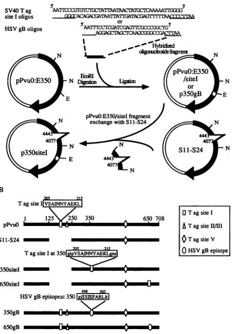
FIG. 1. Cloning strategy and resultant T-antigen constructs. (A) Twopairsofoligonucleotides representing SV40 T-antigen (Tag) site I(residues205to215)orHSVgBepitope (residues 498to505) with EcoRI-compatible endsweresynthesized andhybridized. The EcoRI-compatiblesequencesandadjacent nucleotides addedtomaintain the
correctreadingframeareunderlined.Theoligonucleotide fragments
wereinsertedattheEcoRI site ofpPvuO:E350. Toremovethe original
site I sequencewithin theT-antigenconstructwhichcontains the site Isequenceatamino acidposition 350, the NdeI fragment within the
constructwasreplacedwiththecorresponding fragmentfromS1 1-S24
T-antigen mutant,whichhasa deletion ofnucleotides 4443 to4077 including the site I sequence. The final constructs, p350site I and p350gB, are indicated. Constructs p650site Iand p650gB were
con-structed inasimilarmanner.N, recognitionsite for the endonuclease NdeI;E, recognition sitefor EcoRI.(B)Constructs used in thisstudy andpositionsof the CTLepitopes.Theamino acidsequencesof the CTLepitopesarelisted insingle-lettercode.Uppercaselettersreferto the CTLepitope amino acidsequences, andlowercase letters repre-senttheflankingamino acids encodedbytheEcoRI linkersequence.
products were transfected into competent Escherichia coli DH5o. The recombinant plasmid DNA was purified by the alkalinelysis procedureandanalyzed byrestriction
endonucle-asedigestion (34).The restrictionendonuclease usedtoscreen
recombinant T-antigen constructs containing thesite I insert
was AsnI, which recognizes an ATTAAT sequence located within the site Iepitopesequence,and therestriction endonu-clease usedtoscreenrecombinantscontainingthegB epitope
insertwasEcoRI,because theoligonucleotidesweredesigned
such that the EcoRI site would be destroyed upon the inser-tion. The orientation and authenticity of the inserts were
[antlligenes
FIG. 2. Levels of T-antigen expression in immortalized celllines.
Thesameamountofproteinfrom each celllysatewasfirst
immuno-precipitated with PAb901 and then blottedontonitrocellulose
mem-brane andprobed with PAb901. Bindingof PAb9O1 toT antigenwas
detectedby reacting theblot withgoatanti-mouseimmunoglobulinG conjugatedwithhorseradishperoxidaseandthensubjecting the blotto
enhanced chemiluminescence treatment (27). B6/K-pPvuO cells ex-press awild-type T antigen of about 94kDa, whereasB6/K-S11-S24 cellsexpressa mutantTantigen with amolecularmass of about67
kDaas aresultofdeletionof 124aminoacids.
determined and confirmed byDNA sequencing as described
previously (27). In constructing recombinant T antigens
con-taining
a relocatedsite I epitope, the originalsite I sequence wasremovedby fragment exchangewithaconstructcontaining themutantTantigen, S11-S24. S11-S24producesaTantigen which contains an in-framedeletion from amino acids 127to 250. Therefore, theS11-S24Tantigen lacks the CTL recogni-tionepitopes site1(205to215)andsiteII/III(223to231)(41).A fragment containing the deletion was released from the S11-S24constructby digestion with NdeI andusedto replace thecorrespondingfragment of theconstructsthathavethesite
Isequenceinsertedatpositions 350 and 650 ofTantigen
(Fig.
IA).
Expression of chimeric T antigens in
primary
B6 cells.Primary adult B6 kidney cells or embryo fibroblasts were immortalized by the wild-type orchimericSV40Tantigen as described previously (42). All recombinant T antigens
(Fig.
1B)werecapable of immortalizing B6 primary kidney cells and B6 embryo fibroblasts. Colonies of immortalized cells were picked and expanded into stable cell lines. All cell lineswere foundtosynthesize SV40 T antigen when examined by indirect immunofluorescence with monoclonal antibodies PAb419 and PAb901 (45), which recognize the N and C termini, respec-tively, of T antigen (results not shown). The recombinant T antigen containing site I translocated to residues 350or 650 waslocalizedtothecytoplasm (datanotshown) becauseofthe deletion of the nuclear localization signal. The chimeric T antigencontainingtheHSVgBepitopeatposition350or650 of Tantigenwaslocalizedtothe nucleusasexpected. Allof the
cell lines used in the CTL assayswere examinedby Western blotting (immunoblotting) as described previously (27) with
PAb9O1 and were found to contain comparable levels of T antigen. The expression ofT antigen in one clonally derived immortalized B6 kidneycell line of each typeis shown inFig. 2. The lower molecular weight of the chimeric S11-S24 T antigenthan of wild-type T antigen is dueto the deletion of 124amino acids. The B6mouseembryofibroblasts
immortal-izedbytheseconstructswerefoundtoexpresssimilar levelsof Tantigen (datanotshown).
ExpressionandrecognitionofH-2D"-restricted site I
trans-locatedtoT-antigenpositions 350 and 650. TheSV40 largeT
antigencontains threeH-2Db-restricted CTLepitopes,sitesI, II/III, and V, which are recognized by the site-specific CTL
clonesY-1, Y-2orY-3,andY-5, respectively (10, 40, 41). Site
Iis located in theamino-terminal one-third ofSV40Tantigen
and corresponds to residues 207 to 215 (10). However, the
A
SV40T ag
site Ioligos
HSVgB oligos
B
212 21s
Tagsite IV Y
1 350
pPPv uO
Sl1-S24 -
-205
TagsiteI at3501giPVSARINY)
350siteI _
650siteI -
-215
NOTES 6867
on November 9, 2019 by guest
http://jvi.asm.org/
[image:2.612.359.530.81.157.2] [image:2.612.67.305.85.427.2]6868 NOTES
T-antigen residues containing site Itransferred to
position
350or 650 of Tantigen corresponded to residues 205 to 215. The
decision to include residues 205 and 206 was based on our previousobservationthat a syntheticpeptidecontaining amino acids205 to 215was far more efficient in
sensitizing
H-2h
cellsto lysis by CTL clone Y-1 than was a synthetic
peptide
containing residues 207 to 215 (10). It was
predicted,
but notproved, that site I may actually be a 10-mer consisting of residues 206 to 215. The original site I along with the
neigh-boring siteII/III sequence within the recombinant T antigenswas removed by fragment exchange with a mutant T-antigen sequence, S1I-S24, which has a deletion removing codons 127
to250. SiteII/III hasbeendefined byT-antigen residues223 to 231 (10). The processing, presentation, and recognition of the siteIin these cell lineswereexamined by standard
51Cr
releaseassay asdescribed previously(10, 41). Briefly, target cellswere
treatedwith gamma interferonfor 48 to 72 h prior to the assay
to increase the cell surface expression of MHC class I mole-culesontarget cells and then labeled with 150 1iCiof
5'Cr
for1 h, washed three times, and mixed with CTLs at designated effector/target cell ratios. After centrifugation of the
CTL-target cell mixture at 400 x g for 5 min, the cultures were
incubatedforS h at 37°C in thepresence of 5% CO2. A 100-pLI
sample ofsupernatant from each cell mixture was counted to determine the amount of
5'Cr
released from target cells. The percentageofspecific lysis was calculated as (E - S)/(M -S),
where E represents the average counts per minute released from target cells in the presence of effector cells, S representsthespontaneouscounts per minute released in the presence of
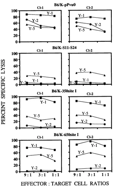
medium only, and M represents the maximum counts per minute releasedinthepresenceof 5% sodium dodecyl sulfate. The results in Fig. 3 show that two clonal isolates immortalized with thewild-typeT antigen
(B6/K-pPvuO
Cl-I and Cl-2)wererecognizedbyH-2D"-restricted CTL clones Y-1, Y-2, and Y-5. The lytic activity ofCTL clone Y-5 is low compared with that
of CTLclone Y-1. Site V is an immunorecessive site and has beenmappedtoSV40 T-antigenresidues 489 to 497 (10). CTL
clone Y-5 shows a consistently lower level of recognition in
5'Cr
releaseassaysin most cell lines (40). The cells (B6/K-S11-S24 Cl-I and Cl-2) immortalized by S11-S24 were not recog-nized and lysed by CTLclones Y-1 and Y-2 as a result of thedeletion of sites I and II/III, as expected, but were lysed by
CTL clone Y-5,which recognizes residues 489 to 497 in the T
antigen.
Cells (B6/K-350 site I Cl-I and Cl-2) expressing chimeric T
antigens containing site I at position 350 were recognized by
CTL clone Y-1 (Fig. 3) at levels comparable to those in cell
lines expressing wild-typeT antigen. Again, siteII/IIIwas not
recognized by the CTL clone, as expected because of the deletion ofresidues 127 to 250, a segment which includes site
II/III (residues 223 to 231). These cells, however, remained
susceptible to the lysis by CTL clone Y-5. Similar results were obtained when site I was translocated to T-antigen position
650. These results indicate that the T-antigen site I CTL
epitope can be transferred to either position 350 or 650 in T antigen without affecting its processing, presentation, or rec-ognition by thesite-specific CTLclone.
Expression andrecognitionof theH-2Kb-restricted HSV gB epitope (498 to 505)transferredto T-antigen positions 350 and 650. Comparison ofCTL clone Y-1's recognition of the site I
CTL epitope at the original and relocated positions clearly
indicated thattransferofthisH-2Dh epitope did not affect the
efficient processingandpresentationof this antigenic determi-nant. We then testedwhether these two positions can be used to express and present an H-2K"-restricted HSV gB CTL
recognition epitope atamino acids498 to 505. This epitope is
B6/K-pPvuO
En U)
0U
)0.4
80' 60 40' 20
0
ci-i
Y-5
1 Y-1
B6/K-Sll-S24
:
Cl B6/K-350siteI
Cl-2
100-6011
40-
1Y- I
Y
20
-D~~~~~~~~~~~~~~~~~~~~
80 60 40 20 0*
Cl-l B6/K-650siteI Cl-2
Y-1
Y-2
9:1 3:1 1:1
1 ~ - Y-2
9*
13*
1 :EFFECTOR: TARGET CELL RATIOS
FIG. 3. Effect of translocation ofSV40T-antigen site I to positions 350 and 650 in T antigen on recognition by the site I-specific CTL clone. B6/K-pPvuO, B6/K-SI -S24, B6/K-350site
I,
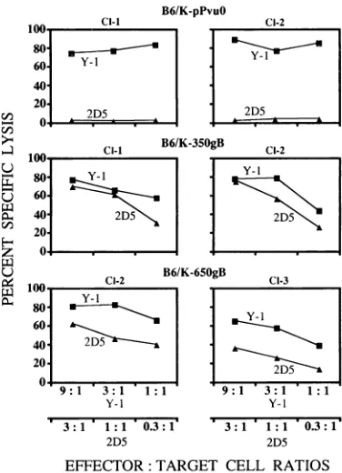
and B6/K-650site I cells were labeled with 5'Cr and reacted with T-antigen site-specific CTL clones Y-1, Y-2, and Y-5 for 5 h at the indicated effector/target cell ratios. Cl-I and CI-2 refer to the twoindependentclonal isolates.recognized by HSV CTL clone 2D5 (6). The data in Fig. 4 show that CTL clone
2D5
can recognize cells immortalized byT antigens containing the gB epitope irrespective of its location at amino acid 350 or 650. These cells were also susceptible to lysis by the CTL clone Y-1 and thus served as positive internal control for the processing of T antigen. It should be noted that the sequences flanking the gB epitope within the recombinant
T antigens are different from those of site I transferred to the same locations (Fig. IB), and neither change in flanking sequences affects the processing, presentation, and recognition
of the transferred CTL determinants. Our results show that CTL epitopes restricted by two different restriction elements can be expressed from the same location in T antigen and efficiently presented to epitope-specific CTLs.
The demonstration that CTLs recognize 8- to
10-amino-acid-long peptides presented by the MHC class I molecules has
led to efforts to utilize CTL epitopes to induce CTL responses and immune protection against viral infections (28, 29, 35, 49,
51, 54). Immunization of hosts with synthetic peptides them-selves or in combination with adjuvants has been shown to induce the generation of CTL (1, 7, 13, 26, 35, 39).
Alterna-tively, the CTL epitopes have been presented by using virus vectors, among which vaccinia virus has been used most effectively (3, 4, 12, 28, 53, 54). In addition, CTL epitopes
expressed within heterologous proteins have been shown to be
processed, presented, and recognized by epitope-specific CTL
(8, 11). In this study, we investigated the possibility of using
J.VlIROL.
on November 9, 2019 by guest
http://jvi.asm.org/
[image:3.612.338.525.77.385.2]cl-i
Ci-2 B6/K-650gB
2D5
9:1 3:1 ' 1
Y-1
3:1 1:1 0.3:1
2D5 EFFECTOR: TARGET CELL RATIOS
FIG. 4. Recognition of the HSV gB epitope expressed at amino
acidpositions350 and 650( of Tantigen bythe HSVgB epitope-specific CTL clone.B6/K-pPvu(, B6/K-350gB,andB6/65OgBcellswerelabeled with ''Crandincubatedwithsite-specificCTLclones2D5 andY-l for
5 h at the indicated effector/targetcell ratios.
SV40 T antigen as a carrier protein to express CTL epitopes. Thereareseveraladvantagesto usingT antigentodeliver CTL epitopes. T antigen is a nonstructural nuclear multifunctional
protein which is well marked antigenically; as a result, the integrity of the protein in the cells expressing the chimeric
protein can be precisely determined by using monoclonal
antibodies with defined specificities (43, 45). The SV40 T-antigen-coding sequence has undergone close scrutiny with respect to theregionsthat aredispensablefor its immortaliza-tion funcimmortaliza-tion(45),andmultiple cloningsiteswhichcanbe used for the introduction of CTLepitopeshave been inserted into T antigen (27). T antigen can immortalize primary mouse and other mammalian cellsin cultureand thus acts asa dominant markerfor the selection of transfectants (19, 27, 30, 42). The cells immortalized by T antigen express high levels of the protein.
Using this knowledge, we have identified at least two locationsin T antigen,positions350 and 650,which appearto be suited for the expression of CTL epitopes restricted by H-2Kb and H-2Dbepitopes.The transferof theT-antigen CTL site I epitope (residues205 to215) topositions350 and 650 of Tantigenandexpression of the HSVgB epitope (aminoacids 498 to 505) in the same locations did not interfere with the processing, presentation, and recognition of these epitopes by therespectiveCTLclones. In thisrespect,ourresultsappearto
be similar to those reported by others (8, 22) who
demon-strated that the flanking sequence changes surrounding the
epitope do not affect its processing. It should be noted that
T-antigen site I transferred to different locations in T antigen carriesatleastoneadditionalresidue, 205, flankingthe amino
terminus of CTLsite; therefore, expressionof this sitemaynot
beinfluencedbythe residuesflankingthis site atpositions350
NOTES 6869
and 650. However, in other instances, epitope location and
flanking sequence have been shown to influence antigen
processing and presentation. Del Val et al. (11) have shown that an H-2L
d-restricted
CTL epitopeof the murinecytomeg-alovirus immediate-early (IEI) protein was not efficiently
processed upon transfer to amino terminus of the hepatitis B
virus core antigen (HBVc), which was expressed in a
recom-binant vaccinia virus. The defect in processing of this epitope
from the amino-terminal regionof HBVc was related to the decreased amounts of natural epitope peptide generated in
vivo. This defect could be overcome by inserting polyalanine
linkers surrounding the epitope at the unfavorable site. The
processing of the same epitope, when expressed at another
location in HBVc or translocated to another position within the murinecytomegalovirusIEIprotein,was notaffected(1 1).
Eisenlohr et al. (17) have shown that the addition of two
residues, 157 and 158,with deletionofarginineatposition 156 to the influenza virus
H-2K'-restricted
CTL epitope, amino acids 147 to155,anddelivered througharecombinantvacciniavirus resultedindefective presentationofthis epitopetoCTL. Thisnegative effectonantigen processingcould bereversed by expressing this epitope, along with the negatively influencing residues, either inthe context offull-length nucleoprotein (17)
orincellsexpressingamembranedipeptidyl carboxypeptidase angiotensin-converting enzyme (16). Hahn et al. recently
re-ported abolitionofrecognition oftheminimum influenza virus hemagglutinin
H-2K"-restricted
epitope (204 to 213) by an amino acid substitution(Val-*Gly)
at position214 (23). The results from these studies appear to showthatflanking residuesin some cases do affect antigen processing, and the residues whichnegatively influence antigen processingare close
neigh-bors ofthe CTL epitope.
It should be mentioned that T-antigen CTL site I (amino acids 205 to 215) relocated to residues 350 and 650 is an
11-mer, longer than the 10-mer predictedon the basis of the
H-2D" binding
motif identifiedby
Falk and coworkers(18).
Although the minimum site I determinant includes residues
207to215,we havepreviouslyshown thatan 1 1-mer
synthetic
peptide
corresponding
toT-antigen
residues 205 to 215 isrecognizedat amuchhigher
efficiency
byCTL clone Y-1 thanisthe 9-mer
synthetic peptide consisting
ofresidues207to215(10). It is possible that other locations in T
antigen
havenegative influenceson antigen processing.
The
H-2Kt-restricted
HSVgBepitope,
in contrast,matchesthe MHC
binding
motif(18,
51).
Although
the immediateflanking
sequencessurrounding
thegBepitope
encodedby
theEcoRI linker sequence are different from those of the trans-ferred site I
determinant,
neither showed anynegative
effecton
antigen processing
andpresentation.
Therefore,
it appears thatT-antigen
locationsatresidues350and 650act asuniver-sal acceptor locations for the
expression
of CTLepitopes
restrictedby variousMHC alleles. The results obtained in this
study
show thatepitopes
restrictedby
bothH-2D"
andH-2K"
can beexpressed
from these two locations in Tantigen.
Theexpression
ofthe HSVgB
epitope
recognized by
CTL clone2D5 in T
antigen
will allow us to determine itsimmunogenic
potential
in vivo and itsability
to inducelong-term
T-cell memory.Preliminary
resultsindicate that B6 cellscarrying
thechimeric T antigen can induce the
generation
of HSVgB
epitope-specific
CTL and activate memory T cells(Sa).
The use of SV40 Tantigen
as a carrier forforeign
CTLepitopes
also will allow one to determine the
genetic
stability
of sequencesencoding
CTLepitopes.
This workwassupported
by
Public Health Service rescarch grantsCA27503,
CA25000,
and CA24694fromtheNationalCanccrInstitute. Voi.. 67, 1993B6/K-pPvuO
CI-2
2D5
80- 60- 40- 20-0.
Y-1
2D5
-.-- ci.i
rJ)
U)
u
VI~
H
z
U1
Y- 1
2D5< 80
60 40 20 0.
80- 60-40
201
Y-2D5
9:1 3:1 1:1
Y-1
3:1 1:1 0.3:1
2D5
on November 9, 2019 by guest
http://jvi.asm.org/
[image:4.612.90.278.76.336.2]6870 NOTES
REFERENCES
1. Aichele, P., H. Hengartner, R. M. Zinkernagel, and M. Schulz. 1990.Antiviral cytotoxic T cellresponseinduced by in vivo priming
with afree synthetic peptide. J. Exp. Med. 171:1815-1820. 2. Attaya, M.,S. Jameson, C. K. Martinez, E. Hermel, C.Aldrich, J.
Forman, K. F. Lindahl, M. J. Bevan, and J. J. Monaco. 1992. Ham-2 corrects the class I antigen-processing defect in RMA-S cells. Nature(London) 355:647-649.
3. Bennink, J. R., and J. W. Yewdell. 1990. Recombinant vaccinia viruses as vectors for studying T lymphocyte specificity and function. Curr. Top. Microbiol. Immunol. 163:153-184.
4. Bennink, J. R., J. W. Yewdell,G. L.Smith,C.Moller, andB. Moss. 1984. Recombinant vaccinia virus primes and stimulates influenza haemagglutinin-specific cytotoxic T cells. Nature (London) 311: 578-579.
5. Bjorkman, P. J., M. A. Saper, B. Samraoui, W. S. Bennett, J. L. Strominger, and D. C. Wiley. 1987. Structure of the human class I histocompatibility antigen, HLA-A2. Nature (London) 329:506-512.
Sa.Bonneau, R. H., T.-M. Fu, and S. S.Tevethia. In vivo priming and activation of memory cytotoxic T-lymphocytes (CTL) by a chi-meric simian virus 40 T antigen expressing an eight amino acid
residue herpes simplex virus gB CTL epitope. Virology, in press.
6. Bonneau, R. H., L. A.Salvucci, D. C. Johnson, and S. S. Tevethia. 1993. Epitope specificity ofH-2Kb-restricted, 1-, and HSV-2-cross-reactive cytotoxic T lymphocyte clone. Virology 195:62-70. 7. Carbone, F.R., and M. J. Bevan. 1989. Induction of ovalbumin-specific cytotoxic T cells by in vivo peptide immunization. J. Exp. Med. 169:603-612.
8. Chimini,G., P. Pala, J. Sire, B. R. Jordan, and J. L.Maryanski. 1989. Recognition of oligonucleotide-encoded T cell epitopes introduced into a gene unrelated to the original antigen. J. Exp. Med. 169:297-302.
9. Cox, J. H., J. W. Yewdell, L. C. Eisenlohr, P. R. Johnson, and J. R. Bennink. 1990. Antigen presentation requires transportof MHC class I molecules from the endoplasmic reticulum. Science 247: 715-718.
10. Deckhut, A. M., J. D. Lippolis, and S. S. Tevethia. 1992. Compar-ative analysis of core amino acid residues of H-2D&-restricted
cytotoxic T-lymphocyte recognition epitopes in simian virus 40 T antigen. J. Virol.66:440-447.
11. Del Val, M., H. J. Schlicht, T. Ruppert, M. J. Reddehase, and U. H. Koszinowski. 1991. Efficient processing ofan antigenicsequence
for presentation by MHC class I molecules depends on its
neighboring residues in the protein. Cell 66:1145-1153.
12. Del Val, M., H. J. Schlicht, H. Volkmer, M. Messerle, M. J. Reddehase,and U. H.Koszinowski. 1991. Protection against lethal cytomegalovirus infection bya recombinant vaccine containing a
single nonameric T-cell epitope. J. Virol. 65:3641-3646. 13. Deres, K., H. Schild, K. Wiesmuller, G.Jung, and H. Rammensee.
1989. In vivopriming of virus-specific cytotoxic Tlymphocytes with synthetic lipopeptide vaccine. Nature (London) 342:561-564. 14. Doherty, P. C., W. Allan,and M.Eichelberger. 1992. Roles ofc43
and y8 T cell subsets in viral immunity. Annu. Rev. Immunol. 10:123-151.
15. Driscoll, J., and D. Finley. 1992. A controlledbreakdown: antigen processing and theturnover of viral proteins. Cell 68:823-825. 16. Eisenlohr, L.C.,I.Bacik,J. R. Bennink, K. Bernstein, and J. W.
Yewdell. 1992. Expression of a membrane protease enhances presentation of endogenous antigens toMHC class I-restricted T lymphocytes. Cell 71:963-972.
17. Eisenlohr, L. C., J.W. Yewdell, and J. R. Bennink. 1992.Flanking
sequencesinfluence the presentation ofanendogenously synthe-sizedpeptide tocytotoxic T lymphocytes. J. Exp. Med. 175:481-487.
18. Falk, K., 0. Rotzschke, S. Stevanovic, G. Jung, and H. G.
Rammensee.1991. Allele-specific motifsrevealed by sequencing of self-peptides eluted from MHC molecules. Nature (London) 351:290-296.
19. Fanning, E. 1992. Simian virus 40large T antigen: the puzzle,the pieces, and the emerging picture.J.Virol.66:1289-1293. 20. Frement, D. H., M.Matsumura, E. A. Stura,P. A. Peterson, and
I. A. Wilson. 1992. Crystal structures of two viral peptides in
complex with murine MHC class I H-2Kh. Science 257:919-927. 21. Gould, K., J.Cossins,J. Bastin, G.G. Brownlee, and A. Townsend.
1989. A 15 amino acid fragment of influenza nucleoprotein synthesized in the cytoplasm is presented to class I-restricted cytotoxic T lymphocytes. J. Exp. Med. 170:1051-1056.
22. Hahn, Y.S., V. L.Braciale,andT.J. Braciale. 1991. Presentation of viral antigen to class I major histocompatibility
complex-restricted cytotoxic T lymphocyte. Recognition of an immu-nodominant influenza hemagglutinin site bycytotoxic T lympho-cyte isindependentof theposition of the site in thehemagglutinin translation product. J. Exp. Med. 174:733-736.
23. Hahn, Y. S.,C. S. Hahn, V. L.Braciale, T. J. Braciale, andC.M. Rice. 1992. CD8+ T cellrecognition ofanendogenously processed epitope is regulated primarily by residues within the epitope. J. Exp. Med. 176:1335-1341.
24. Jorgensen, J. L., U. Esser, P.A. Reay, B. Fazekas deSt.Groth,and M. M. Davis. 1992. Mapping T cell receptor/peptide contacts by variant peptide immunization of single-chain transgenics. Nature (London) 355:224-230.
25. Jorgensen, J. L., P. A. Reay, E. W. Ehrich, and M. M. Davis. 1992. Molecular components of T-cell recognition. Annu. Rev. Immu-nol. 10:835-873.
26. Kast, W. M., L. Roux, J. Curren, H. J. J. Blom, A. C. Voordouw, R. H. Meloen, D. Kolakofsky, and C. Melief. 1991. Protection against lethal Sendai virus infection by in vivo priming of virus-specific cytotoxic T lymphocytes with a free synthetic peptide. Proc.Natl. Acad. Sci. USA 88:2283-2287.
27. Kierstead, T. D., and M. J. Tevethia. 1993. Association of p53 binding and immortalization of primary C57BL/6 mouse embryo fibroblasts by using simian virus 40 T-antigen mutants bearing internal overlapping deletion mutations. J. Virol. 67:1817-1829. 28. Klavinskis, L. S., J. L. Whitton, E. Joly, and M. B. Oldstone. 1990.
Vaccination and protection from a lethal viral infection: identifi-cation, incorporation, and use of a cytotoxic T lymphocyte glyco-protein epitope. Virology 178:393-400.
29. Koszinowski, U. H., M. J. Reddehase, and M. Del Val. 1992. Principles ofcytomegalovirus antigen presentation in vitro and in vivo. Semin. Immunol. 4:71-79.
30. Livingston, D. M., and M. K. Bradley. 1987. The simian virus 40 large T antigen. A lotpacked into a little. Mol. Biol. Med. 4:63-80. 31. Monaco, J. J., S. Cho, and M. Attaya. 1990. Transport protein genes in the murine MHC: possible implications for antigen processing. Science250:1723-1726.
32. Nuchtern, J. G., J. S. Bonifacino, W. E. Biddison, and R. D. Klausner. 1989. Brefeldin A implicates egress from endoplasmic reticulum inclass I restricted antigen presentation. Nature (Lon-don)339:223-226.
33. Rotzschke,O.,K. Falk, K. Deres, H. Schild,M. Norda, J. Metzger, G.Jung, and H. G. Rammensee. 1990. Isolation and analysis of naturally processed viral peptides as recognized by cytotoxic T cells. Nature (London) 348:252-254.
34. Sambrook, J., E. F. Fritsch, and T. Maniatis. 1989. Molecular cloning: a laboratory manual. Cold Spring Harbor Laboratory Press, ColdSpring Harbor, N.Y.
35. Schulz, M., R. M.Zinkernagel, and H. Hengartner.1991. Peptide-induced antiviralprotection by cytotoxic T cells. Proc. Natl. Acad. Sci. USA88:991-993.
36. Spies, T., M. Bresnahan, S. Bahram, D. Arnold, G. Blanck, E. Mellins, D. Pious, and R. DeMars. 1990. A gene in the human major histocompatibility complex class II region controlling the class I antigenpresentation pathway. Nature (London) 348:744-747.
37. Spies, T., V. Cerundolo, M. Colonna,P. Cresswell, A. Townsend, and R. DeMars. 1992.Presentation of viral antigen by MHC class I molecules is dependent on a putative peptide transporter heterodimer. Nature (London) 355:644-646.
38. Sweetser, M. T., L. A. Morrison, V. L. Braciale, and T. J. Braciale. 1989.Recognition of pre-processed endogenous antigen by class I but not class IIMHC-restricted T cells. Nature (London) 342:180-182.
39. Takahashi, H., T. Takeshita, B. Morein, S. Putney, R. N.Germain,
and J. A.Berzofsky. 1990. Induction of CD8+ cytotoxic T cells by immunization with purified HIV-1 envelop protein in ISCOMs. J. VIROL.
on November 9, 2019 by guest
http://jvi.asm.org/
NOTES 6871
Nature(London) 344:873-875.
40. Tanaka, Y., R. W. Anderson, W. L. Maloy, and S. S. Tevethia. 1989. Localization of an immunorecessive epitope on SV40 T antigen by H-2Db-restricted cytotoxic T-lymphocyte clones anda
synthetic peptide. Virology 171:205-213.
41. Tanaka, Y., M. J. Tevethia, D. Kalderon, A. E. Smith,and S. S. Tevethia. 1988. Clustering of antigenic sites recognized by cyto-toxic Tlymphocyte clones in the amino terminal half of SV40 T antigen. Virology 162:427-436.
42. Tevethia, M. J. 1984. Immortalization of primary mouseembryo fibroblasts withSV40virions, viral DNA, andasubgenomic DNA
fragment inaquantitative assay.Virology 137:414-421.
43. Tevethia, S. S. 1990. Recognition of simian virus 40 T antigen by cytotoxic T lymphocytes. Mol. Biol. Med. 7:83-96.
43a.Tevethia, S. S.Unpublished data.
44. Tevethia, S. S., R. S.Greenfield, D. C. Flyer, and M. J. Tevethia. 1980.SV40transplantation antigen: relationshiptoSV40-specific proteins. Cold Spring Harbor Symp. Quant. Biol.44:235-242. 45. Thompson, D. L., D. Kalderon, A. E. Smith, and M. J. Tevethia.
1990. Dissociation of Rb-binding and anchorage-independent growth fromimmortalization and tumorigenicity usingSV40
mu-tantsproducing N-terminally truncated large T antigens. Virology
178:15-34.
46. Townsend,A., J. Bastin, K. Gould, G. Brownlee, M. Andrew, B. Coupar, D. Boyle, S. Chan, and G. Smith. 1988. Defective presentation toclass I-restricted cytotoxic T lymphocytes in
vac-cinia-infected cells is overcome by enhanced degradation of antigen. J. Exp. Med. 168:1211-1224.
47. Townsend, A., and H. Bodmer. 1989. Antigen recognition by class I-restrictedTlymphocytes. Annu. Rev. Immunol. 7:601-624. 48. Townsend,A., C. Ohlen, J. Bastin, H. Ljunggren, L. Foster, and K.
Karre.1989. Association of class Imajor histocompatibility heavy and light chains induced by viral peptides. Nature (London) 340:443-448.
49. Townsend, A. R. M., J. Rothbard, F. M. Gotch, G. Bahadur, D. Wraith, and A.J. McMichael. 1986. The epitopes of influenza nucleoprotein recognized by cytotoxic T lymphocytes can be defined with short synthetic peptides. Cell 44:959-968.
50. Trowsdale, J.,I. Hanson, I.Mockridge,S.Beck, A.Townsend,and A. Kelly. 1990. Sequencesencoded in the class II region of the MHC related tothe 'ABC' superfamily of transporters. Nature (London)348:741-744.
51. Van Bleek, G., and S. G. Nathenson. 1990. Isolation of an endogenouslyprocessed immunodominant viralpeptidefromthe classI H-2Kh molecule. Nature(London) 348:213-216.
52. Van Kaer, L., P. G. Ashton-Rickardt, H. L. Ploegh, and S. Tonegawa. 1992. TAPI mutant mice are deficient in antigen presentation, surface class I molecules, and CD4-CD8+ T cells. Cell 71:1205-1214.
53. Whitton,J. L., and M. B.Oldstone.1989.ClassIMHCcanpresent an endogenouspeptidetocytotoxicTlymphocytes. J. Exp.Med. 170:1033-1038.
54. Whitton, J. L., N. Sheng, M.B.Oldstone, and T. A. McKee. 1993. A"string-of-beads"vaccine,comprisinglinked minigenes, confers protectionfrom lethal-dose virus challenge. J. Virol.67:348-352. 55. Yewdell, J. W., and J.R.Bennink. 1989. Brefeldin Aspecifically inhibits presentation ofprotein antigens tocytotoxic T lympho-cytes. Science 244:1072-1075.
56. Yewdell, J. W., and J. R.Bennink 1992. Cell biology ofantigen processing andpresentation tomajorhistocompatibility complex class I molecule-restricted T lymphocytes. Adv. Immunol. 52:1-123.
57. Zinkernagel, R. M., and P. C. Doherty. 1979. MHC-restricted cytotoxic T cells: studieson the biological role ofpolymorphic major transplantation antigens determining T-cell restriction-specificity, function, and responsiveness. Adv. Immunol. 27:51-177.
VOL.67, 1993