0022-538X/06/$08.00⫹0 doi:10.1128/JVI.00678-06
Copyright © 2006, American Society for Microbiology. All Rights Reserved.
Phosphorylation of MCM4 at Sites Inactivating DNA Helicase
Activity of the MCM4-MCM6-MCM7 Complex during
Epstein-Barr Virus Productive Replication
Ayumi Kudoh,
1Tohru Daikoku,
1† Yukio Ishimi,
2Yasushi Kawaguchi,
3Noriko Shirata,
1Satoko Iwahori,
1Hiroki Isomura,
1and Tatsuya Tsurumi
1*
Division of Virology, Aichi Cancer Center Research Institute, 1-1, Kanokoden, Chikusa-ku, Nagoya 464-8681, Japan1; Department
of Materials and Biological Sciences, Faculty of Science, Ibaraki University, Mito 310-8512, Japan2; and
Institute of Medical Science, University of Tokyo, Shirokanedai, Minato-ku, Tokyo 108-8639, Japan3
Received 4 April 2006/Accepted 25 July 2006
Induction of Epstein-Barr virus (EBV) lytic replication blocks chromosomal DNA replication notwithstand-ing an S-phase-like cellular environment with high cyclin-dependent kinase (CDK) activity. We report here that the phosphorylated form of MCM4, a subunit of the MCM complex essential for chromosomal DNA replication, increases with progression of lytic replication, Thr-19 and Thr-110 being CDK2/CDK1 targets whose phosphorylation inactivates MCM4-MCM6-MCM7 (MCM4-6-7) complex-associated DNA helicase. Expression of EBV-encoded protein kinase (EBV-PK) in HeLa cells caused phosphorylation of these sites on MCM4, leading to cell growth arrest. In vitro, the sites of MCM4 of the MCM4-6-7 hexamer were confirmed to be phosphorylated with EBV-PK, with the same loss of helicase activity as with CDK2/cyclin A. Introducing mutations in the N-terminal six Ser and Thr residues of MCM4 reduced the inhibition by CDK2/cyclin A, while EBV-PK inhibited the helicase activities of both wild-type and mutant MCM4-6-7 hexamers, probably since EBV-PK can phosphorylate MCM6 and another site(s) of MCM4 in addition to the N-terminal residues. Therefore, phosphorylation of the MCM complex by redundant actions of CDK and EBV-PK during lytic replication might provide one mechanism to block chromosomal DNA replication in the infected cells through inactivation of DNA unwinding by the MCM4-6-7 complex.
A number of replication initiation sites that are present in the genome of eukaryotic cells are utilized in a temporal order during the DNA synthesis (S) phase of the cell cycle. Reini-tiation of DNA replication is prevented, and only a single round of DNA replication is performed in a cell cycle. This DNA replication by the so-called replication licensing system is regulated by the loading of the minichromosome maintenance (MCM) complexes on chromatin DNA and their phosphory-lation (37, 50, 51).
During the G1phase of the cell cycle, replication origins in DNA are licensed by the assembly of prereplicative complexes (pre-RC) comprising the origin recognition complex (ORC), Cdc6, Cdt1, and the MCM complex (47, 51). The ORC binds to origins of DNA replication and remains bound during most of the cell cycle (30, 40, 48). Cdc6 and Cdt1 then bind to the complex and facilitate the loading of the MCM2-MCM7 (MCM2-7) complex. Cdt1 itself is regulated by geminin, which blocks the binding of the MCM complex to the pre-RC (39, 46, 54). Activation of the pre-RC occurs at the G1/S boundary after licensing and is mediated by the action of S-phase cyclin-dependent kinases (CDKs), primarily cyclin A/CDK2, cyclin E/CDK2, and Cdc7/Dbf4 (3, 10, 42, 45), which trigger a chain of reactions that lead to the binding of Cdc45 to the
origin and phosphorylation of Cdc6 and the MCM complex. As a result, the DNA duplex unwinds, facilitating loading of the DNA polymerase machinery (24, 41, 52, 59). The phos-phorylation of key components of this process by the CDKs leads to initiation of replication and at the same time helps to prevent rereplication during the S and G2/M phases of the cell cycle (6, 7, 23, 55).
All of the members of the MCM protein family contain highly conserved DNA-dependent ATPase motifs in the cen-tral domain (3, 44) and form several stable subassemblies, including MCM2-3-4-5-6-7, MCM2-4-6-7, MCM4-6-7, and MCM3-5 complexes (35, 49, 56). DNA helicase activity has been identified in the MCM4-6-7 complexes of human, mouse, and fission yeast (Schizosaccharomyces pombe) (19, 35, 36, 56), while MCM2 and MCM3-5 are known to inhibit this activity by converting the double-trimer structure into a heterotetramer or a heteropentamer (43, 56). MCM4-6-7 proteins form trimers or hexamers to function as DNA he-licases in vitro (37). Such DNA helicase activity is not pro-cessive under standard conditions of a DNA helicase assay. During S phase, MCM proteins are released from origins of replication after initiation of DNA replication and move with replication forks, where they are thought to function as DNA helicases. The mechanisms ensuring replication of DNA only once per cycle involve release of MCM proteins from chromatin after firing of the origins of replication and prevention of reloading (2, 11). Moreover, the phosphory-lation of MCM4 with CDK2/cyclin A is associated with inactivation of the DNA helicase (unwinding) activity of the
* Corresponding author. Mailing address: Division of Virology, Aichi Cancer Center Research Institute, 1-1, Kanokoden, Chikusa-ku, Nagoya 464-8681, Japan. Phone and Fax: 81-52-764-2979. E-mail: [email protected].
† Present address: Department of Virology, University of Toyama, 2630 Sugitani, Toyama 930-0194, Japan.
10064
on November 8, 2019 by guest
http://jvi.asm.org/
MCM4-6-7 complex (22) and the sites critical for phosphor-ylation have been determined previously (20, 22).
Primary infection with Epstein-Barr virus (EBV), a human herpesvirus that infects 90% of individuals, targets resting B lymphocytes and induces their continuous proliferation. In B lymphoblastoid cell lines, there is no production of virus par-ticles (this being termed latent infection), but EBV-infected cell lines sometimes switch from a latent stage of infection into a virus-productive lytic stage, featuring an ordered cascade of viral early and late gene expression. The lytic phase of EBV DNA replication is dependent on seven viral replication pro-teins and occurs in discrete sites in nuclei, called replication compartments (8). We have demonstrated previously that in-duction of the EBV lytic program results in inhibition of rep-lication of chromosomal DNA despite reprep-lication of viral DNA (32). However, rather high S-phase CDK activity is as-sociated with progression of lytic infection, indicating that the lytic replication occurs under S-phase-like cellular conditions (31–33).
The EBV BGLF4 gene product is the only viral protein kinase which belongs to the group of herpesvirus-encoded Ser/ Thr protein kinases, conserved in all herpesviruses (27). Ex-pressed at an early stage after viral reactivation and localized in nuclei, the protein is reported to phosphorylate a number of viral and cellular proteins, including the EBV BMRF1 DNA polymerase processivity factor (EA-D) (5), the EBV nuclear antigen leader protein (EBNA-LP) (26), EBNA2 (58), protein kinase itself, and the cellular translation elongation factor 1␦ (26, 28). Most interestingly, EBV-encoded protein kinase (EBV-PK) phosphorylates EBNA-LP and elongation factor 1␦ at the same sites as CDK1 (Cdc2). However, hitherto there has been no evidence that such phosphorylation influences func-tions of target proteins.
We report here that human MCM4, a subunit of the MCM complex, is phosphorylated in B95-8 and Akata cells when lytic replication is induced. The phosphorylation sites of Thr-19 and Thr-110 in MCM4 were identified as the same amino acid residues targeted by CDK2/CDK1, which inactivates DNA he-licase activity of the MCM4-6-7 complex. Furthermore, expres-sion of EBV-PK in HeLa cells phosphorylated the same sites on MCM4. In vitro, the site of the MCM4 protein of the MCM4-6-7 complex was confirmed to be phosphorylated with EBV-PK, with the same loss of enzyme activity as with CDK2/ cyclin A. Although introducing mutations in the N-terminal six Ser and Thr residues of MCM4 reduced the inhibition by CDK2/cyclin A, EBV-PK inhibited the helicase activities of both wild-type (WT) and mutant MCM4-6-7 hexamers. It was found that EBV-PK can phosphorylate MCM6 and other sites on MCM4 in addition to the N-terminal residues. Thus, phos-phorylation of the MCM4-6-7 complex by redundant actions of CDK and EBV-PK during lytic replication might inhibit chro-mosomal DNA replication, including rereplication of chromo-somal DNA and/or DNA fork progression, through inactiva-tion of DNA unwinding by the MCM4-6-7 complex.
MATERIALS AND METHODS
Cells.Tet-BZLF1/B95-8 cells, a marmoset B-cell line latently infected with EBV (32), and Tet-BZLF1/Akata cells, human EBV-positive Burkitt’s
lym-phoma cells (31), were maintained in RPMI medium supplemented with 1g/ml
of puromycin, 250g/ml of hygromycin B, and 10% tetracycline-free fetal calf
serum. To induce lytic EBV replication, a tetracycline derivative, doxycycline,
was added to the culture medium at a final concentration of 2g/ml. HeLa cells
were cultured in Dulbecco’s modified Eagle’s medium (Sigma) containing 10% fetal calf serum.
Protein preparation. Cells were harvested, washed with phosphate-buffer saline (PBS), and treated with lysis buffer (0.02% sodium dodecyl sulfate [SDS], 0.5% Triton X-100, 300 mM NaCl, 20 mM Tris-HCl [pH 7.6], 1 mM EDTA, 1 mM dithiothreitol [DTT]) for 20 min on ice. After multiple protease inhibitors
(25l/ml; Sigma), 200M sodium vanadate, and 20 mM sodium fluoride were
added, samples were centrifuged at 18,000⫻gfor 10 min at 4°C, and clarified
cell extracts were assayed for protein concentration by use of a Bio-Rad kit.
Cellular fractionation.Tet-BZLF1/B95-8 cells (1.5⫻107) were harvested,
washed with cold PBS, and then lysed for 10 min on ice with 1 ml of ice-cold
modified cytoskeleton (mCSK) buffer {10 mM PIPES [piperazine-N,N⬘
-bis(2-ethanesulfonic acid)], pH 6.8, 100 mM NaCl, 300 mM sucrose, 1 mM MgCl2, 1
mM EGTA, 1 mM DTT, 1 mM phenylmethylsulfonyl fluoride, 10g/ml
apro-tinin} containing 0.5% Triton X-100, multiple protease inhibitors (protease
inhibitor mixture for mammalian cell extracts, 25l/ml; Sigma), 0.1 mM ATP,
200 mM sodium orthovanadate, and 20 mM NaF. The samples were then
sub-jected to centrifugation (2,000⫻g, 3 min, 4°C) for separation into a Triton
X-100-extractable fraction and a nuclear pellet. The latter was then washed once with mCSK buffer and resuspended in the same buffer containing 0.1% Triton
X-100, 1 mM ATP, and 4 mM MgCl2at 1.5⫻10
7
nuclei/ml. To separate phosphorylated forms of MCM proteins, samples were subjected to SDS-poly-acrylamide gel electrophoresis (SDS-PAGE) (7.5% polySDS-poly-acrylamide gels [acryl-amide, 72; bisacryl[acryl-amide, 1]).
Antibodies.Primary monoclonal antibodies to MCM6, the BMRF1 protein, and Cdk2 were purchased from Santa Cruz (MCM6 H-300), Chemicon (EBV BMRF1-R3), and BD Biosciences (CDK2 clone 55), respectively. Rabbit poly-clonal antibodies to the BZLF protein and MCM7 proteins were prepared as described previously (13, 32). Anti-BGLF4 protein antibody was provided by Y. Kawaguchi (Tokyo University) (25). Antisera against phosphothreonine at amino acid (aa) 19 or 110 of human MCM4 were also prepared as detailed earlier (21). MCM4 was detected either with a monoclonal antibody (kindly provided by K. Tamai, MBL Co.) or with a commercially available polyclonal antibody (BD Biosciences). Highly cross-absorbed secondary reagents for dual-color detection (Alexa-488 and -594) were from Molecular Probes.
Immunoblot analysis.Equal amounts of proteins were subjected to SDS-10% PAGE (acrylamide, 29.2; bisacrylamide, 0.8). To separate phosphorylated form of MCM proteins, gradient SDS-PAGE (2/15 PAG Mini; Daiichi Pure Chemi-cals) or SDS-7.5% PAGE (acrylamide, 72; bisacrylamide, 1) was applied. The proteins were then processed as described previously (32). Detection of target proteins was done with an enhanced chemiluminescence detection system (Amersham Biosciences).
Immunofluorescence analysis.Cells were treated with 0.5% Triton X-100– mCSK buffer for 10 min on ice, followed by fixation with 70% methanol for 20 min on ice. The fixed cells were washed with PBS and blocked for 20 min in 10% normal goat serum in PBS. Staining with primary antibody (anti-MCM4 or anti-MCM7) was performed overnight at 4°C in PBS-0.5% goat serum. Staining with a mouse monoclonal antibody to BMRF1 protein was carried out for 1 h at room temperature. Species-specific secondary antibodies were then applied for 1 h at room temperature. Slides were mounted with Vectashield (Vector Labs) for analysis under a fluorescence confocal microscope. Images were captured and processed using a Radiance 2000 confocal system (Bio-Rad). All of the primary antibodies were employed at a 1:100 dilution and the secondary antibodies at a 1:500 dilution. All washes after antibody incubations were performed with 0.05% Tween 20 in PBS at room temperature. When cells were stained singly for either antigen with inappropriate combinations of first and second antibodies, no flu-orescence was observed. Also, no immunofluflu-orescence was observed with alter-nate filters.
Preparation of phosphospecific antibodies.Antisera against phosphothreo-nine at amino acids 19 and 110 of human MCM4 were obtained by immunizing
rabbits with a synthetic peptide of NH2-SRRGRA(pT)PAQTPRSEC-COOH
(for aa 19) or NH2-CGTPRSGVRG(pT)PVRQRPDL-COOH (for aa 110) and
purified as described previously (21).
Purification of human MCM4-6-7 complexes.Sf21 insect cells were coinfected with recombinant baculovirus expressing human MCM7 (Bac-MCM7) and virus expressing human histidine-tagged MCM4 and -6 (Bac-MCM4-6) at a multiplic-ity of infection of 10 and harvested 3 days postinfection. MCM4-6-7 complexes containing histidine-tagged MCM4 were purified by nickel-nitrilotriacetic acid column chromatography and then subjected to glycerol density gradient centrif-ugation (56) as described previously (21). Fractions containing the 600-kDa
on November 8, 2019 by guest
http://jvi.asm.org/
MCM4-6-7 hexamer determined by each specific antibody were collected and pooled.
Site-directed mutagenesis of the human MCM4 gene was conducted using a
QuikChange site-directed mutagenesis kit (Stratagene). The oligonucleotide 5⬘
-GATGCCAGGTCAGCTCCCTCTCAGAGACG-3⬘was used as the primer to
introduce a change from Ser to Ala at amino acid 32, and 5⬘-GGTGTTAGGG
GCGCACCTGTGAGACAGAGG-3⬘was used for the change at amino acid
110. The oligonucleotide 5⬘-CATCACGGATCGGCCCCGGCGTCGGCCCCG
AGCCGC-3⬘was used as the primer to change both Ser-3 and Thr-7 to Ala,
5⬘-CGTGGAAGGGCCGCCCCCGCCCAGACG-3⬘was used for the change at
amino acid 19, and 5⬘-CCGATGCCAACCGCGCCTGGAGTGGAC-3⬘ was
used for the change at residue 54. The mutated MCM4 gene forms were cloned into a pAcUW31 vector (Pharmingen) in which the Mcm6 gene had been cloned (56). The entire MCM4 gene has been sequenced, and all of the mutated sites were confirmed. Sf21 cells were coinfected with the recombinant baculovirus expressing proteins from mutated MCM4 and MCM6 (Bac-MCM4a-6) and a virus expressing the MCM7 protein (Bac-MCM7). The 600-kDa mutated MCM4a-6-7 hexamer was purified by nickel-nitrilotriacetic acid column chroma-tography and then by glycerol gradient centrifugation (56).
Purification of GST-fused BGLF4 protein from baculovirus-infected cells.
Sf21 cells were infected with a baculovirus (either BGLF4 or
Bac-GST-BGLF4K102I
, where GST is glutathioneS-transferase) at a multiplicity of
infec-tion of 10 and harvested 3 days thereafter. Cells were suspended in 10 ml of buffer A (50 mM Tris-HCl [pH 7.5], 150 mM NaCl, 1 mM EDTA, 1 mM dithiothreitol, 0.1% Nonidet P-40, 10% glycerol, 1 mM phenylmethylsulfonyl
fluoride, and a protease inhibitor mixture for mammalian cell extracts [25l/ml;
Sigma]) and then sonicated. After the insoluble materials were removed by
centrifugation, the supernatants were reacted with 500l of a 50% slurry of
glutathione-Sepharose beads (Amersham Pharmacia) for 2 h. The beads were then extensively washed with buffer A-2 (50 mM Tris-HCl [pH 7.5], 1 mM EDTA, 1 mM dithiothreitol, 10% glycerol, and 1 mM phenylmethylsulfonyl fluoride) and eluted in buffer A-2 containing 10 mM glutathione. The eluted
proteins (GST-BGLF4 and GST-BGLF4K102I) were finally applied to Mono Q
column chromatography, and peak fractions were collected and pooled.
In vitro kinase assays.Human MCM4-6-7 or MCM4a-6-7 complexes (500 ng) were incubated with various amounts of EBV BGLF4 protein, kinase-negative
BGLF4K102Iprotein, or human CDK2/cyclin A (purchased from Upstate, Inc.) in
a 50-l reaction mixture containing 20 mM Tris-HCl (pH 7.5), 1 mM EDTA, 1
mM dithiothreitol, 10 mM MgCl2, 1 mM ATP (or 100M ATP and 2Ci
[␥-32
P]ATP), and 0.2 mM sodium orthovanadate. The reaction mixtures were incubated for 1 h at 37°C, and the products were subjected to electrophoresis on 7.5% polyacrylamide gels containing SDS and were detected with an image guider (BAS2500; Fujifilm).
DNA helicase assays.For preparation of a substrate for DNA helicase activity,
a 17-mer oligonucleotide (5⬘-GTTTTCCCAGTCACGAC-3⬘) complementary to
the M13mp18(⫹) strand was labeled at the 5⬘end with polynucleotide kinase in
the presence of [␥-32P]ATP and then annealed to M13 DNA. Approximately 50
fmol of the annealed oligomer was incubated at 37°C for 1 h with MCM4-6-7 proteins in 25 mM HEPES-NaOH, pH 7.5, 1 mM DTT, 4 mM ATP, 10 mM
Mg(CH3COO)2, and 0.1 mg of bovine serum albumin per ml. The reaction was
terminated by adding SDS to a final concentration of 0.2%, and an aliquot was electrophoresed on a 12% acrylamide gel in Tris-borate-EDTA. The loaded oligomer in the gel was detected with an image guider (BAS2500; Fujifilm) and quantitated.
To examine the effects of phosphorylation on DNA helicase activity of
MCM4-6-7 complexes, EBV BGLF4 protein, kinase-negative BGLF4K102I
protein, or CDK2/cyclin A was added to the reaction mixture from the start of the DNA helicase assay, and the displaced oligonucleotide was analyzed as described above. Phosphorylation of MCM4 with EBV-PK or with CDK2/cyclin A reached a plateau within 3 min of incubation (data not shown).
Plasmids.The BGLF4 expression plasmid pME-BGLF4(F) and the control plasmid pME18S were constructed as described previously (25, 26, 28). In
pME-BGLF4(F), the expression of the 3⬘Flag epitope-tagged BGLF4 is driven by the
SR␣promoter. HeLa cells were transfected with these plasmid DNAs with
Lipofectamine 2000 (Invitrogen).
RESULTS
Effect of EBV lytic replication on MCM loading onto chro-matin.It has been reported previously that infection of quies-cent primary fibroblasts (G0phase) with human cytomegalovi-rus (HCMV) induces expression of all prereplicative factors,
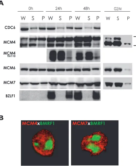
like the ORC, CDC6, Cdt1, and the MCM complex, but that the virus prevents prereplicative complex assembly by inhibit-ing MCM complex loadinhibit-ing onto chromatin (4, 53). Therefore, we examined whether the MCM complexes are detached from chromatin after induction of EBV lytic replication in cycling cells latently infected with EBV. Tet-BZLF1/B95-8 cells were treated with doxycycline to induce lytic replication (32). We have demonstrated previously subcellular dynamics of EBNA1, viral replication proteins, histones, and human chromosomal DNA replication initiation proteins in lytic-program-induced Tet-BZLF1/B95-8 cells by using biochemical fractionation methods with a buffer containing nonionic detergent Triton X-100 at a relatively physiological salt condition (8, 9). The treatment extracts not only cytoplasmic but also nuclear pro-teins not tightly bound to nuclear structures (8, 9). Propro-teins remaining in extracted nuclei have been shown previously to indeed bind to chromatin or viral DNA/nuclear matrix mate-rial (12, 15). With these procedures, the MCM4 and MCM7 proteins were extracted with the buffer, and most of the re-mainder were solubilized after DNase I treatment (8). Thus, the detergent-resistant fractions of MCMs represent their chromatin binding. As shown in Fig. 1A, all of the BZLF1 immediate-early protein became bound to chromatin or viral DNA/nuclear matrix material. In contrast, almost all of the MCM4, -6, and -7 proteins in the G2/M phase were sensitive to the detergent treatment, indicating release of the MCM com-plex from chromatin DNA. These results confirmed reproduc-ibility of the fractionation procedure, consistent with our pre-vious observations (8, 9, 16).
In cells latently infected with EBV, about half of the MCM proteins proved resistant to the detergent treatment, indicating MCM loading onto chromatin DNA (Fig. 1A, 0 h). Although two forms of MCM4 were detected, the slower-migrating band represents the hyperphosphorylated form, as determined pre-viously (16), whereas the faster-migrating band is hypophos-phorylated. Levels of the slowly migrating forms of MCM4, increased with progression of lytic replication by an unknown mechanism, while levels of CDC6 remained constant. The ra-tios of the chromatin-bound to unbound forms of MCM6 and MCM7 were essentially unchanged before and after induction. On the other hand, levels of the chromatin-unbound forms of MCM4 increased to some degree after induction. For example, most of the MCM4 protein phosphorylated at Thr-110 was detached from chromatin (Fig. 1A), but about half of MCM4 phosphorylated at Thr-19 became bound to chromatin (data not shown). Thus, the picture of regulation of chromatin bind-ing of the MCM complex is likely to be very complex. Phos-phorylation of some sites on MCM4 appears to trigger the dissociation of the MCM complex from chromatin. It was dem-onstrated previously that EBV lytic replication arrests cell cy-cle and chromosomal DNA replication (32). Thus, the chro-mosomal DNA replication arrest during lytic infection might be partly due to dissociation of the MCM complex from chro-matin. However, the overall picture of MCM attachment did not change dramatically after induction of EBV lytic replica-tion, suggesting that other mechanisms also operate at stages after loading MCM complex onto chromatin.
MCM proteins are localized outside viral replication com-partments in nuclei.Tet-BZLF1/B95-8 cells were treated with
on November 8, 2019 by guest
http://jvi.asm.org/
doxycycline to induce lytic replication (32), harvested, and treated with a buffer containing the nonionic detergent Triton X-100. This treatment is known to disrupt nuclear envelopes, but the nuclear lamina remains intact, thus maintaining nu-clear structures (18). By confocal immunofluorescence analy-ses, the sites stained with anti-BMRF1 replication protein-specific antibodies completely coincided with foci of newly synthesized viral DNA, as judged by 5-bromodeoxyuridine in-corporation and fluorescence in situ hybridization analyses (8, 33). Thus, BMRF1 protein-localized sites represent loci of viral DNA synthesis, termed viral replication compartments. As shown in Fig. 1B, the detergent-resistant forms of MCM4 and MCM7 proteins were localized outside these compart-ments in nuclei, indicating chromatin binding during lytic infection.
Induction of EBV lytic program elicits phosphorylation of Thr-19 and Thr-110 on MCM4.Phosphorylation of Thr-19 and
Thr-110 on MCM4 in human and mouse MCM4-6-7 com-plexes is associated with inhibition of the associated DNA helicase activity (20). In order to investigate whether chromo-somal replication arrest induced by the EBV lytic replication is partly due to inactivation of the MCM complex, we examined the phosphorylation state of MCM4 on Thr-19 and Thr-110 in Tet-BZLF1/B95-8 cells after induction of lytic replication. Cells were harvested at the indicated times and subjected to Western blot analysis with specific antibodies (Fig. 2). Detailed expression profiles of viral proteins after induction of lytic replication with doxycycline have been described previously for Tet-BZLF1/B95-8 cells (32), with BZLF1 protein becoming detectable 4 hours postinduction (h.p.i.) (32) and reaching a plateau at 24 h.p.i. (Fig. 2, Tet-BZLF1/B95-8). The EBV-PK encoded by the BGLF4 gene also appeared at 6 h.p.i. (data not shown), with a plateau at 24 h.p.i. in lytic replication-induced Tet-BZLF1/B95-8 cells. As shown in Fig. 2, by use of phos-phospecific antibodies raised against two sites (Thr-19 and Thr-110) on MCM4, it was established that induction of lytic replication in Tet-BZLF1/B95-8 and Tet-BZLF1/Akata cells caused a time-dependent increase in the phosphorylation of Thr-19 and Thr-110 on MCM4 (Fig. 2), although their phos-phorylation levels on MCM4 of B95-8 cells treated with
doxy-FIG. 1. Biochemical analysis of the subcellular distribution of CDC6, MCM4, MCM6, and MCM7 in lytic-program-induced Tet-BZLF1/B95-8 cells. (A) Tet-Tet-BZLF1/B95-8 cells were cultured in the presence of 2g/ml doxycycline, harvested at the indicated times, and subjected to biochemical fractionation as described in Materials and Methods. Tet-BZLF1/B95-8 cells were also treated with paclitaxel (20 M) for 24 h to arrest cell cycle at the G2/M phase and processed
[image:4.585.302.534.68.368.2]similarly. The relative abundance of each protein in Triton X-100-extractable supernatants (S) and extracted nuclear pellets (P) was examined by immunoblotting with anti-CDC6, anti-MCM4, phos-phorylated Thr-110 of MCM4, MCM6, MCM7, and anti-BZLF1 antibodies. W, whole-cell lysate. (B) Subnuclear localizations of MCM4 and MCM7 in lytic-program-induced Tet-BZLF1/B95-8 cells. Cells were harvested at 24 h.p.i. and treated with 0.5% Triton X-100–mCSK buffer. Nonionic-detergent-extracted cells were fixed with methanol and then immunostained with MCM4 or anti-MCM7 and anti-BMRF1 antibodies. Shown are merged images of MCM4 (red) or MCM7 (red) and BMRF1 (green) proteins.
FIG. 2. Phosphorylation of Thr-19 and Thr-110 residues of MCM4 upon induction of EBV lytic replication. B95-8, Tet-BZLF1/B95-8, and Tet-BZLF1/Akata cells were cultured in the presence of 2g/ml doxy-cycline and harvested at the indicated times. Equal amounts of pro-teins for each sample (⬃20 to 50g) were subjected to immunoblot analysis with the specific antibodies indicated on the left side of each panel. Anti-CDK2 antibody was used to confirm equal protein loading.
on November 8, 2019 by guest
http://jvi.asm.org/
[image:4.585.49.277.69.347.2]cycline as a control were low and essentially constant (Fig. 2). It has been shown that the amino-terminal region of MCM4 in an MCM4-6-7 complex is extensively phosphorylated with CDK2/cyclin A or CDK1/cyclin B (29) and that this phosphor-ylation inhibits DNA helicase activity of the complex in vitro and in vivo (20, 22). Thus, the finding that MCM4 is phosphor-ylated at Thr-19 and Thr-110 is suggestive of inactivation of DNA helicase activity of the MCM complexes during EBV lytic replication.
Expression of the EBV-PK in HeLa cells leads to phosphor-ylation of Thr-19 and Thr-110 of MCM4.It has been demon-strated previously that during lytic replication levels of cyclin E and cyclin A are elevated, CDK2 activity eventually rises, and high CDK1/cyclin B activity is maintained (31–33). Thus, the MCM4 phosphorylation is conceivably due to high CDK2 or CDK1 activity. However, EBV also expresses viral protein
kinase encoded by the BGLF4 gene, which is known to phos-phorylate the same sites as CDK1 (27). CDK2 also sometimes phosphorylates the target sites of CDK1. Therefore, since there is a possibility that the EBV-PK is involved in MCM4 phosphorylation during lytic infection, we examined phosphor-ylation of MCM4 at Thr-19 and Thr-110 after BGLF4 expres-sion plasmid pME-BGLF4(F) (25) or the control vector pME18S was transfected into HeLa cells. A marked increase in the level of phosphorylation at Thr-19 and Thr-110 was ob-served for cells expressing the BGLF4 protein (Fig. 3A), al-though levels of the slowly migrating forms of MCM4 with expression of EBV-PK were not so distinct. These observations clearly indicate that expression of EBV-PK results in phosphor-ylation of MCM4, including at least Thr-19 and Thr-110 sites in vivo.
[image:5.585.305.532.74.285.2]Expression of the EBV-PK inhibits cell proliferation. To determine whether expression of the EBV-PK inhibits cell proliferation, HeLa cells were seeded at 6⫻105per ml, and the cells were counted 0, 24, and 48 h after BGLF4 expression plasmid pME-BGLF4(F) or the control vector pME18S was transfected into HeLa cells (Fig. 3B). Growth of HeLa cells slowed down following transfection of pME-BGLF4(F). In contrast, when cells were transfected with the control vector,
FIG. 3. Expression of the EBV-PK encoded by the BGLF4 gene in HeLa cells results in phosphorylation of MCM4 at 19 and Thr-110. (A) HeLa cells were transiently transfected with the BGLF4 protein expression vector pME-BGLF4(F) or a control vector, pME18S, and harvested after 2 days. Whole-cell extracts were pre-pared, and equal amounts of proteins for each sample (20g) were separated by gradient SDS-PAGE and subjected to immunoblot anal-ysis with the specific antibodies indicated on the left side of each panel. (B) Effect of expression of the EBV-PK on the proliferation of HeLa cells. HeLa cells (0.6⫻106cells/35-mm dish) were transfected with the
BGLF4 expression plasmid pME-BGLF4(F) or the control plasmid pME18S and were counted with a hemacytometer at the indicated times.
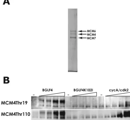
FIG. 4. EBV-PK phosphorylates Thr-19 and Thr-110 residues on MCM4 of the 6-7 hexamer in vitro. (A) Purification of MCM4-6-7 hexamers. Sf21 cells were coinfected with recombinant baculovi-ruses, Bac-Mcm4-6 and Bac-Mcm7, and MCM4-6-7 complexes were purified as described in Materials and Methods, separated by SDS-7.5% PAGE, and stained with silver. The positions of the MCM4, Mcm6, and MCM7 proteins were determined by Western blot analyses (data not shown) and are indicated by arrows. (B) Wild-type or kinase-negative GST-BGLF4 proteins were isolated from Sf21 cells infected with Bac-GST-BGLF4 or Bac-GST-BGLF4K102Ias described in
Ma-terials and Methods. Human MCM4-6-7 hexamer (100 ng) was incu-bated with increasing amounts of wild-type GST-BGLF4, kinase-negative GST-BGLF4K102I, or cyclin A/CDK2 (⫺, none). The
samples were subjected to SDS-7.5% PAGE and analyzed by West-ern blotting using phosphospecific antibodies against MCM4 at Thr-19 and Thr-110.
on November 8, 2019 by guest
http://jvi.asm.org/
[image:5.585.76.255.77.399.2]the cells continued to proliferate normally. Thus, it was dem-onstrated clearly that the expression of the BGLF4 protein inhibits the proliferation of Tet-BZLF1/B95-8 cells.
Both the EBV-PK and CDK2/cyclin A phosphorylate Thr-19 and Thr-110 sites on MCM4 of the MCM4-6-7 complex in vitro.Next, we examined whether the EBV-PK can directly phosphorylate Thr-19 and Thr-110 sites on MCM4 of the MCM4-6-7 complex in vitro (Fig. 4). As shown in Fig. 4A, the recombinant His-tagged MCM4-6-7 hexamer was purified through Ni-nitrilotriacetic acid column chromatography and glycerol density gradient centrifugation from extracts of re-combinant-baculovirus-infected cells and used as a substrate in in vitro kinase assays (Fig. 4A). Also, WT and kinase-inactive BGLF4 proteins were purified from the recombinant-baculo-virus-infected cells. Both the WT BGLF4 protein and the CDK2/cyclin A complex could phosphorylate MCM4 of the MCM4-6-7 hexamer at Thr-19 and Thr-110 sites in a dose-dependent manner, whereas the kinase-inactive BGLF4K102I protein did not (Fig. 4B). The mobilities of MCM4 phosphor-ylated by the BGLF4 protein and by CDK2/cyclin A differed, suggesting that EBV-PK might phosphorylate many sites on MCM4 in addition to those phosphorylated by CDK2. What-ever the case, the data indicate that EBV-PK is able to phos-phorylate at least Thr-19 and Thr-110 of MCM4 in MCM complexes directly.
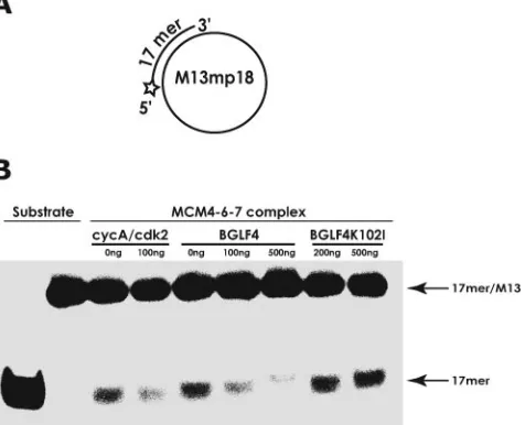
DNA helicase activity of MCM4-6-7 hexamers is inhibited by EBV-PK as well as by CDK2/cyclin A.To date, DNA heli-case activity has been identified for the MCM4-6-7 complex exclusively. The effect of EBV-PK on MCM4-6-7 complex-associated DNA helicase activity was examined using a 32 P-labeled 17-mer annealed to M13 single-stranded DNA as the substrate (Fig. 5A). The purified MCM4-6-7 hexamer was able to effectively unwind the 17-mer primers from annealed M13 DNA substrates (Fig. 5B), and this was inhibited by addition of CDK2/cyclin A, as reported previously (20). Similarly, EBV-PK inhibited the DNA helicase activity of the complex in a dose-dependent manner, whereas kinase-negative GST-BGLF4K102I(up to 200 ng) did not (Fig. 5B).
EBV-PK, unlike CDK2/cyclin A, inhibits helicase activity of MCM4-6-7 hexamers containing mutations in amino-terminal phosphorylation sites of MCM4.To determine whether the suppression of MCM4-6-7 hexamer-associated helicase activity by EBV-PK is actually due to phosphorylation of specific sites on MCM4, mutated MCM4-6-7 hexamers containing six amino acid mutations (to alanine) at amino-terminal phosphorylation sites on MCM4 (aa 3, 7, 19, 32, 54, and 110), which are there-fore resistant to phosphorylation by CDK2/cyclin A, were ex-amined for DNA helicase activity in the presence of EBV-PK. As shown in Fig. 6A, the mutated MCM was as sensitive to the inhibition of DNA helicase activity by EBV-PK as the WT MCM complex. In contrast, the mutated MCM was resistant to the inhibition by CDK2/cyclin A (compare Fig. 5B and 6A). Thus, EBV-PK inhibited the MCM complex-associated heli-case activity more powerfully than CDK2/cyclin A. Although phosphorylation of the N-terminal six amino acids in MCM4 is mainly responsible for inhibition of MCM4-6-7-associated DNA helicase, EBV-PK might further phosphorylate other sites of MCM4 or other subunits of the MCM complex, causing inhibition of the helicase activity.
EBV-PK phosphorylates MCM6 and another site(s) of MCM4 in addition to the amino-terminal six Ser/Thr residues.
[image:6.585.305.543.73.266.2]We examined whether the EBV-PK can directly phosphorylate another site(s) besides the amino-terminal six Ser/Thr residues on MCM4 and other subunits of the MCM4-6-7 complex in vitro (Fig. 6B). Human intact MCM4-6-7 and mutated MCM4a-6-7 hexamers containing six amino acid mutations (to alanine) at amino-terminal phosphorylation sites on MCM4 (aa 3, 7, 19, 32, 54, and 110) were used as substrates for in vitro kinase assays. CDK2/cyclin A phosphorylated mainly MCM4 of the MCM4-6-7 hexamer but hardly the N-terminal-mutated MCM4. Thus, CDK2/cyclin A phosphorylates the N-terminal Ser/Thr res-idues mainly on MCM4. In contrast, EBV-PK could clearly phos-phorylate the mutated MCM4 of the MCM4a-6-7 hexamers, in-dicating that EBV-PK phosphorylates another site(s) in addition to amino-terminal phosphorylation sites on MCM4. Phosphory-lation of MCM6 and -7 by CDK2/cyclin A was scarcely ob-served (Fig. 6B). When intact MCM4-6-7 complex was reacted with EBV-PK, a broad band of MCM4 was observed by SDS-PAGE, probably due to phosphorylation (Fig. 6C). However, when the mutated complex was reacted with EBV-PK, the mobility of the mutated MCM4 by SDS-PAGE was virtually unchanged, distinguishing the mutated MCM4 from MCM6 (Fig. 6C). As shown in Fig. 6B, the EBV-PK phosphorylated both MCM6 and the mutated MCM4. Collectively, EBV-PK inhibits the MCM complex-associated DNA helicase activity through phosphorylation of many sites, including six amino-terminal phosphorylation sites on MCM4 and also MCM6 of the MCM4-6-7 hexamer.
FIG. 5. EBV-PK, like CDK2/cyclin A, inhibits DNA helicase (un-winding) activity associated with the MCM4-6-7 complex. (A) Sub-strate for DNA helicase assays. A 5⬘32P-labeled oligonucleotide
(17-mer) annealed to M13 single-stranded DNA is depicted. (B) DNA helicase assays were performed with 100 fmol of the helicase substrate and human MCM4-6-7 hexamers (100 ng) in the presence of cyclin A/CDK2 (100 ng), BGLF4 protein (100 and 500 ng), or kinase-nega-tive BGLF4K102Iprotein (200 and 500 ng) as described in Materials
and Methods. Positions of the DNA substrate (17mer/M13) and dis-placed oligonucleotide (17mer) are indicated by arrows. The left two lanes show results for heat-denatured and native DNA substrates, respectively.
on November 8, 2019 by guest
http://jvi.asm.org/
DISCUSSION
EBV lytic infection induces hyperphosphorylation of retino-blastoma protein, with elevated levels of E2F-1, cyclin E, and cyclin A and their associated CDK2 activities, while cyclin B/CDK1 activity remains constant (32, 33). The present study provided evidence with phosphospecific antibodies that virus-mediated changes occur in the phosphorylation of MCM4 of the MCM complex, with involvement not only of CDK2/CDK1 but also of EBV-encoded protein kinase. These molecules are likely to act redundantly.
MCM4 exhibits different phosphorylation states that depend on the cell cycle phases and their association with chromatin (16). In the normal cell cycle, MCM4 is phosphorylated by CDK2/cyclin A and CDK1/cyclin B, primarily during S phase and mitosis, respectively (17, 22). The phosphorylation of a mutant complex containing mouse MCM4 mutated at six sites (aa 3, 7, 19, 32, 53, and 109) showed almost no phosphorylation in this region of MCM4 (20). Thus, these three Ser-Pro (aa 3, 32, and 53) and three Thr-Pro (aa 7, 19, and 109) sites are required for phosphorylation of MCM4 in the MCM4-6-7 complex by CDK2/cyclin A. The same sites in the amino-terminal region of MCM4 appeared to be required for the phosphorylation by CDK1/cyclin B in vitro (17), suggesting that both CDK2/cyclin A and CDK1/cyclin B have similar specificities of substrate recognition. Furthermore, Komamura-Kohno et al. have reported recently that phosphorylation of MCM4 at Thr-7, Thr-19, Ser-32, Ser-88, and Thr-110 in the M phase requires CDK1 (Cdc2), although they do not deny the possibility of the phosphorylation of these sites on MCM4 by CDK2 (29). Therefore, it must be noted that there is also a possibility of the involvement of CDK1/cyclin B in MCM4 phosphorylation, since the CDK1/cyclin B activity remains es-sentially constant and high throughout lytic replication (33).
MCM proteins are present in cells in chromatin-bound as well as unbound forms, but at G2/M phase, almost all are heavily phosphorylated and present as the unbound form (16). Hyperphosphorylation of the MCM complex prohibits inap-propriate reloading of MCMs onto chromatin and thereby contributes suppression of rereplication. In heterohexameric Mcm2-7 complexes, either the chromatin-bound or the un-bound form may be formed (16). Although phosphorylation of MCM4 partly resulted in the dissociation of the MCM complex from chromatin, as shown in Fig. 1A, the preparation of the chromatin-bound form of MCM proteins was found to be almost unchanged. It is suggested here that inhibition of the DNA unwinding function of the hexameric MCM complex on chromatin occurs during EBV lytic replication as another role for the phosphorylation of MCM4.
[image:7.585.42.281.68.533.2]In the case of HCMV, it has been reported that the virus prevents prereplicative complex assembly by inhibiting MCM complex loading onto chromatin (4, 53), but quiescent primary
FIG. 6. EBV-PK, unlike CDK2/cyclin A, still inhibits helicase ac-tivity of MCM4-6-7 hexamers containing mutations in amino-terminal phosphorylation sites of MCM4. (A) MCM4-6-7 hexamers containing mutant MCM4 (MCM4a/6/7) (200 ng) or the wild type (MCM4-6-7) (100 ng) were examined for DNA helicase activity in the presence or absence of BGLF4 protein (500 ng) or cyclin A/CDK2 (500 ng) as described in Materials and Methods. The left two lanes show results for heat-denatured and native DNA substrates, respectively. Quanti-tative analysis of the DNA unwinding activities is shown in the graph. The percentage of 17-mer oligonucleotide displaced with each MCM complex in the presence of the BGLF4 EBV-PK or CDK2/cyclin A was calculated from the signal intensity, with that in the absence of the kinase taken as 100%. Data are means⫾standard deviations of three independent experiments. (B) Human MCM4-6-7 or MCM4a-6-7 complexes (1g) were incubated with indicated amounts of CDK2/ cyclin A (left) or the BGLF4 protein (right) in a 50-l reaction mixture containing 20 mM Tris-HCl (pH 7.5), 1 mM EDTA, 1 mM dithiothre-itol, 10 mM MgCl2, 100M ATP, 2 Ci [␥
-32P]ATP, and 0.2 mM
sodium orthovanadate.32P-labeled proteins were separated by
SDS-7.5% PAGE followed by autoradiography. (C) Human MCM4-6-7 or MCM4a-6-7 complexes (1g) were phosphorylated by 500 ng of the
BGLF4 protein in a 50-l reaction mixture containing 20 mM Tris-HCl (pH 7.5), 1 mM EDTA, 1 mM dithiothreitol, 10 mM MgCl2, 1
mM ATP, and 0.2 mM sodium orthovanadate, and products were separated by SDS-7.5% PAGE. Proteins were analyzed by Western blotting using MCM6 and MCM4 antibodies.
on November 8, 2019 by guest
http://jvi.asm.org/
fibroblasts (G0phase) were used so that there was no expres-sion of prereplicative factors like the ORC, CDC6, Cdt1, and the MCM complex. However, both reactivation of EBV from latently infected B cells and infection of HCMV into cycling cells result in cell cycle arrest and blockage of chromosomal DNA replication. Since levels of the chromatin-bound forms of MCM4, MCM6, and MCM7 were essentially unchanged be-fore and after induction in our investigation, the chromosomal DNA replication arrest during EBV lytic infection might not be explained only by dissociation of MCM complexes from chromatin. Overlap of multiple factors might result in the chromosomal DNA replication arrest.
Components of the replication fork include the trimeric, single-stranded DNA binding RPA complex and the DNA helicase. The identity of the replicative helicase which func-tions in the elongation step remains equivocal. Genetic and biochemical evidence supports a role for the MCM2-7 complex providing helicase activity in unwinding DNA at replication origins during initiation (37). The MCM2-7 proteins form a stable complex in vivo (14), although detectable helicase activ-ity is observed only with the MCM4-6-7 subcomplex (19). Cur-rent models suggest that this subcomplex may represent the active helicase, while the remaining subunits may have an es-sential role in regulating its activity (38, 57). Moreover, a role for the MCM complex has also been suggested to occur during the elongation step, and in budding yeast (Saccharomyces cerevisiae), MCM4 appears to move away from replication or-igins after initiation of DNA synthesis (1, 34, 47). Although the MCM2-7-related protein MCM8 recently has been reported to possess ATP-dependent helicase activity (38), the MCM4-6-7 complex is still thought to be a strong candidate for replicative helicase in the elongation step of DNA replication. Taking this into consideration, MCM4 phosphorylation during EBV pro-ductive replication might suppress refiring of chromosomal DNA replication by blocking DNA unwinding activity and also prevent fork movement by blocking replicative DNA helicase activity. EBV might have adopted multiple redundant path-ways to halt the cell cycle. Whatever the underlying mecha-nism, phosphorylation of MCM4 provides one means to block chromosomal DNA replication in lytic infected cells.
ACKNOWLEDGMENTS
We thank Y. Nishikawa for technical assistance.
This work was supported by grants-in-aid for Scientific Research on Priority Areas from the Ministry of Education, Science, Sports, Cul-ture and Technology of Japan (no. 17659138, 16017322, and 15390153 to T.T.) and partly by the Uehara Memorial Foundation. A.K. was supported by a Research Fellowship of the Japanese Society for the Promotion of Science for Young Scientists.
REFERENCES
1.Aparicio, O. M., D. M. Weinstein, and S. P. Bell.1997. Components and dynamics of DNA replication complexes in S. cerevisiae: redistribution of
MCM proteins and Cdc45p during S phase. Cell91:59–69.
2.Bailis, J. M., and S. L. Forsburg.2004. MCM proteins: DNA damage,
mutagenesis and repair. Curr. Opin. Genet. Dev.14:17–21.
3.Bell, S. P., and A. Dutta.2002. DNA replication in eukaryotic cells. Annu.
Rev. Biochem.71:333–374.
4.Biswas, N., V. Sanchez, and D. H. Spector.2003. Human cytomegalovirus infection leads to accumulation of geminin and inhibition of the licensing of
cellular DNA replication. J. Virol.77:2369–2376.
5.Chen, M.-R., S.-J. Chang, H. Huang, and J.-Y. Chen.2000. A protein kinase activity associated with Epstein-Barr virus BGLF4 phosphorylates the viral
early antigen EA-D in vitro. J. Virol.74:3093–3104.
6.Correa-Bordes, J., and P. Nurse.1995. p25rum1 orders S phase and mitosis
by acting as an inhibitor of the p34cdc2 mitotic kinase. Cell83:1001–1009.
7.Dahmann, C., J. F. Diffley, and K. A. Nasmyth.1995. S-phase-promoting cyclin-dependent kinases prevent re-replication by inhibiting the transition
of replication origins to a pre-replicative state. Curr. Biol.5:1257–1269.
8.Daikoku, T., A. Kudoh, M. Fujita, Y. Sugaya, H. Isomura, N. Shirata, and T. Tsurumi.2005. Architecture of replication compartments formed during
Epstein-Barr virus lytic replication. J. Virol.79:3409–3418.
9.Daikoku, T., A. Kudoh, M. Fujita, Y. Sugaya, H. Isomura, and T. Tsurumi.
2004. In vivo dynamics of EBNA1-oriP interaction during latent and lytic
replication of Epstein-Barr virus. J. Biol. Chem.279:54817–54825.
10.Diffley, J. F.1996. Once and only once upon a time: specifying and regulating
origins of DNA replication in eukaryotic cells. Genes Dev.10:2819–2830.
11.Forsburg, S. L.2004. Eukaryotic MCM proteins: beyond replication
initia-tion. Microbiol. Mol. Biol. Rev.68:109–131.
12.Fujita, M., Y. Ishimi, H. Nakamura, T. Kiyono, and T. Tsurumi.2002. Nuclear organization of DNA replication initiation proteins in mammalian
cells. J. Biol. Chem.277:10354–10361.
13.Fujita, M., T. Kiyono, Y. Hayashi, and M. Ishibashi.1996. hCDC47, a human member of the MCM family. Dissociation of the nucleus-bound form
during S phase. J. Biol. Chem.271:4349–4354.
14.Fujita, M., T. Kiyono, Y. Hayashi, and M. Ishibashi.1997. In vivo interaction of human MCM heterohexameric complexes with chromatin. Possible
in-volvement of ATP. J. Biol. Chem.272:10928–10935.
15.Fujita, M., C. Yamada, H. Goto, N. Yokoyama, K. Kuzushima, M. Inagaki, and T. Tsurumi.1999. Cell cycle regulation of human CDC6 protein. Intra-cellular localization, interaction with the human MCM complex, and CDC2
kinase-mediated hyperphosphorylation. J. Biol. Chem.274:25927–25932.
16.Fujita, M., C. Yamada, T. Tsurumi, F. Hanaoka, K. Matsuzawa, and M. Inagaki.1998. Cell cycle- and chromatin binding state-dependent phosphor-ylation of human MCM heterohexameric complexes. A role for cdc2 kinase.
J. Biol. Chem.273:17095–17101.
17.Hendrickson, M., M. Madine, S. Dalton, and J. Gautier.1996. Phosphory-lation of MCM4 by cdc2 protein kinase inhibits the activity of the
minichro-mosome maintenance complex. Proc. Natl. Acad. Sci. USA93:12223–12228.
18.Hozak, P., A. B. Hassan, D. A. Jackson, and P. R. Cook.1993. Visualization
of replication factories attached to nucleoskeleton. Cell73:361–373.
19.Ishimi, Y.1997. A DNA helicase activity is associated with an MCM4, -6, and
-7 protein complex. J. Biol. Chem.272:24508–24513.
20.Ishimi, Y., and Y. Komamura-Kohno.2001. Phosphorylation of Mcm4 at specific sites by cyclin-dependent kinase leads to loss of Mcm4,6,7 helicase
activity. J. Biol. Chem.276:34428–34433.
21.Ishimi, Y., Y. Komamura-Kohno, H. J. Kwon, K. Yamada, and M. Nakanishi.
2003. Identification of MCM4 as a target of the DNA replication block
check-point system. J. Biol. Chem.278:24644–24650.
22.Ishimi, Y., Y. Komamura-Kohno, Z. You, A. Omori, and M. Kitagawa.2000. Inhibition of Mcm4,6,7 helicase activity by phosphorylation with cyclin
A/Cdk2. J. Biol. Chem.275:16235–16241.
23.Itzhaki, J. E., C. S. Gilbert, and A. C. Porter.1997. Construction by gene targeting in human cells of a “conditional” CDC2 mutant that rereplicates its
DNA. Nat. Genet.15:258–265.
24.Jares, P., and J. J. Blow.2000. Xenopus cdc7 function is dependent on licensing but not on XORC, XCdc6, or CDK activity and is required for
XCdc45 loading. Genes Dev.14:1528–1540.
25.Kato, K., Y. Kawaguchi, M. Tanaka, M. Igarashi, A. Yokoyama, G. Matsuda, M. Kanamori, K. Nakajima, Y. Nishimura, M. Shimojima, H. T. Phung, E. Takahashi, and K. Hirai.2001. Epstein-Barr virus-encoded protein kinase BGLF4 mediates hyperphosphorylation of cellular elongation factor 1delta (EF-1delta): EF-1delta is universally modified by conserved protein kinases
of herpesviruses in mammalian cells. J. Gen. Virol.82:1457–1463.
26.Kato, K., A. Yokoyama, Y. Tohya, H. Akashi, Y. Nishiyama, and Y. Kawaguchi.
2003. Identification of protein kinases responsible for phosphorylation of Epstein-Barr virus nuclear antigen leader protein at serine-35, which regulates
its coactivator function. J. Gen. Virol.84:3381–3392.
27.Kawaguchi, Y., and K. Kato.2003. Protein kinases conserved in herpesvi-ruses potentially share a function mimicking the cellular protein kinase cdc2.
Rev. Med. Virol.13:331–340.
28.Kawaguchi, Y., K. Kato, M. Tanaka, M. Kanamori, Y. Nishiyama, and Y. Yamanashi.2003. Conserved protein kinases encoded by herpesviruses and cellular protein kinase cdc2 target the same phosphorylation site in
eukary-otic elongation factor 1␦. J. Virol.77:2359–2368.
29.Komamura-Kohno, Y., K. Karasawa-Shimizu, T. Saitoh, M. Sato, F. Hanaoka, S. Tanaka, and Y. Ishimi.2006. Site-specific phosphorylation of
MCM4 during the cell cycle in mammalian cells. FEBS J.273:1224–1239.
30.Kreitz, S., M. Ritzi, M. Baack, and R. Knippers.2001. The human origin recognition complex protein 1 dissociates from chromatin during S phase in
HeLa cells. J. Biol. Chem.276:6337–6342.
31.Kudoh, A., T. Daikoku, Y. Sugaya, H. Isomura, M. Fujita, T. Kiyono, Y. Nishiyama, and T. Tsurumi.2004. Inhibition of S-phase cyclin-dependent kinase activity blocks expression of Epstein-Barr virus immediate-early and
early genes, preventing viral lytic replication. J. Virol.78:104–115.
32.Kudoh, A., M. Fujita, T. Kiyono, K. Kuzushima, Y. Sugaya, S. Izuta, Y.
on November 8, 2019 by guest
http://jvi.asm.org/
Nishiyama, and T. Tsurumi.2003. Reactivation of lytic replication from B cells latently infected with Epstein-Barr virus occurs with high S-phase cyclin-dependent kinase activity while inhibiting cellular DNA replication.
J. Virol.77:851–861.
33.Kudoh, A., M. Fujita, L. Zhang, N. Shirata, T. Daikoku, Y. Sugaya, H. Isomura, Y. Nishiyama, and T. Tsurumi.2005. Epstein-Barr virus lytic replication elicits ATM checkpoint signal transduction while providing an
S-phase-like cellular environment. J. Biol. Chem.280:8156–8163.
34.Labib, K., J. A. Tercero, and J. F. Diffley.2000. Uninterrupted MCM2-7
function required for DNA replication fork progression. Science288:1643–
1647.
35.Lee, J. K., and J. Hurwitz.2000. Isolation and characterization of various complexes of the minichromosome maintenance proteins of
Schizosaccha-romyces pombe. J. Biol. Chem.275:18871–18878.
36.Lee, J. K., and J. Hurwitz.2001. Processive DNA helicase activity of the minichromosome maintenance proteins 4, 6, and 7 complex requires forked
DNA structures. Proc. Natl. Acad. Sci. USA98:54–59.
37.Lei, M., and B. K. Tye.2001. Initiating DNA synthesis: from recruiting to
activating the MCM complex. J. Cell Sci.114:1447–1454.
38.Maiorano, D., O. Cuvier, E. Danis, and M. Mechali.2005. MCM8 is an MCM2-7-related protein that functions as a DNA helicase during replication
elongation and not initiation. Cell120:315–328.
39.McGarry, T. J., and M. W. Kirschner.1998. Geminin, an inhibitor of DNA
replication, is degraded during mitosis. Cell93:1043–1053.
40.Mendez, J., X. H. Zou-Yang, S. Y. Kim, M. Hidaka, W. P. Tansey, and B. Stillman.2002. Human origin recognition complex large subunit is degraded by ubiquitin-mediated proteolysis after initiation of DNA replication. Mol.
Cell9:481–491.
41.Mimura, S., T. Masuda, T. Matsui, and H. Takisawa.2000. Central role for cdc45 in establishing an initiation complex of DNA replication in Xenopus
egg extracts. Genes Cells5:439–452.
42.Nurse, P.1994. Ordering S phase and M phase in the cell cycle. Cell
79:547–550.
43.Sato, M., T. Gotow, Z. You, Y. Komamura-Kohno, Y. Uchiyama, N. Yabuta, H. Nojima, and Y. Ishimi.2000. Electron microscopic observation and sin-gle-stranded DNA binding activity of the Mcm4,6,7 complex. J. Mol. Biol.
300:421–431.
44.Schwacha, A., and S. P. Bell.2001. Interactions between two catalytically distinct MCM subgroups are essential for coordinated ATP hydrolysis and
DNA replication. Mol. Cell8:1093–1104.
45.Stillman, B.1996. Cell cycle control of DNA replication. Science274:1659– 1664.
46.Tada, S., A. Li, D. Maiorano, M. Mechali, and J. J. Blow.2001. Repression of origin assembly in metaphase depends on inhibition of RLF-B/Cdt1 by
geminin. Nat. Cell Biol.3:107–113.
47.Tanaka, T., D. Knapp, and K. Nasmyth.1997. Loading of an Mcm protein
onto DNA replication origins is regulated by Cdc6p and CDKs. Cell90:649–
660.
48.Tatsumi, Y., S. Ohta, H. Kimura, T. Tsurimoto, and C. Obuse.2003. The ORC1 cycle in human cells. I. Cell cycle-regulated oscillation of human
ORC1. J. Biol. Chem.278:41528–41534.
49.Thommes, P., Y. Kubota, H. Takisawa, and J. J. Blow.1997. The RLF-M component of the replication licensing system forms complexes containing
all six MCM/P1 polypeptides. EMBO J.16:3312–3319.
50.Tye, B. K.1999. MCM proteins in DNA replication. Annu. Rev. Biochem.
68:649–686.
51.Waga, S., and B. Stillman.1998. The DNA replication fork in eukaryotic
cells. Annu. Rev. Biochem.67:721–751.
52.Walter, J., and J. Newport.2000. Initiation of eukaryotic DNA replication: origin unwinding and sequential chromatin association of Cdc45, RPA, and
DNA polymerase alpha. Mol. Cell5:617–627.
53.Wiebusch, L., R. Uecker, and C. Hagemeier.2003. Human cytomegalovirus prevents replication licensing by inhibiting MCM loading onto chromatin.
EMBO Rep.4:42–46.
54.Wohlschlegel, J. A., B. T. Dwyer, S. K. Dhar, C. Cvetic, J. C. Walter, and A. Dutta.2000. Inhibition of eukaryotic DNA replication by geminin binding to
Cdt1. Science290:2309–2312.
55.Wuarin, J., V. Buck, P. Nurse, and J. B. Millar.2002. Stable association of mitotic cyclin B/Cdc2 to replication origins prevents endoreduplication. Cell
111:419–431.
56.You, Z., Y. Komamura, and Y. Ishimi.1999. Biochemical analysis of the
intrinsic Mcm4-Mcm6-Mcm7 DNA helicase activity. Mol. Cell. Biol.19:
8003–8015.
57.You, Z., and H. Masai.2005. DNA binding and helicase actions of mouse
MCM4/6/7 helicase. Nucleic Acids Res.33:3033–3047.
58.Yue, W., E. Gershburg, and J. S. Pagano.2005. Hyperphosphorylation of EBNA2 by Epstein-Barr virus protein kinase suppresses transactivation of
the LMP1 promoter. J. Virol.79:5880–5885.
59.Zou, L., and B. Stillman.2000. Assembly of a complex containing Cdc45p, replication protein A, and Mcm2p at replication origins controlled by S-phase cyclin-dependent kinases and Cdc7p-Dbf4p kinase. Mol. Cell. Biol.
20:3086–3096.