0022-538X/06/$08.00⫹0 doi:10.1128/JVI.00094-06
Copyright © 2006, American Society for Microbiology. All Rights Reserved.
Consistent Effects of
TSG101
Genetic Variability on Multiple Outcomes
of Exposure to Human Immunodeficiency Virus Type 1†
Arman A. Bashirova,
1‡ Gabriela Bleiber,
2Ying Qi,
3Holli Hutcheson,
1Traci Yamashita,
4Randall C. Johnson,
3Jie Cheng,
3Galit Alter,
5James J. Goedert,
6Susan Buchbinder,
7Keith Hoots,
8David Vlahov,
4Margaret May,
9Frank Maldarelli,
10Lisa Jacobson,
4Stephen J. O’Brien,
1Amalio Telenti,
2and Mary Carrington
3*
Laboratory of Genomic Diversity, National Cancer Institute, Frederick, Maryland 217021; Institute of Microbiology and Division of Infectious Diseases, University Hospital of Lausanne, Lausanne, Switzerland2; Laboratory of Genomic Diversity, SAIC-Frederick,
Inc., National Cancer Institute, Frederick, Maryland 217023; Department of Epidemiology, School of Hygiene and Public Health, The Johns Hopkins University, Baltimore, Maryland4; Partners AIDS Research Center, Massachusetts General Hospital,
55 Fruit Street, Charlestown, Massachusetts 021295; Viral Epidemiology Branch, Division of Cancer Epidemiology and Genetics, NCI, Bethesda, Maryland6; University of California, San Francisco, California7; Gulf States Hemophilia Center,
University of Texas Health Science Center, Houston, Texas8; Department of Social Medicine, University of Bristol, Bristol, United Kingdom9; and HIV Drug Resistance Program, National Cancer Institute, Bethesda, Maryland10
Received 13 January 2006/Accepted 16 April 2006
Tumor susceptibility gene 101 (TSG101) encodes a host cellular protein that is appropriated by human immunodeficiency virus type 1 (HIV-1) in the budding process of viral particles from infected cells. Variation in the coding or noncoding regions of the gene could potentially affect the degree of TSG101-mediated release of viral particles. While the coding regions of the gene were found to lack nonsynonymous variants, two polymorphic sites in theTSG1015ⴕarea were identified that were associated with the rate of AIDS progression among Caucasians. These single-nucleotide polymorphisms (SNPs), located at positions ⴚ183 and ⴙ181 relative to the translation start, specify three haplotypes termed A, B, and C, which occur at frequencies of 67%, 21%, and 12%, respectively. Haplotype C is associated with relatively rapid AIDS progression, while haplotype B is associated with slower disease progression. Both effects were dominant over the intermediate haplotype A. The haplotypes also demonstrated parallel effects on the rate of CD4 T-cell depletion and viral load increase over time, as well as a possible influence on HIV-1 infection. The data raise the hypothesis that noncoding variation inTSG101affects the efficiency of TSG101-mediated release of viral particles from infected cells, thereby altering levels of plasma viral load and subsequent disease progression.
One fundamental feature of a successful virus is its ability to utilize the host cellular machinery in order to support its prop-agation. Understanding the interactions between host and viral proteins provides the opportunity to identify means for con-trolling outcomes of viral infection. Recent studies have dis-closed a variety of cellular molecules that are exploited by human immunodeficiency virus type 1 (HIV-1) at different stages of its life cycle, one of which, the tumor suppressor gene 101 (TSG101) protein, is essential for budding of the virus from infected cells (10, 12, 21, 35).
TSG101is an evolutionarily conserved gene located on hu-man chromosome 11p15. It encodes a 46-kDa multidomain protein that contains an N-terminal ubiquitin-conjugating en-zyme E2 variant (UEV) domain, a proline-rich domain, a coiled-coil region including a leucine zipper, and a C-terminal
␣-helical domain. The TSG101 protein has been detected in
the nucleus and cytoplasm, and its localization is cell cycle dependent (38, 39).
A potential role for TSG101as a tumor suppressor that was suggested in an early study of the gene (17) remains controversial (22, 34). Identification of molecules with which TSG101 interacts has suggested its involvement in transcriptional regulation (5, 13, 24, 32) and cell cycle con-trol (6, 18, 27). Targeted deletion oftsg101in mice results in early embryonic death due to a defect in cellular prolifera-tion (29), and reports of cell cycle arrest and death in
TSG101-deficient cells have further confirmed a critical role for TSG101 in cell survival (6, 16).
Many recent studies of TSG101 have focused on its role in endosomal trafficking. TSG101 and its yeast orthologue, Vps23, belong to the so-called “class E” proteins whose func-tions are essential for vacuolar protein sorting (1). TSG101/ Vps23, along with two other proteins, Vps28 and Vps37, form a ⬃350-kDa complex named ESCRT-I (endosomal sorting
complexrequired fortransport) (14). ESCRT-I is involved in a series of protein-protein interactions that result in sorting of ubiquitylated proteins from early endosomes into multivesicu-lar bodies (MVBs). During this process, early endosomes car-rying protein cargo bud into MVBs, organelles that eventually fuse with lysosomes for subsequent protein degradation. This process entails direct binding of the TSG101/Vps23 UEV do-main to ubiquitin, resulting in the delivery of ubiquitylated
* Corresponding author. Mailing address: Laboratory of Genomic Diversity, SAIC-Frederick, Inc., National Cancer Institute, Frederick, Maryland 21702. Phone: (301) 846-1390. Fax: (301) 846-1909. E-mail: [email protected].
‡ Present address: Johns Hopkins University School of Medicine, Baltimore, MD 21231.
† Supplemental material for this article may be found at http://jvi .asm.org.
6757
on November 8, 2019 by guest
http://jvi.asm.org/
proteins (that are destined for degradation) into MVBs (2, 12, 14, 20, 28, 33).
Some viruses have developed an ingenious mechanism for budding from cells that involves the host endosomal sorting process. This became evident after the discovery of the direct interaction between the UEV domain of TSG101 and a highly conserved motif in the p6 region of the HIV-1 Gag protein, Pro-Thr/Ser-Ala-Pro (PTAP), an interaction that was shown to be critical for the release of HIV-1 particles from the cellular membrane (10, 12, 21, 35). The HIV-1 PTAP motif belongs to a family of late (L) domains, so named for their late involve-ment in the viral life cycle. The viral L domains are also characterized by conserved PPXY and YXXL motifs, which, along with PTAP, interact with host proteins involved in the MVB pathway (reviewed in reference 9). The TSG101–HIV-1 relationship has been studied intensively by several laborato-ries, resulting in the identification of additional host proteins involved in the process of HIV-1 budding (30, 31, 36). The TSG101 molecule has also been shown to be involved in the budding of HIV-2 (25), Ebola virus (21), human T-cell leuke-mia virus type 1 (4), and bluetongue virus (37).
Given the central role of TSG101 in release of HIV-1 from infected cells, we hypothesized that genetic variations inTSG101
could potentially affect the functional activity of TSG101 protein in viral budding, thereby altering levels of circulating virus in the blood of infected individuals and the clinical course of AIDS. Here, we report two single-nucleotide polymorphism (SNP) variants, located at positions⫺183 and ⫹181 relative to the translation start site, that associate with differences in viral load dynamics, in CD4 T-cell decline, and, correspondingly, with the rate of AIDS progression after infection. The⫺183 variant has been recently reported to be associated with faster CD4 decline in the Swiss HIV Cohort Study (SHCS) (3). Here, we demonstrate a more detailed analysis of the association between TSG101 variation and several outcomes of HIV-1 infection, which are consistent in the SHCS and a large sample of U.S. AIDS cohorts.
MATERIALS AND METHODS
Subjects.The study group included patients from five U.S. cohorts and a Swiss cohort: the AIDS Linked to the Intravenous Drug Experience Study, the He-mophilia Growth and Development Study, the Multicenter AIDS Cohort Study (MACS), the Multicenter Hemophilia Cohort Study, the San Francisco City Clinic Cohort Study (26), and the SHCS (http://www.shcs.ch). The seroconver-sion date was estimated as the midpoint between the first positive and the last negative HIV-1 antibody test (mean interval, 0.79 years; range, 0.07 to 3.0 years). High-risk exposed uninfected subjects were defined as those who reported en-gaging in behavior that confers high risk for HIV-1 exposure (continual sharing of injection equipment or anal receptive sex with multiple partners) or transfu-sions with factor VIII known to be contaminated with HIV-1. Longitudinal CD4 count values were available for MACS and SHCS, whereas viral load measure-ments were available only for the MACS cohort.
Genotyping. The single-strand conformation polymorphism technique was
performed as described by Cullen et al. (8). TheTSG101SNPs were typed using
TaqMan Genotyping Assays (Applied Biosystems, Foster City, CA) according to the manufacturer’s protocol. Haplotypes were estimated in compound heterozy-gotes using the expectation maximization algorithm (19) and SAS software (SAS Institute, Cary, NC).
Statistical analysis. Four end points reflecting disease progression (AIDS
outcome) were evaluated: time to CD4⬍200 cells/mm3; progression to AIDS
according to the 1987 definition by the Centers for Disease Control and Preven-tion; progression to AIDS according to the 1993 definition by CDC; and AIDS-related death. Defined categorical analysis was performed comparing genotype and haplotype frequencies between groups of patients using a two-tailed Fisher’s
exact test. Analyses presented (see Tables1 to 3, Fig. 2, and tables in the sup-plemental material) were carried out using the SAS software. Participants were stratified by ethnic group, sex, and age at seroconversion (0 to 20, 20 to 40, and over 40 years).
CD4 decline (see Fig. 3) and HIV RNA increase (see Fig. 4) over time were assessed with random effects linear models in which the intercepts were allowed to vary randomly for each participant. These models accounted for the correla-tion between serial measurements from each person and were implemented using the lme 4 package in R (http://www.r-project.org/). The three genotypic categories were included as indicator variables in the models, and both the intercepts and slopes were estimated. These models were adjusted by age, race, and injecting drug use reported at either seroconversion or when CD4 fell
between 500 and 600 cells/mm3for MACS and SHCS, respectively. All CD4 and
HIV RNA data were collected prior to report of highly active antiretroviral therapy use. For MACS patients included in these analyses, the seroconversion date was identified within a 2-year window. For over 90% of the 380 men included in the CD4 decline analysis, the seroconversion was known within a
1-year interval (median [interquartile range]⫽0.51 [0.48 to 0.58] years).
RESULTS
Identification ofTSG101 SNPs and haplotypes. An initial screening of theTSG101coding region in 50 healthy Caucasian blood donors using the single-strand conformation polymor-phism technique indicated the highly conserved nature of the gene, since no nonsynonymous nucleotide changes were iden-tified. Sequencing of 79 randomly chosen Caucasian serocon-verter patients (U.S. AIDS cohorts) was then performed in the 5⬘ area of the gene, which is likely to contain regulatory se-quences. The sequenced fragment consisted of⬃2 kb around exon 1, where nine SNPs were identified (Fig. 1A). Analysis of the SNP genotypes revealed six haplotypes with estimated fre-quencies of ⬎1% that could be defined by four haplotype-tagging SNPs (Fig. 1B) (positions ⫺600 [rs3802966], ⫺518 [rs1857909],⫺183 [rs2292179], and⫹181 [rs1395319] relative to the translation start site.
Analysis ofTSG101variants in the AIDS cohorts.The four
TSG101haplotype-tagging SNPs located in the 5⬘ area were typed in the U.S. AIDS cohorts. Haplotypes based on the four SNPs were estimated in a large population of 1,895 Caucasian individuals (Fig. 1C). The frequencies of the haplotypes in Caucasians differed only slightly from the initial estimation in 79 individuals (Fig. 1B).
The association of the individual SNPs and the five related haplotypes with disease progression was tested using the Cox proportional hazards model (7) and categorical analysis in which frequencies of genotypes were compared in people who developed AIDS during certain time periods. The Cox model did not reveal any significant effect for the four individual SNPs or for the five haplotypes (see Tables S1 and S2 in the supple-mental material). However, we did observe differences in the frequencies of the⫺600G and⫺183C variants among individ-uals who developed AIDS before 7 years compared to those who developed AIDS later (see Table S3 in the supplemental material). In similar analyses, haplotype 2 conferred a suscep-tible effect (odds ratio [OR]⫽1.6 to 2.5;P⫽0.002 to 0.01), haplotypes 3 and 5 were protective (OR⫽0.6 to 0.8,P⫽0.007 to 0.2, and OR⫽0.5 to 0.8,P⫽0.06 to 0.5), while haplotype 4 was relatively neutral (OR ⫽ 0.8 to 1.1; P ⫽ 0.2 to 0.8) compared to a reference haplotype 1 (see Table S4 in the supplemental material). Of note, the⫺600G variant, which is in strong linkage disequilibrium (LD) with the ⫺183T and
⫹181C variants, specifies haplotype 3, and the protective effect
on November 8, 2019 by guest
http://jvi.asm.org/
of this haplotype corresponded to the effect of the individual SNP. Based on these data and the haplotype structures, we concluded that susceptibility and protection are associated with variation at positions⫺183 and⫹181. Three of the four possible haplotypes composed of these two SNPs were ob-served, indicating strong LD between the variants. The corre-sponding haplotypes, T-A, T-C, and C-C, were termed A, B, and C, respectively (Fig. 1D).
Haplotypes B and C are associated with different rates of AIDS progression.The frequencies of haplotypes A, B, and C among individuals who developed AIDS in one of six distinct time periods after seroconversion (ⱕ3, 3 to 5, 5 to 7, 7 to 10, 10 to 12, and⬎12 years) were compared (data not shown). In
this analysis, seroprevalent individuals were included in the last three groups depending on the date of an AIDS-defining out-come after their first HIV⫹visit. This analysis suggested that the effect of the TSG101 haplotypes on HIV disease is not gradual over time, an observation that was further elucidated by the absence of a significant effect of these variants on AIDS progression in survival analysis using the Cox model, where time is a continuous variable starting from seroconversion (see Table S5 in the supplemental material). Rather, differences in susceptible (C) and protective (B) haplotypic frequencies ap-peared somewhat bimodal in that the frequency of the susceptible haplotypic group was significantly greater among individuals who progressed to AIDS within 7 years after seroconversion relative to those who remained AIDS-free for at least 7 years (domi-nant model; OR⫽1.72 to 2.30;P⫽0.0001 to 0.002) (Table 1). Conversely, the protective haplotypes were observed signifi-cantly more frequently among those who remained AIDS free for 7 years or longer after seroconversion compared to those who had progressed within 7 years (dominant model; OR⫽ 0.56 to 0.64;P⫽0.0005 to 0.01). Both the B and C haplotypes appeared to have dominant effects, since a codominant model did not fit the data as well as a dominant model. Haplotype A was relatively neutral (Table 1).
Protective, susceptible, and neutralTSG101haplotype groups.
Haplotypes B and C exhibited opposite dominant effects over the neutral haplotype A (Table 1), so it follows that the hap-logenotypes A/B and B/B would both be protective, and A/C and C/C would both confer susceptibility in terms of AIDS progression before or after 7 years after seroconversion. We hypothesized that the dominant effects of haplotypes B and C would result in a neutral phenotype among B/C heterozygotes similar to the A/A haplogenotype. Thus, the three nonoverlap-ping groups based on genotypic data were tested for their effects on AIDS progression. As expected, the A/C-C/C group-ing showed a strong susceptible effect (OR⫽1.74 to 2.61;P⫽
[image:3.585.48.275.70.448.2]⬍0.0001 to 0.003), A/B-B/B associated with protection (OR⫽
[image:3.585.301.542.519.674.2]FIG. 1. SNPs and the corresponding haplotypes observed in the 5⬘ area ofTSG101. (A) Schematic map of the nine SNPs identified in the 5⬘area ofTSG101. SNP positions are determined relative to the “A” nucleotide of the ATG start codon, which is shown within exon 1 (black box). (B) Haplotypes based on the nine SNPs. The haplotype frequencies were estimated based on sequencing data among 79 ran-domly chosen Caucasian seroconverters. Only haplotypes with fre-quencies of⬎0.01 are listed. Alleles with minor frequencies are shown in boldface. F, frequency, CI, confidence interval. (C) Haplotypes based on four SNPs estimated in 1,895 Caucasian individuals. (D) Struc-ture and frequencies of haplotypes A, B, and C estimated in 2,071 Caucasian individuals.
TABLE 1. Effects ofTSG101haplotypes on AIDS progression among Caucasians from the combined U.S. AIDS
cohorts (dominant model)
Haplotype AIDS
outcome
No.aof fast
progressorsb(F)
No.aof slow
progressorsc(F) OR Pvalue
A CD4⬍200 218 (0.88) 637 (0.89) 0.94 0.71 AIDS-1993 245 (0.87) 775 (0.89) 0.91 0.54 AIDS-1987 132 (0.86) 995 (0.88) 1.06 0.80 Death 92 (0.90) 1169 (0.88) 1.19 0.41
B CD4⬍200 74 (0.30) 286 (0.40) 0.64 0.005 AIDS-1993 82 (0.29) 353 (0.41) 0.60 0.0005 AIDS-1987 44 (0.29) 451 (0.40) 0.60 0.006 Death 27 (0.26) 523 (0.39) 0.56 0.01
C CD4⬍200 77 (0.31) 145 (0.20) 1.78 0.0006 AIDS-1993 89 (0.32) 184 (0.21) 1.72 0.0005 AIDS-1987 52 (0.34) 251 (0.22) 1.78 0.002 Death 40 (0.39) 291 (0.22) 2.30 0.0001
aNumbers of individuals who had at least one of the corresponding haplotypes
(dominant model).
bSeroconverter patients who progressed to an AIDS outcome inⱕ7 years
after HIV-1 infection.
cPatients who avoided an AIDS outcome for⬎7 years after HIV-1 infection
(seroconverters and seroprevalent individuals).
on November 8, 2019 by guest
http://jvi.asm.org/
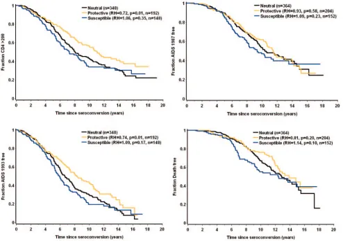
0.51 to 0.61;P⫽ ⬍0.0001 to 0.01), and the A/A-B/C grouping appeared neutral (Table 2). The protective and susceptible haplotype groups were further compared to the neutral group by the Cox proportional-hazards model (Fig. 2). The relative
[image:4.585.45.543.79.237.2]shapes of the curves in the Kaplan-Meier plots confirm that the effects of theTSG101variants are not constant over time, but rather occur most obviously between about 4 and 12 years after seroconversion.
[image:4.585.47.541.354.702.2]FIG. 2. Kaplan-Meier plots for the threeTSG101haplotypic groups. Four AIDS outcomes were analyzed using the Cox proportional-hazards model.
TABLE 2. TSG101haplogenotypes and AIDS progression among Caucasians from the combined U.S. AIDS cohorts
Haplogenotype AIDS outcome No.
aof fast
progressorsb(F)
No.aof slow
progressorsc(F) OR Pvalue
Neutral (A/A or B/C) CD4⬍200 133 (0.54) 357 (0.50) 1.17 0.30
AIDS-1993 147 (0.52) 426 (0.49) 1.14 0.37
AIDS-1987 72 (0.47) 560 (0.50) 0.89 0.55
Death 47 (0.46) 668 (0.50) 0.85 0.47
Protective (A/B or B/B) CD4⬍200 56 (0.23) 251 (0.35) 0.54 0.0003
AIDS-1993 64 (0.23) 307 (0.35) 0.54 ⬍0.0001
AIDS-1987 37 (0.24) 384 (0.34) 0.61 0.01
Death 21 (0.21) 447 (0.34) 0.51 0.006
Susceptible (A/C or C/C) CD4⬍200 59 (0.24) 109 (0.15) 1.74 0.003
AIDS-1993 71 (0.25) 137 (0.16) 1.80 0.0005
AIDS-1987 45 (0.29) 183 (0.16) 2.13 0.0002
Death 34 (0.33) 214 (0.16) 2.61 ⬍0.0001
aNumbers of individuals who had at least one of the corresponding haplotypes (dominant model).
bSeroconverter patients who progressed to an AIDS outcome inⱕ7 years after HIV-1 infection.
cPatients who avoided an AIDS outcome for⬎7 years after HIV-1 infection (seroconverters and seroprevalent individuals).
on November 8, 2019 by guest
http://jvi.asm.org/
Individuals from differentTSG101haplotype groups exhibit different rates of CD4ⴙT-cell decline.The availability of lon-gitudinal CD4 T-cell counts in two of our AIDS cohorts, the MACS and the SHCS (http://www.shcs.ch), provided the op-portunity to determine whether the influence ofTSG101 vari-ants on AIDS progression might involve differential rates of CD4 T-cell decline. CD4 measurements over time were plot-ted, and the slopes of the fitted lines were compared between the three genotypic groups. For 380 MACS patients, CD4 T-cell decline was estimated from measurements obtained over a period of 13 years since seroconversion (Fig. 3A). An average of 12.9 CD4 measurements per individual, ranging from 2 to
32, was considered in the analysis. The SHCS cohort primarily consisted of seroprevalent individuals, so in this case, CD4 T-cell decline was measured over a period of 10 years starting at the point where CD4 counts fell in the range of 500 to 600 cells/mm3(Fig. 3B). For 310 SHCS patients, an average of 11.5
CD4 T-cell count data points (ranging from 2 to 52) per indi-vidual were available for analysis. In both cohorts, the effects of the three genotypic groups on CD4 T-cell decline corre-sponded to the effects these genotypes had on AIDS progres-sion, where the decline was progressively steeper in the follow-ing order: A/C-C/C (susceptible group)⬎ A/A-B/C (neutral group)⬎A/B-B/B (protective group) (Fig. 3). Differences in CD4 T-cell decline between the protective and susceptible groups were highly significant in both cohorts (P⫽ ⬍0.0001 to 0.0002).
Effects of theTSG101haplotypes on viral load increase over time.Given the central role of TSG101 in HIV-1 budding and the known correlation between the viral load and the rate of AIDS progression (23), we tested whether the effect of
TSG101variation on CD4 T-cell decline and AIDS progres-sion was also reflected in viral load changes over time. For this analysis, the increase in viral load over time among 373 MACS patients was measured, stratified by the threeTSG101 geno-typic groups (Fig. 4). An average of 9.6 measurements (ranging from 2 to 28 measurements) of viral load per patient over a period of 13 years since seroconversion were available for the analysis. No significant difference in HIV-1 RNA levels be-tween the three genotypic groups was observed at the time of seroconversion. Strikingly, however, highly significant differ-ences in log10HIV-1 RNA slopes were observed between the
protective and susceptible groups (P⬍0.0001), strongly sug-gesting that variation in theTSG101gene affects the HIV-1 viral load, potentially through the differential efficiency of
TSG101variants to mediate viral budding.
TSG101genotypes and HIV-1 infection.TheTSG101 geno-typic groups were also tested for potential effects on HIV-1
FIG. 3. Decline in CD4 T-cell count among AIDS patients with differentTSG101 haplogenotypes. The data were analyzed using a random-effects linear model (see Materials and Methods). (A) Square-root CD4 counts for 380 individuals from the MACS cohort were plotted as a function of time from seroconversion, and fitted lines for each genotype group were generated based on 4,918 measurements. Slopes for protective, neutral, and susceptible genotypic groups are as follows (95% CI in parentheses):⫺1.11 (⫺1.22,⫺1.00),⫺1.33 (⫺1.42, ⫺1.25), and⫺1.39 (⫺1.50,⫺1.27), respectively. (B) CD4 data from the SHCS patients were plotted starting from the point at which CD4 cell counts fell in the range of 500 to 600 cells/mm3. By chance, the
susceptible haplogenotypic group had a mean CD4 count that was higher in this range than that of the protective or neutral group. The fitted lines are based on 3,551 measurements for 310 people. Slopes (95% CI) for protective, neutral, and susceptible groups are⫺0.52 (⫺0.64, ⫺0.39), ⫺0.57 (⫺0.69, ⫺0.47), and ⫺0.81 (⫺0.99, ⫺0.64), respectively.
FIG. 4. Viral-load increase over time in 373 MACS patients. Fitted lines for the threeTSG101genotypic groups were constructed based on 3,569 measurements of log10HIV RNA over time after
serocon-version. Statistical analysis was performed using the multivariate ran-dom-effects linear model (see Materials and Methods). The best-fit lines for the protective, neutral, and susceptible haplogenotypic groups have slopes (95% CI) of 0.01 (⫺0.01, 0.02), 0.04 (0.03, 0.05), and 0.05 (0.04, 0.07), respectively.
on November 8, 2019 by guest
http://jvi.asm.org/
infection by comparing the distributions of these genotypes in HIV⫹patients with those in seronegative or high-risk exposed uninfected individuals. Although only marginally significant values (P⫽0.02 to 0.08) were determined (Table 3), the effects of these genotypes on HIV-1 infection each paralleled their respective effects on the viral load, on CD4 T-cell decline, and on the rate of progression to AIDS.
DISCUSSION
The consistent protective (haplotype B) and susceptible (haplotype C) influences of TSG101 haplotypes on multiple outcomes after HIV-1 exposure, including longitudinal viral load levels, CD4⫹T-cell decline, and subsequent disease pro-gression, as well as a moderate effect on HIV-1 infection, support a physiological role for genetic variation near/within theTSG101gene in HIV-1 pathogenesis. Whether the (⫺183,
⫹181) haplotypic variants have a direct effect on these out-comes remains in question. We did not find any nonsynony-mous polymorphisms or 3⬘untranslated region SNPs linked to the⫺183,⫹181 variants that could explain the observed effects of these variants on HIV-1 disease. Further, the (⫺183,⫹181) haplogenotypes did not show any significant difference in the levels of TSG101 mRNA transcription in peripheral blood lymphocytes or purified CD4 cells as tested by real-time PCR, and no differences were observed in promoter activity using reporter constructs (data not shown). Likewise, no alterna-tively spliced forms of TSG101 mRNA associating with the genotypes were observed, negating splice variation as the un-derlying mechanism of the associations described. Although we were not able to detect an influence of theTSG101 haplo-types on gene transcription or mRNA splicing, we cannot rule out the possibility that theTSG101SNPs may affect transcrip-tion/splicing under specific physiological conditions.
The protective and susceptible effects ofTSG101haplotypes on AIDS progression are not constant over time, as indicated by the Kaplan-Meier curves (Fig. 2). The relative shapes of the curves suggest that TSG101 variation has little or no effect during the early (⬃4 years) and late (⬎12 years) stages of infection, but rather, only at an intermediate time period. Other host genetic, viral, or environmental factors that affect disease progression during the early and late stages of infection may override effects of TSG101. Alternatively, the TSG101 interaction with HIV and/or consequences of this interaction could be different during these two extreme time intervals compared with the intermediate period. However, the effects ofTSG101haplotypes occurring during the intermediate time
interval were strong enough to be evident when the entire patient cohort was used in the categorical analysis, as well as the longitudinal analyses of CD4 T-cell decline and viral-load increase.
Recently, the⫺183 variant, corresponding to haplotype C, was shown to associate with lower virus production ex vivo, a paradoxical finding given its association with faster CD4 T-cell decline (3) and susceptibility to AIDS reported here. Although this finding appears to contradict the genetic epidemiological findings presented here, the ex vivo assay may not be physio-logically relevant to the described effect ofTSG101on AIDS progression given its time dependence as discussed above.
The HapMap genotype data (http://www.hapmap.org) sug-gest that TSG101 is located in a region of strong LD: the corresponding haplotype block defined according to Gabriel et al. (11) spans 118 kb. This block includes the entireTSG101
gene; its partial paralogue, UEV-3 (15); and two additional gene fragments (the lactate dehydrogenase A-like 6A gene [GenBank accession no. NM144972] and a computationally predicted gene that may encode a protein similar to the mito-chondrial carrier homolog 1 [XM497268]). Therefore, the (⫺183,⫹181) haplogenotypes may mark the true disease vari-ant(s) through LD. In support of this, no effect on disease progression was detected in a smaller sample of African-Amer-ican seroconverters (although longitudinal CD4 and viral-load data from these individuals were not available for analysis; also, the susceptible haplotype group was observed at a fre-quency of only 4%). However, given the requirement of TSG101 for HIV-1 budding in vitro, it seems likely that the effect described herein is due to variation inTSG101and not to polymorphism in a neighboring locus. If so, this study rep-resents the first genetic epidemiological evidence to support previous in vitro studies indicating a primary role for TSG101 in HIV-1 pathogenesis.
ACKNOWLEDGMENTS
This project has been funded in whole or in part with federal funds from the National Cancer Institute, National Institutes of Health, under contract N01-CO-12400. This research was supported in part by the Intramural Research Program of NIH, National Cancer Institute, Center for Cancer Research.
The content of this publication does not necessarily reflect the views or policies of the Department of Health and Human Services, nor does mention of trade names, commercial products, or organizations imply endorsement by the U.S. Government.
We thank Eric Freed, George Nelson, and Marcus Altfeld for help-ful discussions.
REFERENCES
1.Babst, M., G. Odorizzi, E. J. Estepa, and S. D. Emr.2000. Mammalian tumor susceptibility gene 101 (TSG101) and the yeast homologue, Vps23p, both
function in late endosomal trafficking. Traffic1:248–258.
2.Bishop, N., A. Horman, and P. Woodman.2002. Mammalian class E vps proteins recognize ubiquitin and act in the removal of endosomal
protein-ubiquitin conjugates. J. Cell Biol.157:91–101.
3.Bleiber, G., M. May, R. Martinez, P. Meylan, J. Ott, J. S. Beckmann, and A. Telenti.2005. Use of a combined ex vivo/in vivo population approach for screening of human genes involved in the human immunodeficiency virus
type 1 life cycle for variants influencing disease progression. J. Virol.79:
12674–12680.
4.Bouamr, F., J. A. Melillo, M. Q. Wang, K. Nagashima, M. de Los Santos, A. Rein, and S. P. Goff.2003. PPPYVEPTAP motif is the late domain of human T-cell leukemia virus type 1 Gag and mediates its functional interaction with
cellular proteins Nedd4 and Tsg101. J. Virol.77:11882–11895.
5.Burgdorf, S., P. Leister, and K. H. Scheidtmann.2004. TSG101 interacts with apoptosis-antagonizing transcription factor and enhances androgen
re-TABLE 3. Frequencies ofTSG101genotypic groups among HIV-1-positive vs. HIV-1-negative Caucasian individuals
Genotypic group
% SCa
/HREUb
(OR, 95% CI)
% SC/SNc
(OR, 95% CI)
% SC/OSNd
/HREU
(Pvalue for trende
)
Neutral 51/51 (1.0, 0.7–1.5) 51/48 (1.1, 0.8–1.5) 51/45/51 (0.76)
Protective 28/36 (0.7, 0.5–1.0) 28/36 (0.7, 0.5–0.9) 28/36/36 (0.03)
Susceptible 21/13 (1.7, 1.0–2.9) 21/16 (1.5, 1.0–2.2) 21/19/13 (0.04)
aSC, seroconverters (n⫽728).
bHREU, high-risk exposed uninfected people (n⫽134).
cSN, all seronegatives combined (n⫽219).
dOSN, seronegatives other than HREU (n⫽85).
eMantel-Haentzel chi-square test.
on November 8, 2019 by guest
http://jvi.asm.org/
[image:6.585.40.284.89.143.2]ceptor-mediated transcription by promoting its monoubiquitination. J. Biol.
Chem.279:17524–17534.
6.Carstens, M. J., A. Krempler, A. A. Triplett, M. Van Lohuizen, and K. U. Wagner.2004. Cell cycle arrest and cell death are controlled by p53-depen-dent and p53-indepenp53-depen-dent mechanisms in Tsg101-deficient cells. J. Biol.
Chem.279:35984–35994.
7.Cox, D. R., and D. Oakes.1984. Analysis of survival data. Chapman and Hall, New York, N.Y.
8.Cullen, M., J. Noble, H. Erlich, K. Thorpe, S. Beck, W. Klitz, J. Trowsdale, and M. Carrington.1997. Characterization of recombination in the HLA
class II region. Am. J. Hum. Genet.60:397–407.
9.Demirov, D. G., and E. O. Freed.2004. Retrovirus budding. Virus Res.
106:87–102.
10.Demirov, D. G., A. Ono, J. M. Orenstein, and E. O. Freed.2002. Overex-pression of the N-terminal domain of TSG101 inhibits HIV-1 budding by
blocking late domain function. Proc. Natl. Acad. Sci. USA99:955–960.
11.Gabriel, S. B., S. F. Schaffner, H. Nguyen, J. M. Moore, J. Roy, B. Blumenstiel, J. Higgins, M. DeFelice, A. Lochner, M. Faggart, S. N. Liu-Cordero, C. Rotimi, A. Adeyemo, R. Cooper, R. Ward, E. S. Lander, M. J. Daly, and D. Altshuler.2002. The structure of haplotype blocks in the human genome.
Science296:2225–2229.
12.Garrus, J. E., U. K. von Schwedler, O. W. Pornillos, S. G. Morham, K. H. Zavitz, H. E. Wang, D. A. Wettstein, K. M. Stray, M. Cote, R. L. Rich, D. G. Myszka, and W. I. Sundquist.2001. Tsg101 and the vacuolar protein sorting
pathway are essential for HIV-1 budding. Cell107:55–65.
13.Hittelman, A. B., D. Burakov, J. A. Iniguez-Lluhi, L. P. Freedman, and M. J. Garabedian.1999. Differential regulation of glucocorticoid receptor
tran-scriptional activation via AF-1-associated proteins. EMBO J.18:5380–5388.
14.Katzmann, D. J., M. Babst, and S. D. Emr.2001. Ubiquitin-dependent sorting into the multivesicular body pathway requires the function of a
conserved endosomal protein sorting complex, ESCRT-I. Cell106:145–155.
15.Kloor, M., P. Bork, A. Duwe, R. Klaes, M. von Knebel Doeberitz, and R. Ridder.2002. Identification and characterization of UEV3, a human cDNA with similarities to inactive E2 ubiquitin-conjugating enzymes. Biochim.
Bio-phys. Acta1579:219–224.
16.Krempler, A., M. D. Henry, A. A. Triplett, and K. U. Wagner.2002. Targeted
deletion of the Tsg101 gene results in cell cycle arrest at G1/S and
p53-independent cell death. J. Biol. Chem.277:43216–43223.
17.Li, L., and S. N. Cohen.1996. Tsg101: a novel tumor susceptibility gene isolated by controlled homozygous functional knockout of allelic loci in
mammalian cells. Cell85:319–329.
18.Li, L., J. Liao, J. Ruland, T. W. Mak, and S. N. Cohen.2001. A TSG101/ MDM2 regulatory loop modulates MDM2 degradation and MDM2/p53
feedback control. Proc. Natl. Acad. Sci. USA98:1619–1624.
19.Long, J. C., R. C. Williams, and M. Urbanek.1995. An E-M algorithm and
testing strategy for multiple-locus haplotypes. Am. J. Hum. Genet.56:799–
810.
20.Lu, Q., L. W. Hope, M. Brasch, C. Reinhard, and S. N. Cohen.2003. TSG101 interaction with HRS mediates endosomal trafficking and receptor
down-regulation. Proc. Natl. Acad. Sci. USA100:7626–7631.
21.Martin-Serrano, J., T. Zang, and P. D. Bieniasz.2001. HIV-1 and Ebola virus encode small peptide motifs that recruit Tsg101 to sites of particle
assembly to facilitate egress. Nat. Med.7:1313–1319.
22.McIver, B., S. K. Grebe, L. Wang, I. D. Hay, A. Yokomizo, W. Liu, J. R. Goellner, C. S. Grant, D. I. Smith, and N. L. Eberhardt.2000. FHIT and TSG101 in thyroid tumours: aberrant transcripts reflect rare abnormal RNA processing events of uncertain pathogenetic or clinical significance. Clin.
Endocrinol.52:749–757.
23.Mellors, J. W., C. R. Rinaldo, Jr., P. Gupta, R. M. White, J. A. Todd, and L. A. Kingsley.1996. Prognosis in HIV-1 infection predicted by the quantity
of virus in plasma. Science272:1167–1170.
24.Muromoto, R., K. Sugiyama, T. Yamamoto, K. Oritani, K. Shimoda, and T. Matsuda. 2004. Physical and functional interactions between Daxx and
TSG101. Biochem. Biophys. Res. Commun.316:827–833.
25.Myers, E. L., and J. F. Allen.2002. Tsg101, an inactive homologue of ubiquitin ligase e2, interacts specifically with human immunodeficiency virus type 2 gag polyprotein and results in increased levels of ubiquitinated gag.
J. Virol.76:11226–11235.
26.O’Brien, S. J., and G. W. Nelson.2004. Human genes that limit AIDS. Nat.
Genet.36:565–574.
27.Oh, H., C. Mammucari, A. Nenci, S. Cabodi, S. N. Cohen, and G. P. Dotto.
2002. Negative regulation of cell growth and differentiation by TSG101
through association with p21(Cip1/WAF1). Proc. Natl. Acad. Sci. USA99:
5430–5435.
28.Pornillos, O., S. L. Alam, R. L. Rich, D. G. Myszka, D. R. Davis, and W. I. Sundquist.2002. Structure and functional interactions of the Tsg101 UEV
domain. EMBO J.21:2397–2406.
29.Ruland, J., C. Sirard, A. Elia, D. MacPherson, A. Wakeham, L. Li, J. L. de la Pompa, S. N. Cohen, and T. W. Mak.2001. p53 accumulation, defective cell proliferation, and early embryonic lethality in mice lacking tsg101. Proc.
Natl. Acad. Sci. USA98:1859–1864.
30.Strack, B., A. Calistri, S. Craig, E. Popova, and H. G. Gottlinger.2003. AIP1/ALIX is a binding partner for HIV-1 p6 and EIAV p9 functioning in
virus budding. Cell114:689–699.
31.Stuchell, M. D., J. E. Garrus, B. Muller, K. M. Stray, S. Ghaffarian, R. McKinnon, H. G. Krausslich, S. G. Morham, and W. I. Sundquist.2004. The human endosomal sorting complex required for transport (ESCRT-I) and its
role in HIV-1 budding. J. Biol. Chem.279:36059–36071.
32.Sun, Z., J. Pan, W. X. Hope, S. N. Cohen, and S. P. Balk.1999. Tumor susceptibility gene 101 protein represses androgen receptor transactivation
and interacts with p300. Cancer86:689–696.
33.Sundquist, W. I., H. L. Schubert, B. N. Kelly, G. C. Hill, J. M. Holton, and C. P. Hill.2004. Ubiquitin recognition by the human TSG101 protein. Mol.
Cell13:783–789.
34.Trink, B., S. I. Pai, S. L. Spunt, V. Raman, P. Cairns, J. Jen, E. Gabrielson, S. Sukumar, and D. Sidransky.1998. Absence of TSG101 transcript
abnor-malities in human cancers. Oncogene16:2815–2818.
35.VerPlank, L., F. Bouamr, T. J. LaGrassa, B. Agresta, A. Kikonyogo, J. Leis, and C. A. Carter.2001. Tsg101, a homologue of ubiquitin-conjugating (E2) enzymes, binds the L domain in HIV type 1 Pr55(Gag). Proc. Natl. Acad. Sci.
USA98:7724–7729.
36.von Schwedler, U. K., M. Stuchell, B. Muller, D. M. Ward, H. Y. Chung, E. Morita, H. E. Wang, T. Davis, G. P. He, D. M. Cimbora, A. Scott, H. G. Krausslich, J. Kaplan, S. G. Morham, and W. I. Sundquist.2003. The
protein network of HIV budding. Cell114:701–713.
37.Wirblich, C., B. Bhattacharya, and P. Roy.2006. Nonstructural protein 3 of bluetongue virus assists virus release by recruiting ESCRT-I protein Tsg101.
J. Virol.80:460–473.
38.Xie, W., L. Li, and S. N. Cohen.1998. Cell cycle-dependent subcellular localization of the TSG101 protein and mitotic and nuclear abnormalities
associated with TSG101 deficiency. Proc. Natl. Acad. Sci. USA95:1595–
1600.
39.Zhong, Q., Y. Chen, D. Jones, and W. H. Lee.1998. Perturbation of TSG101
protein affects cell cycle progression. Cancer Res.58:2699–2702.