O R I G I N A L A R T I C L E
Disrupted-in-Schizophrenia-1
is essential for normal
hypothalamic-pituitary-interrenal (HPI) axis function
Helen Eachus
1,2, Charlotte Bright
1, Vincent T. Cunliffe
2, Marysia Placzek
2,*
,†,
Jonathan D. Wood
2,3,*
,†and Penelope J. Watt
1,†1
Department of Animal and Plant Sciences, University of Sheffield, Western Bank, Sheffield S10 2TN, UK,
2The
Bateson Centre, Department of Biomedical Science, Firth Court, Western Bank, Sheffield S10 2TN, UK and
3Sheffield Institute for Translational Neuroscience, Department of Neuroscience, University of Sheffield,
Sheffield S10 2HQ, UK
*To whom correspondence should be addressed at: Department of Neuroscience, Sheffield Institute for Translational Neuroscience, University of Sheffield, 385a Glossop Road, Sheffield S10 2HQ, UK. Tel: +44 1142222243; Fax: +44 1142222290; Email: [email protected]; The Bateson Centre, Department of Biomedical Science, Firth Court, Western Bank, Sheffield S10 2TN, UK. Tel: +44 1142222353; Fax: +44 1142222787; Email:
Abstract
Psychiatric disorders arise due to an interplay of genetic and environmental factors, including stress. Studies in rodents have shown that mutants forDisrupted-In-Schizophrenia-1(DISC1), a well-accepted genetic risk factor for mental illness, display abnormal behaviours in response to stress, but the mechanisms through whichDISC1affects stress responses remain poorly understood. Using two lines of zebrafish homozygous mutant fordisc1, we investigated behaviour and functioning of the hypothalamic-pituitary-interrenal (HPI) axis, the fish equivalent of the hypothalamic-pituitary-adrenal (HPA) axis. Here, we show that the role of DISC1 in stress responses is evolutionarily conserved and that DISC1 is essential for normal functioning of the HPI axis. Adult zebrafish homozygous mutant fordisc1show aberrant behavioural responses to stress. Our studies reveal that in the embryo,disc1is expressed in neural progenitor cells of the hypothalamus, a conserved region of the verte-brate brain that centrally controls responses to environmental stressors. Indisc1mutant embryos, proliferatingrx3þ hypotha-lamic progenitors are not maintained normally and neuronal differentiation is compromised:rx3-derivedff1bþneurons, implicated in anxiety-related behaviours, andcorticotrophin releasing hormone (crh)neurons, key regulators of the stress axis, develop abnormally, andrx3-derivedpomcþneurons are disorganised. Abnormal hypothalamic development is associated with dysfunctional behavioural and neuroendocrine stress responses. In contrast to wild type siblings,disc1mutant larvae show alteredcrhlevels, fail to upregulate cortisol levels when under stress and do not modulate shoal cohesion, indicative of abnormal social behaviour. These data indicate thatdisc1is essential for normal development of the hypothalamus and for the correct functioning of the HPA/HPI axis.
†Co-senior authors
Received:January 3, 2017.Revised:February 21, 2017.Accepted:February 23, 2017 VCThe Author 2017. Published by Oxford University Press.
This is an Open Access article distributed under the terms of the Creative Commons Attribution License (http://creativecommons.org/licenses/by/4.0/), which permits unrestricted reuse, distribution, and reproduction in any medium, provided the original work is properly cited.
1992
doi: 10.1093/hmg/ddx076
Introduction
Phenotypes are shaped throughout the life-course by a complex interplay between genes and the environment. When homeo-stasis is threatened by environmental stress, animals respond adaptively by altering their metabolism, physiology and behav-iour. These adaptive responses are co-ordinated by the hypothalamic-pituitary-adrenal (HPA) axis (1). Activation of the HPA axis promotes cortisol release and promotes adaptation (2,3). Circulating cortisol in turn triggers negative feedback sys-tems that limit HPA axis function. However, this circuit can become reprogrammed to trigger responses that are seemingly maladaptive (4,5): in humans, HPA hyperactivity is linked to heightened risk for depression and anxiety disorders (6). Maladaptive stress responses can be triggered through wide-ranging insults, and increasing evidence suggests that insults in developmentally-sensitive periods predispose individuals to later heightened vulnerability to stress. For example, it is well documented that heightened stress in early life can result in the development of adult-onset psychiatric disorders in humans (1,4,7–10). At the same time, the stress response is modulated by an individual’s genetic makeup, and genotype is thought to contribute to individual differences in susceptibility to psychiat-ric disorders (11–14). However, whilst animal models have dem-onstrated that ablation of individual genetic components of the HPA axis can affect stress phenotypes and behaviour (15–18), no study has yet shown a direct link between genetic regulation of HPA axis development and maladaptive stress responses.
One well established genetic risk factor for human psychiat-ric illness, Disrupted-In-Schizophrenia-1 (DISC1), was originally identified at a chromosomal translocation breakpoint in a single Scottish family, in which a high proportion of family members suffered from mental illness (19). Some translocation carriers showed a range of clinical phenotypes, including schizophrenia, major depression and bipolar disorder, whilst other carriers had no psychiatric diagnosis (19). Individuals carrying this translo-cation, including those with no psychiatric condition, exhibited a defect in their cognitive function during decision-making processes (P300 event-related potential), a trait considered to be a marker for risk for schizophrenia (20). The incomplete pene-trance and range of psychiatric presentations make DISC1 a prime candidate for understanding how environmental factors interact with a defined genetic component to yield a variety of behavioural phenotypes.
Studies in mice have shown that DISC1 can impact on behaviour (21,22) and can also modulate reactivity to stress (21,23–28). These studies have utilised either mice withDisc1 point mutations (27), mice carrying a naturally occurring 25 base-pair deletion inDisc1(29,30), or transgenic mice expressing a truncated form of humanDISC1(24,28,31). Depending on the type of mutation or transgene used, varying phenotypes have been found, with many showing an impaired response to stress (23,24,28,31–33). Studies that have investigated the mechanism through which DISC1 and stress interact to modulate behaviour have revealed epigenetic modifications in dopaminergic neu-rons that originate in the ventral tegmental area (24). However, no study has examined whetherDisc1mutation alters develop-ment of the HPA axis in a manner that impacts on stress modulation.
Expression studies in primates and mice have shown that DISC1orthologues are prominently expressed in the hypothala-mus (34–36), a small evolutionarily conserved part of the brain that coordinates responses to stress. Analysis ofDISC1 expres-sion in the human brain has mainly focused on the
hippocampus, but expression patterns here correspond well with those in the primate and rodent hippocampus, suggesting some level of conservation (37). We previously observed strong expression of disc1 in the ventral diencephalon of zebrafish embryos (38). We therefore reasoned thatdisc1may be required for normal hypothalamic development and functioning of the HPA axis or corresponding hypothalamic-pituitary-interrenal (HPI) axis in fish.
To address this hypothesis, we utilised two lines of zebrafish harbouring nonsense mutations indisc1(L115X and Y472X) and analysed baseline and stress-responsive behaviours in the adult. We investigated the developmental origin of mutant behavioural abnormalities, and show thatdisc1is essential for normal development of the early hypothalamus and HPI axis function.
Results
Adultdisc1mutants exhibit anxiety-like behaviour and aberrant behavioural stress responses
The L115X and Y472X mutations both introduce a premature stop codon in theN-terminal head domain of DISC1 (Fig. 1A). We have maintained both lines on a TL background and found that homozygous mutants are born in Mendelian ratios, hatch normally, are viable to adulthood, and fertile. Quantitative RT-PCR demonstrated that Y472X mRNA may be subject to nonsense-mediated decay (Fig. 1B). We first analysed whether, as in mice, the baseline behaviour of the adultdisc1mutants, or their response to an acute stressor, is significantly different to wild type siblings. Adult Y472X mutants were tested for baseline behaviours and adult L115X mutants were tested for response to an established stress paradigm: exposure to alarm substance (Schreckstoff: a zebrafish skin extract that induces a profound fear response (39,40). In an open field test, adult Y472X fish showed increased freezing and increased fast swimming com-pared to wild type siblings (Fig. 1C–D). In a light-dark test, Y472X fish showed no preference for the light compartment, in con-trast to wild type siblings (Fig. 1E). In the tank diving test, L115X mutants did not increase bottom dwell time after treatment with alarm substance, in contrast to wild type siblings (Fig. 1F). Abnormalities in baseline and stress-responsive behaviour have also been described in adult Disc1 mouse models (24,27,28,31–33). These data show that the role of DISC1 in stress responses is evolutionarily conserved.
Hypothalamic progenitors, includingrx3þprogenitors, are not maintained normally indisc1mutant embryos
DISC1 governs neuronal progenitor proliferation (41,42), so we reasoned that early developmental abnormalities may underlie the observed adult phenotypes. Studies in mice have shown that cellular homeostasis is disrupted in the cortex of disc1 mutants where cortical progenitors differentiate prematurely due to compromised Wnt/GSK3 signaling (41,42). Similarly, in zebrafish, zDisc1 promotes brain neurogenesis by promoting Wnt signaling (43), while a study in human induced pluripotent stem cells linked disruption ofDISC1with altered Wnt signaling and neural progenitor cell differentiation (44). To date, however, no study has analysed progenitor cells or differentiating neu-rons in the hypothalamus.
Analysis of disc1 in embryonic zebrafish (24–55 h post-fertilisation (hpf)/2–3 days post-post-fertilisation (dpf)) revealed that expression is most prominent in the basal part of the brain, in
particular the hypothalamus (Fig. 2A–C,F–H). Transverse sec-tions through the 55 hpf hypothalamus show that disc1 is restricted to cells around the lateral recesses and posterior tuberal 3rdventricle (Fig. 2J–M,O), neurogenic zones that harbour proliferating progenitors (45–50). Throughout this period, the expression ofdisc1is largely adjacent to that ofretinal homeobox 3(rx3) a conserved paired-like homeodomain transcription fac-tor (Fig. 2D,I,N), which, in the tuberal hypothalamus, demar-cates progenitor cells that give rise to specific hypothalamic neuronal populations, including neurons of the ventromedial nucleus (VMN) and arcuate nucleus (Arc) (51,52).
To address whether, similar to its role in the mouse cortex, DISC1 maintains hypothalamic progenitor cells, we compared rx3expression in wild type anddisc1mutant embryos and
[image:3.612.148.466.60.472.2]post-hatched (5 dpf) larvae. In both lines, rx3 is altered in disc1 mutants in comparison to wild type siblings (Fig. 3A–L; Supplementary Material, Fig. S1). In Y472X fish,rx3is reduced in mutant fish compared to wild type siblings at all stages exam-ined (24 hpf-5 dpf) (Fig. 3A,C,F,G,I,L). In L115X mutant fish,rx3 transcripts are detected at higher levels at 24 hpf than in wild type siblings (Fig. 3B and H), but from 3 dpf, L115X mutants show a similar reduction to that detected in Y472X mutants (Fig. 3D,E,J,K). Transverse sections show thatrx3is expressed in the 3rdventricle up to 3 dpf and in the lateral recesses up to 5 dpf in wild-type fish (Fig. 3C–F,Q). By contrast, from 2 dpf, both L115X and Y472X mutant larvae show a significant reduction in rx3expression in the lateral recesses, and no expression can be detected in the 3rdventricle (Fig. 3I–L,WandFig. 3legend). This
suggests that DISC1 is required to maintain progenitor cells and predicts thatdisc1mutants will show a premature decline in hypothalamic expression of phosphorylated histone H3 (phosH3), an M-phase marker whose expression correlates with proliferating progenitors in the embryonic zebrafish hypothala-mus (45). At 24 hpf, a significant increase in phosH3þcells is detected in the hypothalamus ofdisc1mutant embryos com-pared to wild type siblings (Fig. 3M,N,R–T). At 3 dpf, however, significantly fewer phosH3þcells are detected in the hypothala-mus of bothdisc1mutant strains compared to wild type siblings (Fig. 3O–Q,U–X). Together our analyses suggest that hypothala-mic progenitors, includingrx3þprogenitors, form, but are not maintained normally indisc1mutant embryos.
Abnormal neuroendocrine differentiation and activity in
disc1mutant embryos
We extended these experiments to determine whether the changes in progenitor proliferation lead to alterations in neuro-nal differentiation. Lineage-tracing studies show thatrx3þ pro-genitors in the tuberal hypothalamus give rise to VMN neurons that express the nuclear receptor,ff1b(also termednr5a1a; an orthologue of mammalianSF1/NR5A1(53)) and to Arc neurons that express pro-opiomelanocortin (pomc) (45,54). We therefore first determined ifdisc1mutant fish showed alterations inff1b andpomc. We analysed both embryos at 2–3 dpf, a time when neuroendocrine cells are being born, and larvae at 5 dpf, a time when the neuroendocrine system begins to respond dynami-cally to external and internal cues (55).
In wild type embryos,ff1bis first detected in the developing hypothalamus at 24 hpf (56). The role offf1bin the zebrafish
hypothalamus has not been determined, but in mice, hypo-thalamicSf1governs anxiety behaviours (57). In both embryos and larvae, expression of ff1b was significantly more pro-nounced in the hypothalamus of L115X and Y472X mutants compared to wild type siblings (Figs. 4A–D, 5A–D; Supplementary Material, Fig. S2A–D) and was detected in greater numbers of cells (Figs. 4Eand5E). Thus, the failure to maintain rx3þprogenitors appears to correlate with an enhanced differentiation of hypothalamicff1bþcells.ff1bis also expressed in steroidogenic cells of the interrenal gland, and is essential for proper development of this tissue (58). We observed normal expression of ff1b in this region of disc1 mutants (Supplementary Material, Fig. S2M–P).
The precursor peptide, proopiomelanocortin (pomc) defines Arc-like neurons in the hypothalamus. In wild type zebrafish, hypothalamicpomcþneurons are detected from 32 hpf, i.e. some hours afterff1bþcells (59). Analysis over 2–3 dpf revealed no signif-icant difference in number of hypothalamicpomcþcells at each time (Fig. 4F–J;Supplementary Material, Fig. S2E–H, K), but at 5 dpf, significantly fewerpomcþneurons were detected in L115X mutants (Fig. 5J). In both L115X and Y472X mutants, pomcþneurons appeared disorganised, and were not detected in the characteristic horseshoe pattern found in the wild type animals (Fig. 5F–I).
[image:4.612.64.549.72.316.2]We next examined whetherdisc1affectscorticotropin releasing hormone (crhb)-expressing neurons, a population that is specified in anrx3-independent manner (60), but that plays an instru-mental role in the stress response. Analysis ofcrhbin Y472X mutants revealed embryonic expression in both the preoptic and tuberal hypothalamus, as shown in previous studies (61,62). At 2–3 dpf, increased numbers ofcrhbþneurons were detected in mutant embryos, compared to wild types, in both domains (Fig. 4K–O, Supplementary Material, Fig. 2I–J, L). By
Figure 2.Expression ofdisc1in the larval zebrafish brain. (A–J) 24 hpf or 55 hpf embryos afterin situhybridization fordisc1(A–C, F–H) orrx3(D, I) shown in ventral whole-mount view (A–D) or side view (F–I). Anterior to left. (B,H) show high power views of boxed regions in (A,G). (E,J) show schematic ventral and side views. Arrows point to expression ofdisc1in hypothalamus. Lines in side views (H–J) indicate planes of sections shown in (K–N). (K–N) Transverse sections taken through 55 hpf embryos afterin situhybridization fordisc1(K-M) orrx3(N). Schematic (O) shows position of 3rdventricle and lateral recesses. Abbreviations: 3V, 3rdventricle; Ant, ante-rior; DT, dorsal thalamus; H, hypothalamus; LR, lateral recess; MO, medulla oblongata; OB, olfactory bulb; PO, preoptic region; PTv, ventral posterior tuberculum; TeO, tectum opticum; Tub, tuberal.N¼6 each. Scale bar: 50lm.
contrast, at 5 dpf, a time when neuroendocrine-responsive tran-scriptional programmes adapt dynamically to the supply and demand for neuropeptides, significantly fewer crhbþneurons could be detected in mutant larvae (Fig. 5O), and where expres-sion was detected, it was weak, relative to that in wild type sib-lings, particularly in tuberal regions (Fig. 5K–S).
Taken together, our results suggest that DISC1 is required to determine appropriate numbers of hypothalamic progenitors, includingrx3þprogenitors, and appropriate numbers/position of differentiated neurons, includingff1bþ, pomcþandcrhþneurons.
disc1mutant larvae show impaired behavioural responses to stress
ff1borthologues are implicated in anxiety-like behaviours in mice (57) and depression and anxiety in humans (63), whilst crh
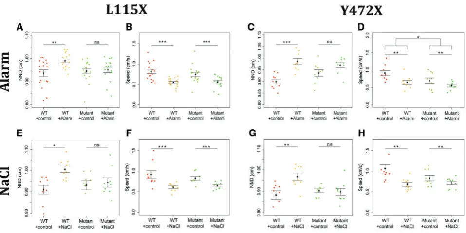
[image:6.612.143.473.61.469.2]orthologues are implicated in anxiety-like behaviours in mice (15,64) and primates (65), and in depression in humans (66). The altered differentiation (and, potentially activity) of ff1bþand crhbþneurons, together with the altered stress reactivity indisc1 adult mutants, led us to ask whether altered behavioural responses to stress indisc1mutants are established early in devel-opment. We analysed each mutant strain for behavioural responses to two established stress paradigms, osmotic stress (67) and exposure to alarm substance (39,40). At 3 dpf embryos already exhibit increased cortisol levels in response to severe stress (61), while at 5 dpf, larvae display anxiety-related behaviours such as thigmotaxis and dark avoidance (68). Acute exposure to either alarm substance or sodium chloride at 5 dpf resulted in a signifi-cant increase in the nearest neighbour distance (NND, a measure of shoal cohesion) in wild type larvae, but did not affect NND in either L115X or Y472X mutants (Fig. 6A,C,E,G). By contrast,
Figure 4.Abnormal neuronal differentiation in the hypothalamus ofdisc1L115X and Y472X embryos. (A–D,F–I) Transverse sections through posterior tuberal hypothal-amus at 52 hpf afterin situhybridisation withff1b(nr5a1a) (A–D) orpomc(F-I).ff1bis expressed more strongly in both L115X (B) and Y472X (D) mutant larvae, compared to wild types (A and C). (E,J) Quantitative analyses offf1bandpomccell number at 52hpf.ff1bcell count was not different in L115X embryos (ttest,t¼ 2.45, df¼3.86,
P¼0.073,N¼3), but was significantly increased in Y472X mutants compared to wild types (ttest,t¼-3.13, df¼14.06,P¼0.007,N¼9–10).pomccell count was not
signifi-cantly altered in L115X (ttest,t¼1.10, df¼5.56,P¼0.318,N¼3–5) or Y472X (ttest,t¼0.54, df¼25.38,P¼0.593,N¼13–16) embryos (J). (K-O) Transverse sections through preoptic (K,L) or posterior tuberal hypothalamus (M,N) at 52 hpf afterin situhybridisation withcrhbin the Y472X line. Quantitative analysis (O) shows significantly
morecrhbþcells in the preoptic and tuberal hypothalamus of mutant larvae (ttest,t¼ 2.53, df¼7.83,P¼0.036).N¼5 each. Abbreviations: 3V, 3rdventricle of the
hypo-thalamus; LR, lateral recess of the hypohypo-thalamus; WT, wild type larvae; mutant, homozygous mutant larvae. Scale bar: 50lm.
exposure to the stressors caused a significant reduction in swim-ming speed of both wild type and mutant disc1 larvae (Fig. 6B,D,F,H). L115X mutant swimming speed was not different to that of wild types, but Y472X mutants swam more slowly than wild types (Fig. 6D). To summarise, whilst both wild type and mutant larvae swam more slowly when under stress, only wild type larvae modulated shoal cohesion in response to stressors.
The failure to modulate shoaling behaviour in response to stress bydisc1mutants could be due to abnormalities in lateral line development or the visual system, either of which could impact on shoaling behaviour (69,70). However, when exposed to a short pulse of darkness, both wild type and mutant larvae exhibit a star-tle response (Supplementary Material, Fig. S3A). Furthermore, anal-ysis of FM1-43, a marker of neuromasts of the lateral line, likewise showed similar numbers in both wild type and mutant larvae (Supplementary Material, Fig. S3B). Thus, gross defects in two sen-sory systems - the visual system and the lateral line – do not appear to account for the impaired shoaling behaviour.
Environmental stress fails to trigger the HPI axis indisc1
mutant larvae
[image:7.612.128.487.61.418.2]The altered behavioural reactivity to stress in the disc1 mutant larvae led us to postulate that endocrine responses might be impaired. We therefore measured cortisol levels with and without stress exposure. As anticipated (67), expo-sure to either alarm substance or sodium chloride led to a significant increase in whole body cortisol levels in wild type larvae. By contrast, exposure to either alarm substance or sodium chloride had no significant effect on whole body cor-tisol levels of eitherdisc1L115X mutant larvae (Fig. 7A and C) or Y472X mutant larvae (Fig. 7B and D). No significant differ-ences were observed in baseline cortisol levels between wild type anddisc1mutant larvae. Together, these results show that mutations in disc1prevent the normal functioning of the HPI axis, in particular, the cortisol-mediated stress response.
Figure 5.Abnormal neuronal differentiation in the hypothalamus ofdisc1L115X and Y472X larvae. (A–E) Transverse sections (A–D) through posterior tuberal hypothal-amus at 5 dpf afterin situhybridisation withff1b.ff1bis expressed more strongly in both L115X and Y472X mutant larvae, and in more cells (E) compared to wild types (L115X,ttest,t¼ 2.20, df¼11.64,P¼0.049, Y472X,ttest,t¼ 2.70, df¼12.91,P¼0.018).N¼8–10 each. (F–J) Ventral whole-mount views at 5 dpf afterin situhybridisation
withpomc.pomcþcells are disorganised in the hypothalamus of mutant larvae (G,I) compared to wild type siblings (F,H). (J) Quantitative analysis at 5 dpf shows
Discussion
Humans that carry a mutation inDISC1present with a variety of psychiatric conditions, including depression, schizophrenia and bipolar disorder (19). Mousedisc1mutant models similarly exhibit behavioural abnormalities (21,22,27). Our studies reveal behaviou-ral abnormalities in adultdisc1mutant zebrafish that include freez-ing in the open field and a reduced preference for the light compartment in the light-dark test. What might these behaviours represent, and are they relevant to understanding the pathobiology of psychiatric disorders? Freezing behaviour is commonly observed in zebrafish after exposure to stressors such as alarm substance and is typically accompanied by darting and erratic movements (39). This combination of behaviours has been considered an anxiety-like behaviour in zebrafish (71). Moreover, freezing in the open field test is a characteristic behaviour of the zebrafish gluco-corticoid receptor mutant (16), where, unaccompanied by darting, it is considered to represent a depressive-like behaviour. Since, in the open field,disc1mutant zebrafish exhibit increased freezing, and high speed darting in between freezing periods, we believe that behaviour in this test indicates increased anxiety. Further sup-port for this conclusion comes through analysis of the response to the light-dark test. Wild type zebrafish strongly prefer the light compartment, a behaviour that is likely to be advantageous, in that the light allows for easier detection of food, mates and preda-tors (68); the light compartment is also more familiar, due to its similarity to the home tank. A reduction or reversal of preference for the light compartment has previously been detected in zebra-fish that have been stressed (72,73). Further, preference for the light compartment is exacerbated by exposure to anxiolytic drugs (74). Together, the decreased preference for the light compartment shown by thedisc1Y472X mutants supports the notion that they exhibit increased anxiety-like behaviour.
DISC1 is known to interact via its N-terminal globular domain with PDE4B, mutation of which has previously been implicated in neurodevelopmental disorders such as schizo-phrenia (75). A recent study revealed that treatment of zebrafish larvae with the PDE4-specific small molecule inhibitor Rolipram elicited robust, anxiety-like and hyperactive behaviours (76). Taken together with our findings, these results suggest that Disc1-PDE4 protein complexes may perform anxiolytic func-tions in the zebrafish brain, disruption of which could increase the risk of developing a psychiatric disorder. Interestingly, anal-ysis ofpde4dhomozygous mutant zebrafish indicates thatpde4d performs an anxiogenic function in wild-type fish (76). Thus, it seems likely that Disc1 protein might interact with a PDE4 orthologue other than Pde4d, such as Pde4b, to limit anxiogenic behaviour in zebrafish. Future biochemical studies of the forma-tion of Disc1-Pde4 complexes in zebrafish may help to address this question.
[image:8.612.67.546.63.303.2]The diversity of psychiatric conditions presented in the DISC1pedigree, suggests that DISC1 function, or downstream effectors, might be modulated by environmental signals. Indeed, mouse models of DISC1 display abnormal stress responses (21,23–28). We found that acute exposure to alarm substance increased bottom dwell time in wild type zebrafish, as previously described (39,77), but had no effect on disc1 mutants. Increased bottom dwell is generally considered to be indicative of increased anxiety, and, indeed, this measure is sensitive to anxiolytic drugs (74). By this logic, a failure to increase bottom dwell duration bydisc1L115X mutants could be considered as reduced anxiety, but this explanation seems at odds with the previously discussed anxiety-like behaviours that were detected in the Y472X mutants in the open field and light-dark tests. We therefore hypothesise thatdisc1mutants may
Figure 6.Shoaling behaviour of 5 dpfdisc1L115X and Y472X larvae is modulated by chemical stressors. (A–D) Effect of alarm substance ondisc1L115X and Y472X larval behaviour. Exposure to alarm substance caused an increase in NND of L115X (P¼0.009) and Y472X (P¼0.0001) wild type larvae but not mutants (L115X,P¼0.985; Y472X,
P¼0.228) (A, C). Exposure resulted in a decrease in swim speed of both wild type and mutant L115X (two-way ANOVA,F¼29.63, df¼1, 64,P¼<0.0001,N¼17) and
Y472X (two-way ANOVA,F¼10.06, df¼1,32,P¼0.003,N¼9) larvae (B, D). Y472X mutants swam slower than wild types (two-way ANOVA,F¼5.98, df¼1,32,P¼0.020,
N¼9) (D). (E–H) Effect of osmotic shock ondisc1L115X and Y472X larval behaviour. Exposure to sodium chloride caused an increase in NND of L115X (P¼0.010) and
Y472X (P¼0.003) wild type larvae but not mutants (L115X,P¼0.968; Y472X,P¼0.996) (E,G). Exposure resulted in a decrease in swim speed of both wild type and mutant L115X (two-way ANOVA,F¼18.49, df¼1, 64,P¼0.0002,N¼9) and Y472X larvae (two-way ANOVA,F¼12.32, df¼1,32,P¼0.001,N¼9) (F,H).
have an impairment in their detection or processing of stressful stimuli.
The observed anxiety-like behaviours and failure to activate appropriate stress responses in the adultdisc1mutant zebrafish led us to question whetherdisc1functions in the hypothalamus, the key regulator of the stress axis, and whether the observed behavioural defects have a developmental origin. We found thatdisc1is expressed in the developing hypothalamus in pro-liferating progenitor cells that line the posterior part of the 3rd ventricle and lateral recesses (45,51). Studies in mice have shown that inDisc1mutants, progenitor cells in the cortex exit the cell cycle, and differentiate, prematurely (41). Our studies suggest that disc1 may play a similar role in the embryonic zebrafish hypothalamus: proliferation is initially enhanced, then prematurely reduced, in mutant embryos compared to wild type siblings. Likewise,in situhybridisation reveals reduced expression of rx3, a marker of anterior/tuberal hypothalamic progenitors, indisc1mutants. Previous studies have shown that rx3is required for specification of ff1b-positive (the zebrafish homologue of SF1/NR5A1) andpomc-positive neurons (51), and we find that alterations inrx3þprogenitor cells indisc1mutants have downstream effects on each of these neuronal classes: ff1b-positive cells, which differentiate early, increase in number, while later-bornpomc-positive neurons are disorganised, and, in the L115X line, reduced in number. In mice and zebrafish, a complete loss ofRax/rx3function leads to loss ofSf1/ff1b -posi-tive cells (45,51), and so at first glance it is surprising that the reduced rx3 expression observed in disc1 mutants correlates with enhancedff1bexpression. However, studies in fish show thatrx3must be downregulated in progenitor cells for them to realize their differentiation programme (45). This further sup-ports the idea thatdisc1mutants show a premature differentia-tion of rx3-positive progenitor cells, and suggests that ff1b-positive neurons (which normally differentiate early) are
preferentially increased in number indisc1mutant fish. In sum-mary, our studies suggest that in zebrafish,disc1is required for proliferation ofrx3-positive progenitors, with loss ofdisc1 func-tion leading to premature differentiafunc-tion and early excessive production offf1b-positive neurons.
Several studies have linkedSF1/NR5A1to anxiety. Central nervous system-specific knockout of Sf1 in mice leads to increased anxiety-like behaviours (57), whilst more recently, down-regulation of glutamatergic output from the VMN, which harboursSf1-positive neurons, was shown to have an anxiety-reducing effect (78). NR5A1 mutations have also been linked with anxiety and depression in humans (63). Therefore, increasedff1b/nr5a1aexpression indisc1mutants might indeed be expected to have behavioural consequences. Further studies are needed to determine whether upregulated expression offf1b in the hypothalamus plays a direct role in the impaired stress response indisc1mutant larvae.
[image:9.612.151.466.63.321.2]An additional possibility is that the impaired stress response that we detect indisc1mutant fish reflects a broader altered hypothalamic development. Expression ofdisc1is not restricted torx3-positive progenitor cells, suggesting that other neuronal subsets, whose differentiation occurs independently of rx3, may develop abnormally in the mutant fish. In support of this idea, we detected a significant increase in the number ofcrh-positive neurons in disc1 mutant embryos, followed by a significant reduction incrhbindisc1mutant larvae. These results are con-sistent with a model in which inappropriately high crh levels in embryos lead to unspent neuropeptide cargo, that feeds back to reduce transcription in larval neuropeptidergic cells just as they become functionally required (55). Mouse models suggest a vital role for appropriate Crh levels in normal stress regulation. Under stress,Crhknockout mice have impaired production of corticosterone, suggesting that Crh is essential for the normal adrenal response to stress (79). These studies raise the
possibility, therefore, that alterations in crh indisc1mutant lar-vae could play a direct role in aberrant stress responses.
Our studies reveal thatdisc1mutant larvae display altered behavioural and endocrine responsiveness to acute stress. Zebrafish are a shoaling fish species, in which individuals aggre-gate, often with a common direction. Shoaling behaviour has been reported in larvae, soon after hatching (80). When exposed to alarm substance or NaCl, wild type larvae reduce shoal cohe-sion, likely a stimulus avoidance response, which, in the case of alarm exposure, would confuse the predator (81). In contrast, mutant larvae appear to have a defect in this behaviour: when stressed,disc1mutant larvae fail to modulate shoal cohesion. Defects in brain and muscle development were previously reported in L115X mutant zebrafish (43). We did not observe morphological abnormalities in L115X homozygotes and their baseline swimming speed was normal (Fig. 6). These differences in phenotype may reflect differences in the genetic backgrounds on which the mutants were maintained (AB vs. TL). Furthermore, our studies indicate normal locomotor behaviour in response to light stimulus, and normal numbers of lateral line neuromasts, arguing against the possibility that the failure to modulate shoal cohesion is underlain by a defect in vision or mechanoreception. Instead, it raises the possibility that failure to modulate shoal cohesion indicates a reduced social interac-tion, as has been demonstrated in a DISC1 mouse model (28). Such changes are likely to impact negatively on the fitness of an animal, and in humans may manifest in psychiatric disease.
In support of this idea, bothdisc1mutants failed to upregu-late cortisol when stressed. Under basal conditions, cortisol lev-els were not significantly different between mutant and wild type larvae, indicating that the differences observed in response to stress represent a failure to activate the HPI axis, rather than an inability to synthesise cortisol. Reduced corticosterone release in response to an acute stressor was seen in DISC1 transgenic mice after maternal prenatal immune activation, compared with control mice (28). In contrast, a different trans-genic DISC1 mouse model showed hyper-responsivity to stress (24). In this gene-environment interaction model, isolation stress did not lead to increased corticosterone levels in wild type mice, but did lead to increased corticosterone levels in DISC1 transgenics. While these mouse models vary in the pro-moters used to drive DISC1 expression and the stress paradigms used, both studies, as well as our studies with zebrafish, support the conclusion that DISC1 interacts with the HPA axis, and that aberrant DISC1 function results in altered responses to stress.
In conclusion, our data suggest thatdisc1is essential for ena-bling normal stress responses, including stress-sensitive social behaviour, which is likely mediated, at least in part, by altered hypothalamic development. Future studies aimed at evaluating stress responses in adults may provide insight into the dynamic action ofdisc1in the HPA axis throughout the life-course. Our studies demonstrate that thedisc1mutant zebrafish is a valua-ble system in which to study gene-environment interactions and the molecular pathways underlying psychiatric disorders.
Materials and Methods
Zebrafish husbandry
Adult zebrafish were maintained with a 14 h light/10 h dark cycle at 28C according to standard protocols and were mated in
groups using spawning tanks or paired using individual cross tanks (82). Both lines ofdisc1mutant zebrafish were identified in an ENU mutagenesis-based screening programme and have
been reported elsewhere (43). We obtained them as an F3 out-cross from Dr Cecilia Moens (Fred Hutchison Cancer Research Center, Seattle, WA) and all fish used in this study were out-crossed with the TL strain to F7/F8 generations prior to in-crossing. We refer to the disc1fh291 allele as L115X and the disc1fh292allele as Y472X throughout. Larvae were obtained from in-crosses ofdisc1wild type and in-crosses ofdisc1homozygous mutant adult siblings. For behavioural analysis, 21 larvae were maintained per petri dish in E3 medium at 28.5C and staged
according to Kimmel’s guide (83). All procedures involving experimental animals were performed in compliance with local and national animal welfare laws, guidelines and policies.
Quantitative RT-PCR
Pools of 40 whole larvae were snap frozen at 2.5 dpf. RNA extraction, cDNA synthesis and qRT-PCR were carried out as previously described in Boydet al.2015 (84).
Behavioural analysis
Zebrafish were transferred to the behavioural analysis room and left to acclimatise for 1 h prior to analysis. Alarm substance, the osmotic stressor (250 mM NaCl) or a control solution (water) were pipetted into the centre of the fish container and move-ments were tracked for 10 min. Alarm substance was extracted according to the method of Schirmer and colleagues (85) and used at a concentration of 200ll/l swimming medium. Zebralab software (Viewpoint, France) was used to track the movement of larval and adult zebrafish and provide quantitative measures of their behaviour.
Adult behaviour
The open field and dark-light test tank was 25 x 15 x 15 cm and filled to 4.1 L, whilst the trapezoid tank diving test tank was 23.5 x 6.2 x 13.5 cm and filled maximally. The open field and tank diving tanks were bare, whilst the light-dark tank was half cov-ered in a black opaque material on all sides. Fish were accli-mated to the open field tank for 1 h prior to filming, whilst filming in the light-dark and tank diving tests began immedi-ately after the fish was transferred to the tank. Light time is the percentage of time each fish spends in the light compartment, whilst bottom dwell is the percentage of time a fish spends in the lower half of the tank diving test tank.
Larval behaviour
Nearest-neighbour distance (NND) was quantified based on the formula provided by Miller & Gerlai (86). Briefly, NND is the mean distance of each larva to the nearest larva and is a meas-ure of shoal cohesion, with lower values indicating greater cohesion. Swimming speed, the mean velocity of all larvae, was also measured in order to determine the motor response of lar-vae to the stressor.
Whole-body cortisol assay
Immediately following behavioural analysis, the larvae in a sin-gle petri dish were pooled in to a sinsin-gle tube and snap frozen in liquid nitrogen. Whole-body cortisol was extracted and meas-ured according to the ELISA-based method developed by Yeh et al.(67). Cortisol standards were analysed in triplicate.
Whole-mount
in situ
hybridisation
Whole-mountin situhybridisation was performed according to standard protocols (87). The following riboprobes that mark hypothalamic regions were used:rx3(88);disc1(38);ff1b/nr5a1a (provided by V. Laudet, Pierre and Marie Curie University, Paris, France), pomc (45), crhb (provided by W. Norton, Leicester University, UK). For analysis ofdisc1, eyes were removed after fixation. After staining, larvae were re-fixed then transferred to 30% sucrose for cryosectioning. Specimens were mounted in OCT and 12lm thick transverse sections through the entire forebrain, were serially collected. Sense probes were routinely used as controls.
Section analysis
Section position was determined on the basis of serial number, and relative to defined morphological landmarks (optic com-missure, optic vesicles, lateral ventricle, posterior hypothala-mus and adenohypophysis). This enabled accurate matching of sections from wild type and mutant siblings. Note that in some cases, optic vesicles were displaced upon cryosectioning. Width across the lateral recess, in whichrx3was expressed, was quan-tified manually by measuring the distance from the end point of the lateral recess, to the point where it joins the 3rdventricle, for each section of the mid hypothalamus. The number of labeled cells in the hypothalamus was manually counted in cry-osections after labeling via whole-mountin situhybridization. The total number of labeled cells for each individual was counted using all hypothalamic sections, identified using mor-phological landmarks as described above.
Immunohistochemistry
Fixed embryos or sections were labeled using anti-phosH3 (06-570, Millipore) at 1:1000, as described by Muthuet al.(45), and mounted in VectaShield.
Image acquisition
Images of whole-mount and sectioned zebrafish were acquired using an Olympus BX60 microscope using Q Capture Pro 7.0 (QImaging). Images were processed using Adobe Photoshop CC 2014.
Statistical analysis
Statistical analysis and graphics were created in ‘R’ Version 3.3.0 (89). Data were tested for equal variance and normality prior to analysis. Statistical significance was tested using unpairedttests, for comparisons between two samples, whilst samples classified by two or more different types of treatment were analysed by Analysis of Variance (ANOVA) andpost hoc analysis via Tukey’s test. For quantitative RT-PCR,DCT values for disc1 were calculated relative to ef1a for each sample. Fold change was calculated for each sample relative to the mean of the control group (wild type). Absorbance readings of cortisol standards were used to create a standard curve. Cortisol con-centrations of experimental samples were determined by inter-polation using a 4-parameter non-linear regression curve fit. In all cases, standard error of the mean is reported and *P<0.05, **P<0.01, ***P<0.001.
Supplementary Material
Supplementary Material is available atHMGonline.
Acknowledgements
We would like to thank colleagues in the University of Sheffield Zebrafish Aquaria for fish husbandry, and the Zebrafish Screening Unit for access to behavioural analysis facilities and technical support.
Conflict of Interest statement.None declared.
Funding
H.E. was supported by a cross-cutting interdisciplinary network studentship provided by the University of Sheffield. Zebrafish facilities were supported through Medical Research Council awards G0400100 and G0802527. The behavioural analysis work was carried out in the Sheffield Zebrafish Screening Unit, sup-ported by Medical Research Council pump priming grant G0802527. Funding to pay the Open Access publication charges for this article was provided by Research Councils UK.
References
1. Chrousos, G.P. (2009) Stress and disorders of the stress sys-tem.Nat. Rev. Endocrinol.,5, 374–381.
2. de Kloet, E.R., Joels, M. and Holsboer, F. (2005) Stress and the brain: From adaptation to disease. Nat. Rev. Neurosci., 6, 463–475.
3. de Celis, M.F.R., Bornstein, S.R., Androutsellis-Theotokis, A., Andoniadou, C.L., Licinio, J., Wong, M.L. and Ehrhart-Bornstein, M. (2016) The effects of stress on brain and adre-nal stem cells.Mol. Psychiatry,21, 590–593.
4. Maniam, J., Antoniadis, C. and Morris, M.J. (2014) Early-life stress, HPA axis adaptation, and mechanisms contributing to later health outcomes.Front. Endocrinol.,5, 73–73.
5. McEwen, B.S. (2007) Physiology and neurobiology of stress and adaptation: Central role of the brain.Physiol. Rev.,87, 873–904.
6. O’Keane, V., Frodl, T. and Dinan, T.G. (2012) A review of atyp-ical depression in relation to the course of depression and changes in HPA axis organization.Psychoneuroendocrinology,
37, 1589–1599.
7. Carr, C.P., Severi Martins, C.M., Stingel, A.M., Lemgruber, V.B. and Juruena, M.F. (2013) The role of early life stress in adult psychiatric disorders a systematic review according to child-hood trauma subtypes.J. Nerv. Ment. Dis.,201, 1007–1020. 8. Mehta, D., Klengel, T., Conneely, K.N., Smith, A.K., Altmann,
A., Pace, T.W., Rex-Haffner, M., Loeschner, A., Gonik, M., Mercer, K.B.,et al. (2013) Childhood maltreatment is associ-ated with distinct genomic and epigenetic profiles in post-traumatic stress disorder.Proc. Natl. Acad. Sci. U.S.A.,110, 8302–8307.
9. De Kloet, E.R. and Derijk, R. (2004) Signaling pathways in brain involved in predisposition and pathogenesis of stress-related disease - Genetic and kinetic factors affecting the MR/GR balance.Ann. N. Y. Acad. Sci.,1032, 14–34.
10. McEwen, B.S. and Gianaros, P.J. (2010) Central role of the brain in stress and adaptation: Links to socioeconomic sta-tus, health, and disease.Ann. N. Y. Acad. Sci.,1186, 190–222. 11. Heim, C. and Binder, E.B. (2012) Current research trends in
sensitive periods, gene-environment interactions, and epi-genetics.Exp. Neurol.,233, 102–111.
12. van Os, J., Rutten, B.P.F. and Poulton, R. (2008) Gene–environ-ment interactions in Schizophrenia: review of epidemiologi-cal findings and future directions. Schizophr. Bull., 34, 1066–1082.
13. Kannan, G., Sawa, A. and Pletnikov, M.V. (2013) Mouse mod-els of gene-environment interactions in schizophrenia. Neurobiol. Dis.,57, 5–11.
14. Klengel, T., Mehta, D., Anacker, C., Rex-Haffner, M., Pruessner, J.C., Pariante, C.M., Pace, T.W.W., Mercer, K.B., Mayberg, H.S., Bradley, B.,et al. (2013) Allele-specific FKBP5 DNA demethylation mediates gene-childhood trauma inter-actions.Nat. Neurosci.,16, 33–41.
15. Bale, T.L., Contarino, A.B., Smith, G.W., Chan, R., Gold, L.H., Sawchenko, P.E., Koob, G.F., Vale, W.W. and Lee, K.F. (2000) Mice deficient for corticotropin-releasing hormone receptor-2 display anxiety-like behaviour and are hypersensitive to stress.Nat. Genet.,24, 410–414.
16. Ziv, L., Muto, A., Schoonheim, P.J., Meijsing, S.H., Strasser, D., Ingraham, H.A., Schaaf, M.J.M., Yamamoto, K.R. and Baier, H. (2013) An affective disorder in zebrafish with mutation of the glucocorticoid receptor.Mol. Psychiatry,18, 681–691. 17. Tronche, F., Kellendonk, C., Kretz, O., Gass, P., Anlag, K.,
Orban, P.C., Bock, R., Klein, R. and Schutz, G. (1999) Disruption of the glucocorticoid receptor gene in the nerv-ous system results in reduced anxiety. Nat. Genet., 23, 99–103.
18. Zelena, D., Pinter, O., Balazsfi, D.G., Langnaese, K., Richter, K., Landgraf, R., Makara, G.B. and Engelmann, M. (2015) Vasopressin signaling at brain level controls stress hormone release: the vasopressin-deficient Brattleboro rat as a model. Amino Acids,47, 2245–2253.
19. Millar, J.K., Wilson-Annan, J.C., Anderson, S., Christie, S., Taylor, M.S., Semple, C.A.M., Devon, R.S., St Clair, D.M., Muir, W.J., Blackwood, D.H.R.,et al. (2000) Disruption of two novel genes by a translocation co-segregating with schizophrenia. Hum. Mol. Genet.,9, 1415–1423.
20. Blackwood, D.H.R., Fordyce, A., Walker, M.T., St Clair, D.M., Porteous, D.J. and Muir, W.J. (2001) Schizophrenia and affec-tive disorders - Cosegregation with a translocation at chro-mosome 1q42 that directly disrupts brain-expressed genes: Clinical and P300 findings in a family.Am. J. Hum. Genet.,69, 428–433.
21. Lipina, T.V., Niwa, M., Jaaro-Peled, H., Fletcher, P.J., Seeman, P., Sawa, A. and Roder, J.C. (2010) Enhanced dopamine func-tion in DISC1-L100P mutant mice: implicafunc-tions for schizo-phrenia.Genes Brain Behav.,9, 777–789.
22. Kuroda, K., Yamada, S., Tanaka, M., Iizuka, M., Yano, H., Mori, D., Tsuboi, D., Nishioka, T., Namba, T., Iizuka, Y.,et al. (2011) Behavioral alterations associated with targeted dis-ruption of exons 2 and 3 of the Disc1 gene in the mouse. Hum. Mol. Genet.,20, 4666–4683.
23. Cash-Padgett, T. and Jaaro-Peled, H. (2013) DISC1 mouse models as a tool to decipher gene-environment interactions in psychiatric disorders.Front. Behav. Neurosci.,7, 113. 24. Niwa, M., Jaaro-Peled, H., Tankou, S., Seshadri, S., Hikida, T.,
Matsumoto, Y., Cascella, N.G., Kano, S., Ozaki, N., Nabeshima, T.,et al. (2013) Adolescent stress-induced epige-netic control of dopaminergic neurons via glucocorticoids. Science,339, 335–339.
25. Porteous, D.J., Thomson, P.A., Millar, J.K., Evans, K.L., Hennah, W., Soares, D.C., McCarthy, S., McCombie, W.R., Clapcote, S.J., Korth, C.,et al. (2014) DISC1 as a genetic risk
factor for schizophrenia and related major mental illness: response to Sullivan.Mol. Psychiatry,19, 141–143.
26. Sullivan, P.F. (2013) Questions about DISC1 as a genetic risk factor for schizophrenia.Mol. Psychiatry,18, 1050–1052. 27. Clapcote, S.J., Lipina, T.V., Millar, J.K., Mackie, S., Christie, S.,
Ogawa, F., Lerch, J.P., Trimble, K., Uchiyama, M., Sakuraba, Y., et al. (2007) Behavioral phenotypes of Disc1 missense mutations in mice.Neuron,54, 387–402.
28. Abazyan, B., Nomura, J., Kannan, G., Ishizuka, K., Tamashiro, K.L., Nucifora, F., Pogorelov, V., Ladenheim, B., Yang, C.X., Krasnova, I.N.,et al. (2010) Prenatal interaction of mutant DISC1 and immune activation produces adult psychopathol-ogy.Biol. Psychiatry,68, 1172–1181.
29. Juan, L.-W., Liao, C.-C., Lai, W.-S., Chang, C.-Y., Pei, J.-C., Wong, W.-R., Liu, C.-M., Hwu, H.-G. and Lee, L.-J. (2014) Phenotypic characterization of C57BL/6J mice carrying the Disc1 gene from the 129S6/SvEv strain.Brain Struct. Funct.,
219, 1417–1431.
30. Gomez-Sintes, R., Kvajo, M., Gogos, J.A. and Lucas, J.J. (2014) Mice with a naturally occurring DISC1 mutation display a broad spectrum of behaviors associated to psychiatric disor-ders.Front. Behav. Neurosci.,8, 253.
31. Ibi, D., Nagai, T., Koike, H., Kitahara, Y., Mizoguchi, H., Niwa, M., Jaaro-Peled, H., Nitta, A., Yoneda, Y., Nabeshima, T.,et al. (2010) Combined effect of neonatal immune activation and mutant DISC1 on phenotypic changes in adulthood.Behav. Brain Res,206, 32–37.
32. Lipina, T.V., Zai, C., Hlousek, D., Roder, J.C. and Wong, A.H.C. (2013) Maternal immune activation during gestation inter-acts with Disc1 point mutation to exacerbate Schizophrenia-related behaviors in mice.J. Neurosci.,33, 7654–7666. 33. Haque, F.N., Lipina, T.V., Roder, J.C. and Wong, A.H.C. (2012)
Social defeat interacts with Disc1 mutations in the mouse to affect behavior.Behav. Brain Res.,233, 337–344.
34. Austin, C.P., Ma, L., Ky, B., Morris, J.A. and Shughrue, P.J. (2003) DISC1 (Disrupted in Schizophrenia-1) is expressed in limbic regions of the primate brain. Neuroreport, 14, 951–954.
35. Schurov, I.L., Handford, E.J., Brandon, N.J. and Whiting, P.J. (2004) Expression of disrupted in schizophrenia 1 (DISC1) protein in the adult and developing mouse brain indicates its role in neurodevelopment.Mol. Psychiatry,9, 1100–1110. 36. Austin, C.P., Ky, B., Ma, L., Morris, J.A. and Shughrue, P.J.
(2004) Expression of disrupted-in-schizophrenia-1, a schizophrenia-associated gene, is prominent in the mouse hippocampus throughout brain development.Neuroscience,
124, 3–10.
37. James, R., Adams, R.R., Christie, S., Buchanan, S.R., Porteous, D.J. and Millar, J.K. (2004) Disrupted in Schizophrenia 1 (DISC1) is a multicompartmentalized protein that predominantly localizes to mitochondria.Mol. Cell. Neurosci.,26, 112–122. 38. Wood, J.D., Bonath, F., Kumar, S., Ross, C.A. and Cunliffe,
V.T. (2009) Disrupted-in-schizophrenia 1 and neuregulin 1 are required for the specification of oligodendrocytes and neurones in the zebrafish brain. Hum. Mol. Genet., 18, 391–404.
39. Egan, R.J., Bergner, C.L., Hart, P.C., Cachat, J.M., Canavello, P.R., Elegante, M.F., Elkhayat, S.I., Bartels, B.K., Tien, A.K., Tien, D.H.,et al. (2009) Understanding behavioral and physio-logical phenotypes of stress and anxiety in zebrafish.Behav. Brain Res.,205, 38–44.
40. Speedie, N. and Gerlai, R. (2008) Alarm substance induced behavioral responses in zebrafish (Danio rerio).Behav. Brain Res.,188, 168–177.
41. Mao, Y., Ge, X., Frank, C.L., Madison, J.M., Koehler, A.N., Doud, M.K., Tassa, C., Berry, E.M., Soda, T., Singh, K.K.,et al. (2009) Disrupted in Schizophrenia 1 regulates neuronal pro-genitor proliferation via modulation of GSK3 beta/beta-catenin signaling.Cell,136, 1017–1031.
42. Singh, K.K., De Rienzo, G., Drane, L., Mao, Y., Flood, Z., Madison, J., Ferreira, M., Bergen, S., King, C., Sklar, P.,et al. (2011) Common DISC1 polymorphisms disrupt Wnt/GSK3 beta signaling and brain development.Neuron,72, 545–558. 43. De Rienzo, G., Bishop, J.A., Mao, Y.W., Pan, L.Y., Ma, T.P.,
Moens, C.B., Tsai, L.H. and Sive, H. (2011) Disc1 regulates both beta-catenin-mediated and noncanonical Wnt signal-ing dursignal-ing vertebrate embryogenesis.faseb J.,25, 4184–4197. 44. Srikanth, P., Han, K., Callahan, D.G., Makovkina, E.,
Muratore, C.R., Lalli, M.A., Zhou, H.L., Boyd, J.D., Kosik, K.S., Selkoe, D.J.,et al. (2015) Genomic DISC1 disruption in hiPSCs alters Wnt signaling and neural cell fate. Cell Reports, 12, 1414–1429.
45. Muthu, V., Eachus, H., Ellis, P., Brown, S. and Placzek, M. (2016) Rx3 and Shh direct anisotropic growth and specifica-tion in the zebrafish tuberal/anterior hypothalamus. Development,143, 2651–2663.
46. Bosco, A., Bureau, C., Affaticati, P., Gaspar, P., Bally-Cuif, L. and Lillesaar, C. (2013) Development of hypothalamic sero-toninergic neurons requires Fgf signalling via the ETS-domain transcription factor Etv5b. Development, 140, 372–384.
47. Lee, J.E., Wu, S.-F., Goering, L.M. and Dorsky, R.I. (2006) Canonical Wnt signaling through Lef1 is required for hypo-thalamic neurogenesis.Development,133, 4451–4461. 48. Wang, X., Lee, J.E. and Dorsky, R.I. (2009) Identification of
Wnt-responsive cells in the zebrafish hypothalamus. Zebrafish,6, 49–58.
49. Wang, X., Kopinke, D., Lin, J., McPherson, A.D., Duncan, R.N., Otsuna, H., Moro, E., Hoshijima, K., Grunwald, D.J., Argenton, F.,et al. (2012) Wnt signaling regulates postembryonic hypo-thalamic progenitor differentiation.Dev. Cell,23, 624–636. 50. Wullimann, M.F., Puelles, L. and Wicht, H. (1999) Early
post-embryonic neural development in the zebrafish: A 3-D reconstruction of forebrain proliferation zones shows their relation to prosomeres.Eur. J. Morphol.,37, 117–121.
51. Lu, F., Kar, D., Gruenig, N., Zhang, Z.W., Cousins, N., Rodgers, H.M., Swindell, E.C., Jamrich, M., Schuurmans, C., Mathers, P.H.,et al. (2013) Rax Is a selector gene for mediobasal hypo-thalamic cell types.J. Neurosci.,33, 259–272.
52. Tessmar-Raible, K., Raible, F., Christodoulou, F., Guy, K., Rembold, M., Hausen, H. and Arendt, D. (2007) Conserved sensory-neurosecretory cell types in annelid and fish fore-brain: Insights into hypothalamus evolution. Cell, 129, 1389–1400.
53. Kuo, M.W., Postlethwait, J., Lee, W.C., Lou, S.W., Chan, W.K. and Chung, B.C. (2005) Gene duplication, gene loss and evolution of expression domains in the vertebrate nuclear receptor NR5A (Ftz-F1) family. Biochem. J., 389, 19–26.
54. Kurrasch, D.M., Cheung, C.C., Lee, F.Y., Tran, P.V., Hata, K. and Ingraham, H.A. (2007) The neonatal ventromedial hypo-thalamus transcriptome reveals novel markers with spa-tially distinct patterning.J. Neurosci.,27, 13624–13634. 55. Kurrasch, D.M., Nevin, L.M., Wong, J.S., Baier, H. and
Ingraham, H.A. (2009) Neuroendocrine transcriptional pro-grams adapt dynamically to the supply and demand for neu-ropeptides as revealed in NSF mutant zebrafish.Neural Dev.,
4, 22.
56. Chai, C. and Chan, W.K. (2000) Developmental expression of a novel Ftz-F1 homologue, ff1b (NR5A4), in the zebrafish Danio rerio.Mech. Dev.,91, 421–426.
57. Zhao, L., Kim, K.W., Ikeda, Y., Anderson, K.K., Beck, L., Chase, S., Tobet, S.A. and Parker, K.L. (2008) Central nervous system-specific knockout of steroidogenic factor 1 results in increased anxiety-like behavior. Mol. Endocrinol., 22, 1403–1415.
58. Chai, C., Liu, Y.W. and Chan, W.K. (2003) ff1b is required for the development of steroidogenic component of the zebra-fish interrenal organ.Dev. Biol.,260, 226–244.
59. Liu, N.-A., Ren, M., Song, J., Rios, Y., Wawrowsky, K., Ben-Shlomo, A., Lin, S. and Melmed, S. (2008) In vivo time-lapse imaging delineates the zebrafish pituitary proopiomelano-cortin lineage boundary regulated by FGF3 signal.Dev. Biol.,
319, 192–200.
60. Burbridge, S., Stewart, I. and Placzek, M. (2016) Development of the Neuroendocrine Hypothalamus. Compr. Physiol., 6, 623–643.
61. Alderman, S.L. and Bernier, N.J. (2009) Ontogeny of the corticotropin-releasing factor system in zebrafish. Gen. Comp. Endocrinol.,164, 61–69.
62. Chandrasekar, G., Lauter, G. and Hauptmann, G. (2007) Distribution of corticotropin-releasing hormone in the developing zebrafish brain.J. Comp. Neurol.,505, 337–351. 63. Suwanai, A.S., Ishii, T., Haruna, H., Yamataka, A., Narumi, S.,
Fukuzawa, R., Ogata, T. and Hasegawa, T. (2013) A report of two novel NR5A1 mutation families: possible clinical pheno-type of psychiatric symptoms of anxiety and/or depression. Clin. Endocrinol.,78, 957–965.
64. Kishimoto, T., Radulovic, J., Radulovic, M., Lin, C.R., Schrick, C., Hooshmand, F., Hermanson, O., Rosenfeld, M.G. and Spiess, J. (2000) Deletion of Crhr2 reveals an anxiolytic role for corticotropin-releasing hormone receptor-2.Nat. Genet.,
24, 415–419.
65. Kalin, N.H., Fox, A.S., Kovner, R., Riedel, M.K., Fekete, E.M., Roseboom, P.H., Tromp, D.P.M., Grabow, B.P., Olsen, M.E., Brodsky, E.K., et al. (2016) Overexpressing corticotropin-releasing factor in the primate amygdala increases anxious temperament and alters its neural circuit.Biol. Psychiat.,80, 345–355.
66. Arborelius, L., Owens, M.J., Plotsky, P.M. and Nemeroff, C.B. (1999) The role of corticotropin-releasing factor in depres-sion and anxiety disorders.J. Endocrinol.,160, 1–12.
67. Yeh, C.-M., Gloeck, M. and Ryu, S. (2013) An optimized whole-body cortisol quantification method for assessing stress levels in larval zebrafish.Plos One,8, e79406
68. Steenbergen, P.J., Richardson, M.K. and Champagne, D.L. (2011) The use of the zebrafish model in stress research. Progress in Neuro-Psychopharmacology & Biological Psychiatry,
35, 1432–1451.
69. Yoshizawa, M. (2015) Behaviors of cavefish offer insight into developmental evolution.Mol. Reprod. Dev.,82, 268–280. 70. Faucher, K., Parmentier, E., Becco, C., Vandewalle, N. and
Vandewalle, P. (2010) Fish lateral system is required for accurate control of shoaling behaviour.Animal Behaviour79, 679–687.
71. Cachat, J., Stewart, A., Utterback, E., Hart, P., Gaikwad, S., Wong, K., Kyzar, E., Wu, N. and Kalueff, A.V. (2011) Three-dimensional neurophenotyping of adult zebrafish behavior. Plos One,6, e17597.
and related mood disorders in a zebrafish model: altered brain proteome profile implicates mitochondrial dysfunc-tion.Plos One,8, e63302.
73. Champagne, D.L., Hoefnagels, C.C.M., de Kloet, R.E. and Richardson, M.K. (2010) Translating rodent behavioral reper-toire to zebrafish (Danio rerio): Relevance for stress research. Behav. Brain Res.,214, 332–342.
74. Gebauer, D.L., Pagnussat, N., Piato, A.L., Schaefer, I.C., Bonan, C.D. and Lara, D.R. (2011) Effects of anxiolytics in zebrafish: Similarities and differences between benzodiaze-pines, buspirone and ethanol.Pharmacol. Biochem. Behav.,99, 480–486.
75. Millar, J.K., Pickard, B.S., Mackie, S., James, R., Christie, S., Buchanan, S.R., Malloy, M.P., Chubb, J.E., Huston, E., Baillie, G.S.,et al. (2005) DISC1 and PDE4B are interacting genetic fac-tors in schizophrenia that regulate cAMP signaling.Science,
310, 1187–1191.
76. Lundegaard, P.R., Anastasaki, C., Grant, N.J., Sillito, R.R., Zich, J., Zeng, Z.Q., Paranthaman, K., Larsen, A.P., Armstrong, J.D., Porteous, D.J., et al. (2015) MEK inhibitors Reverse cAMP-mediated anxiety in zebrafish.Chem. Biol.,22, 1335–1346. 77. Mathuru, A.S., Kibat, C., Cheong, W.F., Shui, G., Wenk, M.R.,
Friedrich, R.W. and Jesuthasan, S. (2012) Chondroitin frag-ments are odorants that trigger fear behavior in fish.Curr. Biol.,22, 538–544.
78. Cheung, C.C., Krause, W.C., Edwards, R.H., Yang, C.F., Shah, N.M., Hnasko, T.S. and Ingraham, H.A. (2015) Sex-dependent changes in metabolism and behavior, as well as reduced anxiety after eliminating ventromedial hypothalamus exci-tatory output.Mol. Metabol.,4, 857–866.
79. Muglia, L., Jacobson, L., Dikkes, P. and Majzoub, J.A. (1995) Corticotropin-releasing hormone deficiency reveals major
fetal but not adult glucocorticoid need. Nature, 373, 427–432.
80. Engeszer, R.E., Alberici da Barbiano, L., Ryan, M.J. and Parichy, D.M. (2007) Timing and plasticity of shoaling behaviour in the zebrafish, Danio rerio.Anim. Behav.,74, 1269–1275.
81. Smith, M.F.L. and Warburton, K. (1992) Predator shoaling moderates the confusion effect in blue-green chromis, chro-mis-viridis.Behav. Ecol. Sociobiol.,30, 103–107.
82. Nu¨sslein-Volhard, C., and and Dahm, R. (2002)Zebrafish: a practical approach. Oxford: Oxford University Press, 2002, Oxford.
83. Kimmel, C.B., Ballard, W.W., Kimmel, S.R., Ullmann, B. and Schilling, T.F. (1995) stages of embryonic-development of the zebrafish.Dev. Dyn.,203, 253–310.
84. Boyd, P.J., Cunliffe, V.T., Roy, S. and Wood, J.D. (2015) Sonic hedgehog functions upstream of disrupted-in-schizophrenia 1 (disc1): implications for mental illness. Biology Open,4, 1336–1343.
85. Schirmer, A., Jesuthasan, S. and Mathuru, A.S. (2013) Tactile stimulation reduces fear in fish.Front. Behav. Neurosci.,7, 167. 86. Miller, N. and Gerlai, R. (2012). Zebrafish Protocols for Neurobehavioral Research. New York: Humana Press, Vol. 66, pp. 217–230.
87. Thisse, C. and Thisse, B. (2008) High-resolution in situ hybridization to whole-mount zebrafish embryos. Nat. Protoc.,3, 59–69.
88. Chuang, J.C., Mathers, P.H. and Raymond, P.A. (1999) Expression of three Rx homeobox genes in embryonic and adult zebrafish.Mech. Dev.,84, 195–198.
89. R Core team. (2016). R: A language and environment for stat-istical computing. R Foundation for Statstat-istical Computing, Vienna, Austria. URL https://www.R-project.org/.