A COMPARATIVE STUDY OF VARIOUS PROGNOSTICS SCORING SYSTEM (RANSON, BISAP, CTSI) IN ACUTE PANCREATITIS
Dissertation submitted in
Partial fulfilment of the regulations required for the award of M.S. DEGREE
In
GENERAL SURGERY – BRANCH - I
THE TAMILNADU
DR. M.G.R. MEDICAL UNIVERSITY CHENNAI
CERTIFICATE
This is to certify that the enclosed work “" A COMPARATIVE STUDY
OF VARIOUS PROGNOSTICS SCORING SYSTEM(RANSON,
BISAP,CISI) IN ACUTE PANCREATITIS” submitted by Dr V SARAVANA RAJA to The Tamilnadu Dr.M.G.R Medical University is based on bonafide cases studied and analyzed by the candidate in the Department of General Surgery, Coimbatore Medical College Hospital during the period of September 2011 – November 2012 under the guidance and supervision of Professor D.N. Renganathan. M.S.( General Surgery) and the conclusions reached in this study are his own.
Guide
Prof.Dr.D.N. Renganathan, M.S., Prof.Dr.P.V.Vasantha kumar, M.S., Professor of surgery Professor & Head of the Department, Department of General Surgery, Department of General Surgery, Coimbatore Medical College,
Coimbatore Medical College, Coimbatore. Coimbatore.
Prof.Dr.R.Vimala, M.D., The Dean,
DECLARATION
I solemnly declare that the dissertation titled "A COMPARATIVE STUDY OF VARIOUS PROGNOSTICS SCORING SYSTEM(RANSON,
BISAP,CISI) IN ACUTE PANCREATITIS” was done by me from September 2011 to November 2012 under the guidance and supervision of Professor D.N. Renganathan M.S.( General Surgery), Coimbatore Medical College, Coimbatore.
This dissertation is submitted to the Tamilnadu Dr. MGR Medical University towards the partial fulfillment of the requirement for the award of MS Degree in General Surgery (Branch I)
I have not submitted this dissertation on any previous occasion to any University for the award of any degree
our digtgaled cpcgTa
hsgkd cpcgTadlpnwuvecitckdasladhr wgagwd cpcgycid.ur dTlTc Bdfceuvd.urdvgeedmgwidascd cpcgTadgwmu blaguw ctl igwtd.ur dkrPbgkkguwB
IlTc dD2 8916711A7
IlTc dagaec CdOMRICVChDESdUhF2odMGdECVDMFUdIVMN(MUhDOUdUOMVD(N UoUhSRd,VC(UM()dfDUCI)dOhUDjdD(dCOFhSdIC(OVSChDhDU
Ckkgtwbcwa
agaec Rcigple
Crasu Ul lylwldVl0ld886-6886dRBUBdNcwc ledUr tc . S@blge yk l0l8---4.lsuuBpub
UrPbgkkguw
agbc 8:@2cp@8-68d683q:IR hualedvu ik 6A616
Gg kad6--dvu ikdumd.ur dkrPbgkkguw
CdOMRICVChDESdUhF2odMGdECVDMFUdIVMN(MUhDOUdUOMVD(NdUoUhSRd,VC(UM()dfDUCI) OhUDjdD(dCOFhSdIC(OVSChDhDUd2gkkc alaguwdkrPbgaacidgwdIl agledmremgebcwadumdascd ctrelaguwkd cLrg ci mu dascdlvl idumdRBUBd2SNVSSdDwdNS(SVC–dUFVNSVodHdfVC(O“d@dDdh“SdhCRD–(C2Fd2VBdRBNBVB RS2DOC–dF(DESVUDhodO“S((CDdCIVD–@d8-6qBdOSVhDGDOChSdhsgkdgkdaudpc agm.dasladascdcwpeukci vu nd"”dCdOMRICVChDESdUhF2odMGdECVDMFUdIVMN(MUhDOUdUOMVD(NdUoUhSR,VC(UM() fDUCI)ODUDjdD(dCOFhSdIC(OVSChDhDUJdkrPbgaacidP.d2 dEdUCVCEC(CdVCzCdaudhscdhlbgewlir 2 BRBNBVdRcigpledFwgyc kga.dgkdPlkciduwdPuwlmgicdplkckdkarigcidlwidlwle.cidP.dascdplwigilacdgwdasc 2cTl abcwadumdNcwc ledUr tc .)dOugbPlau cdRcigpledOueectcd“ukTgaledir gwtdascdTc guidumBBB
ACKNOWLEDGEMENT
I am grateful to professor R Vimala M.D., Dean, Coimbatore Medical College Coimbatore, for providing me resources to complete my dissertation. I would like to thank Prof. P.V.Vasantha Kumar M.S., Professor and Head, Department of General Surgery, Coimbatore Medical College, for allowing me to conduct this study in Coimbatore Medical College Hospital I express my deepest gratitude to Prof. D.N.Renganathan M.S., for his valuable sincere guidance and sincere interest in conducting this study.
I would like to thank Dr.V.S.Venkadesan M.S.D.A , Dr R.Narayanamoorthy M.S and Dr.T.Srinivasan M.S., Surgical registrar for their suggestions and support. Also, I would like to thank all the faculty and staff members of Department of General Surgery for their co-operation and help.
It is my pleasure to thank Chiefs of Laboratory Services, Microbiology Laboratory, for their timely analysis of blood investigations and kind cooperation.
For lending their emotional support, I am eternally grateful to my parents and other family members.
LIST OF CONTENTS
Sl. No
TOPICS Page No
1. INTRODUCTION 1
AIMS OF THE STUDY 2 REVIEW OF LITERATURE 3
METHODOLOGY 43
RESULTS 46
DISCUSSION 70
CONCLUSION 77
SUMMARY 78
BIBLIOGRAPHY ANNEXURES
PROFORMA
LIST OF TABLES
1. RANSON'S CRITERIA ………..38
2. AGE DISTRIBUTION WITH MEAN AND SD……….47
3. ETIOLOGY……….50
4. SAP VS PROGNOSTIC SCORE WITH ANALYSIS………52
5. MORTALITY VS PROGNOSTIC SCORE………55
6. PANCREATIC NECROSIS VS PROGNOSTIC SCORE………...58
7. AUC FOR SAP………61,62 8. AREA UNDER THE CURVE FOR MORTALITY…………...63,64 9. AREA UNDER THE CURVE FOR PAN NEC……….65,66 10. AGE COMPARITION……….71
11. MEDIAN HOSPITAL STAY………..72
12. MORTALITY COMPARITION………..73
13. SENSITIVITY, SPECIFICITY, PPV, NPV OF BISAP SCORE IN PREDICTING THE MORTALITY IN DIFF. STUDIES………73
14. AUC OF BISAP SCORE IN PREDICTING THE MORTALITY IN DIFF STUDIES………74
15. SENSITIVITY, SPECIFICITY, PPV, NPV OF BISAP SCORE IN PREDICTING THE SAP……….74
16. AUC OF BISAP SCORE IN PREDICTING THE SAP IN DIFF STUDIES……….75
17. SEN., SPE., PPV, NPV OF BISAP SCORE IN PREDICTING THE PANCREATIC NECROSIS IN DIFF STUDIES………....76
LIST OF FIGURES
1. ANATOMY OF PANCREAS
2. DEVELOPMENT OF PANCREAS
3. PATHOPHYSIOLOGY OF PANCREATITIS 4. ACUTE PANCREATITIS SPECIMEN 5. X RAY SHOWING COLON CUT OF SIGN 6. SEVERE NECROTIC PANCREATITIS
LIST OF ABBREVATION USED
1. BISAP- bedside index of severity in acute pancreatitis 2. CTSI-CT severity index
3. SAP-severe acute pancreatitis 4. PAN NEC-pancreatic necrosis 5. CEA-carcinoembryonic antigen 6. ROC-Receiver operator curve 7. AUC-Area under curve
8. NPV-Negative predictive value 9. PPV-Positive predictive value 10. SD-Standard deviation
11. NP-Not prevalent
A COMPARATIVE STUDY OF VARIOUS PROGNOSTICS SCORING SYSTEM (RANSON, BISAP, CTSI) IN
ACUTE PANCREATITIS
ABSTRACT
Acute pancreatitis is a sudden inflammation of the pancreas due to many causes. There are many prognostic scoring systems to predict the severity and the
outcome of the disease. The aim of the study is to compare the three scoring
system namely RANSON, BISAP and CTSI in predicting the outcome such as
acute severe pancreatitis, pancreatic necrosis, mortality and number of days stayed
in hospital are considered in the study and to classify the pancreatitis according to
atlanda classification and study its prognostic significance.
METHODS:
Extensive data from consecutive patients with AP admitted o to our
Coimbatore Medical college was collected between September 2011 and
November 2012. The BISAP scores were calculated using data from the first 24 h
from admission and the RANSON score within 48 h from admission. Predictive
accuracy of the scoring systems was measured by the area under the
receiver-operating curve (AUC).
RESULTS:
There were 117 patients with AP (mean age 39+/-11). Incidence in males 94%.
21% patients developed organ failure and were classified as severe AP .
14%developed, and 7 died (mortality 5.9%). AUCs for BISAP in predicting SAP is
0.773 and predicting the mortality is 0.789. The AUC FOR CTSI on predicting the
CONCLUSION
To conclude BISAP score is simple and it is the better scoring system in
predicting the prognosis when compared to other score. The BISAP score has
many advantages when compared to other scoring system. The CTSI score
predicted the pancreatic necrosis well. Atlanta classification also holds good for
the classify the pancreatitis into mild and severe disease also holds good to assess
the prognosis of the acute pancreatitis.
INTRODUCTION
Acute pancreatitis is a sudden inflammation of the pancreas due to many causes. The disease can range from mild to severe disease. More often mild disease can be treated with conservative management like nil by mouth and the pain which is severe, is managed. But severe case is bound to have many complications and they need a careful monitoring. We need to know the prognosis of the patients with severe disease so complications can be anticipated and patients who need monitoring can be found out.
CONCLUSION
To conclude BISAP score is simple and it is the better scoring system in
predicting the prognosis when compared to other score. The BISAP score has
many advantages when compared to other scoring system. The CTSI score
predicted the pancreatic necrosis well. Atlanta classification also holds good for
the classify the pancreatitis into mild and severe disease also holds good to assess
the prognosis of the acute pancreatitis.
AIMS OF THE STUDY
1) TO COMPARE THE SENSITIVITY, SPECIFICITY, POSITIVE
AND NEGATIVE PREDICTIVE VALUE OF VARIOUS SCORING
SYSTEM NAMELY RANSON SCORE, CTSI, BISAP IN PREDICTING
THE SEVERE ACUTE PANCREATITIS, PANCREATIC NECROSIS,
MORTALITY AND TO SUGGEST THE BEST SCORING SYSTEM
AMONG THE THREE PROGNOSTIC INDICATER APPLICABLE TO
OUR POPULATION
2) TO CLASSIFY THE PANCREATITIS ACCORDING TO
ATLANDA CLASSIFICATION AND TO STUDY ITS PROGNOSTIC
REVIEW OF LITERATURE
HISTORICAL REVIEW
• In Greek Pan means "All" and Kreas means "Flesh" and it was described first by Herophilus Chalcedon.
• Rufees and Ephesees-named the pancreas. • Wirsung- described the main pancreatic duct.
• Santorini- illustrated the accessory duct bearing his name.
• Ringor de Graaf- the first reference to pancreatic lithiasis in 1664. • Reiddle- chronic pancreatitis was first described by him.
• Kini- reported the first case of pancreatic calculi In India.
• Elizabeth and Stephen- reported 9 cases of pancreatic calculi from vellore.
• Chuttani and Anand- reported 32 cases of pancreatits from North India with gall bladder disease in 25% of cases and alcoholism in 6%.
• Brocks and Gifferd- performed the first human homotrans plant in 1959, using fragmented pancreatic tissue.
• Lillehei and colleagues- performed the first human pancreatic whole organ transplant in 1967.
ANATOMY OF THE PANCREAS1,2
Pancreas is a retroperitoneal organ and it lies behind the stomach, transverse colon, and mesocolon. The whole, organ measures over 15 cm long weighs about 90 to 120 g in adult, soft in consistency with lobulated surface. It occupies the supracolic and partly the infracolic compartment. It comprises of head, neck, body and tail.
Head:
It is the broadest part, occupies the concavity of the duodenum, and lies over the inferior venacava, right and left renal veins. Its posterior surface is indented by last part of common bile duct. The uncinate process is the wedge shaped lower part of the gland, lies posterior to the superior mesenteric artery and vein and lies anterior to the aorta, at the level of L2.
Neck:
Body:
The body starts from the neck and it runs across the left renal vein, left crus of the diaphragm, aorta, left psoas muscle, hilum or the left kidney and the lower pole of the left suprarenal gland. The Splenic artery passes along the upper border of the body and tail and it is tortuous in course. Splenic vein lies closely applied to its posterior surface. The Splenic vein gets its tributary from the inferior mesenteric vein behind the body of the pancreas. The transverse mesocolon is attached in the lower part of the anterior surface of the body and neck.
Tail:
It passes forwards from the anterior surface of the left kidney at the level of hilum, accompanied by splenic artery, splenic vein and lymphatic in the two layer of lieno renal ligament and then touches the hilum of the spleen.
Ductal system of pancreas:
plan, opens into the duodenum 2 cm proximal to the major papilla and 7 cm distal to the pylorus ". Injury to the duct of Santorini in the pancreatic divisum during gastrectomy results in severe hemorrhagic or recurrent pancreatitis.
Blood supply:
Blood supply is chiefly derived from splenic artery which supplies neck, body and tail by a large branch named as "arteria pancreatica magna". The head is supplied by superior pancreatico duodenal artery (a branch of coeliac artery) and inferior pancreatico duodenal artery (a branch of superior mesenteric artery). The right hepatic artery is a branch of superior mesenteric artery, passes behind the head of the pancreas or within its substance.
Venous drainage is by small veins into the splenic vein and the head of the pancreas drain into the superior pancreatico duodenal vein into the superior mesenteric vein which forms a landmark during pancreatic resection.
Lymphatic drainage:
Lymphatic drainage generally follows venous drainage in all directions. They drain into the following group of lymph nodes
A. Superior nodes drain the anterior and superior upper half of the gland. B. Inferior nodes drain the anterior and posterior lower half.
D. Posterior nodes drain the posterior surface of the head. E. Splenic nodes drain the tail of the pancreas.
Every group of lymph node finally drains into the coeliac and superior mesenteric group lymph nodes.
Nerve supply:
The afferent pain sensation from the pancreas is conducted through the sympathetic fibers from the greater, lesser and lowest splanchnic nerves via the central ganglia. The coeliac branch of the right vagus nerve provides the parasympathetic supply.
Development of pancreas:
The pancreatic alveoli developed by the growth of cells from the terminal part of the branching ducts. The islet cells appear to have as identical origin but become separated from their parent ducts and undergo change of secretary function.
Microscopic anatomy:
This lobulated gland composed of alveoli of serous cells with, very few ducts without islet cells by characteristic staining reaction. In each alveolus the basal part of the cell is deeply stained and basophilic, while the central part is acidophilic. The nucleus is situated towards the basal part. The ducts are lined with simple columnar epithelium.
PHYSIOLOGY18,4
The pancreas has both the endocrine and exocrine functions. Exocrine pancreas - The acinar cells of the pancreas secrete enzymes and small amount of electrolytes. The centroacinar and ductular cells secrete water and electrolytes.
Composition - Total volume - 1500 to 2000 ml/day Protein - 5to8 G pH - alkaline (8.3)
It is isoosmotic and alkalinity is due to the bicarbonate concentration which depends on the secretory rate (100 to 150 mmol/1). The Na+ and K+ concentration is similar to the plasma but other anions and chlorides are inversely related to the bicarbonate concentration and are flow dependant.
Proteins in pancreatic juice:
1. Amylolytic enzymes -alpha amylase 2. Proteolytic enzymes
a)Endopeptidases-Chymotrypsinogen,Trypsinogen,Proelastases b)Exopeptidases- Procarboxypeptidase
4. Other enzymes – Phospholipase A, carbonyl ester hydrolase, Ribonuclease Deoxyribonuclease
5. Other proteins-Immuno globulins, Lactoferrin, CEA
REGULATION OF SECRETION
Both nervous and hormonal control.
I. Cephalic Phase
Stimuli similar to gastric secretion. Efferent fibres - Vagus nerve
Volume of secretion - small Enzyme - High
Hormone - Gastrin from antrum.
II. Gastric Phase
Secretion further stimulated. Both nervous and humoral control
III. Intestinal Phase
Acid chyme enters the duodenum and causes the release of hormone secretin from the endocrine cells of the mucosa. Secretin stimulates watery secretion and an isoosmotic solution of bicarbonates.
Pancreozymin hormone from the I cells in crypts and villi of duodenum and jejunum on release stimulates enzyme rich secretions.
ACUTE PANCREATITIS
Defined as pancreatic inflammation followed by clinical and biological restitution gland if the primary cause is eliminated. Different stages are distinguished in the development of acute pancreatitis. There are a number of known and unknown etiologic factors capable of initiating pancreatic inflammation in a variety of ways that finally results in pancreatic necrosis
ETIOLOGIC FACTORS2
A number of factors either acting alone or a combination of them may be responsible for the pancreatic onslaught, Etiological factors in acute pancreatitis
I. METOB0LIC
• A1cohol
• Hyper1ipoprotenemia • Hypercalcemia
• Drugs
• Scorpion venom
II MECHANICAL
• Choielithiasis
• Post, operative [gastric, biliary] • Post traumatic
III VASCULAR
• Post operative (cardio pulmonary bypass) • Periarteitis nodosa
• Atheroembolism
IV INFECTIONS
• Mumps
• Coxsackies Infection
DEVELOPMENT OF ACUTE PANCREATITIS4,5
Etiological factor described above initiate the process of bile reflux and causes the pancreatic injury. The pancreatic injury is manifested as edema, vascular injury, and pancreatic acinar damage. This injury causes the activation of the pancreatic enzymes such as trypsin, phospholipase A etc. This leads to autodigestion and pancreatic necrosis.
MECHANISM BY WHICH COMMEN ETIOLOGICAL FACTOR CAUSES ACUTE
PANCREATITIS
A) ALCOHOL - MECHANISM OF INJURY
ampullary obstruction.
1. Alcohol is a stimulant of gastric acid sceretion, and the resultant duodenal acidification relases secretin which increases the exocrine pancreatic secretion of water and bicarbonate
2. Alcohol also increases the resistance of sphincter of Oddi causing partial obstruction to the flow of pancreatic secret ion.
3. Alcohol increases the intraductal pressure in pancreatic ducts and also increases permeability of ducts to macromolecules.
• Alcohol initiates enzyme extravasation and cause pancreatic injury as a result of protein obstruction of the pancreatic duct.
• Intermediate state of hypertriglyceredemie following alcohol ingestion. Toxic levels of free fatty acids, produced from the lipolysis of
triglycerides may cause acinar cell or capillary endothelial cell injury in the pancreas.
B) GALL STONES Mechanism
Evidences
• Presence of Gall stones in stools of 90% of patients with acute Gall stone pancreatitis.
• Cholangiographic studies show a common channel between the CBD and pancreatic duct in 90% of patients with Gallstone pancreatitis.
• Intraoperative cholangiogram after cholecystectomy shows that pancreatic duct reflux in 60% of patients with history of pancreatitis.
• Endoscopic recovery of stones impacted at Ampulle of Vater within 48hrs of onset of symptoms.
C) HYPERLIPOPROTENEMIA
D) Pancreatitis associated with various primary hyperlipoprotenemic conditions are as follows.
Disease % occurance of pancreatitis
Fredrickson Type I 30%
Type IV 15%
Since Type IV is the commonest form of Hyperlipoprotenemia, this accounts for mo6t examples of lipid associated pancreatitis. Free fatty acids released by pancreatic lipase may exert a toxic influence as the pancreatic parenchyma.
E) HYPERPARATHYROIDISM AND HYPERCALCEMIA Incidence-7 -19%
Mechanism
• Calcium induced trypsinogen activation and subsequent auto destruction.
• Calcium associated stone precipitation in the duct causing obstruction.
MECHANISM OF ACUTE PANCREATITIS4,5
I. INTRA PANCREATIC ACTIVATION OF PANCREATIC
ZYMOGENS
The cardinal mechanism is the activation of the trypinogen to trypsin and this enzyme activates the other enzymes and the pathology continues. Whatever may be the etiology finally it lands upon the above given mechanism.
A concept known as the intrapancreatic activation of the enzymes in postulated. The release of the pancreatic enzyme is hindered and they join the intracellular lysosomes and this results in the activation of the proenzyme trypinogen to trypsin. This results in activating all known pancreatic zymogens like chymotrypsinogen to active chymotrypsin, proelastase to elastase and prophospholipase to lipase A. Only lipase already synthesized in active form is independent of trypsin. Every activated enzyme has its own function and it is summarized in the flow chart.
• Hyper secretion of the exocrine pancreatic secretion in the presence of the partial ampullary obstruction.
• Enzyme extravasations initiating the pancreatic injury
• Alcoholic usually have hypertrigiyceredemia this also initiates the pancreatitis.
The next common cause in the gallstone pancreatitis. Gall stone migrates into the ampulla of vater which causes the diversion of the bile into the pancreatic duct which results in the bile induced pancreatic injury.
PATHOLOGICAL CHANGES ACUTE PANCREATITIS5
Mildest pathological change - Edema of the gland. May be accompanied by infiltration of the intralobular septa by inflammatory cells.
Microscopy-fat necrosis in the pancreas and surrounding tissues. If extensive necrosis - Whitish yellow plaques occur due to necrosis and calcium deposition.
Vascular thrombosis or disruption results in pancreatic necrosis or gross hemorrhagic infarction.
Increased levels of active pancreatic enzymes occur, 1. within pancreas
2. in the peritoneal exudate
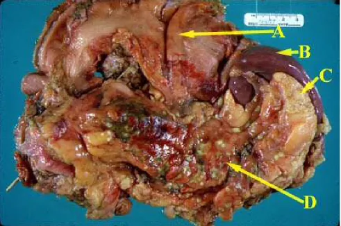
FIG 4 ACUTE PANCREATITIS SPECIMEN
CHANGES IN DIFFRENT SYSTEM IN PANCREATITIS
The pathophysiology alters many system in the body. The changes affects the following system.
• Fluid and electrolyte changes • Cardiovascular changes
• Respiratory changes • Renal changes
• Local changes
I. FLUID AND ELECTROLYTE CHANGES
Circulating blood volume decreased due to loss from intravascular space of plasma into the retroperitoneum and systemically. Additional loss occur following vomiting or naso gastric aspiration. Hypocalcaemia and hypomagnesemia are frequent. Decreased ionised calcium level also occurs due to trapping of calcium in areas of fat necrosis.
II. CARDIOVASCULAR CHANGES
restoration of intravascular volume.
III. RESPIRATORY COMPLICATIONS
• Early feature of acute pancreatitis -arterial hypoxemia
• Pulmonary function studies - Decreased inspiratory lung volume with decreased pulmonary compliance and decreased diffusing capacity.
• Early respiratory failure resolves with subsidence of pancreatitis. • Severe or unresolving pancreatitis may develop progressive
pulmonary insufficiency, infiltrates and pleural effusion.
IV. FACTOR IMPLICATED FOR PULMONARY
COMPLICATIONS
1. Abdominal distension and elevation of diaphragm.
2. Alteration in the lecithin of pulmonary surfactant by circulating pancreatic lecithinase.
3. Pulmonary thromboembolism. 4. Circulating free fatty acids
V. RENAL FAILURE
Major factor is deaths from pancreatitis. Due principally to hypovolemic. So many patients go in for acute renal failure. Pathologically it is due to the deposition of the fibrin complexes in the glomeruli.
VI. OTHER SYSTEMIC FEATURES
Abnormal Liver functions-Elevation of serum bilirubin and liver enzymes such as Alkaline Phosphatase are raised and it is mainly due to the biliary obstruction and pericholangitis.
Early Intravascular thrombosis with decreased platelet count and fibrinogen level occur due to the effects of pancreatic proleolytic enzymes. May be followed by marked thrombocytosis and hyperfibrinogenemia.
VII. LOCAL SEQULAE
Intra abdominal complications 1) Paralytic ileus
2) Duodenal/Biliary obstruction
4) Destruction of tissues adjacent to pancreas.
5) Rarely cause gross disruption of the pancreatic ductal system which is usually self limited.
6) Persistent chronic pseudocyst in 1%
7) Infected pancreatic abscess due to secondary infection occur in 1-9% and organisms are usually enteric.
8) Extension of local necrosis to involve colonic wall causing colonic perforation occurs in 1% and occurs in the left transverse colon or splenic flexure.
CLINICAL MANIFESTATIONS AND DIAGNOSIS2,3,10
The classical feature of acute pancreatitis is its severity of symptoms and paucity of physical signs.
1. Abdominal pain - 85 -100%
Upper abdominal constant pain may radiate to the back and-may be severe. Pain is aggravated by the food or by a drink of alcohol. Pain resistant to analgesics. Patient assumues of various postures in an effort to obtain relief.
2. Nausea and vomiting - 92%
contains gastric and duodenal content and it is not feculent.
3. Physical examination
• Restless patient.
• Rapid pulse and respiratory rate. • Arterial hypotension
• Abdomen- moderately distended with epigastric dullness Tenderness markedly in the upper abdomen.
• Moderate muscle spasm present.
• GREY TURNERS SIGN - Grey green discoloration of the flank in patients with peripancreatic heamorrhage
• CULLEN' S SIGN - bluish discoloration of periumbilical region
4. Extra abdominal manifestations
• Left pleural effusion
• Acute pulmonary failure marked by Tachypnoea, dyspnoea, • cyanosis - due to
a) circulating phospholipase A
b) circulating free fatty acids from triglycerides from 1ipolysis c) Pulmonary surfactant
5. Central Nervous System manifestations-
Nonlateralizing nature, including billigerence, confusion, psychosis and coma. This is due to hyperosmolarity, hypoperfusion, hypoxia, cerebral fat embolism or Disseminated intra vascular coagulation.
LABORATORY DETERMINATION2,18
DIAGNOSIS OF ACUTE PANCREATITIS
Laboratory Tests Radiographic Procedures
• Sr. amylase • Chest X Ray
• Sr. amylase isoenzymes • Plain abdominal X Ray • Urine amylase • Ultrasonography
• Amylase-creatinine clearance
ratio •
Contrast enhanced C T SCAN
I. BLOOD COUNT :
• Leucocytosis - 10,000 to 20,000 occurs early in all cases • Haematocrit - is high in most patients at the onset.
II. SERUM AMYLASE
Elevated in 95% of patients with Acute pancreatitis. But this is not an ideal marker because it is elevated in other conditions such as
A. Perforated peptic ulcer B. Biliary lithiasis
C. Intestinal obstruction D. Mesenteric infarction.
Also in patients with acute pancreatitis, serum amylase in normal levels can occur due to
I. Hyper triglyceredemia - Latescent serum II. Assayed 3 days or more after onset
III. Previous attack has destroyed most glandular tissues
IV. Present attack is associated with massive destruction of gland
Serum amylase in Acute Pancreatitis is elevated within 24 hrs of onset of symptoms and returns to normal in 7 days.
III. SERUM ISOAMYLASE P:
IV. SERUM LIPASE:
Serum lipase is solely of pancreatic origin hence serum lipase level is more specific than amylase. Recent development of an enzyme immuno assay of lipase is reliable and is of great value in Acute Pancreatitis. Duration of Hyper lipasemia exceeds hyperamylossemia.
V. PLURAL AND PERITONEAL FLUID AMYLASE
Pleural effusion shows raised levels of amylolytic activity in pancreatitis. High activities of amylase may also be found in fluids aspirated from peritoneal cavity in patients with acute pancreatitis.
IV. OTHER BIOCHEMICAL INDICES
I. Hyperglycemia and Glycoscuia- Nonspecific, transient Cause - relative hypoinsulinemia and Hyperglucognaemia II. Hypocalcemia - Well recognised entity in acute pancreatitis
but can also occur in perforated peptic ulcer
Cause - Deposition of calcium in areas of fat necrosis Release of glucagon Inadequate parathyroid response Dilutional hypo albuminaemia
i. Methemalbumin:
ii. Liver function tests:
Slight increases in alkaline phosphatase and amino transfer are with raise in serum bilirubin - transitions. Markedly elevated serum aspartate and alanine amino transfer are within 48 hrs after onset discrimianates biliary from non-biliary pancreatitis.
RADIOGRAPHIC FINDINGS2,18
I PLAIN X-RAYS
i). Plain X-Ray abdomen
a) Segmental small bowel ileus or a “SENTINEL LOOP’ in the left upper quadrant.
b) Dilatation of the transverse colon – “COLON CUT OFF SIGN”. A. Increase epigastric soft tissue density
B. Obscured psoas muscle margins. C. Presence of gall stones
D. Pancreatic calcification – may not be an acute pancreatitis
ii). Plain x-ray chest
(a) Pleural effusion (b) Atelectasis (c) Pneumonia
CONTRAST STUDIES WITH WATER SOLUBLE
CONTRAST
Upper Gastrointestinal study (a) widening of “C” loop
(b) Anterior displacement of stomach (c) Subtle duodenal mucosal sign
ABDOMINAL ULTRASONOGRAM
(a) enlargement and edema of pancreas (b) Pseudocysts of pancreas
(c) Delineates pancreatic abscess
(d) Dilatation of Bile duct and presence of stone in gall bladder and common vile duct
COMPUTED TOMOGRAPHY
VI ENDOSCOPY
To detect biliary pancreatitis.
Therapeutically used for papillotomy and removal of stones impacted at the ampulla of Vater.
TREATMENT3,10
MEDICAL MANAGEMENT
A. NUTRITION
1. Enteral nutrition
Previously it was thought that enteral nutrition stimulates the pancreas and results in pain in pancreatitis, but now it is found that pancreas is actually in a state of rest in acute pancreatitis so it better to stimulate the pancreas. So enternal feeding does no harm to the patient. 2. Total parenteral nutrition
TPN is associated with many complications such as arterial injury, pneumothorax, thrombosis, and catheter embolism. Many studies comfirm that enternal nutrition is better than TPN.
Patients suffering from severe AP have risk to develop peptic ulcers or erosive gastritis Histamin2-antagonists IS indicated in patient on mechanical ventilation and patients with adult respiratory distress syndrome (ARDS)
C. FLUID MANAGEMENT
Adequate fluid management is the main stay in the management of acute pancreatitis. If missed it can lead to serious complications. Plenty of fluids are sequestrated in third spaces. So crystalloids and colloids are used in the ratio of 3:1. The fluid loss may be 6-10 liters. It is said that when hematocrit is less than 30% if Dextran is used it improves the microcirculation. Good fluid resuscitation is indicated by adequate urine out put, CVP AROUND 8-12 cm of water, hematocrit of 35-40%.
D. PAIN MANAGEMENT
As a result of the activation of pancreatic enzymes there are increase releases of the inflammatory mediators. These mediators irritate the sensory fibers of the celiac plexus (T5-T9) and cause severe pain radiating to the back. The following drugs are used in management of pain
• meperidine • Tramadol
• thoracic epidural analgesia
E. THE ROLE OF ANTIBIOTICS9
There is a great controversy regarding use of antibiotic in acute pancreatitis. Since there is great risk in developing necrosis in acute pancreatitis, there is also problem in developing abscess in acute pancreatitis. So in order to prevent the abscess formation antibiotics are used. The most common antibiotics used are imipenem, meropenem, metronidazole, fluoroquinolones, and cephalosporins. These antibiotics penetrate the pancreas well, but aminoglycosids do not penetrate the pancreas. Over use of antibiotics result in the fungal infection.
F. SUPPRESSION OF PANCREATIC EXOCRINE SECRETION
These are done by nasogastric suction, histamine H2-receptor antagonists,
antacids,atropine, glucagon, calcitonin, stomatostatin.
SURGICAL MANAGEMENT: INDICATIONS AND TIMING
INDICATIONS14,19,23
2. Treatment of pancreatic sepsis
3. Correction of associated biliary tract disease
4. Progressive clinical deterioration despite optimal supportive care.
5. Infected necrosis 6. Severe sterile necrosis
7. Symptomatic organized pancreatic necrosis
A) BILIARY OPERATIONS IN PATIENTS WITH CHOLELITHIASIS
a. Cholecystostomy
b. Common duct drainage c. Cholecystectomy
d. Early endoscopic papillotomy
B) SURGICAL MANAGEMENT: PROCEDURES19,23
1. RESECTION
2. PANCREATIC DEBRIDEMENT
3. MINIMALLY INVASIVE APPROACHES
• Retroperitoneal approach via dorsal lumbotomy
• Percutaneous necrosectomy and sinus tract endoscopy
RESECTION
Pancreatic resection is primarily of historical interest only and is not recommended currently
PANCREATIC DEBRIDEMENT
All pancreatic débridement and postdébridement care are based on : (1) Wide removal of devitalized and necrotic tissue
(2) the assurance of postoperative removal of the products of ongoing local inflammation and infection
TECHNIQUES OF DÉBRIDEMENT
2. Open packing for pancreatic necrosis
THE COMPLICATIONS OF ACUTE PANCREATITIS2,18
The Complications of Acute Pancreatitis Local Fluid collections
Pancreatic ascites/pleural effusion Pancreatic pseudocyst
Pancreatic necrosis
Infected pancreatic abscess Hemorrhage/pseudoaneurysm Regional Venous thrombosis
Paralytic ileus
Intestinal obstruction
Intestinal ischemia/necrosis Cholestasis
Systemic Systemic inflammatory response syndrome Multiple-organ-dysfunction syndrome ARDS/pulmonary failure
Renal failure
Cardiovascular complications Hypocalcemia
Disseminated intravascular coagulopathy Protein calorie malnutrition
A) PANCREATIC ABSCESS
Pancreatic abscess - Incidence '9%. Most common in patients with post operative pancreatitis.
Clinical features
Persistent of recurrent fever Abdominal distension
Abdominal mass
Hypotension (BP 9 0 mmHg). Pneumonia / Effusion
Renal failure Coma
Elevated serum amylase Leucocytosis ( 10000 /mm^) Radio graphic diagnosis
1. Upper GI contrast studies showing di splacement of
2. Ultra sound abdomen can delineate pancreatic abscess
3. Computed Tomography sensitive and specif.lc 4. Percutaneous aspiration under CT guidance
Treatment Adjuvant
1. Vigorous supportive management
2. Meticulous respiratory care nutritional support
3. Prevention of GIT haemorrhage
Specific
1. Percutaneous drainage by catheter
2. Laprotomy - Debridement and packing of the pancreatic bed. 3. Surgical correction of other complications like
involvement of colon by colostomy
4 Feeding jejunostomy to correct nutritiona1 imbalance B) PSEUDOCYSTS
intervention is needed only when they go in for further complications. It is dealt in detail along with treatment for pseudocysts following chronic pancreatitis.
C)PANCREATIC ASCITES
Pancreatic ascites - more common following chronic pancreatitis. But may also follow acute pancreatitis secondary to trauma, pseudocysts and rarely pancreatic neoplasm.Treatment is by drainage procedures as dealt in pancreatic ascites following chronic pancreatitis.
PROGNOSTIC ASSESSMENT
PROGNOSTIC INDICATORS
Because of the variability and unpredictability of acute pancreatitis, clinical scoring systems have been made to predict the severity of acute pancreatitis
1 . RANSON S CRITERIA6
RANSON's Criteria
RANSON's Criteria
Nonbiliary Acute
Pancreatitis
Biliary Acute
Pancreatitis
Admission
Age (yr) >55 >70
WBC count (×1000/mm3) >16 >18
Glucose (mg/dl) >200 >220
AST (IU/L) >250 >250
LDH (IU/L) >350 >400
Within 48 Hours of Admission
Hematocrit decrease (points)
>10 >10
BUN increase (mg/dl) >5 >2
Deficit in base(mEq/L) >4 >5
Fluid replaced (L) >6 >4
PaO2 (mm Hg) <60 <60
To calculate base deficit first bicarb VD must be calculated Bicarb Vd =(0.4+2.6/HCO3) x Lean Body Weight
Base Deficit= Bicarb Vd x (Normal HCO3- Measured HCO3)
RANSON's score interpretation
According to RANSON score when the score is > 8 it indicates pancreatic necrosis upto 30% of the gland. If the score is >= to three the severe pancreatitis is likely and if it is < than three the severity is unlikely.
Score 0 to 2: The mortality is 2% Score 3 to 4: The mortality is 17% Score 5 to 6: The mortality is 45%
Score 7 to 8: The mortality is nears 100%
In this study the cut of value of the score is 3.the patients are classified under two groups one with RANSON score < 3 another with score >=3 since a score less than three severe pancreatitis is unlikely.
2. BISAP SCORE13
mortality and severity in 24hrs period calculation of risk can improve the clinical care. The score was developed in state of pennyyslvania using a huge cardinal health database.
INDIVIDUAL COMPONENTS OF THE BISAP SCORING SYSTEM
• Impaired conscious level (GCSS < 15) • Blood urea level > 25mg/dl
• Age > 60
• Pleural effusion
• Systemic inflammatory response syndrome
The syndrome contains two or more of the following : (1) Temperature of < 36 or > 38 ° C
(2) Respiratory rate > 20 breaths/min or P a CO2 < 32 mm Hg (3) Pulse > 90 beats/min
(4) WBC < 4,000 or >12,000 cells/mm 3 or >10% immature bands
The total score is 5. In this study the cut of value of the score is 3, the patient are classified into two group one group with score < 3 and the other group with score >= 3.
3) CT SEVERITY INDEX2
pancreatic changes and necrosis. Either contrast or plain ct is used. This is developed by BALTHAZAR and AP is graded from A to E. It gives point according to the following criteria,
ACUTE PANCREATITIS WITH EDEMA
58 Mild pancreatitis, interstitial pancreatitis
Patients with pancreatitis having no collections or necrosis. They have a mild pancreatitis. Balthazar grade A-C come under this group. CTSI is 2
Severe pancreatitis or necrotizing pancreatitis
They occur in 20 of patients. They have uneven clinical course and high mortality rate. They are more than fluid collections. The grade is d or e and this score is usually above 3. The peripancreatic collection is due to fat necrosis. This group of patients with. Necrosis has most complication and they have to be identified. There is a separate type called extra pancreatic necrosis which has on pancreatic necrosis the CTSI is 4.
Balthazar Scoring for the Grading of Acute Pancreatitis
Grade A – normal CT
Grade B – focal or diffuse enlargement of the pancreas
Grade C – pancreatic gland abnormalities and peripancreatic inflammation
Grade D – fluid collection in a single location
Grade E – two or more collections and/or gas bubbles in or adjacent to pancreas
CT severity index = CT grade point + points for necrosis
First grade points are calculated. The grade A, B, C, D, E have 0, 1, 2,
necrosis. When the necrosis is <30% the point is 2, 30-50% the point is 4,>50%
the point is 6.
CT grade points are added to points assigned for percentage of necrosis to
determine the CT severity index. So the patients with score greater than three is
said to manifest severe disease. So in this study two category one with score < 3
and another with score > =3 are taken into consideration
OTHER PROGNOSISTIC SYSTEMS
• The Acute Physiology and Chronic Health Evaluation II (APACHE II) score.
• C-reactive protein (CRP) assays.
• Trypsinogen-activating peptide (TAP) assays.
ATLANDA CLASSIFICATION
1) ACUTE ODEMATOUS PANCREATITIS – MILDER FORM
MORTALITY 1%
2) ACUTE NECROTISING PANCREATITIS – incidence of 20% and
MATERIALS AND METHOD
STUDY AREA :
GOVT. COIMBATORE MEDICAL COLLEGE AND HOSPITAL
STUDY POPULATION :
Patients admitted in CMCH with symptoms suggestive of acute pancreatitis
INCLUSION CRITERIA :
1. Clinical findings suggestive of abdominal pain characteristic of acute pancreatitis, serum amylase raised 3 times above normal value, ct scan with finding suggestive of acute pancreatitis data from the patient admitted with acute pancreatitis was collated. When some values are missed the referred case no mark is given. The data for RANSON is collected for 24 hrs and 48 hrs. CT scan is taken within 48 hrs. BISAP is calculated with 24 hrs data.
The Following criteria are studied
DEFINITION
1. MILD OR SEVERE AP
Depending on the organ failure the patients are classified into mild or severe pancreatitis. The presence of organ failure is again dictated by the presence of following factor
• Pulmonary failure(pco2 < 60mm of hg)
• Renal failure(serum creatinine levels > 2 mg/dl) • Severe shock
2. PANCREATIC NECROSIS
This finding is easily found by the CECT scan. This is detected by the no enhancement in the CECT scan in the pancreatic parenchyma.
3. MORBIDITITY
Length of the hospital is used as the indicator of the morbidity
EXCLUSION CRITERIA :
STUDY PERIOD:
September 2011, November 2012.
SAMPLE SIZE:
All patients eligible by inclusion and exclusion criteria are to be included in the study.
STUDY DESIGN:
RESULTS
STATISTICAL ANALYSIS:
The data are reported as the mean +/- SD or the median, depending on their distribution. The differences in quantitative variables between groups were assessed by means of the Unpaired t test. Comparison between groups was made by the Non parametric Mann - Whitney test. The chi square test was used assess differences in categorical variables between groups. Data were analyzed by diagnostic efficiency derived from the receiver operating characteristic [ROC] Curve and area under the ROC curve. Sensitivity, specificity and predictive values were determined. A p value of <0.05 using a two-tailed test was taken as being of significance for all statistical tests. All data were analyzed with a statistical software package. (SPSS, version 16.0 for windows)
After calculating the sensitivity, specificity, PPV, NPV for the scoring system such as RANSON, BISAP,CTSI in predicting the mortality, pancreatic necrosis, acute severe pancreatitis, the results are compared to derive at the conclusion.
A) AGE SEX DISTRIBUTION
Table 1 – Age Sex Distribution Age Male Female Total <= 30 23 1 24 31 - 40 37 2 39 41 - 50 35 4 39 51 - 60 8 0 8 >= 60 7 0 7 Total 110 7 117
The above tabular column 1 gives the age sex distribution of the disease in the various age group.
Table 2 – Mean SD for Age
Sex N Mean +/- SD Range Male 110 39 +/- 12 16 - 72 Female 7 39 +/- 9 23 - 50 Total 117 39 +/- 11 16 - 72
[image:69.595.162.435.517.660.2]The total patients studied in this study were 117, which comprises of 110 males and 7 females. Among the male population the maximum age group is 31-40. Next comes the 41-50 which includes 35 patients. Among the female population the maximum age group is 41-50 which includes about 4 patients.
The next tabular column 2 clearly shows the mean and the standard deviation of the age group. On the whole it is 39+/-11. It is 39+/- 12 for male and 39+/-9 for female. The bar diagram shows the same in the percentage. Males greater than 60 years of age affected by the disease are 6%. This is the least age group affected.
0% 5% 10% 15% 20% 25% 30% 35%
<= 30 31 - 40 41 - 50 51 - 60 >= 60
Male 20% 32% 30% 7% 6%
Female 1% 2% 3% 0% 0%
Sex distribution is shown in the pie chart. 6 percent is female and the rest is males. In our population males are commonly affected than the female population. This factor has link with the etiology, in our population alcohol is the common cause of the acute pancreatitis. Alcohol consumption is not prevalent in female population so female are not commonly affected by acute pancreatitis.
Male[n=110] 94% Female [n=7]
6%
B. ETIOLOGY
Table 3 - Etiology Etiology No.of cases
Gall Stone 4
Alcohol 90
unknown 23
The above diagram shows the etiology of the acute pancreatitis in the percentage. The orange shaded area shows that the alcohol contributes the 77% of the etiology in acute pancreatitis. The brown shaded area gives the percentage of gall stone contributing to pancreatitis, it is about 3%. Out of 7 females 2 had gall stones pancreatitis. The gall stone pancreatitis is common in females. Unknown causes contribute to about 20%.
Gall Stone 3%
Alcohol 77% unknown
20%
C.SEVERE ACUTE PANCREATITIS(SAP) Vs DIFFEENT
[image:74.595.94.502.186.644.2]PROGNOSTIC SCORE
Table 4 - Severe Acute Pan. vs Prognostic Score
RANSON
SAP
Total P VALUE
Yes No
< 3 14 67 81
NS >= 3 11 25 36
Total 25 92 117
BISAP
< 3 10 87 97
NS >= 3 15 5 20
Total 25 92 117
CTSI
< 3 17 61 78
NS >= 3 8 31 39
Total 25 92 117
Table 5 - Severe Acute Pain vs Prognostic Score
Study SEN SPEC PPV NPV RR ODDS RANSON 44% 73% 31% 83% 1.768 2.106
BISAP 60% 95% 75% 90% 7.275 26.100 CTSI 32% 66% 21% 78% 0.941 0.926
TABLE 4 shows the different scores of patients with SAP. 14 patients with SAP had RANSONs score < 3 and 11 with SAP had RANSON score > 3. But only 10 patients with SAP had BISAP score < 3 and 15 SAP patients had BISAP score > 3. CTSI score in 17 SAP was < 3 and 8 patient with CTSI had > 3.
So out of 25 SAP patient BISAP picked out 15 and RANSON picked 14 and CTSI picked only 8.
The above bar diagram shows the relationship between the severe acute pancreatitis and the other prognostic score. The blue shaded area indicates the presence of the SAP and the red shaded area indicates the absence of the SAP. Among the RANSON score <3, SAP is present in 12% and 57% had no SAP. On the other hand in the patients with BISAP score <3, 9% had SAP and 74% had no SAP. In the patients with BISAP score >=3, 15% had SAP and 4% had no SAP.
< 3 >= 3 < 3 >= 3 < 3 >= 3
Ranson Bisap CTSI
SAP Yes 12% 9% 9% 13% 15% 7%
SAP No 57% 21% 74% 4% 52% 26%
0% 10% 20% 30% 40% 50% 60% 70% 80%
D) MORTALITY Vs THE PROGNOSTIC SCORE
Table 6 - Mortality vs Prognostic Score
RANSON
Mortality
Total P VALUE Yes No
< 3 3 78 81
NS
>= 3 4 32 36
Total 7 110 117
BISAP
< 3 2 95 97
<0.01
>= 3 5 15 20
Total 7 110 117
CTSI
< 3 4 74 78
NS
>= 3 3 36 39
Total 7 110 117
Table 7 - Mortality vs Prognostic Score
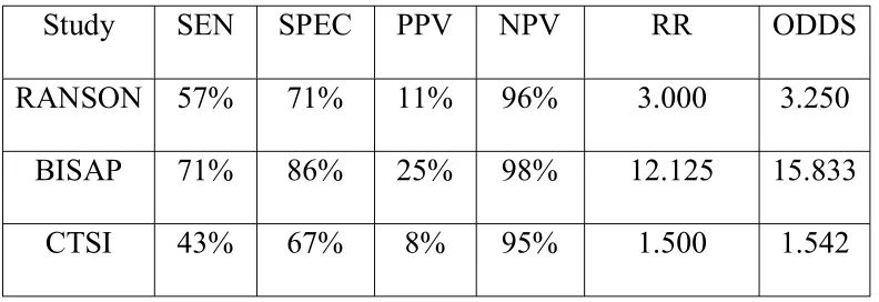
Study SEN SPEC PPV NPV RR ODDS RANSON 57% 71% 11% 96% 3.000 3.250 BISAP 71% 86% 25% 98% 12.125 15.833 CTSI 43% 67% 8% 95% 1.500 1.542
This type of analysis shows which type of score is better in predicting the mortality in acute pancreatitis patient. Out of total 7 deaths 4 patients has RANSON score >=3 , 5 patients had BISAP score >=3 and 3 patients had CTSI score >=3. The p value of the BISAP score is <0.01 which shows the significant relationship between the BISAP score and the mortality.
The above bar diagram shows the relationship between the mortality and the various prognostic score. The mortality is three percent for the patients with RANSON score <3 and >=3. There is same percentage of mortality occurred in the patients with CTSI score <3 and >=3. But the mortality is 4% for the patient with BISAP score >=3. This is high when compared to other score. This indicates predicting accuracy of mortality by the BISAP score.
< 3 >= 3 < 3 >= 3 < 3 >= 3
Ranson Bisap CTSI
Mortality Yes 3% 3% 2% 4% 3% 3% Mortality No 67% 27% 81% 13% 63% 31%
0% 10% 20% 30% 40% 50% 60% 70% 80% 90%
E) PANCRETIC NECROSIS (PANCREATIC NECROSIS) Vs
[image:80.595.94.504.232.660.2]PROGNOSTIC SCORE
Table 8 - Pancreatic Necrosis vs Prognostic Score
RANSON
P Nec
Total
P
VALUE
Present Absent
< 3 8 73 81
NS
>= 3 8 28 36
Total 16 101 117
BISAP
< 3 9 88 97
< 0.05
>= 3 7 13 20
Total 16 101 117
CTSI
< 3 2 76 78
<0.001
>= 3 14 26 39
Total 16 102 117
prognostic score.
Table 9 - Pancreatic Necrosis vs Prognostic Score
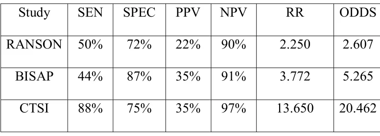
Study SEN SPEC PPV NPV RR ODDS RANSON 50% 72% 22% 90% 2.250 2.607 BISAP 44% 87% 35% 91% 3.772 5.265 CTSI 88% 75% 35% 97% 13.650 20.462
TABLE 8 shows the score which can predict the Pancreatic necrosis. Since pancreatic necrosis is a CT finding naturally it shows that CTSI is good in predicting the pancreatic necrosis. Out of the 117 patients 16 patients had pancreatic necrosis. Out of the 16 patients 14 patients had CTSI score >=3 and only 2 patients had score <3. The P value of the BISAP score in predicting the mortality is <0.05. But the P value of the CTSI score in predicting the pancreatic necrosis is <0.001. This indicates strong relationship between the CTSI score and pancreatic necrosis.
[image:81.595.102.490.165.304.2]The above bar diagram shows that the 12% of the patients with CTSI >=3 had Pancreatic necrosis. Only 2% of the patients with score < 3 had Pancreatic necrosis. 7% of the patients with RANSON score <3 and >=3 had pancreatic necrosis. 8% of the patients with BISAP score < 3 had pancreatic necrosis. Six percent of the patients with score >=3 had pancreatic necrosis. The bar diagram finally shows the highest predicting value for pancreatic necrosis by the CTSI.
F)MORBIDITY
Morbidity is assessed by number of days patients stayed in the hospital. The mean and standard deviation is 9.03+/- 3.42 days. The overall days patients stayed in the hospital has no significant relationship with the scoring system.
< 3 >= 3 < 3 >= 3 < 3 >= 3
Ranson Bisap CTSI
P nec Present 7% 7% 8% 6% 2% 12%
P nec Absent 62% 24% 75% 11% 65% 22% 0%
10% 20% 30% 40% 50% 60% 70% 80%
AREA UNDER CURVES
AUC 1 COMPARISON OF SCORING SYSTEMS IN PREDICATING
SEVERE ACUTE PANCREATITIS
AUC FOR SAP
covered below the line is maximum when compared with other line Source of the graph is RANSON, BISAP, CTSI group. The AUC is 0.773 for BISAP which is the maximum when compared to other scores (table 14). The standard error of the same score is 0.0653 and the 95% confidence interval is 0.650 to 0.896. The AUC is least for CTSI group in predicting the SAP.
Table 10 - Area Under the Curve for SAP
Test Result Variable(s)
Area
Std. Errora
Asymptotic Sig.b
Asymptotic 95% Confidence Interval Lower
Bound
Upper Bound RANSON
grp 0.584 0.066 0.198 0.454 0.714 BISAP grp 0.773 0.063 0.000 0.650 0.896 CTSI grp 0.492 0.065 0.897 0.364 0.619
AUC 2 COMPARISION OF THE SCORING SYSTEM IN
PREDICTING THE MORTALITY
the reference line. The BISAP score line is high above all line, the area covered below the line is maximum when compared with other line.
Table 11 - Area Under the Curve for mortality
Test Result Variable(s)
Area
Std. Errora
Asymptotic Sig.b
Asymptotic 95% Confidence Interval Lower
Bound
Upper Bound RANSON grp 0.640 0.112 0.215 0.420 0.860
BISAP grp 0.789 0.102 0.011 0.589 0.989 CTSI grp 0.551 0.115 0.654 0.326 0.776
The AUC for the BISAP is 0.789 and it has the standard error of 0.102. the AUC for the RANSON and the CTSI is 0.640 and 0.551 respectively. The BISAP score has the CI of 0.589 to 0.989.
AUC 3 COMPARING THE SCORING SYSTEM IN PREDICTING
THE PANCREATIC NECROSIS
shows the RANSON score, the pink colored indicates the CTSI and the violet colored line is the reference line. The CTSI score line is high above all line, the area covered below the line is maximum when compared with other line
Table 11 - Area Under the Curve for pancreatic necrosis
Test Result Variable(s)
Area
Std. Errora
Asymptotic Sig.b
Asymptotic 95% Confidence Interval
Lower Bound
Upper Bound RANSON grp 0.611 0.079 0.153 0.457 0.766 BISAP grp 0.654 0.082 0.048 0.494 0.815 CTSI grp 0.814 0.055 0.000 0.706 0.921
ATLANDA CLASSIFICATION ANALYSIS
The Atlanta classification classifies the acute pancreatitis into necrotic pancreatitis and edematous pancreatitis. The classification uses the above parameter to predict the mortality. In this study of 117 patients necrotic pancreatitis is 16 i.e. 14% and the rest is edematous pancreatitis accounting to about 86%.
The above bar diagram compares the mortality and the pancreatitis necrosis and the mortality. Among 16 patients who had pancreatic necrosis 3 died and the others survived. Among the rest 4 died and 83% survived. Only 11% of the patients who had acute severe pancreatitis survived (bar diagram).
0% 20% 40% 60% 80% 100%
Yes No
Mortality
Present 3% 11%
Absent 3% 83%
This shows there relationship between the pancreatic necrosis and the mortality.
[image:90.595.74.494.233.397.2]PANCREATIC NECROSIS vs SEVERE ACUTE PANCREATITIS
Table 12 - PANCREATIC NECROSIS VS SAP
P Nec
SAP
Yes No Total (%)
Present 8 8 16 14%
Absent 17 84 101 86%
Total 25 92 117 100%
This tabular column 12 gives the analysis between the pancreatic necrosis and the SAP. Out of the 25 SAP patients pancreatic necrosis present in 8 patients and absent in 17 patients. On the other hand out of 92 patients with no SAP, 8 patients had pancreatic necrosis.
PAN NEC VS SAP & MORTALITYAUC
PAN NEC VS SAP & MORTALITYAUC
DISCUSSION
[image:92.595.134.464.233.435.2]1 AGE COMPARISON
Table 13 - Age Comparison
S.No STUDY
MEAN
AGE
1 PRESENT STUDY 39+/-11
2
VIKESH SINGH AND GROUP13
52+/-16
3 GEORGIOS AND GROUP 52+/-14
The mean age with SD of study group is 39+/-11 and inter quartile range is 16-72. This is less when compared to other. This might be due to the etiology alcohol is more prevalent in the younger age group.
2. ETIOLOGY
2. MEDIAN HOSPITAL STAY
Table 14 – Median Hospitals
S.No STUDY
HOSPITAL
STAY(days)
1 PRESENT 9
2 A.O FARRELL 7
Compare to the study done by AO Farrell25 the hospital stay is increased with our study.
4. MORTALITY COMPARITION
Table 15 – Mortality Comparison
S.No STUDY MORTALITY%
1 PRESENT STUDY 5.9
[image:93.595.127.502.505.724.2]Table 16 - SENSITIVITY, SPECIFICITY,PPV,NPV OF BISAP SCORE IN PREDICTING THE MORTALITY IN DIFF. STUDIES
S.NO STUDY SENSITIVITY % SPECIFICITY % PPV% NPV% 1 PRESENT STUDY
71 86 25 98
2
VIKESH SINGH AND GROUP
71 83 17.5 99
3
GEORGIOS AND GROUP15
[image:94.595.67.539.168.431.2]57.1 87.6 15.5 98
Table 17 - AUC OF BISAP SCORE IN PREDICTING THE MORTALITY IN DIFF STUDIES
S.No STUDY AUC
1 PRESENT STUDY 0.78
2
VIKESH SINGH AND GROUP
0.83
The overall mortality of the study is 5.9 it is higher when compared to the Vikesh Singh and group, Georgios and group but lower than the LOSADA M and group. This might be due to the many factors such as availability of the intensive care in the developing country, other co morbid factor. The sensitivity, specificity, PPV, NPV of the BISAP score in predicting the mortality is consistent with study done by Vikesh Singh and group. The sensitivity is higher when compared to the GEORGIOS and group study. This suggests that BISAP score does well in predicting the mortality. AUC for predicting the mortality by the BISAP score is also more or less consistent with the other study.
[image:95.595.66.531.520.655.2]4. SAP COMPARISON IN DIFFERENT STUDIES
Table 18 - SENSITIVITY, SPECIFICITY, PPV, NPV OF BISAP SCORE IN PREDICTING THE SAP
S.NO STUDY SENSITIVITY SPECIFICITY PPV NPV
1 PRESENT STUDY 60 95 75 90
2
GEORGIOS AND GROUP15
35.5 92.4 57.7 84.3
of the BISAP score in predicting the SAP with other study. The sensitivity and specificity in the present study is 60% and 95% respectively. While in the georgious group the same is 35-5% and 92.4% respectively. The specificity is consistent when compared with present study.
Table – 19 AUC OF BISAP SCORE IN PREDICTING THE SAP IN DIFF STUDIES
S.NO STUDY AUC
1 PRESENT STUDY 0.773 2 GEORGIOS AND GROUP 0.8I
Table 20 - SENSITIVITY, SPECIFICITY, PPV, NPV OF BISAP SCORE IN PREDICTING THE PANCREATIC NECROSIS IN DIFF
STUDIES
S. NO SCORE STUDY SENTI SPECI PPV NPV
1
BISAP
PRESENT STUDY 50 72 22 90 GEORGIOS AND
GROUP
33.3 90.6 46.6 84.9
2
CTSI
PRESENT STUDY 88 75 35 97 GEORGIOS AND
GROUP
97.2 75.8 59.3 98.7
Pancreatic necrosis is well predicted by the CTSI in both the study. But BISAP score has increased sensitivity in predicting the pancreatic necrosis when compared to the GEORGIOS and group.
5. ATLANDA CLASSIFICATION ANALYSIS
Table 21 – Atlanta Classification analysis
S.No STUDY NECROTIC
PAN
OTHERS
1 PRESENT STUDY 14 86
[image:97.595.66.531.624.753.2]CONCLUSION
To conclude BISAP score is simple and it is the better scoring system in predicting the prognosis when compared to other score. The BISAP score has many advantages when compared to other scoring system. The advantages are as follows
1) It is simple in calculating the score the components are clinically relevant and easy to obtain.
2) It uses the 24 hrs data to predict the prognosis of the acute pancreatitis patient. But in RANSON score 48hrs data has to be collected.
3) BISAP SCORE gives weightage to the immune response of the injury and includes the age criteria.
4) BISAP score is used to triage the patient to closer observation and to asses which patients need closer monitoring.
SUMMARY
1. In our study occurrence of the pancreatitis is common in the age group is 39+/-11.
2. Most common etiology is alcohol it is about77%. 3. In our community men are most affected than women.
4. Median hospital stay in our community for acute pancreatitis is about 9+/-3days.
5. According to ATLANDA score 86% had mild disease.
6. On the whole BISAP score is better in predicting the mortality an severe disease .
7. CTSI predicted the necrotic pancreatitis in a better way. 8. Among the pancreatic necrosis the mortality is 3%.
9. Among the pancreatic necrosis patient 7% had severe acute pancreatitis.
10. The sensitivity and specificity of the BISAP score in predicting the severe acute pancreatitis is 60%, 95%.
11. The sensitivity and specificity of the BISAP score in predicting the mortality is 71%, 86%.