0022-538X/96/$04.0010
Copyrightq1996, American Society for Microbiology
Analysis of 15 Adenovirus Hexon Proteins Reveals the Location
and Structure of Seven Hypervariable Regions Containing
Serotype-Specific Residues
LETA CRAWFORD-MIKSZA1,2*
ANDDAVID P. SCHNURR1
Viral and Rickettsial Disease Laboratory, Division of Communicable Disease Control, California Department
of Health Services,1and School of Public Health, Program in Infectious Diseases,
University of California at Berkeley,2Berkeley, California
Received 17 July 1995/Accepted 29 November 1995
The first full-length hexon protein DNA and deduced amino acid sequences of a subgenus D adenovirus (AV) were determined from candidate AV48 (85-0844). Comprehensive comparison of this sequence with hexon protein sequences from human subgenera A, B, C, D, F, bovine AV3, and mouse AV1 revealed seven discrete hypervariable regions (HVRs) among the 250 variable residues in loops 1 and 2. These regions differed in length between serotypes, from 2 to 38 residues, and contained >99% of hexon serotype-specific residues among human serotypes. Alignment with the published crystal structure of AV2 established the location and structure of the type-specific regions. Five HVRs were shown to be part of linear loops on the exposed surfaces of the protein, analogous to the serotype-specific loops or ‘‘puffs’’ in picornavirus capsid proteins. The HVRs were supported by a common framework of conserved residues, of which 68 to 75% were hydrophobic. Unique sequences were limited to the seven HVRs, so that one or more of these regions contain the type-specific neutralization epitopes. A neutralizing AV48 hexon-specific antiserum recognized linear peptides that corre-sponded to six HVRs by enzyme immunoassay. Affinity-purification removal of all peptide-reactive antibodies did not significantly decrease the neutralization titer. Eluted peptide-reactive antibodies did not neutralize. Human antisera that neutralized AV48 did not recognize linear peptides. Purified trimeric native hexon inhibited neutralization, but monomeric heat-denatured hexon did not. We conclude that the AV48 neutral-ization epitope(s) is complex and conformational.
The adenoviruses (AVs) comprise a large family of double-stranded DNA viruses found in mammals, amphibians, and birds which have a common genome organization and nonen-veloped icosahedral capsid structure (48). The capsid is made up of two types of capsomeres containing three proteins: 12 vertex capsomeres composed of fiber attachment protein and its penton base, and 240 hexons (10). Each hexon capsomere is
a homotrimer of the hexon protein, a complex protein of.900
residues (42). Electron microscopy and X-ray crystallography of human AV2 hexon revealed a dense pedestal base com-posed of two eight-stranded, antiparallel beta barrels stabilized by an internal loop. A tower region projects away from the surface of the virion formed by three loops, L1, L2, and L4. The L2 and L4 loops coil to interact on either side with the tower regions of the other two copies of the protein in the trimer. This interdigitation of the loops, combined with adja-cent pedestal interactions, gives the trimer extreme structural stability. The L1 loop is the longest and most complex, folding back on itself several times and projecting furthest into the solvent, providing maximal interaction with the environment (1, 31, 42).
The human AVs now number 49 serotypes (16, 36) divided into six subgenera (A through F) on the basis of nucleic acid differences, fiber protein characteristics, and biological prop-erties (50). Neutralization is type specific (15) and is directed against an epitope(s), historically designated eta, on the hexon protein (22). Studies in which the protein sequences of several
AV hexon proteins have been compared have noted that while most of the pedestal regions are conserved, variable regions exist in L1 and L2 (19, 21, 28, 40, 41, 46).
Until recently, complete protein sequence information avail-able on human AVs has been limited to subgenera C and F. Much less is known about the subgenus D AVs. First isolated in the 1950s from epidemic conjunctivitis (20), they are cur-rently recognized as significant pathogens in immunocompro-mised populations (13, 14, 49). They are the largest group, containing 30 of the 49 known serotypes, and the group which appears to be changing the fastest, with the appearance of multiple intermediates and intertypic strains (14, 49). The seven most recent human AVs to be characterized (AV43 to AV49) were all subgenus D serotypes isolated primarily from AIDS patients (13, 16, 36). AV48 first appeared in 1985, iso-lated from AIDS patients in the San Francisco Bay area. Among isolates from subsequent years, only one was not as-sociated with concurrent human immunodeficiency virus infec-tion (5, 36). As part of ongoing investigainfec-tions into the source and evolution of AIDS-associated AVs, we examined a part of the genome undergoing rapid evolution, the major capsid pro-tein.
The purpose of this study was to obtain the first full-length subgenus D protein sequence and perform a comprehensive comparison of hexon protein sequences for fine-mapping of conserved and variable residues in order to define those resi-dues which are responsible for type specificity. We found seven discrete regions which contained unique sequences that
ac-counted for .99% of type-specific variation. These regions
also varied in length between serotypes, sometimes dramati-cally. Overlapping peptides corresponding to the entire L1 and L2 regions of AV48 were synthesized to define the type-spe-* Corresponding author. Mailing address: California Department of
Health Services, Viral and Rickettsial Disease Laboratory, 2151 Berkeley Way, Berkeley, CA 94704. Phone: (510) 540-2813. Fax: (510) 540-3305.
1836
on November 9, 2019 by guest
http://jvi.asm.org/
MATERIALS AND METHODS
PCR and direct DNA sequencing.The complete hexon gene of AV48 (85-0844), including part of the upstream pVI core protein and the hexon 59 non-coding region, was sequenced by generating overlapping PCR products and direct cycle sequencing. A set of 10 pairs of primers were designed and synthe-sized from the published sequences of AV2 (32), AV5 (21), AV40 (41), AV41 (40), and BAV3 (19). Deoxyinosine was used in codons with ambiguity. All products were sequenced from both directions with internal and template prim-ers. A pair of primers consisted of the rightward coding sequence primer and the leftward complement of that sequence. Rightward coding primers were (AV48 numbering): UP, 59-AACAGCATIGTGGGTITGGGIGTG-39(pVI); OR, 59-ATGGCIACCCCITCGATGATGCCG-39(codons 1 to 8); 1R, 59-TACTTTGA CATCCGCGGCGTGCTGGA-39(98 to 106); 2Rm, 59-CCITGCTATGGITCIT TTGC-39(220 to 226); 3R, 59-ATGTGGAAICAGGCIGTIGACAG-39(381 to 388); 4R, 59-TTTGCCATGGAIATIAAICT-39(453 to 459); 5R, 59-ATGGACA AIGTIAAICCITTIAAICACCACCG-39(523 to 533); 6R, 59-GCIGCIAACAT GCTITAICCIATICC-39(643 to 650); 7Rm, 59-ATGTAITCCTTITTICGIAAC TTCCAICCIATG-39(785 to 795); and 8R, 59-TTCTCIGCCGGIAACGCIACI ACATAA-39(938 to 946). Full-length AV48 DNA template was prepared by Hirt extraction of infected A549 cells (17). Standard PCR conditions (35) of 100/pmol of each primer, 50 mM KCl, 10 mM Tris, 1.5 mM MgCl2, 0.01%
gelatin, 2.5 U of Tfl heat-stable DNA polymerase (Epicenter Technologies, Madison, Wis.), 250mM each of the four deoxynucleoside triphosphates (Phar-macia, Piscataway, N.J.), and 1 to 5mg of template DNA in a 100-ml reaction volume were supplemented with 5 to 7% glycerol and 1mg of single-stranded DNA binding protein (Bind-Aid; US Biochemical, Cleveland, Ohio). An initial denaturation of 948C for 2 min was followed by 30 cycles of 948C for 1 min, annealing for 1 min, and extension at 728C for 2 min. The annealing temperature varied with the primer pair, from 45 to 558C. PCR products were isolated directly or from agarose gels by adsorption to silica (GeneClean; Bio 101, La Jolla, Calif.). Cycle sequencing with the ‘‘fmol’’ DNA Sequencing System (Promega, Madison, Wis.) was carried out with 10 pmol of primer and 1 to 6ml of PCR product (30).
Immune serum preparation.Enriched AV48 hexon protein, prepared as de-scribed before (2), was purified by electrophoresis in 4% high-resolution agarose (ProSieve; FMC Corp, Rockland, Maine). A band containing 100mg of 300-kDa hexon trimer was cut from the gel, homogenized with an equal volume of complete Freund’s adjuvant, and injected intraperitoneally into New Zealand White rabbits. Two more 100-mg injections with incomplete Freund’s adjuvant were given at 1-month intervals.
Peptides and ELISAs.A set of 45 biotinylated 15-mer peptides with an overlap of 5 were synthesized for the entire AV48 variable regions of L1 and L2, from residues 121 to 322 and 409 to 454, with an SGSG linker between the biotin and peptide moieties (Chiron Mimotopes Ltd., Victoria, Australia). Peptide enzyme-linked immunosorbent assays (ELISAs) were performed as described before (47), using streptavidin to coat the plates and bind biotin with casein blocker (45), with the AV48 hexon-specific antiserum and with two human sera shown previously to contain neutralizing activity against AV48 (5). Affinity purification of antipeptide antibodies was carried out as reported before (12) with biotiny-lated acrylic beads which were coated with avidin, followed by the biotinybiotiny-lated peptides, and blocked with casein. Five peptides from hypervariable regions (HVRs) 1, 2, 3, 4, and 7 were used individually and in combination to remove antipeptide antibodies from the serum. The peptide core sequences were as follows: peptide 8, AAMGGIEITAKGLQI (HVR 1); peptide 11, GIDATKEED NGKEIY (HVR 2); peptide 16, IGEENWQDSDNYYGG (HVR 3); peptide 26, GEEPKELDIDLNFFD (HVR 4); and peptide 42, GVKVKTTNNTEWEKD (HVR 7).
Neutralization assays and hexon protein competition.Neutralization was as-sessed in a microtiter colorimetric assay with A549 human lung carcinoma cells (6). Native hexon protein was purified as described before (2), and monomeric unfolded hexon polypeptide was obtained by boiling an aliquot for 3 min. Hexon protein competition for neutralizating antibodies was determined by incubating undiluted AV48 hexon-specific antiserum for 1 h with increasing concentrations of native trimeric hexon protein or heat-denatured monomeric hexon protein. Protein-antiserum mixtures were assayed for neutralization as described above. Controls included virus-protein mixtures to rule out protein inhibition of virus infectivity.
Nucleotide sequence accession number.The complete hexon protein DNA sequence from subgenus D candidate AV48 (85-0844) was determined, and the amino acid sequence of 946 residues was deduced. The nucleotide sequences of 2,935 bp of AV48 hexon protein, the hexon 59noncoding region, and the 39 terminus of the pVI core protein have been entered in the GenBank database under accession number U20821.
Deduced protein sequence.The deduced AV48 protein se-quence was aligned and compared with that of 14 other AVs for which sequence data are available (Fig. 1). Partial se-quences for AV9 (26), AV15 (9), and AV31 (29) were omitted from Fig. 1 but were included in the analysis and conform to the structural alignments made here. Secondary structural des-ignations below the sequences were taken from the published coordinates published for AV2 hexon (1). Conservation throughout the protein was greatest within a subgenus (88%) and was reduced between human subgenera (79 to 81%) and between animal serotypes (66 to 68%). Shaded areas represent complete and functional conservation between serotypes within the variable regions from residues 131 to 331 (AV2 numbering) of L1 and residues 427 to 475 of L2. Functional conservation is defined as substitution with amino acids similar in size and charge (basic, R and K; aliphatic, A, C, G, I, L, M, and V; aromatic, F, Y, W, and H; hydrophilic, E, Q, N, and D; hydroxyl, S and T; P has no functional equivalent) (7). Among the 250 residues in variable regions, 82 residues were con-served among human serotypes, 72 were concon-served within bo-vine AV3 (BAV3), and 61 were conserved in mouse AV1 (MAV1). Of these, hydrophobic residues accounted for 68, 71, and 75%, respectively.
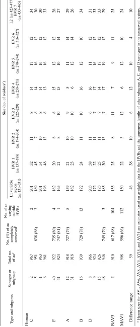
Alignment of conserved residues defined seven HVRs which differed in both length and sequence. Table 1 summarizes these differences. With a few exceptions, the composition and length in each HVR showed subgenus clustering. Serotypes within subgenera A and F were the most closely related to each other, differing in length by at most two residues in all HVRs. The greatest variability was in HVR 1, as has been noted previously (19, 28, 40, 41). Subgenus C serotypes and AV8 (subgenus D) contained an insertion of more than 30 residues in this region. The AV8 insertion was slightly larger, with a more basic and hydrophobic chemical composition than that of the subgenus C serotypes.
HVR 4, which forms the exterior side surface of the L1 tower, had two distinct conformations. Subgenus A and the animal serotypes contained an abbreviated 5 to 7 residues, making them the smallest hexons, while all others had 13 to 17 residues in this region. HVR 5 exhibited the same type of dichotomy, although not as extreme: subgenera A, B, and F and the animal serotypes contained a shorter version (9 to 12 residues), and subgenera C and D (other than AV8) contained longer stretches (16 to 19 residues).
More than 99% of serotype-specific variability was con-tained in the seven HVRs among human subgenera A, C, D, and F. When the sequences were aligned with the published crystal structure of the AV2 hexon (1), five of the HVRs (2, 3, 4, 5, and 7) occurred on the external surface of the protein (Fig. 2). For the most part, they were shown to be loops lacking secondary structure, but a single beta strand was seen in HVR 4. HVR 5 contained a region which remains unresolved crys-tallographically. HVRs 1 and 6 were internal loops beneath HVRs 2, 3, 4, and 5.
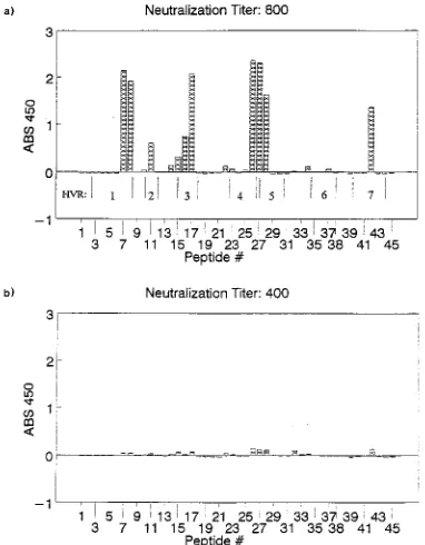
Peptide ELISAs and serum neutralization. Figure 3 and Table 2 summarize the lack of correlation between neutraliza-tion and linear peptide reactivity. AV48 hexon-specific anti-serum, with a neutralization titer of 800, reacted with peptides that corresponded to residues in the five surface HVRs and part of HVR 1 (Fig. 3a). These residues are underlined in the AV48 sequence in Fig. 2. A broad peak from peptides 26 to 28 recognized residues from both HVR 4 and HVR 5. After affinity removal of all peptide reactivity from the serum by adsorption with five peptides, the neutralization titer was 400
on November 9, 2019 by guest
http://jvi.asm.org/
on November 9, 2019 by guest
http://jvi.asm.org/
FIG. 1. Alignment of deduced amino acid (aa) sequences from 12 hexon proteins taken from DNA sequences for AV2 (32), AV5 (21), AV1 and AV6 (28), AV40 (41), AV41 (40), AV12 (38), AV16 (27), AV8 (29), BAV3 (19), and MAV1 (46) and AV48.p, conservation with AV2 protein sequence. Shaded areas indicate complete and functional conservation within the variable regions of L1 and L2. Underlined residues in the AV48 sequence correspond to peptide-reactive epitopes in HVRs. Labeling beneath each line indicates secondary-structure designations (1). Residue numbering is from the AV2 sequence.
on November 9, 2019 by guest
http://jvi.asm.org/
TABLE 1. Hexon protein amino acid conservation and variation among human, bovine, and mouse AVs Type and subgenus Serotype or subgroup Total no. of aa a No. (%) of aa completely conserved b No. of aa
varying outside HVRs
Size (no. of residues c) L1 variable region (aa 131–331) HVR 1 (aa 137–188) HVR 2 (aa 194–204) HVR 3 (aa 222–229) HVR 4 (aa 258–271) HVR 5 (aa 278–294) HVR 6 (aa 316–327) L2 (aa 423–477), HVR 7 (aa 433–465) Human C 2 967 2 201 52 11 8 14 17 12 34 5 951 838 (88) 3 189 45 7 8 14 16 12 30 1 963 201 54 10 8 14 16 12 30 6 961 196 48 13 8 13 16 12 33 F 40 922 735 (80) 4 162 20 8 8 15 12 12 28 41 924 747 (81) 1 165 21 8 10 15 10 14 27 A 12 918 727 (79) 5 159 21 10 9 5 11 14 29 31 918 162 23 10 10 6 12 14 26 B 16 939 729 (78) 13 172 24 11 10 16 12 10 34 D 8 966 203 58 11 6 16 11 12 31 9 924 172 22 11 7 17 16 12 33 15 926 173 25 11 7 14 17 12 33 48 946 745 (79) 3 185 30 13 7 17 19 12 29 BAV3 I 910 617 (68) 104 155 25 8 11 7 9 11 25 MAV1 908 596 (66) 112 150 22 3 12 6 12 10 24 Dif ference range 59 46 38 10 6 12 10 4 10 aHexon sizes for AV1, AV6, AV8, AV9, AV15, and AV31 are estimates based on available data for the HVRs and the consensus lengths of other subgenus A, C, and D serotypes in the conserved regions. bCompared with the AV2 sequence. cResidue numbering is for the AV2 sequence.
on November 9, 2019 by guest
http://jvi.asm.org/
[image:5.612.165.443.78.728.2](Fig. 3b). The single-peptide adsorptions gave comparable ti-ters (data not shown). Serum adsorbed with an unrelated con-trol peptide also neutralized at a titer of 400 while retaining its AV48 peptide reactivity. Eluted antipeptide antibodies reacted with the corresponding peptides but were not able to neutral-ize AV48. Human sera already shown to possess neutralizing activity against AV48 did not recognize any of the linear pep-tides.
Hexon protein competition.Hexon protein in solution oc-curred only in trimeric and multimeric forms (Fig. 4, lane 2). Monomeric hexon obtained by boiling was denatured so that it did not reassociate into the trimer (Fig. 4, lane 3). End-point neutralization titers for each native and denatured pro-tein-antiserum mixture were determined. Neutralization was
eliminated by adsorption with native trimeric protein but was
completely unaffected by up to 18mg of heat-denatured
mo-nomeric protein (Table 2).
DISCUSSION
[image:6.612.143.470.67.510.2]Our sequence analysis of a large number of AV hexon pro-teins showed that unique sequences were limited to seven discrete HVRs that varied in length as well as sequence. HVR 1 to 6 were found in L1, and HVR 7 was found at the tip of L2, in regions previously shown to vary in serotype comparisons (19, 21, 28, 29, 40, 41, 46). That five of the seven HVRs were found to lie on the surface of the molecule was not unexpected. It has been shown with the picornavirus capsid proteins that FIG. 2. Ribbon diagram of hexon protein main chain, indicating HVRs containing serotype-specific residues. A single monomer is represented, viewed from the internal cavity at the center of the trimer. The bottoms of the densely packed pedestal regions (P1 and P2) form the interior surface of the capsid, through which the N-terminal loop (Ln) is threaded. The exterior surface of the capsid is formed by L1, L2, and L4. Six HVRs are seen in the L1 loop: red, HVR 1, residues 137 to 188 (AV2 numbering); orange, HVR 2, residues 194 to 204; blue, HVR 3, residues 222 to 229; yellow, HVR 4, residues 258 to 271; brown, HVR 5, residues 278 to 294; green, HVR 6, residues 316 to 327. HVR7 encompasses most of L2, residues 433 to 465, shown in magenta. Dashed lines indicate disordered regions that are not defined in the crystallographic model. Produced with MOLSCRIPT from coordinates furnished by R. M. Burnett (1).
on November 9, 2019 by guest
http://jvi.asm.org/
type-specific variability is limited to loops that arise on the surface of conserved beta barrel structures. Poliovirus type 1 has three distinct neutralization epitopes (25), and human rhi-novirus type 14 has four (33) that are excursions from the conserved beta barrels of the three external capsid proteins. These surface loops can accommodate variability in length and sequence without disrupting the underlying structure while giving each picornavirus its unique type specificity (34). The same appears to be true of AVs.
Conserved residues form the framework for the HVRs in L1 and L2. The folding of the L1 loop is complex, and the con-servation of residues may represent a concon-servation of interac-tions between adjacent, noncontiguous chains that give the hexon its unique structure. The fact that more than two-thirds of the conserved residues are hydrophobic strengthens the
assertion that this matrix has a structural function (7). Of particular note is the conservation of 12 bulky aromatic resi-dues (histidine, tyrosine, tryptophan, and phenylalanine), 7 proline residues, and 8 glycine residues. Proline and glycine are important for chain folding and reverse-turn conformations (7, 31).
HVR 1 and HVR 6 are internal structures that support the L1 portion of the tower. Variability in these areas may reflect context effects, which are the correctional or compensatory changes or mutations in a protein structure required by muta-tions in the structures that surround them. This topological clustering of mutations during protein evolution has been ob-served for dihydrofolate reductase genes in gram-negative bac-teria (11).
It has been noted previously that subgenus F, BAV3, and MAV1 viruses lack the long stretch of acidic residues in HVR 1 seen in the subgenus C serotypes (19, 40, 41, 46), leading to speculation regarding the possible role of this region in tissue tropism for the respiratory subgenus C viruses (46). AV8 con-tains an even longer insertion in this region, but the chemical composition is very different, somewhat basic, with three times the number of hydrophobic residues as AV2. None of the other AV serotypes studied to date contain this large insertion. Much of what we know about the three-dimensional structure of the hexon is based on AV2, which may not be representative of the AVs as a whole. The absence of 20 to 30 residues in HVR 1 may mean that the tertiary structure of the L1 loop may be significantly different for other AV subgenera.
The variation in length of HVR 4, the primary external support region of the L1 portion of the tower, and HVR 5, the tip of the tower, may indicate that there are significant differ-ences between AVs in the height of the tower, which is deter-mined by these two regions. The nonhuman and subgenus A serotypes are the shortest in this region (16 to 18 combined residues), followed by subgenera B and F (25 to 28), subgenus C (29 to 31), and subgenus D other than AV8 (31 to 36). The newest AV serotype studied (AV48) contains the longest HVR 4 and HVR 5 regions, suggesting an evolutionary mechanism that involves insertions in these regions.
[image:7.612.389.486.71.209.2]BAV3 and MAV1 are surprisingly closely related to the human AVs, 66 to 68% of all residues being completely con-served compared with AV2 and AV48. These similarities in-dicate a constant relationship to human serotypes. The great-est variability between human AVs and nonhuman serotypes comes in the L4 loop. This region is conserved within human FIG. 3. Peptide ELISAs of AV48 hexon-specific rabbit antiserum tested with
[image:7.612.83.274.72.317.2]45 overlapping linear peptides. (a) Serum adsorbed with biotinylated beads only. The neutralization titer is given as the reciprocal of the highest dilution giving complete neutralization of the virus. (b) Serum adsorbed with five reactive linear peptides, showing insignificant reduction of neutralization titer.
FIG. 4. SDS-PAGE of enriched hexon protein solution stained with Coo-massie brilliant blue (Sigma). Lane M, size standards. Lane 2, native protein solution with an equal volume of SDS loading buffer. Protein appears only in trimeric and concatemeric forms. Lane 3, same as lane 2 but boiled for 3 min. Protein appears in monomeric form.
TABLE 2. Neutralization and ELISA titersaof rabbit and human
sera against AV48
Antiserum
Neutrali-zation
titer
ELISA titer
AV48 antihexon
Adsorbed with beads only 800 .15,000
Adsorbed with control peptide 400 15,000 Adsorbed with peptides 8, 11, 16, 26, and 42 400 ,100 Eluate of peptides 8, 11, 16, 26, and 42 ,80 10,000 Adsorbed with native hexon trimer ,10
Adsorbed with monomeric hexon 800
50951-14 (human) 320 ,100
51048-11 (human) 640 ,100
a
Reciprocal of the highest dilution giving (i) complete neutralization of 100 50% tissue culture infective doses or (ii) greater than 2:1 signal-to-noise ratio in the ELISA.
on November 9, 2019 by guest
http://jvi.asm.org/
[image:7.612.57.299.583.701.2]other. As more information on other animal serotypes be-comes available, it will be interesting to learn if the L4 loop contains species-specific residues.
Neutralization of AVs by antihexon antisera is thought to involve prevention of a low-pH-induced conformational change of the capsid in an endocytic vesicle (44, 51). It has been stated that a single antibody molecule per virion is re-quired for neutralization (51). The L1 and L2 loops appeared to be good candidates for linear neutralization epitopes be-cause of their lack of extensive secondary structure (31) and their location on the surface of the virion (23). This has been observed for the V3 loop of the human immunodeficiency virus envelope glycoprotein (37) and the NIM 1A region of rhino-virus type 14 (33). We optimized antibody recognition of peptides by using biotinylated peptides with a long, flexible Ser-Gly-Ser-Gly linker. Theoretically, this provides the least antigen-binding constraint and maximum surface contact with antibody, enhancing peptide recognition by antibody made to native antigens (3).
On the other hand, it has been estimated that only 5 to 10% or less of all antibodies directed against native antigens bind to linear epitopes (4, 43). Complex native antibody epitopes on influenza A virus neuraminidase that contain residues from as many as five discontinuous sites on four separate loops have been identified (24). The folding of the hexon tower region brings several discontinuous peptide chains into close
proxim-ity, within the 20 by 25 A˚ (1 A˚50.1 nm) (43) to 25 by 30 A˚ (8)
range required for an antibody-binding surface. The neutral-ization epitope(s) may consist of two or more surface HVRs, with antibody bridging adjacent strands. Our results demon-strate that the AV48 neutralization epitopes are conforma-tional, not linear. An antibody that bridges several chains and anchors them together would be more efficient in resisting conformational change than an antibody attached to a single linear epitope.
One study has reported that linear peptides corresponding to 12 residues in HVR 5 of AV2 were able to elicit neutralizing antibody in two of two rabbits, and 15 residues of HVR 7 elicited antibody in one of two rabbits. These peptides were recognized in an ELISA by a human serum with neutralizing activity to AV2 (39). Peptides that do not correspond to native or surface epitopes have been shown to elicit neutralizing an-tibody when used as immunogens by cross-reaction with part of the native epitope (43). Such a response might be seen if HVR 5 and HVR 7 were part of complex, discontinuous neutraliza-tion epitopes. Recognineutraliza-tion of AV2 linear peptides by human serum may reflect either (i) significant differences in the struc-ture of HVR 5 in the AV2 hexon versus AV48 or (ii) the problems inherent in the use of human sera in peptide ELISAs with high signal-to-noise ratios and multiple nonspecific cross-reactivities with synthetic peptides (18).
For the most part, our analysis is consistent with the X-ray crystal structure. It is striking that the mobile segments that were the most difficult to resolve in the X-ray crystal structure of AV2 (1) correspond to three of the HVRs, although the significance is unclear. Sequence analysis does reveal a few small ambiguities in these regions. Between HVR 4 and HVR 5, which is near the tip of the loop that projects the furthest away from the virion, is a hydrophobic node between residues 272 and 277 (AV2 numbering), consisting of two completely conserved phenylalanines in the human types and tyrosines in BAV3 and two functionally conserved hydrophobic residues that alternate with hydrophilic residues. Phenylalanine is large and extremely hydrophobic, the aromatic ring comparable to
electron density in this region indicates that the residues are disordered, appearing in different locations in different mole-cules of the protein. The complete conservation of these hy-drophobic residues argues that they are vital. If they are con-served for structural reasons, as in the hydrophobic framework regions, we would expect them to be ordered. Another possi-bility is that there is maximum mopossi-bility in this region to facil-itate virus-environment interactions in this exposed position and that these residues are conserved for host cell receptor binding or fusion. Further investigation into the mechanism of AV penetration and uncoating may reveal their function.
ACKNOWLEDGMENTS
We thank Roger Burnett for kindly providing the coordinates for the ribbon diagram and Jeff Cronk for production of the ribbon diagram; Thomas Alber, Loy Volkman, and Gertrude Buehring for critical read-ing of the manuscript; James Hardy for encouragement and support; and Dale Dondero, Martha Scheer-Moritz, and Chris Cook for primer synthesis and purification.
REFERENCES
1. Athappilly, F. K., R. Murali, J. J. Rux, Z. Cai, and R. M. Burnett. 1994. The refined crystal structure of hexon, the major coat protein of adenovirus type 2, at 2.9Å resolution. J. Mol. Biol. 242:430–455.
2. Boulanger, P. A., and F. Puvion. 1973. Large-scale preparation of soluble adenovirus hexon, penton and fiber antigens in highly purified form. Eur. J. Biochem. 39:37–42.
3. Chiron Mimotopes Pty., Ltd. 1993. Determination of antibody binding pa-rameters using biotinylated peptides. Pinnacles 3:7–11.
4. Chiron Mimotopes Pty., Ltd. 1993. Mapping antibody defined linear epitopes. Pinpoints Sept.:1–8.
5. Crawford-Miksza, L. K., and D. P. Schnurr. 1993. Seroepidemiology of new adenoviruses from AIDS patients, abstr. 780. Program Abstr. 33rd Intersci. Conf. Antimicrob. Agents Chemother. American Society for Microbiology, Washington, D.C.
6. Crawford-Miksza, L. K., and D. P. Schnurr. 1994. Quantitative colorimetric microneutralization assay for characterization of adenoviruses. J. Clin. Mi-crobiol. 32:2331–2334.
7. Creighton, T. E. 1983. Proteins: structures and molecular principles, p. 2–61. W. H. Freeman and Co., New York.
8. Davies, D. R., S. Sheriff, and E. A. Padlan. 1988. Antibody-antigen com-plexes. J. Biol. Chem. 263:10541–10544.
9. Eiz, B. 1993. GenBank submission no. X74667.
10. Ginsberg, H. S. 1979. Adenovirus structural proteins, p. 409–457. In H. Fraenkel-Conrat and R. R. Wagner (ed.), Comprehensive virology, vol. 13: structure and assembly. Plenum Press, New York.
11. Hardies, S. C., and L. D. Garvin. 1991. Can molecular evolution provide clues to the folding code?, p. 69–76. In B. T. Nall and K. A. Dill (ed.), Conformations and forces in protein folding. American Association for the Advancement of Science, Washington, D.C.
12. Harlow, E., and D. Lane. 1988. Antibodies: a laboratory manual. Cold Spring Harbor Laboratory, Cold Spring Harbor, N.Y.
13. Hierholzer, J. C. 1992. Adenoviruses in the immunocompromised host. Clin. Microbiol. Rev. 5:262–274.
14. Hierholzer, J. C., T. Adrian, L. J. Anderson, R. Wigand, and J. W. M. Gold. 1988. Analysis of antigenically intermediate strains of subgenus B and D adenoviruses from AIDS patients. Arch. Virol. 103:99–115.
15. Hierholzer, J. C., Y. O. Stone, and J. R. Broderson. 1991. Antigenic rela-tionships among the 47 human adenoviruses determined in reference horse antisera. Arch. Virol. 121:179–197.
16. Hierholzer, J. C., R. Wigand, L. J. Anderson, T. Adrian, and J. W. M. Gold. 1988. Adenoviruses from patients with AIDS: a plethora of serotypes and a description of five new serotypes. J. Infect. Dis. 158:804–813.
17. Hirt, B. 1967. Selective extraction of polyoma DNA from infected mouse cell cultures. J. Mol. Biol. 26:365–369.
18. Horsfall, A. C., F. C. Hay, A. J. Soltys, and M. G. Jones. 1991. Epitope mapping. Immunol. Today 12:211–213.
19. Hu, S.-L., W. W. Hays, and D. E. Potts. 1984. Sequence homology between bovine and human adenoviruses. J. Virol. 49:604–608.
20. Jawetz, E., P. Thygeson, L. Hanna, A. Nicholas, and S. J. Kimura. 1957. The etiology of epidemic keratoconjunctivitis. Am. J. Ophthalmol. 43:79–83. 21. Kinloch, R., N. Mackay, and V. Mautner. 1984. Adenovirus hexon: sequence
comparison of subgroup C serotypes 2 and 5. J. Biol. Chem. 259:6431–6436. 22. Norby, E. 1969. The structural and functional diversity of adenovirus capsid
components. J. Gen. Virol. 5:221–236.
on November 9, 2019 by guest
http://jvi.asm.org/
23. Novotny, J., M. Handschumacher, E. Haber, R. E. Bruccoleri, W. B. Carlson, D. W. Fanning, J. A. Smith, and G. D. Rose.1986. Antigenic determinants in proteins coincide with surface regions accessible to large probes (antibody domains). Proc. Natl. Acad. Sci. USA 83:226–230.
24. Nuss, J. M., and G. M. Air. 1994. Defining the requirements for an antibody epitope on influenza virus neuraminidase. J. Mol. Biol. 235:747–759. 25. Page, G. S., A. G. Mosser, J. M. Hogle, and M. Chow. 1987. The epitope
structure of poliovirus type 1, p. 521–530. In D. L. Oxender (ed.), Protein structure, folding and design 2. Alan R. Liss, New York.
26. Pring-Akerblom, P. 1993. GenBank submission no. X74664. 27. Pring-Akerblom, P. 1993. GenBank submission no. X74662.
28. Pring-Akerblom, P., and T. Adrian. 1993. The hexon genes of adenoviruses of subgenus C: comparison of the variable regions. Res. Virol. 144:117–121. 29. Pring-Akerblom, P., and T. Adrian. 1994. Type- and group-specific
poly-merase chain reaction for adenovirus detection. Res. Virol. 145:25–35. 30. Promega Corp. 1992. ‘‘fmol’’ DNA sequencing system technical manual.
Promega Corp., Madison, Wis.
31. Roberts, M. M., J. L. White, M. G. Grutter, and R. M. Burnett. 1986. Three-dimensional structure of the adenovirus major coat protein hexon. Science 232:1148–1151.
32. Roberts, R. J., G. Akusjarvi, P. Alestrom, R. E. Gelinas, T. R. Gingeras, D. Sciaky, and U. Pettersson.1986. A consensus sequence for the adenovirus-2 genome, p. 1–51. In W. Doerfler (ed.), Adenovirus DNA. Martinus Nijhoff Publishing, Boston.
33. Rossman, M. G., E. Arnold, J. W. Erickson, E. A. Frankenberger, J. P. Griffith, H.-J. Hecht, J. E. Johnson, G. Kamer, M. Luo, A. G. Mosser, R. R. Rueckert, B. Sherry, and G. Vriend.1985. Structure of a human common cold virus and functional relationship to other picornaviruses. Nature (Lon-don) 317:145–153.
34. Rueckert, R. 1990. Picornaviridae and their replication, p. 507–548. In B. N. Fields and D. M. Knipe (ed.), Fields virology, vol. 1. Raven Press, New York. 35. Sambrook, J., E. F. Fritsch, and T. Maniatis. 1989. Molecular cloning: a laboratory manual, 2nd ed. Cold Spring Harbor Laboratory, Cold Spring Harbor, N.Y.
36. Schnurr, D. P., and M. E. Dondero. 1993. Two new candidate adenovirus serotypes. Intervirology 36:79–83.
37. Seligman, S. J. 1994. Serial deletion mapping by competition ELISA assay: characterization of a linear epitope in the V3 loop of HIV-1. AIDS Res. Human Retroviruses 10:149–156.
38. Sprengel, J., B. Schmitz, D. Huess-Neitzel, C. Zock, and W. Doerfler. 1994.
Nucleotide sequence of human adenovirus type 12 DNA: comparative func-tional analysis. J. Virol. 68:379–389.
39. Toogood, C. I. A., J. Crompton, and R. T. Hay. 1992. Antipeptide antisera define neutralizing epitopes on the adenovirus hexon. J. Gen. Virol. 73: 1429–1435.
40. Toogood, C. I. A., and R. T. Hay. 1988. DNA sequence of the adenovirus type 41 hexon gene and predicted structure of the protein. J. Gen. Virol. 69:2291– 2301.
41. Toogood, C. I. A., R. Murali, R. M. Burnett, and R. T. Hay. 1989. The adenovirus type 40 hexon: sequence, predicted structure and relationship to other adenovirus hexons. J. Gen. Virol. 70:3203–3214.
42. van Oostrum, J., P. R. Smith, M. Mohraz, and R. M. Burnett. 1987. The structure of the adenovirus capsid. III. Hexon packing determined from electron micrographs of capsid fragments. J. Mol. Biol. 198:73–89. 43. Van Regenmortel, M. H. V. 1987. Antigenic cross-reactivity between proteins
and peptides: new insights and applications. Trends Biol. Sci. 12:237–240. 44. Varga, M. J., C. Weibull, and E. Everitt. 1991. Infectious entry pathway of
adenovirus type 2. J. Virol. 65:6061–6070.
45. Vogt, R. F., Jr., D. L. Phillips, L. O. Henderson, W. Whitfield, and F. W. Spierto.1987. Quantitative differences among various proteins as blocking agents for ELISA microtiter plates. J. Immunol. Methods 101:43–50. 46. Weber, J. M., F. Cai, R. Murali, and R. M. Burnett. 1994. Sequence and
structural analysis of murine adenovirus type 1 hexon. J. Gen. Virol. 75:141– 147.
47. Weiner, A. J., H. M. Geysen, C. Christopherson, J. E. Hall, T. J. Mason, G. Saracco, F. Bonino, K. Crawford, C. D. Marion, K. A. Crawford, M. Bru-netto, P. J. Barr, T. Miyamura, J. McHutchinson, and M. Houghton.1992. Evidence for immune selection of hepatitis C virus (HCV) putative envelope glycoprotein variants: potential role in chronic HCV infections. Proc. Natl. Acad. Sci. USA 89:3468–3472.
48. Wigand, R., and T. Adrian. 1986. Classification and epidemiology of adeno-viruses, p. 409–441. In W. Doerfler (ed.), Adenovirus DNA. Martinus Nijhoff Publishing, Boston.
49. Wigand, R., and T. Adrian. 1989. Intermediate adenovirus strains of subge-nus D occur in extensive variety. Med. Microbiol. Immunol. 178:37–44. 50. Wigand, R., A. Bartha, R. S. Dreizin, H. Esche, H. S. Ginsberg, M. Green,
J. C. Hierholzer, S. S. Kalter, J. B. Mcferran, U. Pettersson, W. C. Russell, and G. Wadell.1982. Adenoviridae: second report. Intervirology 18:169–176. 51. Wohlfart, C. 1988. Neutralization of adenoviruses: kinetics, stoichiometry,
and mechanisms. J. Virol. 62:2321–2328.