This is a repository copy of Understanding the roles of the P2X7 receptor in solid tumour progression and therapeutic perspectives.
White Rose Research Online URL for this paper: http://eprints.whiterose.ac.uk/84478/
Version: Accepted Version
Article:
Roger, S, Jelassi, B, Couillin, I et al. (3 more authors) (2015) Understanding the roles of the P2X7 receptor in solid tumour progression and therapeutic perspectives. BBA: Biomembranes, 1848 (10, Pa). 2584 - 2602. ISSN 0005-2736
https://doi.org/10.1016/j.bbamem.2014.10.029
© 2014, Elsevier. Licensed under the Creative Commons Attribution-NonCommercial-NoDerivatives 4.0 International http://creativecommons.org/licenses/by-nc-nd/4.0/
[email protected] Reuse
Unless indicated otherwise, fulltext items are protected by copyright with all rights reserved. The copyright exception in section 29 of the Copyright, Designs and Patents Act 1988 allows the making of a single copy solely for the purpose of non-commercial research or private study within the limits of fair dealing. The publisher or other rights-holder may allow further reproduction and re-use of this version - refer to the White Rose Research Online record for this item. Where records identify the publisher as the copyright holder, users can verify any specific terms of use on the publisher’s website.
Takedown
If you consider content in White Rose Research Online to be in breach of UK law, please notify us by
Highlights
• Extracellular ATP concentration is increased in the tumour microenvironment
• ATP-gated P2X7 receptor expression changes in cancer cells compared to normal epithelial cells
• Assessing P2X7-related activities is critical for understanding carcinogenesis
A
Endogenous ATP
ATP
Exogenous ATP / BzATP
High concentrations
Pore
Pore
Membrane
permeabilization
High [Ca
2+]
i
P2X7
Apoptosis
B
Ca
2+, Na
+K
+Ca
2+, Na
+K
+basal activities
Cancer cell
metabolism
Migration / invasion
P2X7
Understanding the roles of the P2X7 receptor in solid tumour progression and
therapeutic perspectives
Sébastien Roger1,2, Bilel Jelassi1, Isabelle Couillin3, Pablo Pelegrin4, Pierre Besson1 &
Lin-Hua Jiang5
1, Inserm UMR1069 Nutrition, Croissance et Cancer; Université François-Rabelais de Tours,
10 Boulevard Tonnellé, 37032 Tours, France
2, Département de Physiologie Animale, UFR Sciences et Techniques; Université
François-Rabelais de Tours, Avenue Monge, 37200 Tours, France
3, UMR CNRS 7355 Experimental and Molecular Immunology and Neurogenetics;
Université d'Orléans; 3B rue de la Ferollerie ; F-45071 Orléans, France
4, Inflammation and Experimental Surgery Research Unit, CIBERehd, Clinical University
Hospital “Virgen de la Arrixaca”, Murcia’s BioHealth Research Institute IMIB-Arrixaca;
Carretera Cartagena-Madrid s/n, 30120 Murcia, Spain;
5, School of Biomedical Sciences, Faculty of Biological Sciences, University of Leeds,
Leeds, LS2 9JT, United Kingdom
#, Address correspondence should be sent to:
Dr. Sébastien Roger,
UMR Inserm 1069, Université François-Rabelais de Tours, 10 Boulevard Tonnellé, 37032 Tours,
France; Phone: (+33) 2 47 36 61 30; Fax: (+33) 2 47 36 62 26; Email:
Abstract
P2X7 is an intriguing ionotropic receptor for which the activation by extracellular ATP
induces rapid inward cationic currents and intracellular signalling pathways associated with
numerous physiological processes such as the induction of the inflammatory cascade, the
survival and proliferation of cells. In contrast, long-term stimulation of P2X7 is generally
associated with membrane permeabilization and cell death. Recently, P2X7 has attracted great
attention in the cancer field, and particularly in the neoplastic transformation and the
progression of solid tumours. A growing number of studies were published; however they
often appeared contradictory in their results and conclusions. As such, the involvement of
P2X7 in the oncogenic process remains unclear so far. The present review aims to discuss the
current knowledge and hypotheses on the involvement of the P2X7 receptor in the
development and progression of solid tumours, and highlight the different aspects that require
further clarification in order to decipher whether P2X7 could be considered as a cancer
biomarker or as a target for pharmacological intervention.
Key words:
1. Introduction
The purinergic signalling, mediated by multiple receptors called P1 and P2 purinoreceptors
upon extracellular stimulation by adenine nucleoside and nucleotide di- or triphosphates
respectively, represents very dynamic and plastic mechanisms for controlling a diversity of
crucial biological functions of cells and tissues such as cell-to-cell communications, secretory
activities, membrane excitability, cell proliferation, cell differentiation and maturation, cell
adhesion and migration, and cell death. P1 and P2 receptors are expressed in a large number
of eukaryotes and particularly at the plasma membrane of virtually every mammalian cell [1].
P1 receptors are seven-transmembrane segments (7TM) G-protein coupled receptors and
commonly termed adenosine receptors because they are physiologically activated by
nanomolar levels of extracellular adenosine. P2 receptors can be divided into two subfamilies,
P2X and P2Y receptors, based on their pharmacology and the different downstream signalling
pathways they are associated with. The P2Y receptors are also 7TM G-protein coupled
receptors, for which the activating ligands include ATP and some other purine or pyrimidine
di- or tri-phosphate nucleotides such as ADP, UTP or UDP, and activation initiates signalling
pathways dependent on the G-proteins they are coupled to [2]. There are eight P2Y subtypes
in human cells, which can be further subdivided into two groups; P2Y1, P2Y2, P2Y4, P2Y6
and P2Y11 are coupled to Gq and activation of these receptors stimulates the phospholipase C
(PLC)-IP3-Ca2+ signalling pathways, and P2Y12, P2Y13, and P2Y14 are Gi-coupled receptors
and their activation leads to inhibition of adenylyl cyclase (AC). P2Y11 receptor is also
coupled to AC stimulation. [3]. By contrast, P2X receptors are ionotropic receptors which are
physiologically and exclusively activated by extracellular ATP [4]. The P2X receptor family
(TM1 and TM2) separated by a large extracellular loop for the binding of ATP. P2X receptors
were proposed to be homo- or hetero-trimeric [5]. This has been confirmed in recent studies
performed using X-ray crystallography to resolve the three-dimensional structure of the
zebrafish P2X4 receptor [6, 7]. P2X receptors constitute functional non-selective cation
channels and their activation mediates very rapid cellular effects, generally resulting in a
depolarizing inward current due to a major influx of Na+ and Ca2+ into the cytosol at
physiological membrane potential, while a minor but concomitant efflux of K+ is generated.
Besides this direct effect on the transmembrane polarity, P2X receptors further contribute to
important increases in the intracellular Ca2+ concentration by activation of voltage-gated Ca2+
channels as a result of membrane depolarization [8] and therefore activate intracellular Ca2+
-dependent signalling pathways that can have longer-term effects [9], and there are possibly
other signalling pathways following K+ efflux [10]. Some P2X receptors have also been
identified in intracellular compartments, such as the P2X4 which is also localized in the
lysosomes of some cells such as macrophages, microglia and endothelial cells [11, 12].
Having a clear overview on all the signalling pathways activated, in a tissue, as a function of
time is important to better understand the very complex resulting biological responses to
extracellular ATP exposure. Indeed, all cells generally express numerous P1 and P2Y receptor
subtypes and many excitable and non-excitable cells are also known to express P2X receptors.
It has to be considered that P2 receptors are activated in specific ranges of purines in
experimental conditions when agonists are exogenously applied; some receptors such as
P2Y2 receptor and P2X1 receptor are highly sensitive to nanomolar or submicromolar
concentrations of ATP, where some others, like P2X7 receptor, are much less sensitive and
need more than 100 micromolar concentrations to record their activation in vitro [2, 5, 13].
concentration, even a very small one that would only activate a sole receptor subtype in a
particular cell type.
A question naturally arises when one thinks on the purinergic signalling: what is the
biological origin for these nucleotide ligands? Nucleotides can be released by cells
constitutively or in response to either mechanical or chemical stimulations. When only
considering ATP, mechanisms for its release into the extracellular compartment are multiple
[14] and can be classified into i) transmembrane diffusion, ii) vesicular exocytosis and iii) cell
lysis-related release. Depending on the considered release mechanism, extracellular ATP can
reach different concentrations and therefore activates different combinations of P2 receptors,
at least at the initial time. The vesicular release is a physiological cell-to-cell interaction
mechanism that is classically associated with neurotransmission and leads to the exocytosis of
micromolar concentrations of ATP in restricted domains and the activation of post-synaptic
and pre-synaptic P2 receptors in close vicinity. However, the vesicular release of ATP could
occur from many non-neuronal cell types such as endothelial cells [15], urothelial cells [16],
fibroblasts and epithelial lung cancer cells [17], astrocytes [18], osteoblasts [19]
The transmembrane diffusion of ATP through channels, such as pannexins- or
connexins-composed hemichannels, maxi-anion or volume-regulated anion channels, P2X7 receptors or
ATP-Binding Cassettes (ABC) transporters, could result in a pericellular halo of low
micromolar ATP concentration that is involved in the regulation of cell functions via an
autocrine and/or paracrine fashion. In comparison, cell damage is responsible for much higher
extracellular concentrations and the establishment of an ATP gradient possibly activating P2
receptors in multiple cells being in different distances from release site. Furthermore, ATP
can also be synthesized extracellularly by ecto-adenylate kinases or nucleotide diphosphate
kinase [20]. The life time of extracellular ATP is tightly controlled by multiple ecto-enzymes
adenosine [21]. The presence and activity of such ectonucleotidases can terminate
ATP-evoked signalling but can initiate distinctive signalling pathways by the stimulation of
ADP-sensitive P2 receptors and adenosine receptors. The adenosinergic pathway plays an important
role in cancer progression since extracellular adenosine generated after extracellular ATP
hydrolysis by the ectonucleotidases CD39 and CD73 is immunosuppressive, resulting in
tumour progression, metastases appearance, and poor disease outcomes. Indeed adenosine is
an autocrine or paracrine factor with potent immunoregulatory activity which facilitates
tumour escape from the immune control. Targeted therapies toward the adenosinergic
pathway, such as antibodies recognizing CD73 and CD39, have proven efficacy in mouse
tumour models [22, 23]. Therefore, the activation of P2 receptors depends on the
concentration and identity of nucleotides in the extracellular compartment. In such conditions
it is obvious that extracellular ATP can physiologically and successively activate numerous
receptors and their associated intracellular signalling pathways. Each of these signalling
pathways is tightly regulated so that cells and tissues evolve homogeneously. Numerous
pathologies appeared to be dependent on the deregulations of one or multiple elements of the
purinergic signalling cascades. One of these pathologies, which recently has drawn increasing
attention, is the cancer disease, arising from altered purinergic signalling in both cancerous
and non-cancerous cells in the tumour. Several excellent reviews on this topic were published,
some of them being listed here [24-28].
When focusing on solid tumours, several P2 receptors appear to be critical in the promotion
and/or progression of the cancer [24, 25, 29]. It seems clear from numerous studies that some
receptors have pro-cancerous activities and that their pharmacological inhibition, once potent
and specific tools will become available, could represent new opportunities to treat the
disease. This is certainly the case of the highly ATP- and UTP-sensitive P2Y2 receptor. There
[30-32] and resistance to apoptosis [33], thus supporting tumour growth, and in cancer cell
migration [34], invasion [35] and dissemination, therefore promoting tumour metastasis [36,
37]. P2X5, and probably also P2Y12 receptors, are also strongly expressed by tumour cells
and seem to mediate cell differentiation, resulting in an anti-proliferative effect in these cells
(see [25]).
P2X7 is one of the purinergic receptors which has attracted much of the attention during the
last five years in the context of cancer disease. However, while a growing number of studies
show the involvement of this particular receptor in the oncogenic process, multiple aspects
remain unclear so far such as the cells in the tumour which express the P2X7, the conditions
for its activation in tumour sites, its functionality and the cell properties it is regulating. As
such, many questions still remain unanswered that limit consideration of the P2X7 as a cancer
biomarker or as a target for pharmacological intervention. The present review aims to discuss
the current knowledge and hypotheses on the involvement of P2X7 in the development and
progression of solid tumours, and highlight the different aspects that will have to be clarified
with further investigations.
2. ATP in tumour sites
ATP is present in the range of 3-10 mM in intracellular compartments of cells. The
extracellular concentration of ATP is much lower. It was estimated that the physiological
concentration of ATP in blood is in the range of 20-100 nM and is increased after muscular
exercise [38]. However, the concentration of ATP in tissues is hard to measure or estimate.
The team of Francesco Di Virgilio (Ferrarra, Italy) has developed a bioluminescent probe,
called pmeLUC, for the measurement of extracellular ATP both in vitro and in vivo. This
side, which can be transfected in reporter cells that are subsequently inoculated into tissues
[39] or directly into cancer cells [40]. This was used to show that the extracellular
microenvironment of tumours contains relatively high concentrations of ATP, in hundreds of
micromolar range, whereas it is almost undetectable in healthy tissues [41]. While ATP was
thought to be secreted by cancer cells [41, 42], it has been postulated that the main source for
ATP could be dependent on dying cells [43] such as those in the perilesional hypoxic regions
of solid tumours [44].
While exogenously applied ATP was demonstrated to have anti-neoplastic effects [45-50], it
is also proposed that endogenous extracellular ATP could contribute to cancer progression
and promote resistance to anticancer treatments [25]. Endogenously released extracellular
ATP could also play a beneficial role during anti-cancer therapy by stimulating the immune
system through immunogenic cell death [51]. The beneficial or detrimental role of ATP may
depend on the mechanisms of cell death, on the type of ATP release, on the concentrations of
ATP and obviously on the purinergic receptor signalling pathways involved. ATP can derive
from different compartments of the cell, coming from mitochondria, endoplasmic reticulum or
cytoplasm [52]. Tumour cells can undergo different types of cell death with both passive
release or active secretion of ATP. Several mechanisms of ATP secretion in dying, dead or
stressed cells have been reported [53]. Different programmed cell deaths such as apoptosis,
autophagic cell death and pyroptosis promote ATP release by dying cells. Moreover, the
non-programmed cell death called necrosis also leads to ATP release. These four ways to die
promote ATP release via different mechanisms, and can be beneficial with immunogenic
properties or detrimental with increased proliferation and/or migration of tumour cells.
Anti-cancer therapies may reorient these beneficial versus detrimental potential of ATP release. In
particular, the use of immunogenic cell death inducers such as chemotherapy agents in cancer
oncolytic viruses were shown to induce secretion of ATP from human cancer cells [54].
Extracellular ATP, besides acting as a danger-associated molecular pattern (DAMP) that can
stimulate anti-tumour immunity, also functions as a “find me” signal through P2Y2 receptors,
facilitating rapid attraction of antigen presenting cells to sites of extensive apoptotic cell
death.
2.1. Necrosis
ATP may be released by dying cells in response to non-programmed cues, such as the
necrosis produced by pressure disruption, hypoxic injury, toxins, cancer, infection and
trauma. Necrosis is characterized by cellular swelling and rupture of the plasma contents [55].
These processes activate the innate immune system and can promote a strong sterile
inflammation through the NLRP3 inflammasome that activates caspase-1 leading to the
secretion of the mature pro-inflammatory cytokine interleukin (IL)-1 and IL-18. This
activation may be triggered in part by ATP produced by mitochondria and released from
damaged cells [56].
2.2. Apoptosis and autophagy
Cells which die as part of physiological responses, such as apoptotic or autophagic cells, are
removed from tissues to prevent immune reactions and maintain tissue homeostasis. Although
apoptotic cell death has been considered to be non-immunogenic, recent studies unravelled
that several anti-neoplastic agents and irradiations can trigger immunogenic apoptosis in
particular through the active secretion of ATP [57]. Moreover, cells dying via autophagy can
trigger pro-inflammatory responses through the release of ATP driving the inflammasome
activation [40, 51, 58]. Indeed, autophagy has a dual role: it is a survival mechanism
by degradation of long-lived proteins and damaged organelles [59, 60]. However, under
certain circumstances, autophagy constitutes an alternative way for cell to dye through the
autophago-lysosomal pathway [58]. Autophagic cell death induced by nutrient deprivation,
hypoxia and infectious pathogens is a non-lytic process, with cell budding and characterized
by caspase-independent autophagosome formation and action of lysosomal proteases. The
DAMPs released are ATP and the nuclear protein chromatin-binding high mobility group box
1 protein (HMGB1). Phagocytosis of human cancer cells dying through autophagy was shown
to trigger activation of the NLRP3 inflammasome and secretion of mature IL-1β in human
macrophages via an ATP-dependent mechanism [61]. Blockade of K+ efflux during
phagocytosis, addition of P2X7 antagonist, incubation in the presence of apyrase, or silencing
NLRP3 protein expression, inhibit IL-1β secretion response [61]. Moreover, in mouse, the
NLRP3 inflammasome was activated after macrophage engulfment of autophagic dying cells
[62]. In autophagic dying cells, ATP was released via pannexin-1 channels. Moreover, in
phagocytosis of autophagic dying cells, both P2X7 activation and subsequent K+ efflux in
macrophages are obligatory steps for inflammasome activation [62]. A recent report from G.
Kroemer’s group has shown that in response to immunological cell death inducers, ATP
redistributes from lysosomes to autolysosomes and is secreted by a mechanism that requires
the lysosomal protein LAMP1, which translocates to the plasma membrane in a strictly
caspase-dependent manner. These findings suggest that caspase- and pannexin-1
channel-dependent lysosomal exocytosis has an essential role in ATP release in immunological cell
death triggered by immunogenic chemotherapy [63]. In vivo, chemotherapeutic agents induce
tumour autophagic cell death which is immunogenic through recruitment of dendritic cells
In conclusion, it appears particularly important to understand the factors that determine the
beneficial or pathogenic responses to extracellular ATP on cancer evolution. Extracellular
ATP concentrations might be a determinant of the final outcome. Indeed it was shown that
250 nM extracellular ATP, which activates P2Y receptor signalling, does not modulate T
regulatory cells, whereas 1 mM ATP which more likely activates P2X7, stimulates T
regulatory cells, enhancing their ability to proliferate and to be immunosuppressive [64]. In
addition, prolonged release of low amounts of ATP diminishes the capacity of mature
dendritic cells to initiate type 1 immune responses [65, 66].
3. Functional particularities of the P2X7 receptor and relevance to the cancer disease
Among the members of the P2X receptors family, the P2X7 receptor [67, 68] is very unique
by multiple features, from its molecular structure, to its biophysical and pharmacological
properties [69]. Even the cells and tissues expressing P2X7 in physiological conditions are
different from those observed for other P2X. Indeed, P2X1-P2X6 receptors are expressed
widely, even if not exclusively, in the nervous system [70] where they underlie the
neurotransmission at post-synaptic sites generating action potentials or at pre-synaptic sites
modulating neurotransmitter release [9]. In contrast, while the expression of P2X7 receptors
in glial cells is well accepted [71, 72], its expression in neurons remains a topic of debate
because of the use of poorly selective antibodies [73, 74]. The well accepted distribution of
P2X7 receptor is in immune cells coming from the hematopoeitic lineage, such as monocytes,
macrophages, dendritic cells, B and T lymphocytes, mast cells and epidermal Langerhans
cells, in which participation of the receptor in the immune response has been extensively
documented. Particularly, the participation of P2X7 is critical in the inflammation by inducing
the activation of the inflammasome and caspase-1 with the subsequent release of the
macrophages and microglial cells [75, 76]. The P2X7 receptor is expressed in bone cells, in
both osteoblasts [77, 78] and osteoclasts [79], and seems to be critical in the balance
maintained between osteoblast and osteoclast activities in normal bone [80] as well as
deregulations occurring in bone-related cancers [81]. Furthermore, P2X7 is expressed in some
epithelial cells, and especially from exocrine glands. Its expression in submandibular gland
acini and ducts, as well as its involvement in the salivary secretion in rodents is well
documented [82, 83]. P2X7 is also expressed in kidney cells [84], and specifically in
basolateral membranes [85], however its physiological role remains elusive. Taken together,
these different studies suggest that P2X7 receptors seem to be mainly involved in the response
to extracellular ATP in non-excitable cells.
As introduced, P2X7 also demonstrates structural, such as the carboxy-terminal end, and
functional properties, such as the sensitivity to ATP or the membrane permeabilization for
cationic dyes induced by its stimulation, that are very unique among all P2X. Some of these
functional particularities have been considered as being hallmarks of the P2X7 receptor, and
were often used to characterize P2X7 expression or functionality in native cells or in
heterologous expression systems. More than being solely hallmarks, we believe that they
could be of biological relevance, and particularly in the context of the cancer progression.
Furthermore the gene encoding for P2X7 is highly polymorphic and give many splice variants
exhibiting both structural and functional particularities. These aspects are commented in the
following sections.
3.1. Gene, splicing variants and single nucleotide polymorphisms (SNPs)
The human P2X7 is encoded by the P2RX7 (purinergic receptor P2X, ligand-gated ion
Ten naturally occurring alternative splicing variants have been identified in humans and have
been named P2X7A to J [86, 87]. The P2X7A variant is the well-characterized full-length
receptor [68]. Among the ten, five splice variants (P2X7B, P2X7C, P2X7E, P2X7G and
P2X7J) are lacking the C-terminal intracellular tail of the receptor. The truncated P2X7B
seems to display the same pharmacological properties, towards both agonists and antagonists,
as the P2X7A and to be functional as an ion channel but it is unable to trigger membrane
permeabilization for large cationic molecules [86], a property referred to as being the pore
formation and often associated with cell apoptosis. The P2X7I variant is due to a 5’-intronic
splice site and is responsible for a null allele, that is ineffective in inducing membrane
permeabilization to fluorescent dyes such as ethidium [88].
The P2X7J variant is a polypeptide composed of 258 amino acid residues and compared to
the full-length variant it lacks the distal 337 amino acids that correspond to the entire
intracellular carboxyterminal end, the second transmembrane domain and the distal part of the
extracellular loop. This variant, which is unable to induce pore formation, has been proposed
to oligomerize with the full-length P2X7A receptor and to act as in dominant negative fashion
[87]. This variant was identified in cervical cancer cells and was proposed to represent a new
marker for cervical cancer or of defective apoptosis by preventing the cell death due to the
stimulation of P2X7A by the ATP analogue 3'-O-(4-benzoyl)benzoyl-ATP (BzATP) [89].
Splice variants have also been identified in rodents. The first identified was termed P2X7k
and is results from using alternative exon 1 in the rodent P2XR7 gene. This variant is fully
functional and bears a different N-terminal extremity and first transmembrane domain
conferring an increased sensitivity to agonists, and slower deactivation kinetics compared to
the human variants [90]. There are also two variants, P2X7 13B and P2X7 13C, that are
displayed a low membrane addressing, low channel function and no membrane
permeabilization [91].
Therefore, depending on the splice variants considered the biophysical properties of the
receptor, its surface expression, as well as interacting molecules and downstream signalling
pathways could be highly different, and this might be of importance in the cancer.
The human P2X7 receptor is highly polymorphic, and numerous single nucleotide
polymorphisms (SNPs) have been identified in the last years. While a vast majority of these
SNPs are located into intronic sequences, about 150 non-synonymous SNPs (NS-SNPs) have
been reported. Some of these variants have drawn a lot of attention and were particularly
studied following genetic association studies that proposed them as important genetic factors
altering the susceptibility of individuals to various diseases [92, 93]. Some of these NS-SNPs
are responsible for alterations in functional properties of the human receptor, which were
mostly assessed by recording inward currents and/or dye uptake. The V76A, R117W, L191P,
T357S, E496A or G150R, E186K, R276H, R307Q, I568N polymorphisms are responsible for
partial or complete loss-of-function receptors, respectively, while the mutations H155Y,
H270R and A348T are responsible for gain-of-functions [94-100].
Many of these polymorphisms were postulated to be involved in the aetiology of different
pathologies [92], however, only the 1513A>C polymorphism (rs3751143), resulting in the
loss-of-function E496A substitution was further studied in the context of the cancerous
disease. It was initially proposed to be associated with familial B-cell chronic lymphocytic
leukaemia (CLL) and to contribute to the pathology by inhibiting the P2X7-dependent
apoptotic signalling pathway [101, 102]. Authors of these studies found that the 1513C allele
frequency was significantly higher in familial CLL patients than in patients with sporadic
presence of this polymorphism could have a prognostic significance. However, such an
association has not been supported by subsequent studies performed in Swedish [103] or
British cohorts [104, 105], and no correlation was found between the existence of this
polymorphism in the P2RX7 gene in CLL patients and their clinical outcome, characterized
by the time between the first diagnosis of the disease and the initiation of chemotherapy
[106]. Besides, the frequency of this polymorphism studied in samples coming from patients
with multiple myeloma was not significantly different from that found in normal healthy
subjects. There was no effect of the 1513A>C polymorphism on the clinical prognostic
markers and survival in multiple myeloma [107].
Only two studies were performed to study the prevalence of SNPs in the P2X7 gene in solid
tumours. The first one was performed in papillary thyroid cancer (PTC) in which the
expression of two P2X7 polymorphisms was analyzed in lymphocytes from 121 patients.
While the heterozygous expression of the 1513A>C polymorphism was not significantly
different in the PTC patient group versus healthy subjects, it was found a very strong increase
of the homozygous expression in patients suffering from the follicular histological variant of
PTC. In detail, 13.6% of patients with follicular PTC were homozygous for the 1513C
polymorphism compared with 2.6% of patients with the classical variant of PTC and 2% of
healthy subjects. Furthermore, a positive correlation was identified between the 1513A>C
polymorphism and TNM stage and overall tumour aggressiveness [108]. This would suggest
that a loss-of-function of the receptor could be favourable for cancer progression.
The second study was very recently performed in the context of prostate cancer, and led to an
opposite hypothesis. Indeed a genetic analysis was performed in the P2X7 gene for studying
well characterized SNPs, and it was found a significant association of the 1513A>C
polymorphism leading to a loss-of-function of the receptor in low grades of prostate cancers
3.2. General structure of the P2X7 receptor
The P2X7 receptor, like other P2X, is functional under a multimeric assembly of three
subunits [110, 111]. Each subunit bears a dolphin-like three-dimensional topology as
predicted by the X-ray crystallography analyses performed on the zebrafish P2X4 receptor in
the closed and ATP-bound open states [6, 7] and subsequent structural modelling [69]. In line
with other P2X, the P2X7 subunit consists of a large, glycosylated and cysteine-rich
extracellular domain composed of 285 amino acids, two transmembrane-spanning helices
called TM1 and TM2 domains, a short intracellular N-terminal domain and an intracellular
C-terminal domain. The TM1 and TM2 domains in the human P2X7 comprise residues T28-S47
and N332-L354, respectively. One structural particularity of P2X7, being responsible for
multiple functional specificities, is the fact that with 595 amino acid residues the P2X7
protein is significantly bigger than any other P2X subunit. This difference in the protein size
is solely due to the C-terminal domain which is longer than those of all other P2X subunits by
containing from 70 to 220 more residues [5].
The C-terminus of P2X7 has been implicated in regulating receptor function and sub-cellular
localization, but also in protein-protein interactions and the initiation of intracellular
signalling cascades [112]. Of importance, it was demonstrated that the P2X7 C-terminus
possesses a microdomain (P582-Y595) containing a canonical motif Yxxφ
(Y588-S589-G590-F591). This motif was critical for the receptor targeting to basolateral membranes of
epithelial cells, and that its deletion or mutations of key amino acid residues induced the
targeting to the apical membrane [85]. Since P2X7B and P2X7J variants are truncated in the
C-terminus, they are devoid of this basolateral motif, and changes in their specific expression
in epithelial cancer cells could interfere with epithelial cell physiology and could be
3.3. Sensitivity to ATP and allosteric modulation of P2X7
While the mammalian P2X7 orthologs exhibit important differences in their agonist
sensitivity, the rat P2X7 being 3~10-fold and 30~100-fold more sensitive to ATP and BzATP,
respectively, than the human [68, 113] and mouse receptors [114], they all constitute the less
sensitive P2 receptors to extracellular ATP. Residues and motifs involved in P2X7 sensitivity
to both ATP and BzATP in the different mammalian orthologs were recently reviewed [69].
Experimentally, when acutely activated by exogenous ATP, hundreds of micromolars are
required to record P2X7-mediated currents, Ca2+ increases or P2X7-related dye uptake [5] and
there are only few biological conditions for which such high concentrations can be reached,
the more likely being due to the local release of ATP content by dying cells or when
ectonucleotidases are down-regulated [115]. Thus P2X7 was thought to mostly be activated in
threatening situations such as infections and tissue injuries.
However, it has been proposed that P2X7 could also be allosterically modulated in different
manners. In such conditions, the receptor could display a higher sensitivity and could
potentially be activated in vivo in lower concentrations of ATP in the extracellular
compartment. As an example, the mouse P2X7 can be activated by extracellular nicotinamide
adenine dinucleotide (NAD). This activation of the receptor can be independent of, or in
synergy with, ATP binding via ADP-ribosylation of the residue R125, catalyzed by the
activity of ADP-ribosyltransferase 2 ecto-enzymes [116, 117]. Residue R125 is proposed to
be located in close vicinity with the putative ATP-binding site and ADP-ribosylation would
favour the binding of the ADP moiety to this site and thereby activate the receptor [117]. The
corresponding residue in the human P2X7 is also predicted to project towards the
The antimicrobial peptide LL-37 coming from the maturation of cathelicidin hCAP18, the
only cathelicidin found in human, was also postulated to activate the P2X7 receptor through a
mechanism that is not fully understood. Indeed, LL-37 was initially demonstrated to promote
the processing and release of IL-1 from monocytes [119] and to prevent the apoptosis of
neutrophils through the activation of P2X7 [120]. Later on, it was shown that LL-37 could
activate human P2X7 receptors, as assessed as P2X7-dependent dye uptake and intracellular
calcium increase, heterologously expressed in HEK293 cells and native receptors expressed in
murine NIH3T3 fibroblasts and this way leading to an increase in cell proliferation [121].
Importantly, LL-37 exhibits a helix-forming propensity due to the specific distribution of
positively and negatively charged residues allowing for the formation of intramolecular salt
bridges [122]. Thanks to this helicoidal structure, LL-37 is postulated to insert deeply into,
and to affect eukaryotic membrane integrity [123]. It was therefore suggested that the
aggregation of LL-37 in the plasma membrane may be involved in the activation of P2X7
[121]. Interestingly, it has been shown that while LL-37 is present at the surface of normal
epithelial cells, it is highly over-expressed in both mRNA and proteins in different tumours
such as breast, lung, prostate, ovary cancers and melanoma. Furthermore, it seems that it is
synthesized by cancer cells and not by stromal cells, and that its expression is significantly
higher in biopsies of high than in low histological grades of tumours [124-128]. LL-37 was
shown to increase metastatic development in immunodeficient SCID mice inoculated with
human cancer cells [129]. Conversely, LL-37 could participate in the activation of immune
cells and thereby the anticancerous response of the organism [130, 131]. These different
properties have been postulated to be under the regulation of P2X7 receptors (see section 5),
either expressed by cancer or immune cells of the tumours. Thus, it can be postulated that
LL-37 exerts its cancerous effects through the allosteric modulation of P2X7 receptors.
microenvironment, not yet identified, could act on P2X7 to increase its sensitivity to ATP in
vivo.
3.4. P2X7 receptor function : ion channel and pore dilatation
3.4.1. P2X7 receptor ion channel properties
Activation of P2X7, like all other members of the P2X receptor family, opens within
milliseconds a transmembrane ion-conducting pathway that is selectively permeable to small
cations such Ca2+, Na+ and K+ [67, 68]. As mentioned above, opening of the P2X7 receptor
ion channel can mediate extracellular Ca2+ entry into the cell to elevate the intracellular Ca2+
concentrations, and also intracellular K+ efflux or movement out of the cell. For ligand-gated
Ca2+-permeable ion channels like the P2X receptors including the P2X7, the ion channel
functional properties can be studied using the patch-clamp recording technique to directly
measure agonist-induced ionic currents through open channels (e.g., [113]). An alternative
and widely-used means is using fluorescent Ca2+ indicators, such as Fura-2 or Fluo-4, to
monitor agonist-induced increases in the intracellular Ca2+ concentrations resulting from
extracellular Ca2+ influx and thereby indirectly characterize the Ca2+-permeable ion channel
functions (e.g., [132]). The ion channel properties of P2X7 have several biophysical
properties that contrast with the other P2X receptors. A unique functional property of the
P2X7 receptor ion channels is that they display no or little desensitization in the presence of
agonist even for several tens of seconds or minutes, in contrast with the complete
desensitisation of the P2X1 and P2X3 receptors within a few seconds and the strong
desensitization of P2X2, P2X4 and P2X5 receptors during tens of seconds [5, 132]. Also, the
P2X7 receptor ion channels exhibit no discernible rectification, that is, the open channels
The most striking particularity of P2X7-mediated currents, compared to all other members of
the P2X family [5] and to all other ligand-gated ionotropic receptors, is the absence of current
desensitization to agonist application. Instead, P2X7-mediated currents are incredibly
increasing in amplitude upon repeated brief applications or sustained application of agonists,
and this is associated with an increase in the sensitivity to the agonist and was called
facilitation [133-135]. As detailed previously, a structural particularity of P2X7 compared to
all other P2X members, is the existence of a 200 residues-long intracellular carboxyterminal
end that is involved in both receptor functional regulations, by post-translational
modifications, cellular localization and protein interactions, and signalling pathway activation
[112]. From the initial studies describing P2X7, it was demonstrated that the C-terminus was
critical in regulating gating properties [67, 68]. The offset (deactivation) kinetics of
P2X7-mediated currents were also known, from the initial characterization of the receptor, to be
dependent on the C-terminus and to differ from rat to human orthologs [68]. It was also
known for long that the P2X7 was the only P2X receptor to show such incredible current
amplitude and onset kinetics increases during both prolonged and repeated agonist
stimulations [67, 68, 136]. These increases in current amplitudes and activation kinetics are
also accompanied by an increase in the agonist sensitivity during the facilitation process
[135]. However, insights into the molecular mechanism supporting such biophysical
properties appeared quite recently and remain so far incomplete. It was demonstrated that the
rat P2X7 presented both Ca2+-dependent and Ca2+-independent facilitation processes. Indeed
the Ca2+-dependent facilitation was due to the binding of Ca2+-calmodulin (CaM) to a
17-residues-long 1-5-16 calmodulin binding motif between I541 and R557. Such a motif is
different from the well-known “IQ” or “IQ-like” motifs as it is characterized as being Ca2+
-dependent, meaning that CaM needs to coordinate four Ca2+ ions at each of its EF-hands,
the rat P2X7 resulted in the complete loss of CaM binding to the receptor and the Ca2+
-dependent facilitation, without affecting the Ca2+-independent facilitation or the increase in
agonist sensitivity [135]. Interestingly, only the rat receptor, among all mammalian and
non-mammalian P2X7 cloned up to now, seems to bear such a CaM-binding site. Indeed, the
human receptor was shown to only possess an important current facilitation process that was
Ca2+-independent. However, when substituting three residues of the human C-terminus,
T541I, C552S and G559V, a CaM-binding motif and a Ca2+-dependent facilitation, similar to
what is observed with the rat receptor, were introduced. This indicates that the change in
human receptor conformation, following Ca2+-CaM binding and responsible for the increase
in current amplitude, was also possible [134]. The molecular process responsible for the Ca2+
-independent facilitation is still unknown but seems to be -independent on the receptor
addressing to the membrane. However, another domain of the C-terminus, close to the TM2
and composed of 18 amino acids (from C372 to V389) and containing 6 cysteine residues,
was described to be determinant. This cysteine-rich domain is highly conserved between
species, and its deletion in the rat sequence resulted into the complete loss of facilitation. This
mutant also showed higher agonist sensitivity and slower deactivation kinetics compared to
the WT receptor [134]. Thus, it is speculated that the increase in agonist sensitivity during
current facilitation is correlated to the Ca2+-independent process, found in all orthologs. It
could also be postulated that this cystein-rich domain acts as a hinge region for the control of
conformational changes of the receptor during both Ca2+-dependent and Ca2+-independent
facilitation. This same region was also demonstrated to be critical in receptor dilatation and
permeability to N-methyl-D-glucamine (NMDG), but not in the dye uptake [137]. A separate
study describes such increased current responses as sensitization, and proposes that it results
from the difference in the conductive state of the ion channel arising from the different
While the molecular mechanisms involved in P2X7 facilitation / sensitization are not fully
characterized, such a process, along with the absence of desensitization, must be taken into
account when thinking on the biological functions of P2X7 in vivo. Indeed, this could explain
an activity of the receptor in vivo under low but constant concentrations of extracellular ATP
that could be released through an autocrine/paracrine mechanism, and not only it would
maintain but exacerbate plasma membrane depolarization and signalling pathways dependent
on P2X7 activation.
3.4.2. P2X7 receptor activation-induced pore formation
P2X7 is perhaps best known for its capacity to induce a phenomenon called membrane
permeabilization that refers to the formation of large pores that allow passage of large cationic
molecules up to 900 Da across the plasma membrane [67, 68]. Due to this unique functional
property, this receptor was initially named P2Z receptor to separate from other P2X receptors
before molecular cloning [139]. The large pores appear after the activation of P2X7 for tens
of seconds to minutes. The time-course or kinetics of the large pore formation or dilation can
be determined using patch-clamp recording in bi-ionic extracellular and intracellular
solutions; upon prolonged application of agonist, there is a progressive increase in the
permeability of the plasma membrane to organic cations, such as NMDG [67, 68, 137], which
are much larger in size than the physiological cations such as Ca2+, Na+ and K+. The large
pore formation can be demonstrated under more physiological conditions by monitoring
progressive efflux of preloaded fluorescent dyes, for example, calceine [140], and more
commonly, accumulation of extracellular fluorescent dyes, such as YO-PRO-1 (e.g., [141])
and ethidium (e.g., [98]), inside the cells during receptor activation. The latter is widely
Two fundamentally distinctive pore-forming mechanisms have been proposed to explain how
the large pore forms following P2X7 receptor activation [5]. The first and widely recognized
mechanism is that separate protein(s) constitute the NMDG-permeable or dye uptake pores
and the pores open upon activation of P2X7 via physical interactions or conformational
coupling. There is evidence to support the hemichannel composed of pannexin-1 proteins as
being the pore-forming unit [140, 142-144]. The second mechanism entails that the large pore
is an intrinsic functional property of the P2X7 receptor, resulting from progressive dilatation
of the small ion-conducting pathway. Such a mechanism offers single and satisfactory
interpretations of the remarkable effects on the large pore-forming capacity and dynamics
induced by diverse structural difference or disturbances [67, 90, 145, 146]. This is also
supported by a recent study showing that P2X7 receptor ion channels carrying mutations in
the ion-conducting pathway are able to pass molecules with sizes up to 14 Å [133].
Therefore, both mechanisms have gained strong support by various studies so far. It is worth
pointing out that these mechanisms are mutually exclusive and in fact, a multiple of
pore-forming mechanisms have been proposed [147]. It is also worth mentioning that formation of
large pores occurs after activation of some other homomeric or heteromeric P2X receptors,
including P2X2, P2X4, P2X2/3 and P2X2/5 [148-150] and via different pore-forming
mechanisms. For example, there is evidence to suggest no major role of pannexin-1 channel
in the pore formation induced by activation of the recombinant P2X2 receptor in HEK293
cells [151] or endogenous P2X4 receptor in microglia [152].
3.5. P2X7 pharmacology and ligands: potential cancer therapeutic strategies?
The first three mammalian P2X7 receptors were molecularly identified in 1996-1997 [67, 68,
136]. Compelling evidence emerged from subsequent studies using transgenic P2X7 deficient
ATP-induced generation of IL-1β, a key proinflammatory cytokine, and in the pathogenesis of
arthritis and chronic pain [153-155]. These findings prompted huge interest in search for
potent and selective P2X7 receptor ligands. ATP and BzATP still remain the agonists
available up to date. As mentioned above, BzATP, an ATP synthetic analogue, exhibits a
greater potency at the P2X7 receptor than ATP with an opposite order of potency at the other
P2X receptors [13]; such a unique agonist profile has been widely used to indicate functional
expression of P2X7 receptors, but both ATP and BzATP are not useful therapeutic agents,
because of their rapid degradation by ubiquitously expressed and membrane-delimited
enzymes, such as ecto-nucleoside triphosphate diphosphohydrolases. In contrast, several
potent P2X7 antagonists have been discovered over the past decade, some of which show
striking selectivity among P2X7 receptors of different mammalian species [69]. KN-62,
initially identified as a Calmodulin-kinase II inhibitor [156], was also characterized as being
the first potent human P2X7 selective antagonist versus other P2X receptors with an IC50 of
40-100 nM, to which the rat P2X7 receptor is largely insensitive [157]. Brilliant blue G
(BBG) is the first potent rat P2X7 selective antagonist with an IC50 of 10 nM, which is
slightly less potent at the P2X7 receptor of other species origins [158]. Systemic
administration of BBG in rodents reduced tissue loss and improved motor function following
spinal cord injury [159], and prevented development of morphine tolerance [160], suggesting
that pharmacological intervention of P2X7 receptor is a promising therapeutic approach. A
number of medicinal chemistry programmes have led to discovery of several series of
drug-like compounds with distinctive structural properties as highly selective human P2X7
antagonists with nanomolar potency. Representative examples shown in Figure 1 include
A438079 and A83997 (tetrazole or trizole-based compounds); A740003, A759029 and
A804598 (cyanoguanidine derivatives); AZ10606120 (an adamantane amide); AZ11645373
As discussed above, prolonged activation of P2X7 receptors causes cell death and there is
some evidence suggesting such a cytolytic effect may bring therapeutic benefits to certain
cancerous conditions such as human hematopoietic malignancies including chronic
lymphocytic leukaemia (CLL) [161]. Heterologous expression in K562 cells of the N187D
mutant P2X7 receptor, identified in human J6-1 leukaemia cells, accelerated cell proliferation
and reduced BzATP-induced cell death [162]. Moreover, the tumours developed faster, with
the size and weight becoming larger and heavier, in immune-deficient mice implanted with
cells expressing the mutant receptor. These observations suggest enhancing P2X7 receptor
activity may slow down the pathogenesis and progression of CLL and thus be beneficial.
Our recent studies, on the other hand, have shown that selective inhibition using KN-62 and
A740003 of the human P2X7 receptor endogenously expressed in the highly invasive breast
cancer MDA-MB-435s cells and human lung cancer A549 cells prohibited ATP-induced cell
migration and particularly cell invasiveness in vitro and in vivo [132, 163]. In addition to
synthetic chemicals developed by medicinal chemistry, active compounds from traditional
remedies prepared using plants and other natural sources could be used in therapies. For
example, emodin (1,3,8-trihydroxy-6-methylanthraquinone) is an anthraquinone derivative,
extracted from the rhubarb Rheum officinale Baill that has been used in Chinese traditional
medicine for centuries, and is known to exhibit antitumour properties as well as
anti-inflammatory and immunosuppressive properties, albeit the underlying mechanisms
appearing multiple [132, 164-166]. Our initial study showed that emodin is a potent P2X7
antagonist, inhibiting ATP-induced macrophage cell death [167]. In the following study, we
have further demonstrated that emodin suppressed the invasiveness of breast MDA-MB-435s
and lung A549 cancer cells through antagonism of the human P2X7 receptor [132]. These
mediating cancer cell metastasis, the major cause for high mortality. These studies also
showed proof of concept that pharmacological inhibition of the P2X7 receptors as a
promising therapeutic strategy. P2X7 antagonists have been tested in clinical trials for
treatment of rheumatoid arthritis, osteoarthritis, chronic obstructive pulmonary disease and
inflammatory bowel disease. It is clear that it becomes interesting to test in pre-clinical studies
and clinical trials P2X7 antagonists, which have proved to be clinically safe such as
CE-224535 and emodin, in treatment of some epithelial cancers, such as breast cancer.
4. Expression and functional properties of P2X7 receptors in solid tumours
In most types of cells endogenously or heterogeneously expressing P2X7, the
pharmacological activation of the receptor using high concentrations of ATP or BzATP was
generally associated with dramatic cellular events such as membrane permeabilization and
blebbing, loss of asymmetric distribution in phosphatidyl serine, cell swelling, increase of
internal Ca2+, loss of mitochondrial potential [168, 169]. These entire phenomena were
demonstrated to be reversible when the agonist applications were brief, and were associated to
what was called “pseudoapoptosis”. However, these events were eventually leading to cell
death upon a prolonged stimulation (more than 30 minutes) [169]. This latter aspect was
reminiscent to the initial properties associated with the cytolytic function of extracellular ATP
due to this same receptor [170-173]. These particularities initially led to formulate the
hypothesis that P2X7 over-expression could be deleterious for cells [67, 169, 174, 175] and
that stimulating its activity could represent new clinical therapies for treating cancers [50].
During the last decade, multiple studies assessed the expression levels of P2X7 in different
cancer biopsies and cancer cell lines. P2X7 expression seemed to be decreased in some
studies while others reported a very high increase of mRNA, proteins, or both in several
characterization of the receptor was very often questioned and rarely provided, thus limiting
the hypotheses on its participation in oncogenic processes.
4.1. Expression levels in solid tumours and association with tumour grade/tumour
progression
4.1.1. Breast cancer
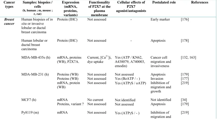
There are contradictory results in assessing the expression of P2X7 in both biopsies and cell
lines. An initial study on breast cancer was performed to identify the protein expression of
P2X7 by immunohistochemistry (IHC) on 40 haematoxylin and eosin (H&E) stained samples
of different histological categories including normal, hyperplastic tissues as well as lobular
carcinoma in situ, ductal carcinoma in situ, invasive lobular and invasive ductal carcinoma
[176]. Normal and hyperplastic breast epithelial samples were completely devoid of P2X7
expression, while all studied cases of in situ or invasive lobular or ductal carcinoma were
intensely labelled. However, only cancer cells from invasive carcinomas had membrane
labelling of P2X7, while cancer cells from in situ carcinoma seemed to only show
intracellular immunoreactivity. While this initial study was very important, it was not
specified what molecular part of the receptor was used as an antigen for the antibody used. As
such it is not clear whether the IHC signal obtained is referring to one or multiple P2X7
variants, and particularly if this antibody was also recognizing the C-terminus truncated
variants. Although the functionality of P2X7 was not assessed in invasive carcinomas, authors
were still under the influence of the cytolytic hypothesis on the cellular role of the receptor.
They postulated that “an attempt was being made to induce apoptosis” [176], suggesting in a
deterministic evolutionary conception, that following the carcinogenetic transformation
proposed that P2X7 expression could be used in order to identify and differentiate early stages
of breast cancers from hyperplastic lobules [176].
Correlatively to the initial IHC, some studies proposed that P2X7 could be over-expressed
during the breast cancer initiation or progression and become functional at the plasma
membrane of cancer cells. Indeed, the expression of both mRNA and proteins for P2X7 was
identified in the highly invasive human breast cancer cell lines MDA-MB-435s, while it was
very weakly or not expressed in non-cancerous mammary epithelial cells HMEC, MCF-10A
and 184A1 [132, 163]. Furthermore, P2X7 was found to be fully functional at the plasma
membrane of MDA-MB-435s cells, giving rise to ATP-induced inward currents, increases in
intracellular calcium concentrations ([Ca2+]i) and ethidium uptake, that were prevented by
antagonists of P2X7 or by specific small interfering RNA [132, 163]. It was indicated that the
variant expressed was the full length P2X7A. However, the expression of other variants was
not addressed. In line with this hypothesis, P2X7 proteins were also detected in highly
invasive MDA-MB-231 breast cancer cells [177]. In weakly invasive MCF-7 cancer cells,
messengers RNA were amplified and while proteins could be detected in Western blotting
experiments [177], the stimulation of cells with high concentrations of ATP was ineffective in
inducing P2X7-related inward currents suggesting that the receptor could not be functional at
the plasma membrane [34].
In contrast with these studies, an IHC study performed with 26 breast carcinoma samples, 13
ductal carcinomas and 3 lobular carcinomas indicated a tendency for a reduced expression of
the full-length P2X7 in cancer biopsies [178]. Another recent study examined the protein
expression of P2X7 by IHC, using an antibody recognizing the carboxyterminal end of the
receptor, in 80 paired-samples breast biopsies and found that P2X7 immunostaining was
reduced in cancer tissues compared to adjacent non tumour mammary tissues [179]. In the
aggressive MDA-MB-231 cancer cells, but its expression was proposed to be reduced,
similarly as in cancer biopsies, by the overexpression of the mature oncogene miRNA
miR-150 [179].
4.1.2. Prostate cancer
An initial study of a cohort of 116 biopsies, obtained from men aged from 47 to 86 years, and
confirmed to be cancerous in the range of the Gleason sum score 4–9, assessed the expression
of P2X7 proteins by IHC [180]. It was found that all cancer biopsies were positive for P2X7
expression, with no correlation with the age of patients or with the Gleason score. P2X7 was
also identified in non-cancerous epithelial cells adjacent to the margins of the tumour;
however the labelling seemed to be distinct depending on the stage/vicinity of cells to the
tumours. Indeed P2X7 appeared to be expressed in the nucleus of non-cancerous cells, then
into the cytoplasm and finally on the apical membrane of cells progressing into the cancer
phenotype. In comparison, P2X7 was totally absent from prostate tissues coming from
patients with no evidence of cancer. These results raised the possibility that P2X7 appears
during the cancerous progression [180]. In a subsequent study, the same team assessed by
IHC the expression of P2X7 and compared this to levels of the prostate-specific antigen
(PSA) in prostate cancer biopsies from 174 patients. They found a positive correlation
between the P2X7 expression and increasing levels of PSA in patients who were later on
diagnosed to be cancerous by H&E staining. These results further sustained the hypothesis by
which the expression of P2X7 could represent an early marker for the diagnosis of prostate
cancer [181]. In these two initial studies, it is not clear whether the P2X7 variant expressed is
the full-length P2X7A or another alternative splicing variant, and its functionality was not
cancer cells, the authors still characterised the receptor as being “apoptotic” and suggested
that it might be non-functional.
Ravenna and collaborators assessed the mRNA and protein expression of different markers
generally associated with inflammation, in both cancer and non-cancer prostate samples
coming from laser-capture microdissection of biopsies and showing no or low leukocyte
infiltrate. They found that P2X7 was highly up-regulated in cancer cells compared to non–
cancer cells from the adjacent host tissue and that this was correlated to the over-expression of
cancer proliferative receptors Epidermal Growth Factor Receptor (EGFR) and Estrogen
Receptor (ER)α. Authors suggested that a whole pattern of proteins usually associated with
inflammatory or reparative functions are up-regulated in cancer cells of transformed tissues,
devoid of detectable leukocyte infiltrate, and could contribute to the cancer progression [182].
4.1.3. Lung cancer
To date there is no paper assessing the expression of P2X7 in cancer versus non-cancer lung
biopsies with the aim of studying possible correlations with cancer progression and clinical
grades. However, there are several in vitro studies relating the expression of P2X7 in human
lung cancer cells, such as in A549, PC-9 and H292 but not in non-cancerous BEAS-2B
bronchial epithelial cells [132, 183, 184], suggesting that P2X7 could be up-regulated during
the malignant transformation of lung epithelial cells.
4.1.4. Pancreatic cancer
The mRNA expression of P2X7 and the presence of proteins were analysed in chronic
pancreatitis (samples from 11 patients) and pancreatic cancer (samples from 28 patients) and
compared to normal pancreas tissues. P2X7 was significantly up-regulated at the mRNA level
the protein level, P2X7 was not significantly increased in both pancreatic cancer and chronic
pancreatitis biopsies compared to normal pancreas. In conclusion, P2X7 was not significantly
overexpressed in pancreatic cancer, and seemed to be mainly expressed by infiltrating
leukocytes in the pancreas [185].
4.1.5. Papillary Thyroid Cancer (PTC)
In an initial study performed on the thyroid cancer, the mRNA for P2X7 was found to be
about two times more abundant in 37 human papillary thyroid cancer samples when compared
with contralateral non-cancer tissues. Furthermore, immunochemistry was also performed
with anti-P2X7 antibodies and P2X7 proteins were shown to be strongly expressed in all
cancerous tissues tested, both in classical and in follicular histological forms of the cancer,
while they were not present in normal thyroid tissue of the same individual. Malignant
thyrocytes showed a diffuse P2X7 labelling into the cytoplasm as well as intense labelling at
the cell periphery. P2X7 mRNA and functional proteins were also expressed in FB1 and FB2
human thyroid cancer cell lines in which stimulation by both ATP and BzATP enhanced the
release of the IL-6 [186]. IL-6 local expression is related to the aggressiveness of PTC [187]
and P2X7 could therefore participate in the development of such a cancer phenotype. The
over-expression of the full-length P2X7 in PTC was confirmed in another IHC study
performed with 4 samples [178]. In a subsequent IHC study performed with 43 PTC samples
compared to 19 samples coming from nodular goiter, it has been demonstrated that the
expression of both the X-linked inhibitor of apoptosis (XIAP) and the P2X7 was increased in
PTC samples. Furthermore, P2X7 expression was associated with an increased in the tumour
size as well as with capsular infiltration and lymph node metastases. Authors of the study
proposed that P2X7 may represent a predictor of the aggressiveness of PTC [188]. Recently,
having the chronic autoimmune thyroid disease Hashimoto’s thyroiditis (HT) and the other 86
without HT. The expression of P2X7 was significantly higher in the absence of HT and
correlated with poor prognostic factors such as tumour multifocality, lymphovascular
invasion and extra-thyroid extension [189]. In conclusion, P2X7 seems to be over-expressed
in PTC and associated with the aggressiveness of the disease.
4.1.6. Skin cancers
Skin cancers are histologically subdivided into non-melanomas, which comprise basal cell
and squamous-cell carcinomas, and melanomas. Squamous cell carcinomas are more frequent
and more aggressive than basal cell carcinomas since they can give rise to distant metastases.
Melanoma is the more dangerous and aggressive skin cancer, which initiates after the
cancerous transformation of melanocytes and has a high potential for metastasis.
Basal-cell and squamous-cell carcinomas
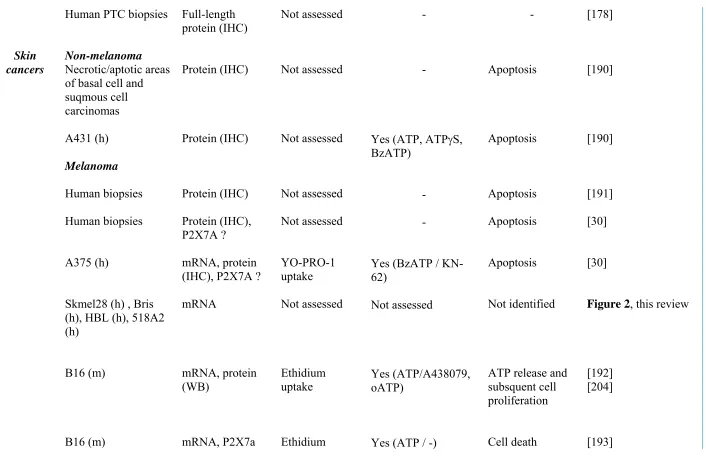
The expression of P2 receptors was assessed by IHC analysis in human biopsies of basal cell
and squamous cell carcinomas, and P2X7 was found to be expressed, especially in the
necrotic center of nodular basal cell carcinomas and in apoptotic cells in superficial multifocal
and infiltrative basal cell carcinomas. P2X7 is also expressed in the human A431 squamous
cell carcinoma cell line [190] and, while its functionality was not demonstrated, stimulation
with high doses of ATP, ATPγS and BzATP reduced cell numbers in vitro [190]. With these
results, authors claimed that P2X7 was involved in the ATP-mediated apoptosis in cancer
cells.
In an IHC study performed on 80 biopsies coming from patients with superficial spreading
melanomas, the P2X7 protein was reported to be over-expressed in cancer versus non-cancer
samples. This increase in P2X7 labelling was also extended to 2µm layer of non-cancerous
keratinocytes of the epidermis surrounding the tumour [191]. Another study performed in 14
specimens of human melanoma with an antibody recognizing a C-terminal epitope in P2X7,
came to confirm these initial results. Indeed, it showed that all melanoma samples expressed
P2X7, and over 75% of cancer cells in the biopsy were stained positively [30]. Again, these
results could suggest that P2X7 is over-expressed during the neoplastic transformation of
keratinocytes. Authors of these two separate studies indicated P2X7 as a potential melanoma
marker. The receptor was associated with apoptosis [30] and was even proposed that
“melanoma cell proliferation overwhelmed the apoptotic defence” [191].
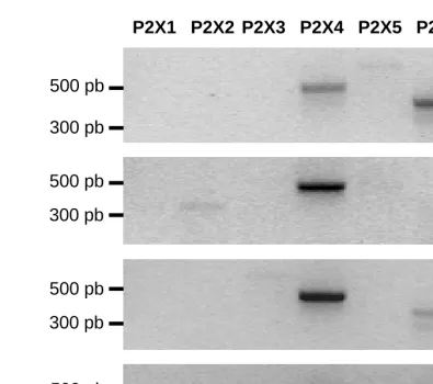
Data obtained from our group indicated that mRNA expression of P2X7, among other P2X,
can be detected by RT-PCR in Skmel28, Bris, HBL, 518A2 human melanoma cell lines
(Figure 2). The presence of proteins and their activity at the plasma membrane was not
assessed so far. P2X7 mRNA and proteins were also identified in human melanoma cell lines
such as the A375 [30], and in the murine B16 melanoma cell line [192] in which the variant
expressed is P2X7a and not the P2X7k variant [193] that exhibits a higher ligand sensitivity
[90].
4.1.7. Brain cancers
Neuroblastoma
Neuroblastomas are malignant tumours of the childhood that mainly originate from
embryonic nerve cells in the sympathetic nervous system. In 2006, it was shown, by IHC
using an antibody targeting the last 20 amino acids of the receptor, that P2X7 proteins were
displayed an intense labelling. The number of the samples analyzed was moderate but the
majority of tumours showed high P2X7 expression irrespectively of the histological grade of
the disease (localized versus metastatic). P2X7 proteins were also detected in the cytosol of
numerous human neuroblastoma cell lines such as ACN, GI-CA-N, HTLA-230, GI-ME-N,
LAN-5, LAN-1, SK-N-BE-2 and SH-SY-5Y, and at the plasma membrane of at least ACN
cells [42]. Even though more characterizations are needed, it can be nonetheless postulated
that this immunostaining is representative of the expression of a non-C-terminus truncated
variant, such as the P2X7A.
Glioma
Gliomas are malignant primary tumours of the central nervous system, deeply infiltrating
adjacent tissues in the brain, and patients bearing these tumours have a very poor prognosis.
The principal therapy actually used for the management of disease is the surgical resection of
the tumour, and there are attempts in improving the efficacy of other treatments such as
radiotherapy by manipulating the purinergic signalling. In this context there are several in
vitro experiments that have been performed in either human or rat glioma cell lines. As such
P2X7 is expressed, in mRNA and proteins, in human U-138MG, U-251MG and M059J, in rat
C6 and in mouse GL261 glioma cells [194-197]. To date, there is no study comparing the
levels of P2X7 in human glioma versus non-glioma samples.
4.1.8. Colorectal cancers
In an IHC study performed with a limited number of human samples, full-length P2X7
immunoreactivity was identified in the basolateral side of the normal epithelium, while in
colon adenocarcinoma, the immunoreactivity was distributed throughout the cells. In all the