0022-538X/90/062884-11$02.00/0
Copyright X3 1990, American Society forMicrobiology
cis-Active Elements from
Mouse
Chromosomal
DNA
Suppress
Simian Virus 40 DNA Replication
MARKUS HARTL,t THOMAS WILLNOW, AND ELLENFANNING*
InstituteforBiochemistry, Karlstrasse 23, D-8000 Munich2,FederalRepublic ofGermany
Received 12February1990/Accepted 26 February 1990
Simian virus 40(SV40)-containing DNA wasrescued after the fusion ofSV40-transformed VLM cellswith permissive COS1 monkey cells and cloned, and prototype plasmidclones were characterized. A 2-kilobase mouse DNA fragment fused with the rescued SV40 DNA, and derived from mouse DNAflanking the single insert ofSV40 DNA in VLM cells, was sequenced. Insertion ofthe intact rescued mouse sequence, ortwo nonoverlapping fragments of it, into wild-type SV40 plasmidDNAsuppressedreplication oftheplasmidin TC7 monkey cells, although the plasmids expressed replication-competentT antigen. Rat cells were transformed with linearized wild-type SV40 plasmidDNA with or withoutfragments ofthe mouse DNA in cis.Althoughall of the rat cell lines expressedapproximately equalamountsofTantigenandp53, transformantscarrying SV40 DNA linked to eitherof the same two replication suppressorfragments producedsignificantlylessfree SV40 DNAafterfusion with permissive cells than thosetransformed by SV40DNAwithoutacellular insertor with a cellular insertlacking suppressor activity.Theresultssuggestthat twoindependent segmentsof cellularDNA act in cisto suppress SV40 replication in vivo,either as aplasmid orintegrated in chromosomal DNA.
Mammalian cells transformed by simian virus 40 (SV40) harbor viral DNA sequences integrated in their chromo-somal DNA and, in general, constitutively express SV40 largeTantigen (for areview, seereference48).Tantigen is able to induce and maintain cell transformation, in part through its association with cellularproteinssuchasp53and theretinoblastoma susceptibility gene productRblO5(7; for a review, see references 24 and 48), although our under-standingof thetransformation processremains very incom-plete. Clearly, however, activities ofT antigen required for productive infection of monkey cells, such as initiation of viral DNA replication, are not essential for initiation or maintenance of celltransformation (48). The fact that many SV40-transformedcells expressTantigens defective in their abilitytoinitiateviral DNAreplicationhas ledtospeculation that celltransformation by SV40 selects for expression ofT antigens that maintain transformation butarenolonger able toreplicateSV40 DNA, perhaps thereby avoiding rearrange-ments of chromosomal DNA induced by T antigen (48). Alternatively, because T-antigen replication functions are not required to initiate or maintain cell transformation, replication-defective mutations inTantigen can accumulate in transformed cells (48).
Upon fusion of many SV40-transformed cell lines with permissive monkey cells, viral origins areactivated by the resident T antigen, followed by amplification and recombi-nation to yield free rescued SV40 DNA (for a review, see reference 48). This mobilization depends on the presence of anintactorigin region and a replication-competent T antigen in the transformed cells (20, 41). In the event that the resident T antigen is replication defective, SV40 DNA can be rescued by fusion with COS1 monkey cells, which constitu-tively express a replication-proficient T antigen (20, 21). A number of replication-defective T-antigen mutants isolated by this method have proved useful in elucidating the bio-chemical activities of T antigen required for viral DNA
*Correspondingauthor.
tPresent address: Department of Microbiology, University of Southern California, Los Angeles, CA 90033-1054.
replication,suchasSV40 DNA binding and ATPase-helicase activities (fora review, see references 8 and55).
The SV40-transformed mouse line VLM (66) expresses large T and superTantigensabletobindspecificallytoboth major sites in the SV40 control region and to cleave ATP (33). Fusion of VLM cells with TC7 cells yielded little free SV40DNA, whereas viral DNAwasefficientlyrescued from VLM-COS1 fusions, suggesting that the VLM T antigens were almost inactive in replication (33). Thus, it seemed likely thatidentificationof thepresumedmutation in VLM T antigencouldprovidegeneticevidence for other biochemical activities possibly involved in SV40 replication.
We report here the characterization ofaprototype clone rescued from VLM-COS1 fusions and compare it with genomic VLM DNA. Contrarytoprediction,nodefect in the origin function, early gene expression, orreplication profi-ciencyof therescuedT-antigen genecould be found. How-ever,wedemonstrate thatmouseDNA sequences present in theSV40 lateregion ofthe rescued clone and derived from sequences flanking theSV40 integration site in VLM DNA suppress replication of SV40 plasmid DNA in uninfected monkey cells inan orientation-dependent manner. Further-more, integrated viral DNA carrying the suppressor se-quences in cis was rescued fivefold less efficiently from newly created transformed rat cell lines than viral DNA lackingthe suppressorelements.
MATERIALS ANDMETHODS
Cells. Allcellswere cultured in Dulbecco modifiedEagle medium (DMEM) (GIBCO-Bethesda Research Laborato-ries, Inc., Eggenstein-Leopoldshafen, Federal Republic of Germany) supplemented with5%fetal calf serum (Hyclone; Greiner, Nurtingen, Federal Republic of Germany). VLM (67), TC7, a subline of CV1 African green monkey kidney cells (49), Rat2 (60), and COS1 (20) cells were described. VLMcellswere cloned insoft agar prior to construction of genomic libraries. Seven of eight cell clones analyzed by genomic blotting with SV40DNAprobes revealed the same viral DNA integration pattern described previously (33) (data notshown).
2884
on November 10, 2019 by guest
http://jvi.asm.org/
cis-ACTIVE ELEMENTS SUPPRESS SV40 DNA REPLICATION 2885
A L 13.2
(18 kb)
L8.1 (1 6 kb)
E B EEE B E B B E B E B
I lii4H--rr-r-l iI I+
B
E EE K B E
TAg ori ori
E CIK
E EE K B(2716) (196)
II
,' " TAg ori inBamH site
:% ofpBR 322
VLM
pV 8
fragments
pV 4
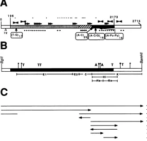
FIG. 1. Restriction maps of the SV40 integration site in VLM
DNA andtherescuedplasmid clones pV4 and pV8. (A) Diagram of the EcoRI (E) and BamHI (B) cleavage sites in thegenomicDNAof
arepresentative clone of VLM cells. The map wasdeduced from
Southern blotting of VLM DNA fragments separated by pulse-field gel electrophoresis and analysis ofgenomic clones (Willnow and Fanning, unpublished data). Mouse DNA is depictedasablack line andSV40DNAas an openbar. Twogenomic clones carrying the
SV40-mouse DNA junction regionsareindicated above. (B) Region rescuedinpV4 and pV8. Both circular DNAswere inserted in the BamHI siteofpBR322,asindicated below. Open bars indicateSV40 sequences, horizontal lines indicatemouseDNA, and dashed lines representplasmid vectorDNA. The arrow under the early region shows the directionof transcription of the large-T-antigen (T Ag)-codingsequence.Mouse fragments E, C, and K, commontoboth rescued clones,weresequenced and characterized (cf. Fig. 2A and
B). The diagrams show the cloned DNA linearized atthe junction
betweenviral and cellular DNA(SV40 nucleotide 196),as shown. ori, Origin of replication. kb,Kilobases.
Plasmids. pSVwt is wild-type SV40 DNA (strain SV-S) cloned inthe BamHI site ofpAT153 (14, 62). pV4 andpV8
are prototype viralgenomes rescued after VLM-COS1 cell fusionand cloned into the BamHI site ofpBR322 (Fig. 1B) (B. Huber, Ph.D. dissertation, Ludwig-Maximilian-Uni-versity of Munich, Munich, Federal Republic of Germany, 1986). pKl contains theSV40genomecloned intothe EcoRI
siteofpMK16/6 (21).
Genomiccloning. Agenomic library wasconstructed with partially digested MboI fragments ofgenomic DNA froma
subcloned line of VLM cells. DNA was cloned into the
lambda replacement vectorEMBL3 (15), asdescribed
pre-viously (34). Bacteriophage clones were grown in
Esche-richia coliK803 (65).
Plasmid constructions. Standard cloning techniques were
used for the constructions (40), and all plasmids were
propagatedin E. coliHB101.
The pV4 mouse DNA fragments El (1,028 base pairs [bp]), E2 (129 bp), C (254 bp), K (958 bp), Hi (484 bp), Ha
(206 bp), and Sa(278 bp) (Fig. 2B) were subcloned in pUC
vectors (66). For replication studies, fragments C, K, Hi, Ha,and Sawereclonedinto the late region ofpSVwt (Fig.
3).
All SV40 nucleotide numbers are given in the BBB system (59). The pV4 nucleotide numbering is simply continued from the viral-cellular DNA junction site (SV40 nucleotide 196)(Fig. 2A). Thus, the pV4 BamHI site in the SV40 region of pV4 isatpV4nucleotide 2716 rather than at 2533,as in the BBBsystem.
In pSV-KV4, the SV40 KpnI-BamHI fragment (nucleo-tides 294 to 2533) was replaced by the pV4 K segment (KpnI-BamHI; 1758 to 2716). pSV-CV4 harbors the C frag-ment(EcoRI-KpnI; 1504 to 1758) in the opposite orientation to thatof pV4,replacing the SV40 KpnI-EcoRI (294 to 1782) sequences.InpSV-AN, the1,482-bp BanII fragment (778to 2258) wassimplydeleted. The subcloned Sa segment (AccI-HincII; 1964to2242)inpUC-SaV4wasexcisedbyusingthe KpnIand Hindlll sitesofthepUC18 polylinker and inserted inplaceof the SV40KpnI-HindIII fragment (294to1708) of pSVwt, resulting in pSV-SaV4.
pSV-sSaV4contains the Safragmentshifted 1.3 kilobases upstream of the origin-promoter region. For this purpose, theSV40fragment of pSVwt AccI-BamHI(nucleotides1628 to 2533) was replaced by the pV4 AccI-BamHI segment (1964 to 2716). pSV-HaV4 bears the Ha sequences KpnI-AccI (1758 to 1964) in pSVwt instead of the original SV40 segment (KpnI-AccI; 294 to 1628). In pSV-iHV4, the Hi fragment (KpnI-HincII; 1758 to 2248) was inserted into plasmid pSV-AN in the opposite orientation to that in pSV-KV4 by replacingthe SV40 KpnI-PvuII fragment (294 to270).
Constructions derived from pV4 are pV-AE, from which the El and E2fragments (EcoRI-EcoRI; nucleotides 293 to 1504) were deleted, and pV-Kwt, which contains pSVwt late-region sequences(KpnI-BamHI; 294 to 2533) insteadof thepV4 Kfragment(KpnI-BamHI; 1758 to2716).
The Hi (KpnI-HincII; 1758 to 2242) and Ha (KpnI-AccI; 1758 to 1964) segments were inserted into the KpnI site (nucleotide 294) of pSV2Cat(22). Thus, pSV-HiV4Cat and pSV-HaV4Cat contain these fragments upstream of the SV40enhancer repeats (Fig. 4). InpSV-pV4Cat, the entire origin-promoter region of pV-AE (HindIII-PvuII; 5171 to 381)replaces theoriginal sequences (HindIII-PvuII; 5171to 270)ofpSV2Cat.
DNAreplication. SemiconfluentTC7 cells
(106
per100-mm dish) werewashedwith Tris-buffered saline and transfected with precisely determined concentrations ofuncut plasmid DNA (0.25to1.0 ,ug) in DEAE-dextran, asdescribed previ-ously (61). After 45 minat37°C,thecellswerewashedagain
and supplied with fresh medium. Low-molecular-weight DNA(30) was isolatedafter 2 days.
Asacontrol, input DNAswerelinearized andanalyzed by gel electrophoresis in parallel with the Hirt supernatant DNAs. SV40plasmidDNAwasdetected byblot
hybridiza-tion(54)byusing 32P-nick-translated (47) pBR322orpAT153 DNA as a probe to ensure that all plasmids would yield a
hybridization signal in proportion to their concentrations, regardless of the different SV40 and cellular sequences present inthe variantlate-region constructs.
DNA sequencingandanalysis. Sequencingwasperformed by using thedideoxy-chain extension method with double-stranded DNA (6). The large fragment of E. coli DNA polymerase I
(Pharmacia, Freiburg,
FederalRepublic
of Germany)orchemicallymodified T7 DNApolymerase
(U.S.
Biochemicals-Renner, Dannstadt, Federal
Republic
of Ger-many) was appliedasdescribedpreviously (57).
The late region ofpV4
containing
the cellular insertion wassubcloned in thesequencingvectorspUC18
andpUC19
(66). The sequence of the fragments was determined from
VOL. 64,1990
on November 10, 2019 by guest
http://jvi.asm.org/
[image:2.612.62.294.71.298.2]A
196 * ,.. ... . 2173
01
271 6B
co I
IT
7T A lA T TT tI E E1E IE2+ C- I K
I-Hl -I
CI
rv
+
* +
No +
FIG. 2. Organization and sequence features of the pV4 late region. (A) =, SV40 DNA startingat theBglI site (nucleotide0) and extendingtotheviralBamHIsite; EJ, M , and _,mouseDNAsequencesreplacingtheSV40lateregionfrom nucleotides 196to2173. The different shadingpatternsindicate thefragmentsEl plus E2, C,and thecellularsegmentofthe Kfragment.One copyof the72-bprepeat ismissinginpV4(A72). 4,Positions of di-andtrinucleotide repeats; " and _,locations ofinverted and directrepeatedsequencesof
11 to 19nucleotides, respectively; *,CpGdinucleotides;+and0,AT-andGC-richregions,respectively. (B)Thesameregionasthat inpanel
Aisshown, indicatingtherestrictionfragmentsanalyzedin this report(belowthebars). =, SV40sequences; _,mousesequenceswith nodetectable homology toSV40: M , mouse sequences with up to25% homology with SV40 DNA; *,possible topoisomerase IIcleavage sites(13matcheswith the15-nucleotideconsensus) (51); AandT, AandTboxes,respectively, found in SARsequencesofDrosophilaspp. (9matches with a 10-nucleotide consensus sequence) (17). (C) SV40 DNAreplication-suppressing activity of each ofthe mouse DNA segments tested isgivenattheright (+,suppression; -,nosuppression). Theorientation of eachfragmenttested is indicated(-_ , -- ).
bothdirections by usingthepUCreverseprimer(agiftfrom P. Heinrich) (5'-CAGGAAACAGCTATGAC-3') and spe-cific internal primers. Sequence analysis and homology searches were carried out by using previously described programs (18, 37, 45). The entire sequence will be made available upon request(GenBank accession no. M33654).
CAT assay. Calcium phosphate DNA precipitates were prepared with 15
pLg
ofplasmid DNA (23) and applied to60% confluent TC7cells (100-mm dishes) by publishedmethods (22).After4 h, the cells were shocked with 20% dimethylsul-foxide inDMEMfor 2minandincubated foranother 48 h. Proteinextractswerepreparedasdescribed previously (35), and 150 ,ul ofclarified lysate corresponding to 300 ,ug of protein was mixed with 20
RI
of 4 mM acetyl coenzyme A (Pharmacia)and2,ulof[14C]chloramphenicol
(New England Nuclear, Dreieich, Federal Republic of Germany). After 1 h ofincubationat37°C,chloramphenicol and acetylated deriv-atives were analyzed by ascending thin-layer chromatogra-phyasdescribed previously (35).RESULTS
Isolationandcharacterization ofSV40 genomes from VLM cells. A subcloned line of VLM cells was fused with COS1 monkey cells (20). Supercoiled SV40 DNA rescued from
VLM-COS1 fusionswas purified by centrifugation in equi-librium density gradients as described previously (21), lin-earized with BamHI, and ligated into the BamHI site of pBR322toyieldtwomajorfamiliesofplasmidDNA(Huber, Ph.D. dissertation). Prototypeplasmids pV4 and pV8were chosen for further characterization. Restrictionmappingand blot hybridization experiments (Fig. 1B) revealed thatpV4 bearsasubsetof the SV40 sequencesfoundinpV8and that bothclones carry thesame2-kilobasemouseDNA sequence in place of the SV40 lateregion (M. Hartl,Ph.D. disserta-tion, Ludwig-Maximilian-University of Munich, Munich, Federal Republic of Germany, 1989). pV4 contained an apparently intact SV40 early region and origin-promoter-enhancerregion (Hartl,Ph.D. dissertation).
The structure of the rescued clones was then compared with that of theintegratedSV40 DNA in VLM cells.Asingle integration site oftandemly repeated SV40 DNA was de-tectedbydetailedblothybridization analysisof VLM DNA (33; Hartl, Ph.D. dissertation; Huber, Ph.D. dissertation). The mapping was confirmed and extended by restriction mapping analysisofgenomic clones of VLM DNAbearing SV40 sequences (Fig. 1A) (T. Willnow and E. Fanning, unpublished data). Apparently, the rescued clones were createdby recombination between cellular sequences adja-cent to the E fragmentand viral sequences adjacentto the
on November 10, 2019 by guest
http://jvi.asm.org/
[image:3.612.169.455.76.351.2]cis-ACTIVE ELEMENTS SUPPRESS SV40 DNA REPLICATION
A r INPUT DNA INPUT DNA
25 33 BamHl
-rm
late L
region i >
>0r0
>°
Bg//U B es
coX>
> >LJ
ii
) co 0B
:-I1;4
7.4kb -
-1 2 3 4 1 2 3 4 5 6 7 5 6 7
Dpn I Dpn I 1 2 3 4 5 6
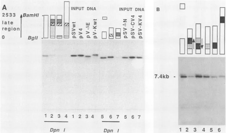
FIG. 3. ReplicationofSV40 plasmid DNA bearing different parts of the pV4 cellular sequence. (A) Late-region constructs are depicted by bars aboveeach lane. Open bars represent wild-type SV40 DNA, and different shaded bars represent the various arrangements of pV4 fragments(note the reverseorientation of the C-V4 segment in lanes 6). Numbers at the bottom of each lane denote the corresponding samples ofeach Hirtsupernatantandinput DNA. Light shading indicates fragmentElplus E2;hatching indicates fragment C and its orientation; dark shading indicatesmouse sequencesinfragmentK.Supercoiled plasmid DNA wastransfected into TC7 cells and low-molecular-weight DNA was isolated after 2 days, as described inMaterials and Methods. One-fifth of the sample was digested with DpnI prior to linearization with
SalI. A3-ng sample ofeach type of Sall-digested inputDNA was analyzed in parallel by electrophoresis in a 1% agarose gel and blot hybridization againstnick-translated pBR322 DNA (5 x 10⁷cpm4/Lg). Autoradiography was done for 6 h. Theamount of newly replicated DNA wasevaluatedby microdensitometry and expressed as a percentage of the wild-type DNA. Lanes: 1, 100%;2,34%; 3,18%;4, <10%; 5,100%;6,98%;7,8%. (B)Supercoiled DNA (250 ng) was transfected into semiconfluent TC7 cells, andlow-molecular-weightDNA was isolated, digestedwithDpnIandClaI,andanalyzed as in panel A. Lane 1,pSV-AN (7.4kilobases);lane 2,pSV-KV4;lane3,pSV-iHV4;lane 4,pSV-HaV4;lane 5,pSV-SaV4;lane 6,pSV-sSaV4.Thesmear visible above the bands in some lanes was causedby a slight displacement duringblotting. Microdensitometryof the autoradiogram was used to compare the amounts of newly replicated DNA. Lane 1,100%;lane 2, 25%;lane 3,120%; lane 4,70%;lane 5,24%;lane 6,27%. kb, Kilobases. Open bars represent SV40 DNA;light shadingindicates the Ha fragment; solidbars show theSa fragment(seeFig. 2B for a map).
latesideof the enhancerregion(Fig. 1B). Thus, the rescued clones pV4 and pV8 are colinearwith VLM chromosomal sequencesand spanthe mouse-SV40junction regiononone side oftheintegration site.
Theorigin region and late region of pV4 were character-ized in greater detail by determinationofthe DNA sequence (Hartl, Ph.D.dissertation). Interestingly,muchof the mouse sequence displayed up to 25% homology with SV40 se-quences,whereas theremaindersharednosequence homol-ogy with SV40 DNA(Fig. 2B). Repetitive sequences were dispersed throughout mostof the mouse-derived portionof thelate region (Fig. 2A; Hartl,Ph.D. dissertation). Repeti-tive (GT)n and trinucleotide stretches were located at both endsofthemousesequence (Fig.2A). However, restriction fragment C (Fig. 2B) carried only single-copy mouse se-quences, as shown by blot hybridization analysis of VLM and mouseL-cell DNAs(Hartl, Ph.D. dissertation). Exten-sive regions of direct and inverted repeats were found, particularly clustered inA+T-rich portionsof the sequence (Fig. 2A). Alarge cluster ofCpG dinucleotideswas located in the central portionof the mouse sequence.
In the early region and control region of pV4, several nucleotide changes with respectto SV-S strain SV40 DNA were also noted.Nucleotide 4839waschanged from C toT, creating an AccI site but no amino acid change in the T-antigen-coding sequence. Nucleotide 5209 was changed from C to T, and nucleotide 81 was deleted. All these sequencechangeswerealso found insequencingtheBaylor
SV40 strain usedto derive the VLM line and thusare strain specific (67; Hartl,Ph.D.dissertation; R.Lanford, personal communication). In addition, one of the 72-bp repeats was
deleted (nucleotides 179 to 250). Both the origin region of pV4 (Hartl, Ph.D. dissertation) and the enhancer (Fig. 4) were fullyfunctional in TC7 cells.
CellularDNAsequences suppressSV40 plasmidreplication. Contrary to our expectations from the fusion
experiments,
the early region of pV4 was able to replace wild-type T-antigen-coding sequences ofplasmid pKl in DNA repli-cation assays in TC7 cells, demonstrating that it was
fully
functional when placed in a wild-type SV40
background
(Hartl,Ph.D. dissertation).
The apparentcontradictionbetween this result and those from the cell fusion experiments (33) raised the
question
whether the mousesequencesin the lateregions of pV4and pV8 couldaffect SV40DNAreplicationormobilizationupon cell fusion. To test this
idea,
SV40plasmids carrying pV4
mouse DNA in whole or in part were constructed and assayed for
replication
inmonkey
cells. Thefollowing
twogroups ofconstructs were used: one set in which cellular sequences were deletedfrom pV4 or
replaced by
wild-type
viral sequences andasecondsetin whichpV4cellular DNA wasinserted intoawild-typeSV40
plasmid.
Each clone had an intact origin ofreplication
and encoded a functional T antigen, derived from eitherpSVwt
orpV4.
All the clones expressedTantigen
aftertransfection of TC7cells,
asjudged
VOL.64, 1990 2887
on November 10, 2019 by guest
http://jvi.asm.org/
[image:4.612.125.491.76.292.2]_-Hlnctf
Ac cl _
I K|
40
qtr
CL
0-X
:I I
Q m
CL)
ccellulaDNA
-Vlt:,jDMAA
0
C CS C) O 0
O ~
> N
( n x
QL
.i
:GP
S.,F
*
gF 'S1 2 3 4 5 6
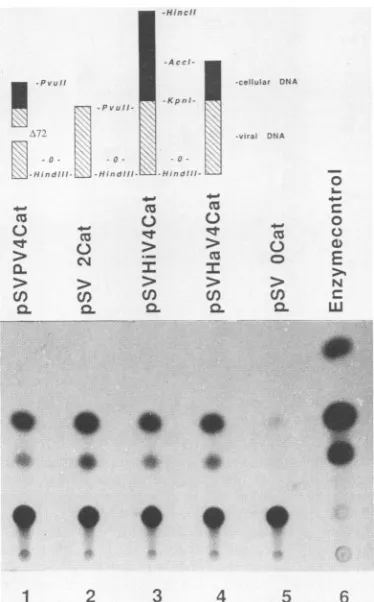
FIG. 4. CAT activity ofconstructscontaining cellularsequences linkedtothe SV40 earlypromoter. Thestructureof the CATclones
is depictedabove eachlane. Thenegative controlpSVOCat lacksthe
entire SV40earlypromoter(36).Transfection andCATassays were
performed as described in Materials and Methods. Acetylated products weredetected by thin-layerchromatography followed by
autoradiographyfor48 h.
by immunoperoxidase stainingwith anantibody specificfor
T antigen(datanot shown).
Theclonesweretested forreplication by transfection into
TC7cells, followed 2days laterbyisolation and analysis of
Hirt supernatant DNA (Fig. 3A). Replication of pV4 was
significantly reduced compared with pSVwt, as
demon-strated by the smaller amount ofDpnI-resistant pV4 (Fig.
3A, lane2) compared withpSVwt (lane 1) DNA. Compari-sonofthe inputDNAs (Fig.3A,lanes 1and2) demonstrated
thatequalamountsofDNAweretransfected. Toidentifythe
sequencesresponsibleforthe observedreduction in
replica-tionefficiency, thetwoEcoRI fragments weredeleted from
pV4DNA. Replication ofthe resulting plasmid dropped to an even lower level than that of pV4 (Fig. 3A, lane 3),
indicatingthatthe distalpartof the cellularregion(fragment
K) wassufficientto suppressreplication. Conversely, theE
fragment (El, E2, and C) was also sufficient to suppress
plasmidreplication (Fig. 3A,lane 4)intheabsence oftheK
fragment.
The clones ofthe second set are almost identical in size
and bear the early region and origin-promoter region of
pSVwt DNA. The plasmid pSV-AN (Fig. 3A, lane 5), from
which viral sequences in the late region were deleted, was
usedas acontrol,demonstratingthat the reduced replication
observedwithpV4 andits derivativeswasnotcaused bythe
absence of thelate region. Insertion of the pV4 C fragment
into pSVwt DNA didnot cause anydetectable suppression
of replication (Fig. 3A, comparelane 6with lane5). Insertion of the pV4 K fragment into the pSVwt plasmid severely reduced DNA replication (Fig. 3A, lane 7). Figure 3A thus demonstrates that two independent segments of pV4cellular mouse DNA suppress
SV40
DNA replication when located in cis upstream of the origin and enhancer.Since the pV4 K fragment carries 484 bp of mouse sequences in addition to
SV40
sequences (Fig. 2B), we wished to test whether the entire sequence, or only part of it, was required to suppress wild-typeSV40
replication. A third series of plasmids, in whichportionsof the pV4 Kfragment were inserted into the late region of pSVwt DNA, was constructed and tested for replication activity after transfec-tion into TC7 monkey cells (Fig. 3B). The plasmid pSV-AN carrying a deletion in the late region was used as awild-type control (Fig. 3B, lane 1). As expected, aplasmidbearing the entire pV4 K fragment replicated poorly (Fig. 3B, lane 2) compared with the control (lane 1). However, inversion of the K-V4 mouse DNA sequences (Fig. 2B, fragment Hi) relative to the origin restored replication activity to a level slightly greater than that of the wild type (compare lane 3 with lane 1), indicating that the suppression was orientation dependent.The cellular sequences of the K fragment were dissected into two portions, the Ha (206 bp) and Sa (278 bp) fragments (Fig. 2B), and each was inserted into pSVwt DNA. The plasmid bearing the Ha fragment replicated with nearly wild-type efficiency (Fig. 2B, lane 4), whereas the Sa-bearing plasmid (lane 5) replicated more weakly than the wild type but better than pSV-KV4. Insertion of the Sa fragment 1.3 kilobases more distal to the origin-enhancer region resulted in the same reduction in replication observed with the origin-proximal Sa plasmid (Fig. 2B, compare lanes 5and 6). These results demonstrate that a 278-bp segment of the pV4 K fragment is sufficient to suppress replication of wild-type SV40 DNA, in a manner independent of distance but depen-dent on orientation relative to the origin-enhancer region.
SV40 promoter-enhancer activity is not suppressed by pV4 mouse DNA sequences. Although the pV4 origin and early regions were shown to be functional (Hartl, Ph.D. disserta-tion), it was conceivable that the expression of replication-competent T antigen could be limited by the presence of the pV4 cellular sequences upstream of the promoter-enhancer region. To address this question, the pV4 origin-promoter-enhancer region was placed upstream of the bacterial chlor-amphenicol acetyltransferase (CAT) gene and CAT enzyme activity was assayed after transfection of plasmid DNA into TC7 cells. The CAT activity expressed from the pV4Cat construct was comparable to that expressed from the wild-type control pSV2Cat (Fig. 4, lanes 1 and 2). The Hi fragment bearing the entire cellular portion of the pV4 K fragment which showed replication suppression activity, or the suppressor-negative Ha fragment (Fig. 3A and B), was placed upstream of the enhancer in pSV2Cat. CAT activity measured with both these constructs was indistinguishable from that measured with the pSV2Cat control plasmid (Fig. 4, compare lanes 3 and 4 with lane 1). These results indicate that the replication suppression activity of the pV4 mouse DNA sequences is unlikely to be due to their effect on promoter-enhancer activity.
Mouse DNA sequences suppress mobilization of chromo-somalSV40DNA. The ability of pV4 mouse DNA sequences to suppress replication of wild-typeSV40plasmid DNA (Fig. 3A and B) suggested that these sequences might also affect the efficiency of rescue ofSV40genomes from transformed rodent cells by cell fusion. This idea was tested by
on November 10, 2019 by guest
http://jvi.asm.org/
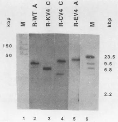
[image:5.612.90.277.69.370.2]cis-ACTIVE ELEMENTS SUPPRESS SV40 DNA REPLICATION 2889
R-WT A
R-KV4
C
R-CV4
C
R-EV4 A
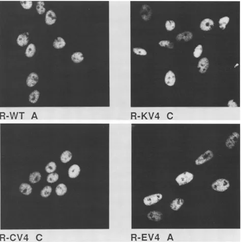
FIG. 5. Transformation of Rat2 cells by SV40 DNA carrying mouse DNA sequences in cis. Rat2 cells were transfected with Sall-Clal-linearizedSV40plasmidDNAcarrying theK,C,or Efragment ofpV4 orwithoutanadditional fragment (Fig. 1B).Foci,picked after 20days, were purifiedin soft agar as described previously (52). Single colonies weretakentoestablish the cell linesR-WT, R-CV4, R-KV4,and R-EV4. Several colonies of each type (lettered A and C) werecharacterized in detail. Semiconfluent dishes (2 x 10⁵cells per 60-mmdish)werefixedbyusing3%paraformaldehyde-0.1%Triton X-100 inphosphate-buffered saline. T antigen wasvisualizedby indirect immunofluorescencestaining (53)using4
pLg
of Pab416 and Pab 419 (27) per ml as the first antibody. Cells were photographed by using a Zeiss IM35microscope equippedwith aPlan-Neofluar63 oil immersionobjective.ing a series of SV40-transformed Rat2 cell lines with or without pV4 mouse sequences in cis. Wild-type SV40 plas-mid DNA or SV40 plasplas-mids carrying insertions of the pV4 E (El + E2 + C), C, or K fragments in the late region were transfected into Rat2 cells. The plasmid DNAs showed nearly equal transformation activity, and all four types of transformants grew insoftagar. Colonies isolated from soft agarwereexpandedinto cell lines and characterized. All the
cells expressed nuclear T antigen (Fig. 5). Immunoblot analysis ofprotein extracts from four representative
trans-formed lines demonstrated that all of the lines
expressed
similar amounts ofT antigen of wild-type size and similar amounts ofp53 (Fig. 6A and B).
Blot hybridization analysis ofgenomic DNA from each transformed linedigestedwithBglII, which does notcleave any of the transfectedDNAs, revealed thatoneof the lines
VOL.64, 1990
on November 10, 2019 by guest
http://jvi.asm.org/
[image:6.612.70.546.75.551.2]o
mC
Y
(&&
Cr a: Cr
B :1.
li:
0J
me3
<:
H >rt lq> >etr
~e 0 0
&f
.E
Iw
1 80
11 6 8 4 58
48. 5 If e
Y.
36.5
2 6 . 6 ^t.
1 2 3 4 5 6 1 2 3 4 5 6
FIG. 6. Immunoblots of proteinextracts fromratcell linestransformed by SV40 DNA with and withoutpV4 mouse DNA in cis.(A) Proteinextracts wereprepared from each cell line (lanes2through 5),and 125 ,gof eachwerefractionatedbysodiumdodecylsulfate-gel electrophoresis,asdescribedpreviously (53). Prestainedmarkerproteins(Sigma, Munich,FederalRepublicofGermany)of knownMr(listed
atthe left inkilodaltons)wereelectrophoresed in parallel (lane M). Immunoblotting (12)andstainingforSV40 largeTantigen (53)werecarried
outexactlyasdescribedpreviouslyby using KT3 immunoglobulinGasthefirstantibody(39). (B)The immunoblotanalysiswasrepeatedby
using Pab 122 culturesupernatant(25)asthe first antibodytostainp53.
(Fig. 7, lane 5) hadone majorintegration site of viralDNA,
whereas the others had two insertions of viral sequences
(Fig. 7, lanes 2 through4). Blothybridizationanalysis with BamHI-cleaved genomic DNAs confirmed these results (datanotshown).
Each ofthe transformed lineswasfused with TC7monkey
cellstorescue theintegrated viral DNA (Fig. 8). Abundant
X
150 ;
0t 'C:
) us
!n
c x
low-molecular-weight DNA was generated from the
wild-type-transformed line (Fig. 8, lane 1) and the line carrying the pV4 C fragment (lane 2). Theamount of low-molecular-weight SV40 DNA rescued from the lines bearing the K and Efragments was reduced by more than 80to90% (Fig. 8, lanes 3 and 4), asshownby microdensitometric scanning of theautoradiograms. Similar resultswereobtainedwith
sev-eral independent lines of eachtypeof transformant (Hartl,
TC 7 x
~0 w
* , I
c: a:rM
23.5 9.5
6.8 - 5.2kb
3.7kb
2.2
[image:7.612.136.477.70.270.2]1 2 3 4 5 6
FIG. 7. Genomic blots ofratclones transformed bySV40DNA
with and without pV4 mouseDNAin cis. Genomic DNA (20 ,ug) isolated as described previously (56) from cloned rat lines was digested with BglII and fractionated by pulse-field gel
electrophore-sis. MarkerDNAs(lanes M)wereelectrophoresedin parallel (lane
1, ligated lambda DNA; lane 6, HindIll-digested lambda DNA).
Lanes 2through5wereblotted and hybridized with random-primed
(Amersham, Braunschweig, Federal Republic of Germany) 32p_ labeledpSVwt DNA. kbp, Kilobase pairs.
1 2 3 4 5
FIG. 8. RescueofSV40DNAfromtransformedratcell linesby fusionwithTC7monkey cells. TC7 cells (2 x 10⁶)werefused(x)
with each of the indicated ratlines (106)as described previously
(33). Half the low-molecular-weight DNA obtained from each fusionwasdigestedwithBamHI andanalyzed by blot hybridization with nick-translated pSVwt DNA (specific activity, 108 cpm/pu.g) (lanes 1through 4). BamHI-digested pSVwt DNA servedas asize
marker (lane M). The blot was autoradiographed for 30 h. kb,
Kilobases. A
-Ca
180 1 16 84 58 48.5 36.5 26.6
.J
_s
,. M
on November 10, 2019 by guest
http://jvi.asm.org/
[image:7.612.85.275.438.635.2] [image:7.612.356.517.447.635.2]cis-ACTIVE ELEMENTS SUPPRESS SV40 DNA REPLICATION 2891
2058 2107 HOMOL.
ACAATAACAATAGTAACAATAGTAACAATAACAATAATGATAACAACAAC SaV4
TAGTtACAACA
TAGTtACAAcA
terC core
TAACAATAATaAT ACA
AGTtAACAACAACAAT
TAACAACAAC
mt DNA 820-805 10, 63 CSB1(hu)
SV40 2665-2680 2668-2677
32
ACAA AACgATAGTcAaAATA
TAACAATctAATGAaAA E1V4 668-684 937-956
FIG. 9. Replication suppressorfragment Sa (209 bp) carries sequenceshomologous tothe pV4El fragment, human (hu)mitochondrial (mt) D loop DNA, E. coli replication terminatorcore sequence, and SV40 termination region. Nucleotide matchesaregiven inuppercase
letters,and mismatches aregiven in lowercase letters. Fordetails, seetext. Homol., Homology;NT.,nucleotides;REF.,reference.
Ph.D. dissertation), indicating that thepresenceofaplasmid
replication suppression element flanking the integrated SV40 DNA is correlated with inefficient mobilization of the viral DNA after cell fusion and suggesting that these elements
maybe responsible for theinefficient rescue observed.
DISCUSSION
MouseDNAsequencesrescuedtogetherwithSV40 DNA fromtheSV40-transformed cellline VLMhave been shown
to severely suppress viral DNA replication in cis in TC7
monkey cells (Fig. 3) and the mobilization of integrated SV40 DNAuponfusionwithTC7 cells (Fig. 8). A
compara-bledegreeofsuppressionwasobserved inboth approaches. TwoseparatesegmentsofthemouseDNAwereshownto havereplication suppression activity,asassayed byplasmid
replication and by viral DNA rescue from cell fusions.
Control experiments ruledout an effect of the mouse
repli-cationsuppressorelementsontheactivity of theSV40 early
promoter-enhancer in transient assays (Fig. 4) and on the
steady-state level ofTantigen in the transformed rat lines used for viral DNA rescue (Fig. 5 and 6A). The supply of
replication-competent T antigen in the cell fusion
experi-ments could be limited by complex formation with p53, which hasbeen showntoinhibit SV40 replication (3, 16,58, 64). However,all thetransformedratlinescontainedsimilar steady-state amounts ofp53 (Fig. 6B), thus excluding this explanation for theratcell fusion results.
Thesupplyof active Tantigencould also belimiting if it
was sequestered by thecellularDNA sequences. A similar mechanism has been proposed toexplain the limited SV40 plasmid replication observed in cells transformed by SV40-bovine papillomavirus hybrid plasmids (50). If a similar mechanism was operating in the experiments presented here, we would expect the cellular sequences to suppress
plasmid replication in a mannerindependent of orientation
relative tothe origin and also when located in trans.
How-ever,inversion ofonecellular suppressor element restored
wild-type replication (Fig. 3B, lane 3) and no inhibition of
plasmid replication was detected when the suppressor
ele-ments were cotransfected in trans with the SV40 plasmid (Hartl,Ph.D.dissertation). Thus, T-antigen sequestration by the cellular suppressor elements can be ruled out as an
explanationfor theirsuppression activity.
Insummary,then,thepresence ofpV4cellularsequences
in cis appears to be directly responsible for the reduced
replication of SV40 plasmid DNAs in TC7 cells and the inefficientmobilizationof viral DNA in transformedratcells afterfusion.
DNAsequencesinthepV4 replicationsuppressorelements.
ThegeneralfeaturesofpV4mouseDNAaredepictedinFig.
2A and B.Those regions of the sequencetested for
replica-tion suppression activityinboth the plasmidand viral DNA
rescue assays are summarized in Fig. 2C, which showsthe
orientation of each sequence tested. In general, the
frag-ments with suppression activity are rich in A and T (Fig.
2A). The shortest segment with suppressor activity (Sa)
contains 278 bp, of which 209 bp are mouse sequences.
Although it is not yet clearwhich ofthe 209-bp sequences
areresponsible for thesuppressoractivity,we searched for
homologybetweenthesesequences and theotherregionsof thepV4 late region. Twosequenceswithgood homologyto the Sa fragment were identified inthe El fragment, within the replication-suppressing E fragment (Fig. 9). Several otherhomologieswith sequences in thereplication termina-tion regions ofSV40 (32), R6K, and E. coli chromosomal DNA (28, 31)aswellas ahighly conservedsequenceinthe D loop of mammalian mitochondrial DNA (10, 63) (Fig. 9)
were identified.
On the basis of these homologies, it istemptingto
specu-late thatthereplication suppressors may actas terminators orpause sites forreplication forks. The sequences
respon-sibleforreplicationforkpausinginvivo and the mechanisms involved are notwell understood in eucaryoticcells (8). In contrast, in procaryotic chromosomes, not only the
se-quences required for replication termination but also the proteins involved and their function have been elucidated (28, 29, 31, 34a). Like thepV4 replicationsuppressorin the Hifragment(Fig. 3B), the E. coli chromosomalterminators
are inactive in the inverted orientation (29). Moreover, preliminary evidence suggests that a trans-acting factor in
monkey cells is required to observe the Hi s'uppressor
activity (M. Hartl and E. Fanning, unpublished data). Ter-mination regions of mammalian chromosomal replication
were shown to overlap with matrix or scaffold attachment
regions (9, 26), characterized inmanycasesbythepresence
ofpotential topoisomerase II cleavage sites and sequences
known as A and T boxes (17, 43). Such sequences are
clustered at both ends of the pV4 mouse sequences (Fig. 2B).Two-dimensionalgel electrophoresisofreplicating
plas-NT. REF.
28, 31 VOL. 64, 1990
on November 10, 2019 by guest
http://jvi.asm.org/
[image:8.612.74.473.73.221.2]mid DNA(4)should reveal whether thesuppressionis in fact causedby termination orpausingofreplication forks.
Possible implications for chromosomal replication and re-combination. The colinearity of the rescued plasmids with VLMchromosomalDNA suggests,but doesnotprove, that VLM cells express a wild-type T antigen. Assuming that
they do,
thequestion
ariseswhythe VLM-TC7 cell fusionsyield
so little rescued viral DNA relative to the amounts fromVLM-COS1 fusions (33).Asimilar resultwasreported for cell fusions with MKS-A cells, another SV40-trans-formedmousecell line(11, 33).TheSV40 genomes rescued fromthesecells also encodedreplication-competentT anti-gen (11), suggesting that a common mechanism may beresponsible
for the inefficient rescueobserved.Itseemsunlikelythatrodent cellsorevenmousecellsare
generally
unabletopermitefficient SV40rescueafter fusion withmonkey
cells, since more mouse cells mobilize SV40 DNAequallywell afterfusionwithCOS1and othermonkey cells (19, 33). Dora et al. (11) suggested that murine p53, present in elevated amountsin MKS-A cells andcapable ofinhibiting
SV40replication (3, 16, 58, 64),couldexplain theparadox,
anexplanation
thatwould alsoapply
toVLM-TC7 fusions. Theimproved
mobilization observed in COS1 fu-sions would then be attributedtotheexcessofTantigen
notcomplexed
top53
or moreweakly
associated withmonkey
p53 (13;
D.Lane, personal communication).
Another plau-sibleexplanation
for the poorrescuefrom VLM-TC7fusionscomesfrom the presenceofsuperT
antigens
in VLMcells,
which in other cell lines havealwaysbeenfoundreplication defective
(for
areview,
see reference 48). If any ofthese aberrant VLM Tantigens
interfered with theactivity
ofthe functional Tantigen
(68 andreferencestherein),
onemight
expectto overcome theinterference
by supplying
an excessof
replication-competent
Tantigen through
a VLM-COS1fusion,
in agreement with the observations reportedprevi-ously (33).
The results in this paper suggest that a third mechanism may contribute to the
efficiency
ofSV40 rescue from cell fusions. The nature of the host DNAflanking
the SV40integration
site has been demonstrated to influence SV40rescuefromtransformedratlines withequivalentlevels ofT
antigen
andp53 (Fig.
6through
8). However, it is not yet clear whether this mechanism is involved in the differentrescueefficiencies inVLM-TC7 andVLM-COS1 fusions.
Nevertheless,
the pV4 suppressor elements may play arole in theregulation ofmouse chromosomalDNA replica-tion. For
example,
ratDNAflanking
theintegrated
polyo-mavirus DNA in transformed rat cells harbors sequences that terminate
polyomavirus amplification
in chromosomal DNAand,
like the pV4elements,
reduce the level of SV40 DNAreplication
inmonkey
cells (1, 2,46).
Moreover,the roleofthepV4
mouseDNA sequences maynotberestrictedtochromosomal
replication.
The remarkablefrequency
with which thepV4-like
DNAwasrescued suggests that recom-binationmaybefavored in thisregionof themouseDNA(5,
38, 42).
Unusual recombination events associated with the mobilization of integrated polyomavirus DNA from trans-formedmouse cells have also been noted(44).ACKNOWLEDGMENTS
We thank Bernd Huber forisolationandpreliminary characteri-zationofclones pV4and pV8. WearegratefultoAvril Arthurfor helpful advice and a critical reading of the manuscript, Michael Duchene for help with sequence analysis, Andrea Schweizer for oligonucleotidesynthesis, RobertLanford forsequenceinformation
on Baylor strain SV40, and the members of our laboratory for
stimulating and supportive discussions. We especially thank Silke DehdeandBirgitPoschfor excellent technicalassistance and Maria Ihmsenfor invaluablesecretarialassistance.
The financial support of the Bundesministerium fur Forschung undTechnologie (Genzentrum),DeutscheForschungsgemeinschaft, and Fonds derChemischen Industrie isgratefully acknowledged.
LITERATURECITED
1. Baran, N., A.Lapidot,and H. Manor.1987. Unusual sequence element found at the end of an amplicon. Mol. Cell. Biol. 7:2636-2640.
2. Baran, N., A. Neer, and H. Manor. 1983. "Onion skin" repli-cation ofintegrated polyoma virusDNAandflankingsequences inpolyoma-transformedratcells.Termination withinaspecific
cellular DNA segment. Proc. Natl.Acad. Sci.USA 80:105-109. 3. Braithwaite, A., H.-W. Sturzbecher, C. Addison, C. Palmer, K. Rudge, and J. R. Jenkins. 1987. Mouse p53 inhibits SV40 origin-dependent DNA replication. Nature (London) 329:458-460.
4. Brewer, B., and W. L. Fangman. 1987. The localization of replication origins on ARS plasmids in S. cerevisiae. Cell 51:463-471.
5. Bullock, P., J. Miller, and M. Botchan. 1986. Effects of poly[d(pGpT) d(pApC)]andpoly[d(pCpG) d(pCpG)]repeats
onhomologousrecombination in somatic cells. Mol. Cell. Biol. 6:3948-3953.
6. Chen,E.J.,andP. H.Seeburg. 1985. Supercoil sequencing: a
fast and simple method for sequencing plasmid DNA. DNA 4:165-170.
7. DeCaprio, J. A., J. W. Ludlow, J. Figge, J.-Y. Shew, C.-M. Huang, W.-H.Lee,E. Marsilio,E. Paucha, andD. M.
Living-ston. 1988. SV40largetumorantigenforms aspecificcomplex withtheproduct oftheretinoblastomasusceptibilitygene. Cell 54:275-283.
8. DePamphilis, M. L., and M. K. Bradley. 1986. Replicationof SV40 and polyoma virus chromosomes, p. 99-246. In N. P. Salzmann(ed.), The papovaviridae, vol. 1. PlenumPublishing Corp., NewYork.
9. Dijkwel, P. A., and J. L. Hamlin. 1988. Matrix attachment regions are positioned near replication initiation sites, genes, and an interamplicon junction in the amplified dihydrofolate reductase domain ofChinese hamster ovary cells. Mol. Cell. Biol. 8:5398-5409.
10. Doda, J., C. Wright, and D. A. Clayton. 1981. Elongation of displacement-loop strands inhuman and mousemitochondrial DNAis arrestednearspecific templatesequences. Proc. Natl. Acad. Sci. USA 78:6116-6120.
11. Dora, S., C. Schwarz, M. Baack, A. Graessmann, and R. Knippers. 1989. Analysisofalarge-T-antigenvariantexpressed in simian virus40-transformedmousecell linemKs-A.J. Virol. 63:2820-2828.
12. Dunn,S. D. 1986.Effects ofthemodification of transferbuffer composition and the renaturation of proteins in gels on the recognition of proteins onWestern blots by monoclonal anti-bodies. Anal. Biochem. 157:144-153.
13. Fanning, E.,C.Burger,and E.Gurney.1981.Comparisonof T
antigen-associated host phosphoproteins from SV40-infected and -transformed cells of different species. J. Gen. Virol. 55:367-378.
14. Fanning, E.,K.-H. Westphal, D. Brauer, and D. Corlin. 1982. Subclasses of simian virus 40 large T antigen: differential binding of two subclasses of T antigen from productively infectedcellstoviral andcellularDNA.EMBO J. 1:1023-1028. 15. Frischauf, A.-M.,H.Lehrach,A.Poustka,and N.Murray.1983. Lambdareplacementvectorscarrying polylinkersequences. J. Mol. Biol. 170:827-842.
16. Gannon, J. V.,and D. P. Lane.1987.p53and DNApolymerase alphacompeteforbindingtoSV40 Tantigen.Nature(London) 329:456-460.
17. Gasser, S. M., and U. K. Laemmli. 1986. Cohabitation of scaffold binding regions with upstream/enhancer elements of threedevelopmentally regulatedgenes of D. melanogaster.Cell 46:521-530.
on November 10, 2019 by guest
http://jvi.asm.org/
cis-ACTIVE ELEMENTS SUPPRESS SV40 DNA REPLICATION 2893 18. George, D. G., W. C. Barker, and L. T. Hunt. 1986. Theprotein
identificationresource (PIR). Nucleic AcidsRes. 4:11-15. 19. Gerard, R. D., R. A. Guggenheimer, and Y. Gluzman. 1987.
Analysis of nonpermissivity in mouse cells overexpressing simian virus 40 Tantigen. J.Virol.61:851-857.
20. Gluzman, Y. 1981. SV40-transformed simian cells support the replication of earlySV40mutants.Cell23:175-182.
21. Gluzman, Y., and B. Ahrens. 1982.SV40earlymutantsthatare
defective for viral DNA synthesis but competentfor transfor-mation of culturedratandsimian cells. Virology123:78-92. 22. Gorman, C. M., L. F. Moffat, and B. H. Howard. 1982.
Recombinant genomes which express chloramphenicol acetyl-transferasein mammalian cells. Mol. Cell. Biol. 2:1044-1051. 23. Graham, F. L., and A. J. van der Eb. 1973. A newtechnique for
the assay ofinfectivityof human adenovirus5 DNA. Virology 52:455-456.
24. Green, M. R. 1989. When the products of oncogenes and anti-oncogenesmeet. Cell 56:1-3.
25. Gurney, E. G., R.0.Harrison, and J. Fenno.1980.Monoclonal antibodies against simian virus 40 T antigens: evidence for distinct subclasses of largeTantigenand for similarities among nonviral Tantigens. J. Virol. 34:752-763.
26. Handeli, S., A. Klar, M. Meuth, and H. Cedar. 1989. Mapping replication units in animal cells. Cell 57:909-920.
27. Harlow, E., L. Crawford, D. Pim, and N. Williamson. 1981. Monoclonal antibodies specific for simianvirus 40 tumor anti-gens.J. Virol. 39:861-869.
28. Hidaka, M., M. Akiyama, and T. Horiuchi. 1988. Aconsensus
sequenceofthree DNAreplication terminus sitesontheE.coli chromosomeishighly homologoustothe terR sites of the R6K plasmid. Cell55:467-475.
29. Hill, T. M., J. M. Jenson, and P. L. Kuempel. 1987. The terminus region ofthe Escherichia coli chromosome contains two separate loci that exhibit polar inhibition of replication. Proc. Natl. Acad. Sci. USA 84:1754-1758.
30. Hirt, B. 1967. Selective extraction of polyoma DNA from infectedmousecell cultures.J. Mol. Biol.26:365-369. 31. Horiuchi, T., and M. Hidaka. 1988. Core sequence of two
separable terminus sites ofthe R6Kplasmid thatexhibitpolar inhibition of replication in a 20 bp inverted repeat. Cell 54: 515-523.
32. Hsieh, C. H., and J. D. Griffith. 1988. Theterminus ofSV40 DNA replicationand transcription contains asharp sequence-directedcurve. Cell52:535-544.
33. Huber, B., E. Vakalopoulou, C. Burger, andE. Fanning. 1985. Identification and biochemical analysis of DNA replication-defective largeTantigensfromSV40-transformed cells. Virol-ogy146:188-202.
34. Kaiser,K., and N. E. Murray. 1985.Theuseofphage lambda replacementvectors in the construction ofrepresentative ge-nomic DNA libraries, p. 1-47. In D. M. Glover (ed.), DNA cloning,vol. 1. IRLPress, Oxford.
34a.Khatri, G. S., T.MacAllister, P.S. Sista, and D. Bastia. 1989. Thereplication terminatorproteinofE.coliisaDNA sequence-specific contra-helicase. Cell59:667-674.
35. Kruczek, I.,and W.Doerfler. 1983. Expressionof the chloram-phenicol acetyltransferase gene in mammalian cells under the control of adenovirus type 12 promoters: effect ofpromoter methylationongene expression. Proc. Natl. Acad. Sci. USA 80:7586-7590.
36. Laimins, L. A., G. Khoury, C. Gorman, B. Howard, and P.
Gruss. 1982. Hostspecificactivation oftranscription bytandem repeatsfrom simian virus 40 andMoloneysarcomavirus. Proc. Natl. Acad. Sci. USA 79:6453-6457.
37. Lipman, D. J., andW. R. Pearson. 1985. Rapidand sensitive protein similaritysearches. Science 227:1435-1441.
38. Lowenhaupt, K., A.Rich,andM. L.Pardue. 1989.Nonrandom distribution oflong mono- and dinucleotide repeats in Droso-phila chromosomes: correlations with dosage compensation, heterochromatin, and recombination. Mol. Cell. Biol. 9:1173-1182.
39. MacArthur, H., and G. Walter. 1984. Monoclonal antibodies
specific for the carboxy terminus of simian virus 40 large T
antigen. J. Virol. 52:483-491.
40. Maniatis,T., E. F.Fritsch, andJ.Sambrook. 1982. Molecular cloning: alaboratorymanual. ColdSpringHarborLaboratory,
ColdSpring Harbor,N.Y.
41. Miller,J.,P.Bullock,and M. Botchan. 1984.Simian virus 40 T
antigenisrequiredfor viralexcision from chromosomes.Proc. Natl. Acad.Sci. USA81:7534-7538.
42. Pardue,M.L.,K.Lowenhaupt,A.Rich,and A.Nordheim. 1987.
(dC-dA) (dG-dT)n sequences have evolutionarily conserved chromosomal locationsinDrosophilawithimplicationsforroles in chromosomestructureand function. EMBO J. 6:1781-1789. 43. Phi-Van, L.,and W. H.Stratling. 1988. The matrix attachment
regionsofthechickenlysozymegene co-mapwith the bound-aries of thechromatin domain. EMBO J. 7:655-664.
44. Piche, A.,and P.Bourgaux.1987.Resolution ofa
polyomavirus-mousehybrid replicon:releaseofgenomicviral DNA. J. Virol. 61:840-844.
45. Queen, C., and L. C. Korn. 1984. Acomprehensive sequence
analysis program for the IBM computer. Nucleic Acids Res. 12:581-599.
46. Rao,B.S.,H.Manor,andR.G. Martin.1988.Pausinginsimian virus 40 DNA replication by a sequence containing (dG-dA)27. (dT-dC)27. Nucleic AcidsRes. 16:8077-8094.
47. Rigby, P. W.J.,M.Dieckmann,C. Rhodes,and P. Berg.1977.
Labelingdeoxyribonucleicacidtohigh specific activityin vitro
by nick-translation with DNA polymerase I. J. Mol. Biol. 98:503-515.
48. Rigby, P., and D. Lane. 1983. The structure and function of SV40largeTantigen. Adv. ViralOncol. 3:31-57.
49. Robb, J. A.,andK. Huebner. 1973. Effect of cell chromosome number on simian virus 40 replication. Exp. Cell Res. 81: 120-126.
50. Roberts, J. M., and H. Weintraub. 1988. Cis-acting negative
controlofDNAreplicationineukaryoticcells. Cell52:397-404. 51. Sander, M., and T. Hsieh. 1985. Drosophila topoisomerase II double-strand DNA cleavage: analysis ofDNA sequence
ho-mologyatthecleavagesite. Nucleic AcidsRes. 13:1057-1071. 52. Schneider, J.,and E.Fanning.1988.Mutationsin the phosphor-ylationsites ofsimian virus 40 (SV40)Tantigenalter itsorigin DNA-bindingspecificityfor sites Ior IIandaffect SV40DNA
replication activity.J. Virol. 62:1598-1605.
53. Schneider, J.,C.Schindewolf,K.vanZee,and E.Fanning.1988. AmutantSV40largeTantigeninterferes with nuclear localiza-tion ofaheterologousprotein. Cell 54:117-125.
54. Southern, E. M. 1975. Detection ofspecific sequences among DNAfragments separated by gelelectrophoresis.J. Mol.Biol. 98:503-517.
55. Stahl, H., and R. Knippers. 1987. The simian virus 40 large
tumorantigen. Biochim. Biophys. Acta 910:1-10.
56. Sutter, D., M. Westphal, and W. Doerfler. 1978. Patterns of
integrationofviral sequences in the genomesof adenovirustype 12.Cell 14:569-585.
57. Tabor, S., and C. Richardson. 1987. DNA sequence analysis
withamodifiedbacteriophageT7 DNApolymerase. Proc. Natl. Acad. Sci. USA84:4767-4771.
58. Tack,L.C.,J. H. Wright, S. P. Deb,and P. Tegtmeyer. 1989. Thep53complexfrommonkeycellsmodulates thebiochemical activities of simian virus 40 large T antigen. J. Virol. 63: 1310-1317.
59. Tooze, J. 1981. Molecularbiology oftumor viruses, 2nd ed.,
part 2. DNA tumorviruses. Cold Spring HarborLaboratory, ColdSpring Harbor,N.Y.
60. Topp,W.C.,D. B.Rifkin,and M.J.Sleigh.1981.SV40mutants
with an altered small-t protein are tumorigenic in newborn hamsters.Virology 111:341-350.
61. Traut,W.,and E.Fanning.1988.
Sequence-specific
interactions betweenacellularDNA-binding proteinand thesimian virus 40originof DNAreplication. Mol.Cell. Biol. 8:903-911. 62. Twigg, A. J., and D. Sherratt. 1980.
Trans-complementable
copy-numbermutantsof
plasmid
ColEl. Nature(London)283: 216-218.63. Walberg, M. W., and D. A. Clayton. 1981.
Sequence
andproperties of the human KB cell and mouse L-cell D-loop
VOL.64, 1990
on November 10, 2019 by guest
http://jvi.asm.org/
regions of mitochondrial DNA. Nucleic Acids Res. 9:5411-5420.
64. Wang, E. H., P. N. Friedman,andC.Prives. 1989. Themurine p53 blocks replication of SV40 DNA in vitro by inhibiting the initiation functions of SV40 large T antigen. Cell 57:379-392. 65. Wood, W.B.1966. Hostspecificity of DNA produced byE.coli.
J. Mol. Biol. 16:118-133.
66. Yanisch-Perron, C., J. Vieira, and J. Messing. 1985. Improved M13 phage cloning vectors and host strains: nucleotide
se-quencesof the M13mpl8 andpUC19vectors.Gene 33:103-119. 67. Zarling, J., andS. Tevethia. 1973. Transplantationimmunityto
simian virus 40-transformed cells in tumor-bearing mice (I). J. Natl. Cancer Inst. 50:137-145.
68. Zhu, J., and C. N. Cole. 1989. Linker insertion mutants of simian virus 40 large T antigen that show trans-dominant interference with wild-type large T antigenmaptomultiple sites within theT-antigengene.J. Virol. 63:4777-4786.