Open Access
Research
Characteristic abnormalities in cerebrospinal fluid biochemistry in
children with cerebral malaria compared to viral encephalitis
SR Jakka
1, S Veena
2, RM Atmakuri
2and M Eisenhut*
3Address: 1Whiston General Hospital, Whiston, Warrington Road, L355DR, UK, 2Government General Hospital, Kakinada, India and 3Institute of
Child Health, University of Liverpool, Eaton Road, Liverpool, L12 2AP, UK
Email: SR Jakka - [email protected]; S Veena - [email protected]; RM Atmakuri - [email protected]; M Eisenhut* - [email protected]
* Corresponding author
Abstract
Background: In developing countries where Plasmodium falciparum malaria is endemic, viral encephalitis and cerebral malaria are found in the same population, and parasitemia with Plasmodium falciparum is common in asymptomatic children. The objective of this study was to investigate the cerebrospinal fluid (CSF) biochemistry in children with cerebral malaria compared to those with presumed viral encephalitis.
Methods: We studied the following CSF parameters: cell count, glucose, protein, lactic dehydrogenase (LDH) and adenosine deaminase (ADA) levels, in children with cerebral malaria, with presumed viral encephalitis, and in control subjects who had a lumbar puncture after a febrile convulsion with postictal coma.
Results: We recruited 12 children with cerebral malaria, 14 children with presumed viral encephalitis and 20 controls prospectively, over 2 years in the Government General Hospital in Kakinada, India. Patients with cerebral malaria had significantly lower CSF glucose, and higher protein, LDH, CSF/blood LDH ratio and CSF ADA levels but a lower CSF/serum ADA ratio compared to controls (p < 0.01). Patients with cerebral malaria had lower CSF white cell count, glucose, protein, LDH levels and CSF/serum ADA ratio compared to patients with presumed viral encephalitis. CSF/serum ADA ratio was lower in patients with cerebral malaria due to the fact that serum ADA levels were significantly higher in patients with cerebral malaria compared to the other two groups. A CSF/serum ADA ratio of <0.38 and a CSF glucose level of <3.4 mmol/l were selected as the cut-off values with the highest sensitivities and specificities for comparing the two conditions.
Conclusion: CSF/serum ADA ratio and CSF glucose levels were the best discriminators of cerebral malaria from presumed viral encephalitis in our study. Further studies are needed to explore their usefulness in epidemiological studies.
Background
Previous studies have compared cerebrospinal fluid (CSF) features of bacterial meningitis and cerebral malaria [1,2]. This distinction is in most cases not difficult, because the
majority of patients with bacterial meningitis have marked CSF pleocytosis, in contrast to cerebral malaria where this is uncommon. More difficult is the distinction of cerebral malaria from viral encephalitis on clinical Published: 09 June 2006
Cerebrospinal Fluid Research 2006, 3:8 doi:10.1186/1743-8454-3-8
Received: 05 November 2005 Accepted: 09 June 2006
This article is available from: http://www.cerebrospinalfluidresearch.com/content/3/1/8
© 2006 Jakka et al; licensee BioMed Central Ltd.
grounds or from a CSF cell count. Rapid virological diag-nostic methods are not available in most developing countries. With the benefit of acyclovir in herpes encepha-litis for prevention of death or neurological sequelae [3], and the need to recognize the presence of possible arbovi-rus encephalitis with its public health implications [4], it has become more important to differentiate viral encephalitis from cerebral malaria. The objective of our study was, therefore, to investigate the distinguishing fea-tures of a variety of CSF parameters in patients with cere-bral malaria as opposed to those with presumed viral encephalitis. We have chosen for our investigation CSF cell count, glucose and protein levels, together with CSF and serum lactic dehydrogenase (LDH) and adenosine deaminase (ADA). Previous investigations have found that these parameters may be useful in discrimination of infectious diseases of the central nervous system such as meningitis, encephalitis and cerebral malaria [5,6]. Lactic dehydrogenase is an intracellular enzyme that is released from damaged cells. Its level in the CSF reflects the degree of damage to cells in the central nervous system. CSF- ade-nosine deaminase, an enzyme mainly produced by devel-oping immature T-lymphocytes, is increased in the body fluids of patients with conditions associated with stimula-tion of cellular immunity and was evaluated in this study for its use in differentiation of parasitic and viral infection of the brain.
Methods
Patients
In a 2-year prospective study at Government General hos-pital in Kakinada, India we recruited with ethical approval and informed consent by parents or guardians, children with a clinical diagnosis of presumed viral encephalitis, cerebral malaria and controls. The definition of presumed viral encephalitis was a reduced level of consciousness and pyrexia that could not be explained by a metabolic abnormality, dehydration or shock, negative blood and CSF cultures, and a negative blood slide for malaria para-sites with recovery without antibiotics or lack of response to broad-spectrum antibiotics [7]. Cerebral malaria was defined as coma and pyrexia with a positive thick film for asexual P. falciparum blood stages and no other identified cause of an encephalopathy following the WHO defini-tion [8]. We recruited control subjects from a populadefini-tion of patients who had lumbar punctures to exclude menin-gitis in the context of reduced consciousness following febrile convulsions and who recovered from the postictal state without further signs of central nervous system ill-ness or septicaemia.
Sample collection and analysis
CSF was obtained by lumbar puncture as soon as possible after admission if there were no contraindications, and after informed consent by the parents. CSF cell count,
glu-cose, protein and CSF and serum LDH levels were deter-mined by standard methods as in a previous study published by this group [9]. ADA levels were determined using the Berthelot reaction, through the ammonia released when adenosine is broken down to inosine. After incubation of plasma or CSF with a buffered solution of adenosine, the ammonia is reacted with a Berthelot rea-gent to form a blue colour, which is proportional to the amount of enzyme activity [10].
Data analysis
Results
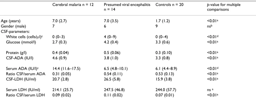
46 children were recruited over a 2-year period. Twelve patients had cerebral malaria, 14 patients presumed viral encephalitis and 20 patients were controls. Demographic and laboratory parameters including LDH and ADA CSF/ serum ratios are listed in Table 1. There was no significant difference in age between patients with cerebral malaria and presumed viral encephalitis, although both groups were both significantly older than controls. The CSF white cell count was significantly higher in the group with
pre-sumed viral encephalitis compared to patients with cere-bral malaria or controls. The mean CSF glucose level was significantly lower in patients with cerebral malaria com-pared to the other two groups and the range for glucose levels in patients with cerebral malaria did not overlap with that for patients with presumed viral encephalitis (Figure 1). Patients with cerebral malaria had significantly higher CSF protein, LDH and ADA levels and CSF/serum LDH ratio but a lower CSF/serum ADA ratio, compared to controls (Table 1). Patients with cerebral malaria had sig-Table 1: Comparison of demographic and cerebrospinal fluid parameters between patients with cerebral malaria, presumed viral encephalitis and controls.
Cerebral malaria n = 12 Presumed viral encephalitis
n = 14
Controls n = 20 p-value for multiple
comparisons
Age (years) 7.0 (2.7) 7.0 (3.5) 1.7 (1.2) <0.01a
Gender (male) 7 6 9 nsb
CSF-parameters:
White cells (cells/µl)c 0 (0–3) 4 (0–9) 0 (0–4) <0.01d
Glucose (mmol/l) 2.7 (0.3) 4.2 (0.4) 3.3 (0.6) <0.01a
Protein (g/l) 0.4 (0.04) 0.5 (0.06) 0.3 (0.10) <0.01a
CSF-ADA (IU/l) 4.6 (0.9) 3.8 (1.0) 3.3 (0.8) <0.01a
Serum ADA (IU/l)c 14.4 (11.6–17.5) 6.5 (4.8–10.1) 6.1 (4.4–8.9) <0.01d
Ratio CSF/serum ADA 0.31 (0.05) 0.54 (0.11) 0.53 (0.13) <0.01a
CSF-LDH (IU/ml) 20.7 (2.8) 26.5 (5.8) 15.9 (3.8) <0.01a
Serum LDH (IU/ml) 214.1 (25.7) 247.5 (46.8) 244.0 (57.7) ns a
Ratio CSF/serum LDH 0.09 (0.02) 0.11 (0.02) 0.07 (0.01) <0.01a
LDH = Lactic dehydrogenase, ADA = Adenosine deaminase. Results where applicable are given as mean (SD). a Analysis of variance: Post hoc
analysis results with p < 0.05 (Tukey HSD test): Age: Cerebral malaria and presumed viral encephalitis versus controls. CSF white cell count: Cerebral
malaria versus presumed viral encephalitis and presumed viral encephalitis versus controls. Glucose and protein: Each group versus the 2 other
groups. CSF-ADA: Cerebral malaria versus controls. Serum ADA: Cerebral malaria versus the 2 other groups. Ratio CSF/serum ADA: Cerebral malaria
versus the two 2 groups. CSF-LDH: Each group versus the other 2 groups. Ratio CSF/serum LDH: Cerebral malaria and presumed viral encephalitis
[image:3.612.57.554.109.303.2]versus controls. b ns = not significant by chi-square test. c Median and range. d Kruskal-Wallis test.
Table 2: Cerebrospinal fluid parameters as discriminators between cerebral malaria and presumed viral encephalitis
Cut-off Sensitivity1 (%) Specificity2 (%) Negative predictive Value3
(%)
Positive predictive Value4
(%)
(95% CI) (95% CI) (95% CI) (95% CI)
CSF white cell count <4 cells/microl
100 (76–100) 57 (32–78) 100 (67–100) 66 (43–83)
CSF protein <0.43 g/l 92 (64–98) 78 (52–92) 91 (64–98) 78 (52–92)
CSF glucose <3.4 mmol/l 100 (78–100) 100 (75–100) 100 (75–100) 100 (78–100)
Ratio CSF/serum ADA <0.385
91 (64–98) 100 (78–100) 93 (70–100) 100 (74–100)
CSF LDH < 22.5 IU/ml 75 (46–91) 71 (44–88) 76 (49–92) 69 (42–87)
1Sensitivity is the true positive rate in percent. It is calculated as the ratio of the number of true positive over the sum of true positive and false
negative patients. True positive meant in the context of this study the presence of cerebral malaria in a group of patients with presumed viral
encephalitis or cerebral malaria below a certain value of a parameter, the "cut-off". 2Specificity is the true negative rate in percent. It is calculated as
the ratio of the number of true negative over the sum of false positive and true negative patients. True negative meant the patient has presumed
viral encephalitis if the parameter is above the "cut-off" chosen. 3The negative predictive value is the ratio of the number of true negative over the
sum of false negative and true negative patients. Positive predictive value is the post-test probability of a positive test and negative predictive value
the post-test probability of a negative test. 4The positive predictive value is calculated as the ratio of the number of true positive over the sum of
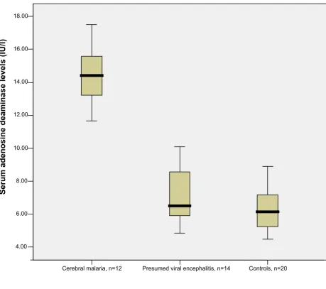
[image:3.612.56.553.522.653.2]nificantly lower CSF glucose, protein and LDH levels and CSF/serum ADA ratios compared to patients with pre-sumed viral encephalitis (Table 1). Patients with cerebral malaria had a significantly higher serum ADA level com-pared to patients with presumed viral encephalitis and to controls (Mann-Whitney test, p < 0.05, Figure 2). Serum ADA levels in patients with presumed viral encephalitis were not significantly different from controls (p > 0.05). Serum LDH levels were not significantly different between groups (p > 0.05). However, CSF/Serum LDH ratios for both patients with cerebral malaria and presumed viral encephalitis were significantly higher than controls.
For markers with a significant difference between cerebral malaria and the presumed viral encephalitis groups, we determined cut-off values of CSF parameters with maxi-mum sensitivities and specificities and positive and nega-tive predicnega-tive values. The best cut-off values taken from the co-ordinates of the ROC curve (see methods section)
below which cerebral malaria was more likely than pre-sumed viral encephalitis were: CSF white cell count, 4 cells/µl; CSF protein level, 0.43 g/l; CSF glucose, 3.4 mmol/l; CSF LDH, 22.5 IU/ml; CSF/serum ADA ratio, 0.385 (Table 2).
CSF white cell count, protein and glucose levels and CSF/ serum ADA ratio below these cut-off values indicated cer-ebral malaria with a sensitivity of > 90%. This resulted in the probability of having presumed viral encephalitis in our group of children with cerebral malaria or viral encephalitis (or negative predictive value for having cere-bral malaria) being above 90% if the values of these parameters were above this cut-off. Specificities for the parameters were 100% for CSF glucose and 100% for CSF/ serum ADA ratio and better than for other parameters, i.e. presumed viral encephalitis was always present in any patient with a value above the cut-off. This resulted in a probability (positive predictive value) of 100% of having
Scatter plot of CSF glucose levels in patients with cerebral malaria, presumed viral encephalitis and controls
Figure 1
Scatter plot of CSF glucose levels in patients with cerebral malaria, presumed viral encephalitis and controls.
5.00
4.00
3.00
2.00
CSF
-g
lu
cose
l
eve
l
(m
mol/
l)
Cerebrospinal fluid glucose levels in patients with cerebral malaria,
h liti
presumed viral encephalitis and controls
Cerebral malaria, n=12
Presumed viral
encephalitis, n=14
cerebral malaria in children with parameters below this cut-off (Table 2).
Discussion
This is to our knowledge the first study to investigate dif-ferences in CSF parameters between presumed viral encephalitis and cerebral malaria. Asymptomatic parasi-taemia with Plasmodium falciparum is common in regions hyperendemic for this parasite. In many of these areas, particularly in South East Asia, viral encephalitis is also a public health problem. It is therefore important to inves-tigate discriminating CSF features between encephalitis and cerebral malaria. Our study is the most detailed study
on CSF parameters in cerebral malaria reported so far. We could not, however, exclude the possibility that patients labeled as having cerebral malaria had a combination of encephalitis and parasitaemia with P. falciparum, but the lack of white cells in their cerebrospinal fluid make this possibility seem very unlikely.
CSF-glucose
A CSF glucose level below 3.4 mmol/l was the best dis-criminator of cerebral malaria from presumed viral encephalitis. This was partly due to the fact that in cerebral malaria CSF glucose levels were below normal range. Low CSF glucose is a well-known phenomenon in cerebral
[image:5.612.74.533.109.516.2]Box plot of serum adenosine deaminase levels in patients with cerebral malaria, presumed viral encephalitis and controls
Figure 2
Box plot of serum adenosine deaminase levels in patients with cerebral malaria, presumed viral encephalitis and controls. The line inside the box represents the median, the box the quartiles, and the whiskers the extreme values.
Serum adenosine deaminase levels (IU/l)
Controls, n=20 Presumed viral encephalitis, n=14
Cerebral malaria, n=12 4.00
malaria and CSF glucose has been found to be lower in patients with fatal cerebral malaria as compared to survi-vors [13]. However, the low CSF glucose may be partly due to low plasma glucose levels not measured in this study, but previously found in falciparum malaria [14]. The previous study found CSF glucose levels of up to 7 mmol/l in patients with cerebral malaria and mean levels of 4.3 mmol/l in survivors, which indicates that a discrim-inator of 3.4 mmol/l between cerebral malaria and encephalitis may not be universally applicable, but dependent on disease severity [13]. Febrile convulsions were an indication for lumbar puncture in the control patients. Consequently the control patients in our study were significantly younger than the disease cases, due to the young age at which febrile convulsions usually occur. CSF glucose levels are independent of age above 2 months of age with lower levels found in infants below this age [15]. All the children in our study were older than 3 months and therefore the observed differences were not age related. Elevated CSF glucose has been described in cases of viral encephalitis [16] and this may have contrib-uted to the significant difference found here between the two groups.
CSF-protein
Previous studies with data on CSF in cerebral malaria also found increased total protein levels [17,18]. Our result of increased cerebrospinal fluid protein levels in presumed viral encephalitis has also been found in patients with her-pes encephalitis [19,20].
Adenosine deaminase
We found that a CSF/serum ADA ratio of <0.38 was the best discriminator of cerebral malaria from presumed viral encephalitis. CSF ADA levels were measured in 3 patients with cerebral malaria previously, and found to have a mean (SD) of 6.6 (1.03) IU/l [5], slightly higher than the levels in our study (4.6 (0.9) IU/l). There has been no previous investigation of serum ADA levels in P. falciparum malaria but serum ADA levels were found to be elevated to more than twice the level of controls in a pre-vious study on patients with Plasmodium vivax malaria [21]. CSF ADA levels in a previous study containing data on 10 patients with viral encephalitis were found to have a mean (SD) of 6.15 (2.93) [6], which is higher than the levels reported here (3.8 (1.0) IU/l). Although the etiolog-ical agent of the presumed viral encephalitis was not determined, it was most likely to be a heterogeneous group over the 2-year period during which the patients were recruited [22].
Lactic dehydrogenase
Mean CSF LDH levels of 26.5 U/ml in our patients with encephalitis were similar to the means of 22.6 and 22.3 U/ ml found in two previous studies, respectively, [23,24].
Our controls had a slightly lower mean CSF LDH level than the 20 controls (adults and children without neuro-logical illness) of a previous study with a mean level of 21.8 U/ml [24]. There is to our knowledge no previous study on CSF LDH levels in cerebral malaria.
CSF lactate, which is characteristically elevated in cerebral malaria [13], is another parameter for future studies look-ing at discriminatlook-ing features between cerebral malaria and encephalitis.
Patients with clinical features of encephalitis and a posi-tive malaria blood slide should still be treated as suffering from cerebral malaria regardless of the CSF findings, as our study cannot exclude a possible overlap in CSF/serum ADA ratios or CSF glucose levels between patients with encephalitis and cerebral malaria in other settings. The same applies to epidemiological surveillance pending fur-ther studies.
Conclusion
CSF adenosine deaminase alone was not a useful discrim-inator between encephalitis and cerebral malaria. How-ever, the CSF/serum ADA ratio was lower in patients with cerebral malaria due to the high levels of ADA in periph-eral blood. This ratio should be evaluated in future, larger-scale, investigations into discriminatory factors between encephalitis and cerebral malaria. Although CSF glucose was significantly reduced in malaria patients, the results should be interpreted cautiously and require confirma-tion in a larger study. It would be preferable to use CSF/ serum glucose ratios for future studies. Large-scale pro-spective studies in areas where P. falciparum and for exam-ple, Japanese encephalitis viruses, are both endemic are needed to validate our findings in different settings.
Competing interests
The author(s) declare that they have no competing inter-ests.
Authors' contributions
SRJ participated in the design of the study, recruitment of patients, collection of samples and data-analysis. SV par-ticipated in the laboratory analysis and data analysis. RMA participated in design of the study. ME participated in data-analysis, interpreted the data and wrote the paper.
References
1. Berkley JA, Mwangi I, Mellington F, Mwarumba S, Marsh K: Cerebral malaria versus bacterial meningitis in children with impaired consciousness. Q J Med 1999, 92:151-157.
2. Wright PW, Avery WG, Ardill WD, McLarty JW: Initial clinical assessment of the comatose patient: cerebral malaria vs. meningitis. Pediatr Infect Dis J 1993, 12:37-41.
Publish with BioMed Central and every scientist can read your work free of charge "BioMed Central will be the most significant development for disseminating the results of biomedical researc h in our lifetime."
Sir Paul Nurse, Cancer Research UK
Your research papers will be:
available free of charge to the entire biomedical community
peer reviewed and published immediately upon acceptance
cited in PubMed and archived on PubMed Central
yours — you keep the copyright
Submit your manuscript here:
http://www.biomedcentral.com/info/publishing_adv.asp
BioMedcentral 4. Rao BL, Basu A, Wairagkar NS, Gore MM, Arankalle VA, Thakare JP,
Jadi RS, Rao KA, Mishra AC: A large outbreak of acute encepha-litis with high fatality rate in children in Andhra Pradesh, India, in associated with Chandipura virus. Lancet 2003, 364:869-874.
5. Gambhir IS, Mehta M, Singh DS, Khanna HD: Evaluation of CSF-adenosine deaminase activity in tubercular meningitis. Jour-nal Assoc Phys India 1999, 47:192-194.
6. Mishra OP, Loiwal V, Ali Z, Nath G, Chandra L: Cerebrospinal fluid adenosine deaminase activity for the diagnosis of tubercu-lous meningitis in children. J Trop Ped 1996, 42:129-132. 7. Whitley RJ, Gnann JW: Viral encephalitis: familiar infections
and emerging pathogens. Lancet 2002, 359:507-514.
8. World Health Organisation, Communicable Diseases Cluster: Severe falciparum malaria. Trans R Soc Trop Med Hyg 2000, 94(suppl 1):S1-S90.
9. Jakka S, Veena S, Rao AR, Eisenhut M: Cerebrospinal fluid adeno-sine deaminase levels and adverse neurological outcome in pediatric tuberculous meningitis. Infection 2005, 33:264-266. 10. Giusti G, Galanti B: Adenosine 5-monophosphate deaminase
and adenosine deaminase activity of the blood in experimen-tal and human hepatic pathological conditions. Boll Soc Ital Biol Sper 1965, 41:614-617.
11. Armitage P, Berry G, Matthews JNS: Statistical methods in med-ical research. 4th edition. Oxford, United Kingdom, Blackwell Sci-ence; 2002:697.
12. Greenhalgh T: Papers that report diagnostic or screening tests. BMJ 1997, 315:540-543.
13. White NJ, Warrell DA, Chanthavanich SLP, Phillips RE, Pongpaew P: Pathophysiological and prognostic significance of cerebros-pinal-fluid lactate in cerebral malaria. Lancet 1985, 6:776-778. 14. White NJ, Warrell DA, Chanthavanich P, Looareesuwan S, Warrell
MJ, Krishna S, Williamson DH, Turner RC: Severe hypoglycemia and hyperinsulinaemia in falciparum malaria. N Eng J Med
1983, 309:61-66.
15. Lermann-Sagie T, Shohat M, Nitzan M: CSF glucose levels in febrile infants. Eur J Pediatr 1988, 147:416-417.
16. Schmidt RM: Liquorveraenderungen bei Enzephalitiden. Z Aer-ztl Fortbild 1978, 72:60-61.
17. Das BS, Mohanty S, Mishra SK, Patnaik JK, Satpathy SK, Mohanty D, Bose TK: Increased cerebrospinal fluid protein and lipid per-oxidation products in patients with cerebral malaria. Trans R Soc Trop Med Hyg 1991, 85:733-734.
18. Warrell DA, Looareesuwan S, Phillips RE, White NJ, Warrell NJ, Warrell MJ, Chapel HM, Areekul S, Tharavanij S: Function of the blood-cerebrospinal fluid barrier in human cerebral malaria: rejection of the permeability hypothesis. Am J Trop Med Hyg
1986, 35:882-889.
19. Koskiniemi M, Vaheri A, Taskinen E: Cerebrospinal fluid altera-tions in herpes simplex virus encephalitis. Rev Infect Dis 1984, 6:608-618.
20. Toth C, Harder S, Yager J: Neonatal herpes encephalitis: a case series and review of clinical presentation. Can J Neurol Sci 2003, 30:36-40.
21. Ozcan E, Abdurrahim K, Adnan S, Senel A, Necmeddin A: Serum erythrocyte and leukocyte adenosine deaminase activities in patients with vivax malaria in Turkey. J Egypt Soc Parasitol 1997, 27:445-454.
22. Kumar R, Mathur A, Kumar A, Sethi GD, Sharma S, Chaturvedi UC: Virological investigations of acute encephalopathy in India.
Arch Dis Child 1990, 65:1227-1230.
23. Beaty HN, Oppenheimer S: Cerebrospinal-fluid lactic dehydro-genase and its isoenzymes in infections of the central nerv-ous system. New Eng J Med 1988, 279:1197-1202.