Kaposi’s Sarcoma-Associated Herpesvirus K8 Is an RNA
Binding Protein That Regulates Viral DNA Replication in
Coordination with a Noncoding RNA
Dongcheng Liu,
a,cYan Wang,
a,bYan Yuan
a,caInstitute of Human Virology and Ministry of Education Key Laboratory of Tropical Disease Control, Zhongshan
School of Medicine, Sun Yat-Sen University, Guangzhou, Guangdong, China
bGuanghua School of Stomatology, Guangdong Provincial Key Laboratory of Stomatology, Sun Yat-Sen
University, Guangzhou, Guangdong, China
cDepartment of Microbiology, University of Pennsylvania School of Dental Medicine, Philadelphia,
Pennsylvania, USA
ABSTRACT
Kaposi’s sarcoma-associated herpesvirus (KSHV) lytic replication and
constant primary infection of fresh cells are crucial for viral tumorigenicity. The
virus-encoded bZIP family protein K8 plays an important role in viral DNA replication in
both viral reactivation and
de novo
infection. The mechanism underlying the
func-tional role of K8 in the viral life cycle is elusive. Here, we report that K8 is an RNA
binding protein that also associates with many other proteins, including other RNA
binding proteins. Many protein-protein interactions involving K8 are mediated by
RNA. Using a UV cross-linking and immunoprecipitation (CLIP) procedure combined
with high-throughput sequencing, RNAs that are associated with K8 in BCBL-1 cells
were identified, including both viral (PAN, T1.4, T0.7, etc.) and cellular (MALAT-1,
MRP, 7SK, etc.) RNAs. An RNA binding motif in K8 was defined, and mutation of the
motif abolished the ability of K8 to bind to many noncoding RNAs, as well as viral
DNA replication during
de novo
infection, suggesting that the K8 functions in viral
replication are carried out through RNA association. The functions of K8 and
associ-ated T1.4 RNA were investigassoci-ated in detail, and the results showed that T1.4
medi-ates the binding of K8 to ori-Lyt DNA. The T1.4-K8 complex physically bound to
KSHV ori-Lyt DNA and recruited other proteins and cofactors to assemble a
replica-tion complex. Deplereplica-tion of T1.4 abolished DNA replicareplica-tion in primary infecreplica-tion.
These findings provide mechanistic insights into the role of K8 in coordination with
T1.4 RNA in regulating KSHV DNA replication during
de novo
infection.
IMPORTANCE
Genomewide analyses of the mammalian transcriptome revealed that
a large proportion of sequence previously annotated as noncoding regions is
actu-ally transcribed and gives rise to stable RNAs. The emergence of a large number of
noncoding RNAs suggests that functional RNA-protein complexes, e.g., ribosomes or
spliceosomes, are not ancient relics of the last ribo-organism but would be well
adapted to a regulatory role in biology. K8 has been puzzling because of its unique
characteristics, such as multiple regulatory roles in gene expression and DNA
replica-tion without DNA binding capability. This study reveals the mechanism underlying
its regulatory role by demonstrating that K8 is an RNA binding protein that binds to
DNA and initiates DNA replication in coordination with a noncoding RNA. It is
sug-gested that many K8 functions, if not all, are carried out through its associated
RNAs.
KEYWORDS
7SK RNA, K8, Kaposi’s sarcoma-associated herpesvirus (KSHV), PAN RNA,
T0.7 RNA, T1.4 RNA, viral lytic DNA replication, human herpesvirus 8 (HHV-8),
noncoding RNA, ori-Lyt
Received14 December 2017Accepted3
January 2018
Accepted manuscript posted online10
January 2018
CitationLiu D, Wang Y, Yuan Y. 2018. Kaposi's
sarcoma-associated herpesvirus K8 is an RNA binding protein that regulates viral DNA replication in coordination with a noncoding RNA. J Virol 92:e02177-17.https://doi.org/10 .1128/JVI.02177-17.
EditorRichard M. Longnecker, Northwestern
University
Copyright© 2018 American Society for
Microbiology.All Rights Reserved. Address correspondence to Yan Wang, [email protected], or Yan Yuan, [email protected].
GENOME REPLICATION AND REGULATION
OF VIRAL GENE EXPRESSION
crossm
on November 6, 2019 by guest
http://jvi.asm.org/
K
aposi’s sarcoma-associated herpesvirus (KSHV), also referred to as human
herpes-virus 8 (HHV-8), is the causative agent of several AIDS-associated malignancies,
including Kaposi’s sarcoma (KS), primary effusion lymphoma (PEL), and multicentric
Castleman’s disease (MCD) (1–3). KSHV has two alternative life cycles, namely, latent
and lytic replication cycles (4, 5). In order to evade host immune surveillance, KSHV
establishes latent infection in most infected cells. However, in KS lesions, latent viral
episomes can be quickly lost as cells divide (6). To sustain the virus-infected cell
population, a small percentage of infected cells undergo spontaneous lytic replication
and produce infectious progeny virions (7–9). Thus, unlike other oncogenic viruses,
where the latent life cycle is primarily responsible for the oncogenic activities of the
virus, KSHV lytic replication and constant
de novo
infection of fresh cells are essential
for viral pathogenesis and tumorigenesis (6).
During
de novo
infection, a set of viral genes are expressed and participate in
immune evasion, epigenetic modification, and transcriptional reprogramming before
the latent infection is established (10–12). Short-term viral DNA replication occurs in the
period between virus infection and latency establishment, which allows the KSHV
genome to be amplified from 1 to 50 to 100 copies. This phase of viral DNA replication
is termed abortive lytic replication as, unlike lytic replication, it does not produce
infectious virions. We and others have found that viral abortive replication is sensitive
to phosphonoacetic acid (PAA), a specific inhibitor of herpesvirus DNA polymerase (11,
13), suggesting that the DNA replication in this phase is different from latent
replica-tion, which is cellular DNA polymerase dependent but utilizes lytic DNA replication
machinery. However, the mechanism that controls abortive replication during
de novo
infection is elusive.
KSHV DNA replication initiates at an origin (such as ori-Lyt) and requires
trans
-acting
elements, both viral and cellular. Two duplicated copies of lytic DNA replication origin,
namely, ori-Lyt (L) and ori-Lyt (R), were identified in the KSHV genome (14, 15). These
two ori-Lyts share almost identical 1.1-kb core component sequences and 600-bp
GC-rich repeats, which are represented as 20-bp and 30-bp tandem arrays. Each 1.7-kb
ori-Lyt sequence is necessary and sufficient as a
cis
-acting signal for KSHV lytic DNA
replication (15). Furthermore, both ori-Lyts carry a transcriptional promoter that directs
transcription of a 1.4-kb RNA in ori-Lyt (L) and a 0.7-kb RNA in ori-Lyt (R) (16). The
ori-Lyt-associated transcriptions are absolutely required for viral DNA replication, but
the functions of the RNAs are unknown (17). Two virus-encoded proteins, namely,
replication and transcription activator (RTA) and K8 (K-bZIP), were found to be ori-Lyt
binding proteins, and both were essential for KSHV lytic DNA replication in a
transient-transfection assay (15, 17, 18). K8 associates with ori-Lyt DNA and recruits DNA
replication-relevant proteins, both viral and cellular, to ori-Lyt to form viral core
replication machinery complexes (15, 19, 20). Recently, we and others observed that
DNA replication still proceeds in K8-null virus during the lytic reactivation phase,
suggesting that the function of K8 in viral DNA replication can be substituted for by
another protein(s) (13, 21). However, K8 was found to be absolutely essential for viral
DNA replication during
de novo
infection (13).
In addition to its role in viral DNA replication, K8 has been shown to be a global gene
expression repressor, inhibiting both viral and cellular gene expression (22, 23). Several
studies have uncovered linkages between K8 and epigenetic regulation, such as
inhibiting the H3K9me3 demethylase JMJD2A and mediating SUMO-2/3 modification of
the KSHV chromosome (24, 25). In addition, K8 was reported to inhibit the activity of
CDK2, which plays a pivotal role in cell cycle progression. Inhibition of CDK2 activity by
K8 resulted in a prolonged G
1phase with concomitant induction of p21, P27, and
C/EBP
␣
(26, 27).
To elucidate the biological functions of K8 in the KSHV life cycle and the
mecha-nisms underlying these actions, we attempted to identify components that interact
with K8 in the belief that knowing the K8 binding partners would provide clues to
reveal K8 function and its underlying mechanism. In this effort, we found that K8 is a
novel RNA binding protein and that its RNA binding ability is essential to its role in viral
on November 6, 2019 by guest
http://jvi.asm.org/
DNA replication during
de novo
KSHV infection. Furthermore, a viral RNA, namely, T1.4
or ori-Lyt-associated RNA, was found to mediate the interaction between K8 and ori-Lyt
DNA and to be absolutely required for viral DNA replication. Through this investigation,
a novel mechanism of viral DNA replication by coordinates actions of K8 protein and
viral noncoding RNA was revealed.
RESULTS
K8 predominately interacts with RNA binding proteins.
In order to elucidate the
mechanisms underlying the K8 actions in viral DNA replication and other events in the
viral life cycle, a proteomics approach was undertaken to identify the proteins that
interact with K8 in KSHV-infected cells during lytic replication. BCBL-1 cells were
induced for reactivation with tetradecanoyl phorbol acetate (TPA) for 48 h and
sub-jected to coimmunoprecipitation (co-IP) with an antibody against K8. The resultant
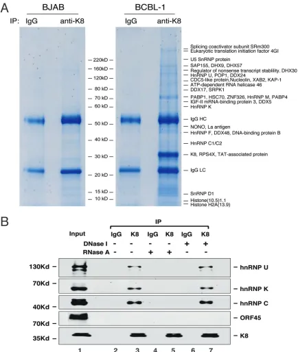
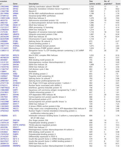
proteins were resolved on SDS-PAGE and revealed by Coomassie blue staining (Fig. 1A).
Numerous protein bands were shown in the K8 immunoprecipitated samples but not
visually detected in the control samples (mouse immunoglobulin G [IgG] precipitate of
induced BCBL-1 nuclear extract and KSHV-free BJAB B lymphoma cells precipitated with
anti-K8 antibody). The discrete bands were excised and subjected to mass spectrometry
(MS) analysis. This approach led to identification of a series of proteins, including a few
viral proteins (RTA, ORF21, and ORF57) (22, 23, 28) and many cellular proteins (Fig. 1A
and Table 1). Interestingly, most of the identified proteins were potential RNA binding
proteins, including DEA(D/H) box helicases (DDX5, DDX17, DDX24, DDX46, DDX48,
DHX9, DHX30, and DHX57), members of the heterogeneous ribonucleoprotein family
(hnRNPs U, M, Q, K, F, C1/C2), and RNA-splicing proteins. The finding that K8
predom-inately interacts with RNA binding proteins suggests that some regulatory function(s)
of K8 is involved with RNA.
To confirm the MS results and explore the mode of binding between K8 and these
RNA binding proteins, we analyzed the interaction of K8 with several representative
RNA binding proteins, such as hnRNPs U, K, and C, in TPA-induced BCBL-1 cells using
coimmunoprecipitation, followed by Western blotting. The results showed that K8
indeed interacts with hnRNPs U, K, and C, but not with ORF45, a viral protein known not
to interact with K8 (17, 29) and serving as a negative control (Fig. 1B). Since these three
hnRNPs are RNA binding proteins, we asked if the interactions of K8 with the hnRNPs
are mediated by RNA. To this end, RNase A was added in washing buffer during the
coimmunoprecipitation procedure. The results showed that treatment with RNase A
eliminated the interaction between K8 and the hnRNPs (Fig. 1B, lane 5). In contrast,
treatment with DNase I did not affect these interactions (Fig. 1B, lane 7). Thus, we
conclude that the interactions of K8 with hnRNPs are mediated by RNA molecules.
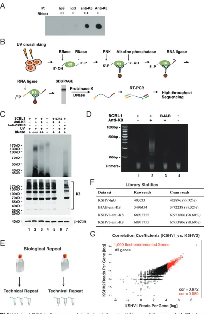
K8 is a novel RNA binding protein.
Since K8 predominately interacts with RNA
binding proteins and the interactions are mediated by RNA, we asked whether K8 itself
is an RNA binding protein. To address this question, we performed UV cross-linking and
immunoprecipitation (CLIP) followed by dot blotting. Briefly, TPA-induced BCBL-1 cells
were UV irradiated for protein-RNA cross-linking. Then, the cells were lysed and
subjected to immunoprecipitation with K8 antibody (mouse IgG was included as a
negative control). RNase A at different concentrations was added to the washing buffer.
The immunoprecipitated components were labeled with biotin and spotted on a nylon
membrane. Biotin-labeled RNA was then detected with a biotin chromogenic detection
kit. As shown in Fig. 2A, the K8 immunoprecipitates showed strong RNA-biotin signals,
which were not detected in the IgG control. Furthermore, the RNA-biotin signal
decreased with increasing concentrations of RNase A in the CLIP experiment, indicating
that K8 was associated with RNA.
Identification of K8 binding RNAs.
To comprehend the biological function and
process mediated by K8 and its associated RNAs, we attempted to identify the RNAs
that are associated with K8. To this end, cross-linking and immunoprecipitation
fol-lowed by high-throughput sequencing (CLIP-seq) (30) was employed to determine K8
binding RNAs. The CLIP procedure is schematically illustrated in Fig. 2B. In brief,
Coordinate Regulation by K8 and Noncoding RNA Journal of Virology
on November 6, 2019 by guest
http://jvi.asm.org/
TPA-induced BCBL-1 cells were UV irradiated to form protein-RNA adducts. Cell lysates
were prepared and treated with RNase A to digest the RNAs that were not protected
by proteins. Then, the lysates were immunoprecipitated with K8 antibody or mouse
IgG. The precipitated RNAs were ligated with linkers and radiolabeled at the 5
=
ends
with [
␥
-
32P]ATP. In SDS-PAGE, a smeared band around 100 kDa was observed in the
FIG 1Identification of proteins that interact with K8 in BCBL-1 cells. (A) Nuclear extracts of TPA-treated KSHV-positive BCBL-1 cells and KSHV-negative BJAB cells were immunoprecipitated with anti-K8 antibody. Immunoprecipitated proteins were resolved on SDS-PAGE, followed by Coomassie blue staining. The discrete bands were excised and subjected to mass spectrometric analysis. The resultant MS-MS spectra were run against a sequence database with the SEQUEST program. Matched cellular proteins are indicated at the right of the gel. (B) Interaction between K8 and hnRNPs is RNA mediated. Coimmunoprecipitation of TPA-treated BCBL-1 cells was performed with anti-K8 antibody. RNase A or DNase I was added during the washing steps. The precipitates were Western blotted with antibodies against hnRNPs U, K, and C. The Western blot was also probed with an antibody against ORF45, a protein that was known not to interact with K8.
on November 6, 2019 by guest
http://jvi.asm.org/
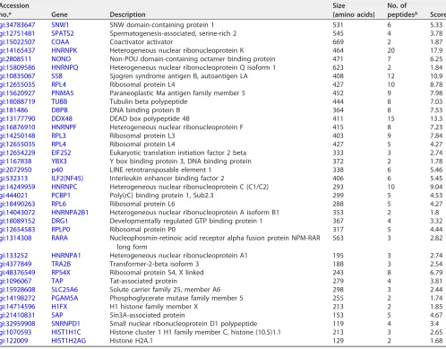
[image:4.585.41.475.69.583.2]TABLE 1K8-associated proteins identified by mass spectrometry
Accession
no.a Gene Description
Size (amino acids)
No. of
peptidesb Score
gi:19923466 SRRM2 Splicing coactivator subunit SRm300 2,752 17 15
gi:21655146 EIF4GI Eukaryotic translation initiation factor 4 gamma 1 1,600 10 8.62
gi:12667788 MYH9 Myosin-9 1,960 7 6.19
gi:28174906 VIRMA Vir-like M6A methyltransferase associated 1,198 2 1.74
gi:37589132 EPRS Glutamyl-prolyl-tRNA synthetase 869 3 2.69
gi:100913206 DHX9 DExH-box helicase 9 1,270 22 19.6
gi:4033735 SAP155 Spliceosome-associated protein 155 1,304 19 16.8
gi:3882183 LARP1 La ribonucleoprotein domain family member 1 1,096 11 9.93
gi:39777586 DEAH 57 DExH box helicase 57 1,386 7 6.14
gi:15215317 RRP12 rRNA processing 12 homolog 1,297 5 4.14
gi:27882034 DHX8 DEAH box helicase 8 1,214 3 2.69
gi:1575536 RENT1 Regulator of nonsense transcript stability 1,118 18 15.8
gi:40254861 UBAP2L Ubiquitin-associated protein-2 like 1,087 9 8.34
gi:20336294 DHX30 DExH box helicase 30 1,194 10 8.54
gi:66347698 C9ORF10 Chromosome 9 open reading frame 10 1,118 7 6
gi:2498883 SF3B2 Splicing factor 3b subunit 2 872 7 6.27
gi:34222504 FNBP3 Formin-binding protein 3 957 2 1.9
gi:18071115 ATXN2L Ataxin-2-related domain protein 1,075 3 2.63
gi:13124451 POP1 Ribonucleases P/MRP protein subunit 1,024 2 1.86
gi:12803113 EFTUD2 Elongation factor Tu GTP binding domain containing 2, U5 SnRNP
component
972 26 23
gi:10047339 MOV10 Mov10 RISC complex RNA helicase 1,032 9 8.16
gi:40788339 MATR3 Matrin 3 853 6 5.57
gi:4050087 RBM25 RNA binding motif protein 25 735 6 4.98
gi:14141161 HNRNPU Heterogeneous nuclear ribonucleoprotein U 806 4 3.49
gi:11526793 DHX36 DEAH box helicase 36 1,008 5 4.57
gi:14250756 DDX24 DEAD box helicase 24 859 4 3.56
gi:11067747 CDC5L Cell division cycle 5 like 802 12 10.5
gi:128841 NCL Nucleolin 707 7 6.08
gi:10566459 XAB2 XPA binding protein 2 855 9 7.9
gi:1699027 TRIM28 Tripartite motif containing 28 835 8 7.01
gi:5032087 SF3A1 Splicing factor 3a subunit 1 793 7 5.96
gi:29881667 SFPQ Splicing factor proline/glutamine-rich 707 4 3.59
gi:27477136 ZC3HAV1 Zinc finger CCCH-type antiviral protein 1 isoform 1 902 5 4.61
gi:5762315 NFAR-2 Nuclear factor associated with dsRNA 2 894 6 5.44
gi:168776622 IFI-16 Interferon, gamma-inducible protein 16 736 2 1.87
gi:10863889 SART1 Squamous cell carcinoma antigen recognized by T cells 1 800 2 1.81
gi:21619831 TFIP11 Tuftelin-interacting protein 11 837 2 1.81
gi:2696613 DDX46 ATP-dependent RNA helicase 46 813 20 18.4
gi:1857944 SRPK2 Serine/arginine-rich protein-specific kinase 2 686 4 3.43
gi:2498733 GP137 GPI-anchored protein p137 709 4 3.67
gi:14252988 SRPK1A Serine/arginine-rich protein-specific kinase 1a 826 7 76.45
gi:32880087 DDX17 DEAD box helicase 17 729 8 7.25
gi:12653845 AKAP8L A kinase (PRKA) anchor protein 8 like 646 2 1.86
gi:10863945 XRCC5 X-Ray repair cross complementing 5, ATP-dependent DNA helicase II 732 3 2.49
gi:37925422 NUFIP2 Nuclear fragile X mental retardation-interacting protein 2, proliferation-inducing gene 1
695 4 3.48
gi:1082856 ILF3 Interleukin enhancer binding factor 3 isoform a, transcription factor
NF-AT 90K chain
894 2 1.74
gi:47125447 ZNF326 Zinc finger protein 326 508 14 12
gi:73974124 PABP1 Polyadenylate binding protein 1 690 19 17.1
gi:55632315 HSP70(HSPA5) 78-kDa glucose-regulated protein 654 11 10.3
gi:2832596 DDX17 DEAD box helicase 17 729 13 11.5
gi:14141152 HNRNPM Heterogeneous nuclear ribonucleoprotein M isoform a 730 15 14
gi:5454064 RBM14 RNA binding motif protein 14 669 5 4.48
gi:73976763 PABP4 Polyadenylate-binding protein 4 644 13 11.6
gi:31542501 DEF6 Differentially expressed in FDCP 6 631 4 3.63
gi:30795212 IGF-II 3 Insulin-like growth factor 2 mRNA binding protein 3 579 18 16.1
gi:56237027 IGF-II 1 Insulin-like growth factor 2 mRNA binding protein 1 577 8 7.07
gi:30584123 DDX5 DEAD box helicase 5 615 10 8.51
gi:228008400 HNRNPQ Heterogeneous nuclear ribonucleoprotein Q isoform 6 562 13 11.6
(Continued on next page)
Coordinate Regulation by K8 and Noncoding RNA Journal of Virology
on November 6, 2019 by guest
http://jvi.asm.org/
UV-cross-linked K8-immunoprecipitated BCBL-1 samples (Fig. 2C, top). Given that the
molecular mass of K8 is around 35 kDa, the K8-RNA complexes over 100 kDa apparently
resulted from homomultimer K8 (31) associated with RNAs, as evidenced in Western
blotting (Fig. 2C, bottom). No band was found in control samples (without UV
cross-linking) or BJAB cells (Fig. 2C, lanes 5 and 6). Such bands were also undetectable with
the sample from anti-ORF45 immunoprecipitation (lane 7). RNA was purified from the
gel and subjected to reverse transcription-PCR (RT-PCR), which yielded a smeared band
from 100 to 500 bp long (Fig. 2D, lane 2). Control samples of IgG (lane 1) and BJAB (lane
3) yielded almost no PCR product. The cDNAs in the range of 100 to 200 bp were
purified and subjected to high-throughput deep sequencing. Experimental design and
library statistics of the RNA sequencing are illustrated in Fig. 2E to G.
The CLIP-seq reads were first mapped to the KSHV genome (GenBank ID
U75698.1
).
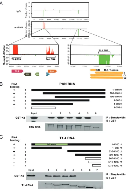
The alignment showed three major peaks in the KSHV genome in the loci of PAN RNA,
T1.4 RNA in ori-Lyt (L), and T0.7 RNA in ori-Lyt (R) regions (Fig. 3A). To confirm the
CLIP-seq data, PAN RNA and T1.4 RNA were selected for
in vitro
RNA pulldown assays
to verify their interaction with K8 (Fig. 3B and C). Glutathione
S
-transferase (GST)-K8
protein prepared from
Escherichia coli
was mixed with biotinylated PAN or T1.4 RNA
[image:6.585.43.541.84.473.2]and subjected to a pulldown procedure with streptavidin beads. Associated protein
was analyzed by Western blotting. The results confirmed that K8 directly interacts with
TABLE 1(Continued)
Accession
no.a Gene Description
Size (amino acids)
No. of
peptidesb Score
gi:34783647 SNW1 SNW domain-containing protein 1 531 6 5.33
gi:12751481 SPATS2 Spermatogenesis-associated, serine-rich 2 545 4 3.78
gi:15022507 COAA Coactivator activator 669 2 1.87
gi:14165437 HNRNPK Heterogeneous nuclear ribonucleoprotein K 464 20 17.9
gi:2808511 NONO Non-POU domain-containing octamer binding protein 471 7 6.25
gi:15809586 HNRNPQ Heterogeneous nuclear ribonucleoprotein Q isoform 1 623 2 1.84
gi:10835067 SSB Sjogren syndrome antigen B, autoantigen LA 408 12 10.9
gi:12655035 RPL4 Ribosomal protein L4 427 10 8.78
gi:15620927 PNMA5 Paraneoplastic Ma antigen family member 5 452 9 7.98
gi:18088719 TUBB Tubulin beta polypeptide 444 8 7.03
gi:181486 DBPB DNA binding protein B 364 8 7.53
gi:13177790 DDX48 DEAD box polypeptide 48 411 15 13.3
gi:16876910 HNRNPF Heterogeneous nuclear ribonucleoprotein F 415 8 7.23
gi:14250148 RPL3 Ribosomal protein L3 403 9 7.84
gi:12655035 RPL4 Ribosomal protein L4 427 5 4.27
gi:12654229 EIF2S2 Eukaryotic translation initiation factor 2 beta 333 3 2.74
gi:1167838 YBX3 Y box binding protein 3, DNA binding protein 372 2 1.78
gi:2072950 p40 LINE retrotransposable element 1 338 6 5.46
gi:532313 ILF2(NF45) Interleukin enhancer binding factor 2 406 6 5.45
gi:14249959 HNRNPC Heterogeneous nuclear ribonucleoprotein C (C1/C2) 293 10 9.04
gi:444021 PCBP1 Poly(rC) binding protein 1, Sub2.3 299 5 4.53
gi:18490263 RPL6 Ribosomal protein L6 288 5 4.27
gi:14043072 HNRNPA2B1 Heterogeneous nuclear ribonucleoprotein A isoform B1 353 2 1.8
gi:18089152 DRG1 Developmentally regulated GTP binding protein 1 367 4 3.32
gi:12654583 RPLP0 Ribosomal protein P0 317 5 4.44
gi:1314308 RARA Nucleophosmin-retinoic acid receptor alpha fusion protein NPM-RAR
long form
563 3 2.82
gi:133252 HNRNPA1 Heterogeneous nuclear ribonucleoprotein A1 195 3 2.74
gi:4377849 TRA2B Transformer-2-beta isoform 3 188 3 2.54
gi:48376549 RPS4X Ribosomal protein S4, X linked 243 8 6.79
gi:1096067 TAP Tat-associated protein 279 4 3.81
gi:15928608 SLC25A6 Solute carrier family 25, member A6 298 3 2.44
gi:14198272 PGAM5A Phosphoglycerate mutase family member 5 255 2 1.74
gi:14714596 H1FX H1 histone family member X 213 2 1.85
gi:21410831 SAP Sin3A-associated protein 153 5 4.67
gi:32959908 SNRNPD1 Small nuclear ribonucleoprotein D1 polypeptide 119 4 3.4
gi:1070593 HIST1H1C Histone cluster 1 H1 family member C, histone (10.5)1.1 213 3 2.65
gi:122009 HIST1H2AG Histone H2A.1 129 2 1.68
aAccession numbers may be found in the NCBI Protein database (https://www.ncbi.nlm.nih.gov/protein/). bNumber of polypeptides detected for the protein by MS.
on November 6, 2019 by guest
http://jvi.asm.org/
FIG 2Validation of K8 RNA binding property and identification of K8-associated RNAs using a CLIP-seq approach. (A) TPA-induced BCBL-1 cells were UV irradiated and subjected to immunoprecipitation with K8 antibody. Different amounts of RNase A (⫹⫹, 2g/ml; (Continued on next page)
Coordinate Regulation by K8 and Noncoding RNA Journal of Virology
on November 6, 2019 by guest
http://jvi.asm.org/
[image:7.585.41.464.68.716.2]PAN RNA and T1.4 RNA in a specific manner. Furthermore, both RNAs were mapped for
the regions that are required for K8 binding by using truncation mutants of the two
RNAs. It was found that K8 binds to PAN RNA in the region between nucleotides 400
and 589 (Fig. 3B) and interacts with T1.4 RNA in both 5
=
GC repeat and 3
=
nonrepeat
regions (Fig. 3C).
Next, the CLIP-seq reads were mapped to the human genome. About half (49%) of
K8 binding RNAs are intronic and intergenic sequence, and 11% are annotated
non-coding RNAs, suggesting K8 predominately associates with nonnon-coding RNAs (ncRNAs).
Thirty percent of the RNAs were from exons of mRNAs (Fig. 4A). These data imply that
ncRNA might play a role in the function of K8. The top six of the abundant cellular
ncRNAs that are associated with K8 are MRP, 7SL, 7SK, MALAT1, U1, and U3 RNAs (Fig.
4B). All of them are associated with RNA metabolism and gene regulation, suggesting
that K8 might play roles in RNA processing and gene regulation.
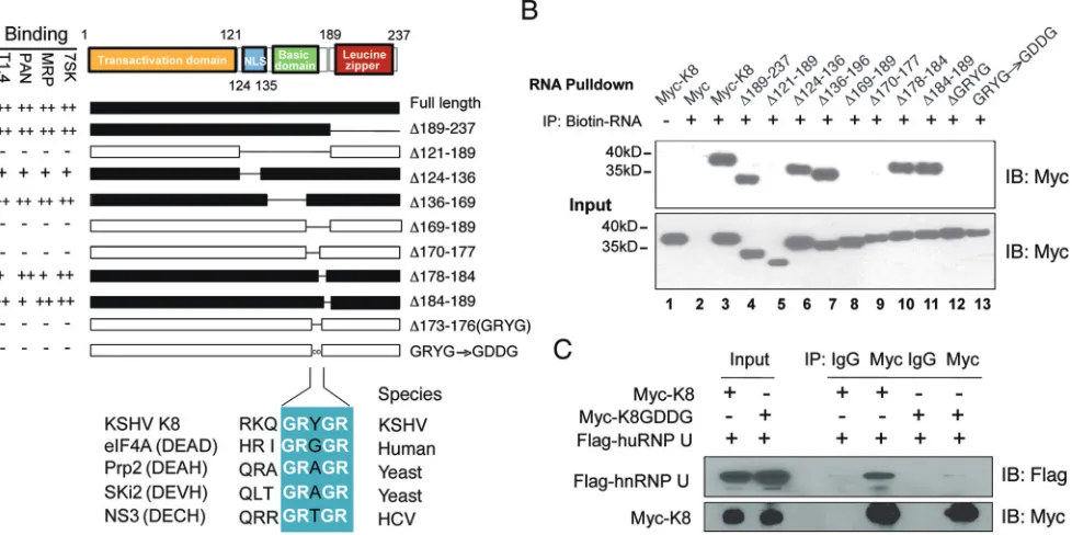
Mapping of the K8 RNA binding region identified a novel RNA binding motif.
Based on its homology to Epstein-Barr virus (EBV) ZTA protein, K8 protein is divided into
three domains: the transactional domain at the N terminus (amino acids [aa] 1 to 121)
(32), the basic domain (DNA binding domain; aa 121 to 189) (23), and a leucine zipper
domain at its C terminus (aa 189 to 237) (Fig. 5A) (31, 33, 34). To identify the domain(s)
in K8 that is required for RNA binding, a series of truncation and deletion mutants of
K8 were constructed and examined for RNA binding ability in a biotinylated RNA
pulldown assay. 293T cells were transfected with Myc-tagged K8 or its mutants, and
whole-cell lysates were prepared. The lysates were incubated with biotinylated
tran-scripts, and the RNA-protein complexes were isolated using streptavidin-coupled
Dyna-beads (ThermoFisher). RNA-associated proteins were eluted in SDS-PAGE buffer and
analyzed by Western blotting with an anti-Myc antibody. Several potential K8 binding
RNAs identified in our CLIP-seq were used to map K8 protein for the RNA binding
region(s), including the PAN and T1.4 viral RNAs, as well as the 7SK and MRP cellular
RNAs. Interestingly, the results with these different RNAs were consistent, as shown in
Fig. 5A. A representative Western blot of 7SK pulldown is presented in Fig. 5B. The
results showed that the basic domain (aa 121 to 189), but not the leucine zipper
domain, was responsible for K8 binding to all four RNAs tested. Further mutagenesis
analysis narrowed down the RNA binding motif to 4 amino acids, GRYG (aa 173 to 176),
as deletion of these 4 amino acids from K8 led to total loss of RNA binding activity.
When GRYG was mutated to GDDG, two negatively charged aspartic acids were
introduced into the motif, and K8 completely lost the ability to associate with all the
RNAs (Fig. 5B, lane 13). Interestingly, GRYG appears to be part of the RNA binding
consensus motif (GRXGR) found in DEAD box family proteins (35–37).
Since the interactions of K8 and hnRNPs are RNA mediated (Fig. 1B), we examined
whether the GDDG mutation in K8 affects the association between K8 and hnRNPs.
Myc-tagged K8 or its GDDG mutant was cotransfected with Flag-tagged hnRNP U into
293T cells. Coimmunoprecipitation was performed with anti-Myc antibody and
ana-lyzed by Western blotting using anti-Flag antibody. It was found that hnRNP U
FIG 2Legend (Continued)
⫹, 1g/ml) were added to the washing buffer during immunoprecipitation. Coprecipitated RNA was labeled with biotin at the 3=end and spotted onto a nylon membrane. The biotinylated RNA was then analyzed with a chemiluminescent nucleic acid detection module kit. (B) Schematic illustration of the CLIP-seq procedure. TPA-induced BCBL-1 cells were UV irradiated and immunoprecipitated with K8 antibody. Different amounts of RNase A were added to the washing buffer. The coprecipitated RNAs were phosphorylated at the 5=end with T4 polynucleotide kinase (PNK) and dephosphorylated with alkaline phosphatase at the 3=end. The RNAs were ligated to the 3=linker and then to the 5=linker labeled with␥-32P. The RNA-protein complex was resolved in SDS-PAGE, and RNAs were isolated and amplified by RT-PCR. The PCR products were analyzed by high-throughput sequencing. (C) Autoradiogram of␥-32 P-labeled RNA cross-linked to K8. Immunoprecipitation was performed with anti-K8 or anti-ORF45 antibody. (Bottom) Input of K8 and
-actin. (D) RT-PCR products from K8-CLIP. (E) Experimental design of CLIP-seq. For each set of CLIP experiments (IgG, KSHV-negative [BJAB], KSHV1 [BCBL-1], and KSHV2 [BCBL-1]), two biological repeats were carried out. Each biological repeat contained three technical repeats. (F) Library statistics of CLIP-seq. Two biological repeats from K8-CLIP-seq (KSHV1 and KSHV2) contained 67,953,806 and 81,632,254 clean reads, respectively. In contrast, there were only 402,896 and 3,472,238 clean reads in the IgG control and KSHV negative (BJAB) data sets, respectively. (G) Libraries from two biological repeats were highly similar. The correlation coefficient between two biological repeats (KSHV1 and KSHV2) was above 0.97.
on November 6, 2019 by guest
http://jvi.asm.org/
FIG 3Three viral noncoding RNAs were identified and confirmed to be directly associated with K8 protein. (A) CLIP-seq reads mapped to the KSHV genome. Enlargement of the K8 cross-linked RNA in the KSHV genomic location showed two peaks (T1.4 RNA and PAN RNA) from the forward strain (left) and one peak (T0.7 RNA) from the reverse strain (right). (B and C) PAN (B) and T1.4 (C) RNAs and
(Continued on next page)
Coordinate Regulation by K8 and Noncoding RNA Journal of Virology
on November 6, 2019 by guest
http://jvi.asm.org/
[image:9.585.43.464.69.700.2]interacted with wild-type (WT) K8 but failed to bind the K8 GDDG mutant, indicating
that K8 was unable to interact with the hnRNP without the GRYGR-dependent RNA
binding activity (Fig. 5C).
The RNA binding ability of K8 is essential for viral DNA replication during
primary infection.
K8 is known to bind on ori-Lyt DNA and plays a role in viral lytic
(including abortive lytic) DNA replication. We asked if the function of K8 in viral DNA
replication requires RNA binding activity. To address this question in the context of
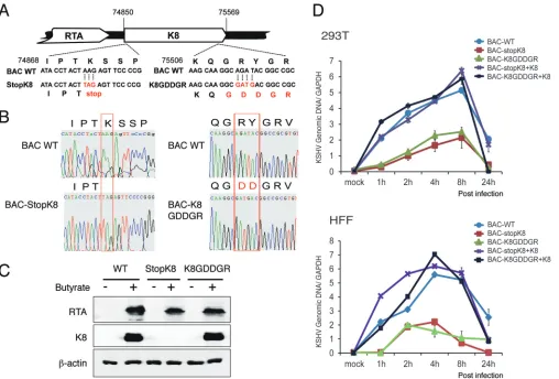
virus, we generated a recombinant virus with the K8 GRYGR motif mutated to GDDGR
in the bacterial artificial chromosome (BAC)-cloned KSHV (BAC16) genome (38, 39)
using two-step lambda Red-mediated seamless recombination (40), which is
desig-nated BAC-K8GDDGR (Fig. 6A). A BAC-stopK8 mutant was also generated as a control
(13). Restriction enzyme digestion (data not shown) and sequencing confirmed that the
mutants had the desired sequences (Fig. 6B). BAC-cloned viral genomic DNAs were
introduced into 293T cells. After validation of the expression of the viral proteins RTA
and K8 in 293T cells at 48 h postinduction (Fig. 6C), virions from the cells infected with
the BAC-cloned viruses (wild-type BAC16, BAC-stopK8, and BAC-K8GDDGR) were
pre-pared at 5 days postinduction. The wild-type and mutant viruses were used to infect
293T cells and human foreskin fibroblasts (HFF) at a multiplicity of infection (MOI) of 50
(viral genomic DNA copy equivalents). Viral DNA contents of infected 293T cells and
HFF with different BAC-cloned viruses were determined at different time points
postin-fection using quantitative PCR (qPCR) analysis. As expected, BAC-stopK8 virus could not
support viral abortive DNA replication in both types of cells, but the lost DNA
replica-tion activity could be restored by ectopic expression of K8 (Fig. 6D) (13). Similarly, viral
DNA replication of BAC-K8GDDGR decreased significantly in comparison to that of
wild-type virus, suggesting that the function of K8 in abortive lytic replication during
primary infection is dependent on its RNA binding activity.
FIG 3Legend (Continued)
their truncation mutants were synthesizedin vitroand labeled with biotin. Anin vitroRNA pulldown assay was performed to evaluate the abilities of the RNAs and their mutants to be bound by K8. Biotinylated transcripts were mixed with purified GST-K8 protein, and the RNA-protein complexes were precipitated with streptavidin-coupled Dynabeads. The precipitates were analyzed by Western blotting for RNA-bound GST-K8 using anti-GST antibody and by Northern blotting for RNAs using the chemiluminescent nucleic acid detection module.
FIG 4Cellular RNAs identified through K8 CLIP-seq. (A) Percentages of cDNAs from K8 –CLIP-seq that mapped to different types of RNAs. UTR, untranslated region. (B) List of K8-associated cellular noncoding RNAs that were identified through K8 –CLIP-seq and their known functions. RPKM, reads per kilobase of transcript per million mapped reads.
on November 6, 2019 by guest
http://jvi.asm.org/
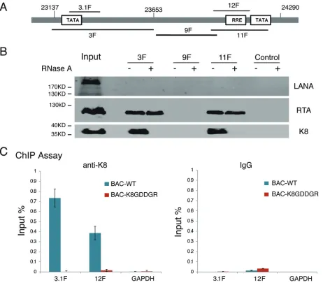
[image:10.585.44.477.66.289.2]K8 binds to ori-Lyt DNA in an RNA-dependent manner.
Following the
observa-tion that the RNA binding activity of K8 is important for viral DNA replicaobserva-tion, we
explored the mechanism underlying this K8 function. In KSHV ori-Lyt-dependent DNA
replication, K8 and RTA recruit six core replication proteins and other cofactors to
ori-Lyt DNA (15, 18, 20). Although K8 binds to the ori-Lyt core region (15–17, 20), this
DNA binding was found to be indirect through piggybacking on another component(s)
(15, 34). Therefore, we wondered whether RNA contributes to the association of K8 with
ori-Lyt DNA. To answer this question, we first adapted a DNA affinity assay (17, 20) to
examine the interaction between K8 and the ori-Lyt core domain. Three overlapping
DNA fragments representing the core domain of KSHV ori-Lyt labeled with biotin at
their 5
=
ends were coupled to streptavidin-conjugated magnetic beads and incubated
with nuclear extract derived from TPA-induced BCBL-1 cells (Fig. 7A). The
affinity-purified materials were then analyzed by Western blotting using antibodies against
LANA, RTA, and K8. As expected, both RTA and K8 were able to bind to 3F and 11F
fragments (RTA-responsive element [RRE] and TATA box), but not 9F, while LANA did
not interact with ori-Lyt DNA at all (20, 41) (Fig. 7B). However, we found that treatment
with RNase A during the DNA affinity assay abolished the association of K8 with ori-Lyt
but did not affect RTA binding to ori-Lyt DNA (Fig. 8B), suggesting that the interaction
between K8 and ori-Lyt DNA is mediated by certain RNAs.
This notion was further tested using mutant K8 with an RNA binding defect
(K8GDDGR) in a chromatin-immunoprecipitation (ChIP) assay in the context of virus.
293T cells carrying BAC16 (wild-type K8) and BAC-K8GDDGR mutant viruses were
induced for reactivation and treated with formaldehyde for cross-linking at 48 h
postinduction. Fragmented chromatin was immunoprecipitated with anti-K8 antibody
or IgG, and K8-bound DNAs were quantified by qPCR. The results showed that K8
bound to ori-Lyt DNA (3F and 11F regions), but not to GAPDH DNA, while mutant
K8 in BAC-K8GDDGR failed to associate with ori-Lyt DNA (Fig. 7C), suggesting that K8
FIG 5Mapping of K8 for the RNA binding domain. (A) Schematic presentation of K8 truncation, deletion, or amino acid substitution mutants that were used for mapping the RNA binding domain. The binding properties with T1.4, PAN, MRP, and 7SK RNAs are shown on the left of each construct. NLS, nuclear localization signal. GRXGR is the consensus RNA binding motif of the DEAD box protein. The symbols⫹⫹,⫹, and⫺represent strong, moderate, and no binding, respectively. (B) Expression vectors of Myc-K8 and mutants were introduced into 293T cells. Cell lysates were prepared and mixed with streptavidin beads coated with biotinylated RNA. The RNA pulldown materials were analyzed by Western blotting with an anti-Myc antibody. Shown are the results from the 7SK RNA pulldown assay. IB, immunoblotting. (C) Effects of GDDG mutation on binding of K8 with hnRNP U determined by immunoprecipitation with anti-Myc antibody and Western blotting using anti-Flag antibody.
Coordinate Regulation by K8 and Noncoding RNA Journal of Virology
on November 6, 2019 by guest
http://jvi.asm.org/
[image:11.585.56.545.73.317.2]binding to ori-Lyt DNA is mediated by RNA(s) that associates with K8 through the
GRYGR motif.
K8 binding to ori-Lyt DNA is mediated by virus-borne T1.4 RNA.
Among the
three virus-borne RNAs identified as encoding K8 binding protein in our CLIP-seq, two,
namely, T1.4 and T0.7 RNAs, are ori-Lyt-associated RNAs (16, 17). Previous studies
demonstrated that T1.4 RNA is absolutely essential for KSHV ori-Lyt-dependent DNA
replication (16, 17) and is expressed at a very high level during KSHV
de novo
infection
(11). Therefore, we asked if this RNA also plays a role in viral DNA replication during
de
novo
infection and, furthermore, whether T1.4 RNA is the RNA that mediates the K8
association with ori-Lyt DNA. To address these questions, a recombinant KSHV with the
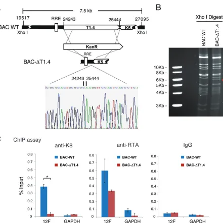
ori-Lyt (L)-associated T1.4 RNA locus deleted from the BAC16-cloned KSHV genome was
constructed and designated BAC-ΔT1.4. The mutant virus was verified by restriction
enzyme digestion and sequencing (Fig. 8A and B). The deletion did not affect viral gene
expression (LANA, RTA, and K8) in lytic replication, but viral DNA replication and virion
production were depleted, consistent with a previous study (data not shown). 293T
cells carrying BAC16 wild-type and BAC-ΔT1.4 viruses were used for ChIP assay with
anti-K8 and anti-RTA antibodies. The results showed that K8 could not bind to ori-Lyt
FIG 6Mutation in the RNA binding motif GRYGR abolishes KSHV DNA replication in the viral context. (A) Schematic diagrams of the structures of K8 in BAC-stopK8 and K8-GDDGR recombinant viruses. The nucleotide sequences refer to GenBank accession numberU75698.1. The mutated sequences on the stop codon and the GRYGR motif are indicated in red. (B) Sequence chromatograms of the clones. The mutations from AAG to the stop codon TAG in BAC-stopK8 and from GRYGR to GDDGR in BAC-K8GDDGR are boxed. (C) Expression of RTA and K8 genes in the wild-type and mutant viruses. (D) Viral DNA contents in 293T cells and HFF that were primarily infected by BAC16 (WT), BAC-stopK8, and BAC-GDDGR at an MOI of 50 (viral genomic DNA equivalents). A complementary experiment with ectopic expression of K8 was performed to examine whether the defects caused by K8 mutation could be rescued by wild-type K8. 293T cells and HFF were transfected with K8 expression vector (pCR3.1-K8␣). Twenty-four hours posttransfection, the cells were infected with BAC16 (WT), BAC-stopK8, and BAC-GDDGR viruses. At different time points postinfection, total DNA was isolated, and viral genomic DNA was quantified by qPCR with primers specific to ORF73. Viral DNA copy numbers were calculated from a standard graph generated using known concentrations of BAC16 DNA. The error bars indicate standard deviations (SD).
on November 6, 2019 by guest
http://jvi.asm.org/
[image:12.585.43.551.70.414.2]DNA without T1.4 in BAC-ΔT1.4. In contrast, RTA was able to bind to ori-Lyt regardless
of the presence of T1.4 RNA in BAC16 and BAC-ΔT1.4 cells (Fig. 8C), indicating that the
interaction between K8 and ori-Lyt DNA is indeed T1.4 RNA mediated.
Then, we further investigated how T1.4 mediates K8 binding to ori-Lyt DNA.
Sequencing alignment identified potential base pairing between T1.4 RNA and ori-Lyt
DNA (Fig. 9A). This led to the hypothesis that T1.4 associates with the viral genome
through RNA-DNA base pairing and recruits K8 to ori-Lyt DNA. To test this hypothesis,
a chromatin isolation by RNA purification (ChIRP) assay (42) was designed (Fig. 9B). In
brief, TPA-induced BCBL-1 cells were cross-linked with 1% glutaraldehyde, and
chro-matin was sheared by sonication to the size range of 100 to 500 bp. Biotinylated probes
complementary to T1.4 and LacZ RNAs (a negative control) were added to the
chro-matin preparation. T1.4-associated chrochro-matin fragments were purified with streptavidin
beads, and T1.4-bound DNA was quantified by qPCR analysis. To validate the T1.4 probe
for specific RNA enrichment, a ChIRP assay was performed with T1.4 and LacZ probes.
The precipitated RNAs were subjected to RT-qPCR with T1.4 primers, and the result
FIG 7Association of K8 with ori-Lyt DNA is mediated by RNA. (A) Schematic illustration of the KSHV ori-Lyt core domain and DNA fragments that were used in the DNA affinity assay. (B) Three biotinylated ori-Lyt DNA fragments and an irrelevant DNA fragment from the ORF45 coding region as a control were prepared by PCR, conjugated on magnetic beads, and incubated with TPA-induced BCBL-1 nuclear extract with and without treatment with RNase A. After washing, samples were assayed by Western blotting with antibodies as indicated. (C) Binding of K8 to ori-Lyt DNA was determined in BAC16 (BAC WT) and BAC-K8GDDGR by ChIP assay with anti-K8 antibody. The positions of the amplicons (3.1F and 12F) are shown in panel A. The error bars indicate SD.
Coordinate Regulation by K8 and Noncoding RNA Journal of Virology
on November 6, 2019 by guest
http://jvi.asm.org/
[image:13.585.42.504.70.475.2]showed the T1.4 RNA level was 1,700-fold higher in the preparation with T1.4 probe
than in that with LacZ probe, indicating that the T1.4 probe can specifically enrich T1.4
RNA (Fig. 9C). Then, two sets of T1.4 probes (odd and even) were used for two
independent ChIRP experiments. Both sets of probes gave rise to similar results
showing that T1.4 associates with ori-Lyt chromatin (Fig. 9D). Furthermore, treatment
with RNase H after chromatin pulldown greatly reduced the association between T1.4
RNA and ori-Lyt DNA in the preparation (Fig. 9D). In contrast, treatment with trypsin did
not reduce the interaction much. Taken together, these results indicate that T1.4 RNA
associates with ori-Lyt DNA through RNA-DNA base pairing rather than mediation by
protein(s).
T1.4 is essential for KSHV abortive lytic replication following
de novo
infection.
Using the BCBL-1 KSHV reactivation system, we have shown that T1.4 RNA associates
FIG 8Binding of K8 to ori-Lyt DNA is mediated by T1.4 RNA. (A) Schematic diagrams of the T1.4 locus in the KSHV genome and strategy for construction of BAC-ΔT1.4. The nucleotide sequences refer to GenBank accession numberU75698.1. The sequence chromatogram of the deletion junction in BAC-ΔT1.4 is shown. (B) Electrophoretic analysis of the BAC-ΔT1.4 mutant viral genomes. BAC16 (BAC WT) and BAC-ΔT1.4 DNAs were digested with XhoI, resolved on a 0.8% agarose gel, and stained with ethidium bromide. The red arrow indicates the changes of restriction pattern caused by the deletion of T1.4 in BAC-ΔT1.4. (C) BAC16 and BAC-ΔT1.4 were introduced into 293T cells, and the cells were induced with sodium butyrate for 48 h. A ChIP assay was performed with anti-K8 and anti-RTA antibodies or IgG and detected by PCR with primers for the 12F region or GAPDH. The data are shown as the means⫾SD of the results of three separate experiments.*,P⬍0.05.
on November 6, 2019 by guest
http://jvi.asm.org/
[image:14.585.51.492.65.507.2]with ori-Lyt DNA and facilitates K8 binding to the KSHV genome. However, whether
T1.4 functions in viral DNA replication during KSHV
de novo
infection needs to be
confirmed. The BAC-ΔT1.4 mutant virus does not produce infectious virions and
therefore cannot be used to study viral
de novo
infection. Therefore, we generated a
T1.4 conditional expression system in which the RRE in the T1.4 promoter was replaced
by a
Saccharomyces cerevisiae
Gal4 binding motif (GATCCGGAGGACTGTCCTCCGG) (Fig.
10B). Thus, RTA could no longer bind to ori-Lyt to transcribe T1.4. However, the T1.4
transcription and ori-Lyt-dependent DNA replication could be restored by ectopic
expression of an RTA-Gal4 chimeric protein (Fig. 10A) (17). The recombinant mutant
virus, termed BAC-Gal4, was shown to be unable to support T1.4 transcription, and
overexpression of RTA was barely able to induce T1.4 transcription. However, ectopic
expression of RTA-Gal4 chimeric protein in the cells restored the transcription of T1.4,
especially when combined with sodium butyrate treatment (Fig. 10C), as well as virion
production (data not shown). BAC16 and BAC-Gal4 virions were prepared and used to
infect 293T cells and HFF at an MOI of 50 (viral genomic-DNA equivalents). Viral
genomic content in the infected cells at different time points was determined by qPCR.
In contrast to BAC16 wild-type virus (in that viral DNA replication continued for 4 h
[13]), BAC-Gal4 virus displayed much lower viral DNA copy numbers in both types of
infected cells, indicating that T1.4 was essential for viral abortive lytic replication after
de novo
infection. However, in the presence of RTA-Gal4 protein, viral DNA replication
was recovered (Fig. 10D). To rule out the possibility that the lower DNA replication in
the mutant virus was caused by the infection rate, we compared infectivity between
BAC-WT and BAC-Gal4 recombinant viruses by detection of green fluorescent protein
(GFP)-expressng cells with a fluorescence microscope 24 h postinfection. The results
FIG 9T1.4 RNA associated with ori-Lyt DNA. (A) Alignment of T1.4 RNA with ori-Lyt DNA sequences in the KSHV genome revealing potential base pairing between them. (B) Schematic illustration of the ChIRP assay procedure. Glutaraldehyde was applied to induce DNA-RNA cross-linking. Chromatin was sonicated and then incubated with biotinylated oligonucleotide probe specifically to the target RNA. Streptavidin beads were added to pull down the biotinylated RNA and bound DNA. qPCR was used to detect the ori-Lyt DNA. (C) Validation of the efficiency of T1.4 probe in pulldown of T1.4 RNA. A ChIRP assay was performed using probes targeting T1.4 or LacZ. Precipitated RNAs were purified and subjected to RT-qPCR with primer against T1.4. (D) T1.4 RNA associates with ori-Lyt DNA. Two sets of probes were used to perform ChIRP experiments. RNase H, RNase A, or trypsin was added in washing buffer during the experiments to digest the RNA or protein. The pulled down DNA was amplified by primers for the 12F region of ori-Lyt. The error bars indicate SD.
Coordinate Regulation by K8 and Noncoding RNA Journal of Virology
on November 6, 2019 by guest
http://jvi.asm.org/
[image:15.585.38.498.68.360.2]FIG 10T1.4 RNA is essential for KSHV DNA replication followingde novoinfection. (A) Schematic presentation of the experimental design for conditional expression of T1.4 to demonstrate its role in viral DNA replication using RTA-Gal4 mutant virus. In WT virus, RTA binds the RRE to induce the expression of T1.4 RNA and lytic replication, followed by virion release. Duringde novoinfection, RTA binds to the RRE to induce T1.4 RNA for the initiation of DNA replication. In BAC-Gal4 mutant virus, the RRE in the T1.4 promoter has been
(Continued on next page)
on November 6, 2019 by guest
http://jvi.asm.org/
[image:16.585.41.465.65.687.2]showed that the infectivities of these viruses were similar, indicating that T1.4 does not
involve virion attachment or entry (Fig. 10E). Taken together, these data showed that
T1.4 expression and association with ori-Lyt DNA are critical for KSHV DNA replication; T1.4
RNA mediates the interaction of K8 and the viral genome and, as a result, facilitates the
assembly of the viral replication complex and DNA replication during
de novo
infection.
DISCUSSION
In this study, we investigated the functional role of K8 in the KSHV life cycle. The
salient features of the outcomes are as follows. (i) The study discovered that K8 is an
RNA binding protein and that the majority of K8-associated RNAs are noncoding RNAs,
both viral and cellular. This suggests that some of the functions of K8 in the viral life
cycle may be carried out in coordination with noncoding RNAs. (ii) A function of K8 in
viral DNA replication following KSHV
de novo
infection (abortive lytic replication) was
studied in detail, which led to the conclusion that viral DNA replication following
de
novo
infection initiates at an ori-Lyt and relies on coordinated actions of K8 and the
noncoding RNA T1.4.
Unique characteristics and roles of K8 in the KSHV life cycle as an RNA binding
protein.
K8 is a structural and positional homolog of EBV ZTA (31, 43). However, unlike
ZTA, which is a transcription activator and is capable of initiating an EBV reactivation
cascade (44–46), K8 has no transcriptional activity and is not able to initiate viral lytic
replication. Instead, K8 is a transcriptional repressor that affects the expression of a
subset of viral and cellular genes (22, 23). Unlike EBV ZTA, which binds directly to ZRE
DNA within the ZTA promoter (47) and other promoters (45) to regulate gene
expres-sion, K8 does not directly bind to viral genomic DNA (15, 34). However, K8 was found
to bind to the origin of viral lytic DNA replication (ori-Lyt) of KSHV in an indirect manner,
and the binding is absolutely required for viral DNA replication (15, 34). In addition, K8
causes cell cycle arrest at the G
1phase through induction of C/EBP
␣
and p21 (48).
Therefore, K8 is a multifunctional protein with its own characteristics. In this study, the
unique characteristics of K8 as an RNA binding protein were unveiled, and some of the
functions of K8 were found to be executed in coordination with its associated RNAs.
K8-associated RNAs, which include both viral and cellular RNAs, were identified
using a CLIP-seq approach. Interestingly, 49% of the K8 binding RNAs are intronic and
intergenic sequences, and 11% are annotated noncoding RNAs, suggesting K8
pre-dominately associates with noncoding RNAs. Identification of these noncoding RNAs
has provided some clues for us in searching for mechanisms underlying K8 functions in
the viral life cycle. For example, K8 is known to function as a global gene expression
repressor, but its mechanism remains unknown. Among K8-associated noncoding
RNAs, 7SK RNA has also been reported to be a transcriptional repressor. It inhibits gene
expression through inactivating positive transcription elongation factor b (P-TEFb) (49).
It was shown that P-TEFb–7SK RNA interaction is regulated by hnRNP K, as depletion of
hnRNP K increases the binding of 7SK RNA to P-TEFb (50). It can be speculated that K8
may repress global gene expression through modulating the dynamics of the P-TEFb–
7SK complex and strengthening the inhibition of P-TEFb. Interaction of K8 with PAN
RNA may suggest that K8 represses RTA-mediated transcription of viral delayed-early
genes (22, 23), possibly through targeting PAN RNA and blocking its function in
FIG 10Legend (Continued)
replaced with a Gal4 binding sequence. T1.4 expression is induced by ectopic expression of RTA-Gal4 fusion protein, and BAC-Gal4 mutant virus is produced. When BAC-Gal4 virions infect fresh cells, RTA is unable to bind to the Gal4 motif in the T1.4 promoter, so no T1.4 is transcribed. (B) Construction of BAC-Gal4, where the RRE sequence in ori-Lyt has been replaced with a Gal4 binding motif. The mutant virus was confirmed by sequencing. (C) Validation of the expression of T1.4 RNA in BAC16 (BAC WT) and BAC-Gal4. (D) BAC-Gal4 does not support viral DNA replication followingde novoinfection. 293T cells and HFF were infected by BAC16 (WT) or BAC-Gal4 at an MOI of 50 (viral genomic DNA equivalents). To induce the expression of T1.4 in BAC-Gal4 virus, 293T cells and HFF were transfected with Gal4-RTA expression vector. Twenty-four hours posttransfection, the cells were infected with BAC16 and BAC-Gal4 viruses. The viral DNA content was determined at different time points postinfection by qPCR analysis. The viral genomic DNA copy number was normalized by GAPDH. The error bars indicate SD. (E) The infectivity of BAC-WT and BAC-Gal4 viruses was examined 24 h postinfection by GFP expression (green fluorescence) under a fluorescence microscope at⫻5 magnification.
Coordinate Regulation by K8 and Noncoding RNA Journal of Virology
on November 6, 2019 by guest
http://jvi.asm.org/
recruiting histone demethylases (UTX and JMJD3) to the gene loci (51, 52). These
hypotheses warrant future investigation.
The basic region of K8 is an RNA binding domain.
Based on its homology to EBV
ZTA, K8 has been proposed to be divided into several functional domains: a
transcrip-tion activatranscrip-tion domain at the N terminus (aa 1 to 121), a DNA binding domain (aa 121
to 189) that contains a basic region (aa 169 to 185), and a leucine zipper domain at the
C terminus (aa 191 to 237) (31, 43, 53). The leucine zipper domain has been known to
be essential for certain functions of K8, including homodimerization of K8 (31),
asso-ciation with RTA (22, 23), and interaction with histone modification proteins JMJD2A
and histone deacetylase (HDAC) (24). In the current study, we demonstrated that the
basic region within the previously designated DNA binding domain is actually
respon-sible for RNA binding. Further analysis identified a GRYGR motif (aa 173 to 177) that is
required for RNA binding. Interestingly, this motif appears to be a conserved RNA
binding motif (motif VI) of the DEAD box protein family (Fig. 5A), and such motifs in
other DEAD box family proteins have been shown to be involved in ATPase activity and
RNA binding (35–37). When the GRYGR motif in the K8 protein was mutated to GDDGR,
the mutant K8 completely lost the ability to bind to the RNAs that we tested (T1.4, PAN,
MRP, and 7SK RNAs), as well as viral DNA replication, demonstrating the importance of
this motif, as well as the RNA binding property of K8 in the viral life cycle. Thus, this
region should be named the RNA binding domain instead of the DNA binding domain,
as K8 has never been demonstrated to directly bind to DNA.
The GRYGR motif was found to be responsible for binding of K8 to the diverse
noncoding RNAs (T1.4, PAN, MRP, and 7SK RNAs), suggesting that K8 binds to these
RNAs through the same RNA recognition mode. A structure analysis of the K8 basic
domain with different noncoding RNAs is warranted to reveal the K8 RNA binding
mechanism. Since K8 is known as a multifunctional protein, we may further hypothesize
that K8 executes different cellular functions through coupling with different RNAs. In
the current study, we demonstrated that the function of K8 in viral DNA replication is
carried out in coordination with T1.4 RNA. We expect that the functions of K8 in
association with PAN and 7SK RNAs will be revealed soon and that it is possibly related
to K8-mediated global gene repression. In addition, other functions of K8 have been
reported to be associated with the K8 basic domain. It was shown that the basic region
is required for cell cycle arrest and cell growth control. Deletion of aa 121 to 189 from
the K8 protein resulted in loss of the activity of cell cycle regulation (26). Furthermore,
K8 SUMOylation at lysine 158, which was reported to be crucial for the gene repression
function of K8, is dependent on the basic domain, as K8 could not be SUMOylated
when the basic region was deleted (54). It is worthwhile to discover if the GRYGR
mutation affects these events (cell cycle and K8 SUMOylation) and to identify the RNAs
that bind to K8 and are responsible for these functions.
KSHV DNA replication following
de novo
infection initiates at the ori-Lyt and
requires coordinated functions of K8 protein and T1.4 RNA.
Besides latent and lytic
viral DNA replication, there is a third phase of DNA replication in herpesviruses. After
a herpesvirus particle enters a host cell, the virus undergoes DNA replication that
results in accumulation of the viral genome to 50 to 100 copies per cell prior to
establishment of latency. There is no virion particle produced in this process, so it is
called abortive lytic replication. Using K8-null recombinant virus, we previously
dem-onstrated a role of K8 in initial viral DNA replication following
de novo
KSHV infection.
K8-null viruses exhibit much lower viral genome copy numbers than wild-type viruses
(13). However, little is known regarding the mode of abortive viral replication and the
regulation mechanism. Where does the abortive lytic DNA replication initiate, ori-Lyt,
the origin of plasmid replication (ori-P), or another locus in the viral genome? In this study,
our data confirmed that KSHV abortive lytic DNA replication initiates at ori-Lyt. In addition,
we found that T1.4 noncoding RNA and its mediated K8 binding to ori-Lyt DNA are essential
for abortive lytic DNA replication. RTA and its association with ori-Lyt DNA are also critical
for DNA replication. These results demonstrate a novel mechanism that regulates DNA
on November 6, 2019 by guest
http://jvi.asm.org/
replication through coordinated actions of a protein and a noncoding RNA, presenting a
new paradigm of eukaryotic DNA replication.
Potential role of T1.4 RNA in KSHV ori-Lyt-dependent DNA replication and a
possible mechanism.
T1.4 is highly expressed during
de novo
infection (11), and its
transcription is essential for ori-Lyt-dependent DNA replication (17). In this study, using
a Gal4-RTA conditional T1.4 expression system, we demonstrated that KSHV abortive
lytic DNA replication is dependent on the expression of T1.4. BAC-Gal4 recombinant
virus was unable to initiate DNA replication during
de novo
viral infection, but the
defect could be rescued by ectopic expression of Gal4-RTA, indicating that T1.4
noncoding RNA plays a critical role in ori-Lyt-dependent DNA replication in KSHV
primary infection. In addition, our
in vitro
assay showed that T1.4 mediates K8 binding
to ori-Lyt DNA, presenting a new paradigm for noncoding RNA regulating a biological
process by mediating protein-DNA interaction.
The mechanism of T1.4 mediation of K8 binding to ori-Lyt DNA has not been fully
elucidated. One model is that T1.4 may function as a guide RNA, bringing its associated
protein (K8) to DNA by recognizing the DNA sequence through base pairing. Potential
base pairing between T1.4 and ori-Lyt DNA was proposed, and an RNase H assay
provided supporting evidence for this model (Fig. 9C and D). Another model is that T1.4
may form a G-quadruplex (G4) structure that associates with ori-Lyt DNA and recruits
proteins to the DNA. T1.4 RNA is a noncoding RNA with high GC content. QGRS
(quadruplex-forming G-rich sequences) Mapper (55) (
http://bioinformatics.ramapo.edu/
QGRS/index.php
) predicts that T1.4 is a potential G-quadruplex RNA. G quadruplexes
are noncanonical secondary structures found in guanine-rich regions of DNA and RNA.
There are more than 370,000 G-quadruplex-forming sequences in the human genome
(56). G-quadruplex structure has also been found in the genomes of many viruses,
including HIV-1 (57), KSHV (58), and EBV (59). In EBV latent DNA replication, EBNA1
binds to ori-P and recruits the cellular origin recognition complex (ORC). The interaction
between EBNA1 and the ORC is mediated by a noncoding G-rich RNA, which is
predicted to possess a G4 structure and which forms a stable complex with EBNA1 and
the ORC (60). By treatment with a G-quadruplex-interacting compound, BRACO-19,
EBNA1-dependent viral latent DNA replication was inhibited (59). In KSHV, it was also
shown that treatment of latently infected cells with a quadruplex-interacting
com-pound, PhenDC3, led to loss of KSHV episomes (58). Whether the function of T1.4 in
binding to ori-Lyt and recruiting K8 is dependent on its G-quadruplex structure
needs further investigation. If the G-quadruplex hypothesis is proven to be true for
T1.4 RNA, it will inform a novel therapeutic strategy to block KSHV primary
infection, and possibly lytic replication, as well, using small molecular compounds
that interrupt RNA G-quadruplex formation.
MATERIALS AND METHODS
Cell culture.BCBL-1, a primary effusion lymphoma cell line that carries latently infected KSHV (5), and BJAB, a KSHV-free Burkitt lymphoma B cell line, were maintained in RPMI 1640 medium with 10% heat-inactivated fetal bovine serum (FBS). Human embryonic kidney (HEK) 293T cells, obtained from the ATCC, and HFF (HFF2441), kindly provided by Meenhard Herlyn at the Wistar Institute, were cultured in Dulbecco modified Eagle medium (DMEM) supplemented with 10% FBS. All cultures contained penicillin-streptomycin (50 U/ml) and amphotericin B (1.25g/ml).
Antibodies.Mouse anti-Myc tag, anti-Flag (M2), and anti--actin antibodies were purchased from Sigma-Aldrich. Rabbit anti-hnRNP U, anti-hnRNP K, and anti-hnRNP C antibodies were purchased from Abcam. IRDye 680LT/800CW goat anti-rabbit IgG or anti-mouse IgG antibodies were purchased from Li-Cor Biosciences. The mouse monoclonal antibodies against K8 and ORF45 were generated and purified in our laboratory. Mouse monoclonal anti-RTA antibody was provided by Erle Robertson (University of Pennsylvania). Mouse monoclonal anti-LANA antibody was provided by Ke Lan (Wuhan University, China).
Co-IP assay.TPA-induced BCBL-1 cells (1⫻107) were collected and lysed in 1 ml of immunopre-cipitation (IP) buffer (50 mM Tris-HCl, pH 7.4, 150 mM NaCl, 1% NP-40, 30 mM sodium fluoride, 1 mM sodium orthovanadate [Na3VO4], 1 mM phenylmethylsulfonyl fluoride [PMSF], protease inhibitor cocktail) on ice for 30 min. The cell lysates were clarified by centrifugation at 4°C for 10 min. Immunoprecipitation was performed by addition of 2l of the antibodies of interest or IgG to cell lysates with gentle agitation at 4°C for 2 h. Then, 30 l of washed protein G-coated beads was added, and the mixtures were incubated at 4°C for 4 h. The beads were then washed four or five times with cold IP buffer (without
Coordinate Regulation by K8 and Noncoding RNA Journal of Virology
on November 6, 2019 by guest
http://jvi.asm.org/
protease inhibitor). To eliminate RNA-mediated or DNA-mediated protein interactions, RNase A (100
g/ml) and DNase I (100 U/ml) were added to washing buffer with 5 mM MgCl2. The precipitates were resuspended in 60l of SDS-PAGE loading buffer, boiled for 10 min, and then loaded onto SDS-PAGE gels for Western blot assay.
Western blotting.Cells were lysed with lysis buffer (50 mM Tris-HCl, pH 7.4, 150 mM NaCl, 1% NP-40, 30 mM sodium fluoride,1 mM Na3VO4, 1 mM PMSF, protease inhibitor cocktail [1 tablet in 50 ml lysis buffer]), homogenized for 30 min on ice, and centrifuged at 13,000 rpm for 10 min. The whole-cell extract was resolved by SDS-PAGE and transferred onto nitrocellulose membranes. The membranes were blocked in 5% nonfat milk in 1⫻Tris-buffered saline (TBS) for 1 h and incubated with diluted primary antibodies overnight at 4°C. IRDye 680LT/800CW goat anti-rabbit or anti-mouse antibody (Li-Cor Biosci-ences) was used as a secondary antibody. The blots were visualized in an Odyssey system (Li-Cor).
Mass spectrometric analysis.Proteins coimmunoprecipitated with K8 were resolved on 4 to 12% bis-Tris NuPAGE gels (Invitrogen) and stained with a Coomassie G-250 staining kit (Invitrogen). The protein bands were excised and subjected to trypsin digestion. A portion of the peptide digest was injected onto a nanocapillary reverse-phase high-performance liquid chromatograph coupled to a nanoelectrospray ionization source of an ion trap mass spectrometer (ThermoFinnigan LCQ). Mass spectrometry measured peptide masses and then fragmented individual peptides to produce liquid chromatography–tandem-MS (MS-MS) spectra of fragments that reflected the peptide sequence. The MS-MS spectra were run against a nonredundant sequence database using the program SEQUEST. The mass spectrometry was carried out in the protein microchemistry/mass spectrometry facility at the Wistar Institute.
Dot blotting.RNA was isolated with TRIzol reagent, and the RNA was biotin labeled with the Pierce RNA 3=end biotinylation kit (Thermo). Briefly, RNA was incubated at 85°C for 3 to 5 min to relax the secondary structure and chilled on ice. Biotinylated cytidine (Bis) phosphate was ligated to the 3=end of the RNA with T4 RNA ligase at 16°C for 4 h. The biotinylated RNA was purified with chloroform and precipitated with ethanol and glycogen. Biotin signal was detected with a chemiluminescent nucleic acid detection module (Thermo). Two microliters of RNA sample was spotted onto the nitrocellulose mem-brane. The membrane was dried and blocked with bovine serum albumin (BSA)-TBS with Tween 20 (TBST) and incubated with primary antibody against biotin in BSA-TBST for 30 min at room temperature. The membrane was incubated with secondary antibody conjugated with horseradish peroxidase (HRP) for 30 min at room temperature and then exposed to X-ray film.
CLIP.CLIP was performed as previously described (30, 61, 62). Briefly, 3⫻107TPA-induced BCBL-1 cells and BJAB cells were collected by centrifugation, followed by washing with ice-cold phosphate-buffered saline (PBS). The cells were irradiated at 150 mJ/cm2at 254 nm and then mixed and irradiated again. The cells were spun down and resuspended in 1 ml lysis buffer (50 mM Tris-HCl, pH 7.4, 100 mM NaCl, 1% NP-40, 0.1% SDS, 0.5% sodium deoxycholate, protease inhibitor cocktail). Two microliters of Turbo DNase (Ambion) was added to the cell lysate. The lysate was incubated on ice for 30 min and spun down at 20,000⫻gat 4°C for 10 min to clear the lysate. Two microliters (⬃2g) of the antibody of interest and 50l washed protein A Dynabeads were added per 500l lysates. Samples were rotated for 2 h at 4°C. The supernatant was discarded, and the beads were washed twice with 900l high-salt buffer (50 mM Tris-HCl, pH 7.4, 1 M NaCl, 1 mM EDTA, 1% NP-40, 0.1% SDS, 0.5% sodium deoxycholate). Then, the beads were washed again twice with 900l washing buffer (20 mM Tris-HCl, pH 7.4, 10 mM MgCl2, 0.2% Tween 20) with different amounts of RNase A (high RNase concentration, 2g/ml; moderate RNase concentration, 1g/ml; and low RNase concentration, 0.2 g/ml) and an additional 900 l RNase-free washing buffer twice. The RNAs were dephosphorylated at the 3=ends. Linker RNA 3=adapter (Table 2) was ligated to the 3=ends of the RNAs, and [␥-32P]ATP-labeled linker RNA 5=adapter (Table 2) was ligated to the 5=ends of the RNAs. The protein-RNA complexes were resolved on a 4 to 12% NuPAGE Bis-Tris gel and transferred to a nitrocellulose membrane. K8-RNA complexes were cut and subjected to proteinase K digestion. The RNAs were purified with TRIzol and subjected to RT-PCR. Sequencing was performed on an Illumina HiSeq 2500 sequencer at the Beijing Genomics Institute (BGI).
RNA pulldown assay.To synthesize biotinylated transcripts (7SK, MRP, PAN, and T1.4 RNAs)in vitro, DNA templates were prepared by PCR using forward primers that contained the T7 RNA polymerase promoter sequence. Biotinylated transcripts were synthesized using MEGAscript kits (Thermo Fisher) with biotin-14-CTP (Thermo Fisher). Since T1.4 RNA contains high-GC-repeat sequence, biotinylated T1.4 was generated by adding biotin at the 3=end ofin vitro-transcribed T1.4 RNA with the Pierce biotin 3=end DNA-labeling kit (Thermo Fisher). The expression vectors for K8 and its mutants were transfected into 293T cells, and cell lysates were prepared 48 h posttransfection in lysis buffer (150 mM KCl, 25 mM Tris-HCl, pH 7.4, 0.5 mM dithiothreitol [DTT], 0.5% NP-40, RNase inhibitor, and protease inhibitor cocktail). Two micrograms of biotin-labeled RNAs and 2l yeast tRNA were added to 1 ml cell lysate (or purified GST-K8 protein fromE. coliBL21) and incubated with rotation at 4°C for 1 h. Complexes were isolated with streptavidin-coupled Dynabeads, and the RNA-bound proteins were detected by Western blotting. Biotinylated RNAs could be detected by Northern blotting with the chemiluminescent nucleic acid detection module (Thermo Fisher Scientific).
Genetic manipulation of BAC-cloned KSHV genome.The mutagenesis of BAC16 was performed using a recombineering system as described by Brulois et al. and Tischer et al. (38, 40). In brief, the Kan/I-SceI cassettes were amplified from plasmid pEPKan-S by PCR with oligonucleotides of interest for BAC mutants. The oligonucleotides used for PCR are listed in Table 2. The purified PCR fragments were transformed by electroporation into BAC16-containing GS1783 cells that had been induced at 42°C for 15 min (38). The recombinant clones were selected at 32°C on LB plates containing 34g/ml chloram-phenicol and 50g/ml kanamycin. Positive clones were analyzed by miniassay and restriction enzyme
on November 6, 2019 by guest
http://jvi.asm.org/
digestion. Confirmed clones were cultured with 1%L-arabinose, induced at 42°C again, and plated on LB plates containing 1%L-arabinose and 34g/ml chloramphenicol for secondary recombination. Colonies that survived on theL-arabinose plates were cultured with 34g/ml chloramphenicol alone and with both 34g/ml chloramphenicol and 50g/ml kanamycin. The kanamycin-sensitive clones were analyzed by restriction enzyme digestion, and proper mutations were further confirmed by DNA sequencing.
Quantification of viral genomic DNAs.Total DNAs were prepared from KSHV-infected cells with a GeneJet genomic DNA purification kit (Fermentas Life Sciences). KSHV genomic DNA was measured by qPCR, and genomic DNA copy numbers were calculated based on external standards of known concentrations of BAC16 DNA. The primers ORF73-LCN and ORF73-LCC were previously described by Krishnan et al. (63). The viral DNA copy numbers were normalized to glyceraldehyde-3-phosphate dehydrogenase (GAPDH) DNA.
[image:21.585.43.546.81.561.2]KSHV virion purification and primary infection.293T cells carrying BAC16 wild-type or mutant virus were induced by 3 mM sodium butyrate for 4 to 5 days. The culture supernatant was filtered through a 0.45-m filter and centrifuged at 100,000⫻gfor 1.5 h at 4°C. The pellet was resuspended in 1/100 volume of 1⫻PBS or DMEM and stored at⫺80°C until use. The infection process was previously described (13). Briefly, cells were incubated with concentrated virus in the presence of 4g/ml Polybrene
TABLE 2Oligonucleotides used in this study
Purpose Name of oligonucleotide Sequence (5=-3=)
qPCR KSHV ORF73-LCN CGCGAATACCGCTATGTACTCA
KSHV ORF73-LCC GGAACGCGCCTCATACGA GAPDH-F ACATCATCCCTGCCTCTAC GAPDH-R TCAAAGGTGGAGGAGTGG
Construction of plasmids hnRNP U-F gcgggatcccaccATGAGTTCCTCGCCTGTTAATGTA hnRNP U-R gggatccTCAATAATATCCTTGGTGATAATGCTGA GST-K8 F cggaattcCGATGCCCAGAATGAAGGACATAC GST-K8 R atagtttagcggccgctttTCAACATGGTGGGAGTGGC
ChIP assay 3.1F TAGGTGGGACCGTGAGCGACT
3.1R CCATAATCCTCTGCCCCGC
RRE 12F ACGGGCCTGGAATCTCGCCTCTGG RRE 12R ATGGGCGTAACCGTAGGACAAGCTG