Processivity Factor ORF59 by a Viral Kinase Modulates Its Ability To
Associate with RTA and
ori
Lyt
Maria E. McDowell, Pravinkumar Purushothaman, Cyprian C. Rossetto, Gregory S. Pari, Subhash C. Verma
Center for Molecular Medicine, Department of Microbiology & Immunology, University of Nevada, Reno, School of Medicine, Reno, Nevada, USA
ORF59 of Kaposi’s sarcoma-associated herpesvirus (KSHV) plays an essential role in viral lytic replication by providing DNA
processivity activity to the viral DNA polymerase (ORF9). ORF59 forms a homodimer in the cytoplasm and binds and
translo-cates ORF9 into the nucleus, where it secures ORF9 to the origin of lytic DNA replication (
ori
Lyt) in order to synthesize long
DNA fragments during replication. ORF59 binds to
ori
Lyt through an immediate early protein, replication and transcription
activator (RTA). Here, we show that viral kinase (ORF36) phosphorylates serines between amino acids 376 and 379 of ORF59
and replacement of the Ser378 residue with alanine significantly impairs phosphorylation. Although mutating these serine
resi-dues had no effect on binding between ORF59 and ORF9, viral polymerase, or ORF36, the viral kinase, it significantly reduced
the ability of ORF59 to bind to RTA. The results for the mutant in which Ser376 to Ser379 were replaced by alanine showed that
both Ser378 and Ser379 contribute to binding to RTA. Additionally, the Ser376, Ser378, and Ser379 residues were found to be
critical for binding of ORF59 to
ori
Lyt and its processivity function. Ablation of these phosphorylation sites reduced the
produc-tion of virion particles, suggesting that phosphorylaproduc-tion is critical for ORF59 activity and viral DNA synthesis.
K
aposi’s sarcoma (KS)-associated herpesvirus (KSHV), also
known as human herpesvirus 8 (HHV8), is a member of the
Gammaherpesvirus
family. KSHV is associated with KS as well as
several lymphoproliferative diseases, including primary effusion
lymphoma (PEL) and multicentric Castleman’s disease (MCD)
(
1
,
2
). Similar to other herpesviruses, KSHV establishes a lifelong
latency following a primary infection (
3
). During latency, the
KSHV genome is maintained as a nonintegrated episome tethered
to the host chromosome via viral latency-associated nuclear
anti-gen (LANA) protein, encoded by ORF73 (
4
). Limited numbers of
viral genes are expressed during latency, and replication of the
virus is dependent on host cell replication machinery (
5
). KSHV
latency can be disrupted by various stimuli, and disruption results
in the expression of various lytic genes and the production of
infectious virions. KSHV replication and transcription activator
(RTA), encoded by ORF50, is necessary and sufficient for the
ac-tivation of KSHV lytic replication and production of viral particles
(
6
–
8
). Several chemical inducers, such as 12-
O
-tetradecanoyl-phorbol-13-acetate (TPA) and sodium butyrate (NaB), have
suc-cessfully been used to trigger lytic replication (
9
).
In KSHV-induced malignancies, the majority of the tumor
cells are latently infected with the virus; however, a small
propor-tion (2 to 5%) of the latent tumor cells undergoes lytic replicapropor-tion
(
10
,
11
). This spontaneous reactivation is proposed to produce
homologs of cellular cytokines, which act in a paracrine manner
for tumor progression, as well as generation of virions for further
spread of infection (
10
). These cytokines are capable of promoting
tumorigenesis by activating pathways involved in inflammation,
angiogenesis, and enhanced proliferation, invasion, and
dissemi-nation of tumor cells (
12
–
23
). Consequently, the study of lytic
replication is essential not only for understanding the mechanism
of lytic DNA replication but also for developing new treatment
strategies for preventing KSHV-associated malignancies.
KSHV lytic replication requires eight viral proteins, including
ORF9 (DNA polymerase), ORF6 (single-stranded DNA binding
protein), ORF40/41 (primase-associated factor), ORF44
(heli-case), ORF56 (primase), ORF59 (processivity factor), ORF50
(RTA), and ORF K8 (K-bZIP) (
24
). RTA is the most important
protein required for the initiation of lytic replication, similar to
the requirements of other herpesviruses. RTA binds to the
C/EBP
␣
and the RTA response element (RRE) of the origin of lytic
replication (
ori
Lyt) and thereby initiates lytic replication through
transcription activation as well as recruitment of additional
fac-tors (
20
,
25
–
30
).
One of the factors recruited by RTA is the viral processivity factor,
ORF59. Rossetto et al. have shown that the binding of ORF59 to the
C/EBP
␣
binding motif within
ori
Lyt is crucial for its function and is
dependent on the presence of RTA (
31
). ORF59 forms a homodimer,
which translocates viral polymerase (ORF9) into the nucleus for
effi-cient synthesis (processivity function) of DNA fragments (
32
). The
ORF59 homolog in another herpesvirus, human cytomegalovirus
(hCMV), ppUL44, is phosphorylated by a viral Ser/Thr kinase,
ppUL97, which modulates ppUL44’s ability to localize to the nucleus
(
33
). BMRF1, the Epstein-Barr virus (EBV) processivity factor, is
phosphorylated by BGLF4 viral kinase within a hinge region-like
do-main known to be important for transmitting conformational
changes (
34
). As a result, BGLF4 phosphorylation enhances the
trans-activation activity of Zta (an RTA homolog) and the synergistic
acti-vation of lytic replication at
ori
Lyt (
35
). Similarly, ORF59 is a
phos-phoprotein which is phosphorylated by KSHV viral Ser/Thr kinase
(ORF36), but the consequences of this modification were not
deter-mined previously (
36
). Here, we show that ORF36 phosphorylates
Received17 December 2012Accepted5 May 2013
Published ahead of print15 May 2013
Address correspondence to Subhash C. Verma, [email protected]. Copyright © 2013, American Society for Microbiology. All Rights Reserved.
doi:10.1128/JVI.03460-12
on November 7, 2019 by guest
http://jvi.asm.org/
ORF59 primarily at Ser378, which is critical for ORF59’s ability to
bind to RTA and
ori
Lyt. Furthermore, replacing the phosphorylating
serines of these sites with alanines significantly reduced viral
produc-tion.
MATERIALS AND METHODS
DNA constructs. ORF36-Flag/Myc, ORF59-GST full-length, and ORF59-GST segments were constructed by PCR amplification and clon-ing usclon-ing a wild-type (wt) KSHV genome as the template. ORF59 deletion mutants were assembled from two PCRs: (i) one using sense ORF59 primer and antisense ORF59 mutant primer and (ii) a second using sense ORF59 mutant primer and antisense ORF59 primer. Mutants with spe-cific deletions were screened and confirmed by sequencing. ORF59 point mutants were constructed using a QuikChange Lightning Multi site-di-rected mutagenesis kit as disite-di-rected by the manufacturer (Agilent Technol-ogies Inc., Santa Clara, CA). These ORF59 deletion and point mutants were cloned into glutathione S-transferase (GST), pxi-hemagglutinin (HA), and pLVX-Flag vectors by PCR amplification. An ORF36 kinase-dead (kd) mutant (ORF36 K108Q) was generated in a two-step approach similar to the approach used for the ORF59 deletion mutants. The result-ing PCR product was ligated, transformed, and screened for the K108Q mutation. All primers used for cloning and targeted mutagenesis are listed inTable 1. Bacterial artificial chromosomes (BAC) containing the KSHV genome but lacking ORF59 (BAC36⌬ORF59) and plasmids ORF9-Flag, K8-Flag, and 8088sc were gifts from Gregory Pari (University of Nevada, Reno, NV).
Cell culture, transfection, and transduction by lentivirus.293T cells were grown in Dulbecco’s modified Eagle’s medium (DMEM) supple-mented with 10% bovine growth serum (HyClone, Logan, UT), 2 mM L-glutamine, 25 U/ml penicillin, and 25g/ml streptomycin. All cultures were incubated at 37°C in a humidified chamber supplemented with 5% CO2. Cells were transfected by the calcium phosphate method as previ-ously described (37), with minor modification. Briefly, 500l 1⫻HBS (140 mM NaCl, 0.75 mM Na2HPO4·2H2O, 25 mM HEPES, 5 mM KCl, 6 mM dextrose, pH 7.1) was added to DNA and the components were mixed. Thirty microliters of 2.5 M CaCl2was added to the solution and the components were mixed. After 20 min at room temperature, the so-lution was evenly added dropwise onto the 60 to 70% confluent 293T cells. For generating lentivirus, 10g of lentiviral vector, 7.5g CMV-dR8.2 packaging plasmid, and 2.5g pCMV-VSVG envelope plasmids were transfected into a 100-mm dish of 293T cells using 1⫻HBS. At 24 h posttransfection, virus was induced with 1 mM NaB in DMEM (with 5% bovine growth serum) with 100 mM HEPES for 10 h. The supernatant was collected three times thereafter at 12- to 16-h intervals. The supernatant was filtered through a 0.45-m-pore-size filter and ultracentrifuged at 25,000 rpm for 1.5 h using a Beckman Coulter Optima L-90K ultracentri-fuge (Beckman Coulter, Inc., Brea, CA). The virus pellet was resuspended in DMEM and added to the target cells.
[image:2.585.39.558.76.428.2]BACmid DNA was transfected with the Metafectene Pro reagent (Biontex Laboratories GmbH, San Diego, CA) due to the larger size of the plasmid and the fragility of the DNA. Ninety percent confluent cells on a 6-well plate were transfected with 5 to 10g of BACmid DNA in 200l
TABLE 1Sequences of primers used in this study
Clone or mutant Sensea Primer oligonucleotide sequence (5=–3=)
Clones
ORF36-pA3F/pA3 M S TTTGAATTCCGCCATGCGGTGGAAGAATGGAGAG
AS TTTGCGGCCGCGAAAACAAGTCCGCGGGTGTGGGG
pLVX-ORF59-Flag S TTTGAATTCTCCTGTGGATTTTCACTATGGGGTC
AS TCAGCGGCCGCGAAAACAAGTCCGCGGGTGTGGGG
ORF59-HA-pxi S TTTGAATTCCACCATGCCTGTGGATTTTCACTATGGG
AS TTTGGATCCTTAGCCCTAAGCGTAATCTGGAACATCGTATGGGTACCAA
CCCGGGACTTTACACAGTCT
ORF59-GST S TTTGGATCCATGCCTGTGGATTTTCACTAT
AS TTTGAATTCGTAGGAAATGGTGGTCCTGA
ORF59 1-132aa GST S TTTGAATTCCACCATGCCTGTGGATTTTCACTATGGG
AS TTTGAATTCGTAGGAAATGGTGGTCCTGA
ORF59 133-264aa GST S TTTGGATCCGGGGACAACCTCACCTCCACC
AS TTTGAATTCCCAACCCGGGACTTTACACAGTCT
ORF59 265-396aa GST S TTTGGATCCTTTACCCCCGGGCTGATTTGG
AS TTTGAATTCGTAGGAAATGGTGGTCCTGA
Mutants
ORF36-K108Q-pA3F S GTGTGCAGCAGTTTGATAGCCGCCGGG
AS AACTGCTGCACACACAGATCCTCGGAGA
ORF59-d304-318 (DM1) S CCGATCTGTGATACCGGACTCCAGGAGGCA
AS AGTCCGGTATCACAGATCGGTTTACCTCTA
ORF59-d333-349 (DM2) S GTCTCCGGATGAGGACGCTGCCTCTGTCAC
AS CAGCGTCCTCATCCGGAGACTCCAATTCCG
ORF59-d376-379 (DM3) S CAAGAGGCGCCAGTCCAGGGATCGTGGGAA
AS CCCTGGACTGGCGCCTCTTGTGAGGCCTCT
ORF59 S305A, S312A, S318A (PM1) AACCGATCTGTGGCAGCCGAGGGAGGCGAGTCTGCACAAAAG
GTTCCCGATGCAATACCGGACTCC
ORF59 S333A, S349A (PM2) TTGGAGTCTCCGGATGCACCCCCTCTCACACCA
ORF59 S376A, S378A, S379A (PM3) CCTCACAAGAGGCGCGCAGACGCGGCCCAGTCCAGGGATCGT
ORF59 S376A CCTCACAAGAGGCGCGCAGACTCGAGCCAGTCCAGGGATCGT
ORF59 S378A CCTCACAAGAGGCGCTCAGACGCGAGCCAGTCCAGGGATCGT
ORF59 S379A CCTCACAAGAGGCGCTCAGACTCGGCCCAGTCCAGGGATCGT
a
S, sense; AS, antisense.
on November 7, 2019 by guest
http://jvi.asm.org/
serum and antibiotic-free medium, after mixing with 6l Metafectene Pro reagent. The mixture was added onto the cells dropwise after 15 to 20 min of incubation at room temperature. The BACmid contained green fluorescent protein (GFP), which allowed efficient evaluation of transfec-tion efficiency.
Immunoprecipitation, Western blotting, and antibodies. Trans-fected cells were harvested, washed with ice-cold phosphate-buffered sa-line (PBS), and lysed in 0.5 ml ice-cold radioimmunoprecipitation assay (RIPA) buffer (1% Nonidet P-40 [NP-40], 10 mM Tris [pH 7.5], 2 mM EDTA, 150 mM NaCl) supplemented with protease inhibitors (1 mM phenylmethylsulfonyl fluoride, 1g/ml aprotinin, 1g/ml pepstatin, 1 g/ml leupeptin). Cell debris was removed by centrifugation at 13,000⫻
g(10 min and 4°C), and lysates were precleared by 1 h of rotation at 4°C with 30l of protein A-protein G-conjugated Sepharose beads. After approximately 5% of the lysate was saved for use as an input control, the protein of interest was captured by rotating the remainder of the lysate with 1g of the appropriate antibody overnight at 4°C. Immune com-plexes were captured with 30l of protein A-protein G-conjugated Sep-harose beads by rotating for 2 h at 4°C. The beads were pelleted and washed three times with RIPA buffer. Proteins immunoprecipitated for kinase assay were washed with RIPA buffer containing 300 mM NaCl to reduce any contaminating proteins. For Western blot assay, input lysates and immunoprecipitated (IP) complexes were boiled for 5 to 10 min in Laemmli buffer, resolved by SDS-PAGE, and transferred as per the man-ufacturer’s recommendation (Bio-Rad Laboratories). The nitrocellulose membrane was probed with appropriate antibodies, followed by incuba-tion with infrared dye-tagged secondary antibody, and viewed on an Od-yssey imager (LICOR Inc., Lincoln, NE).
The following antibodies were used: mouse anti-Flag (M2; Sigma-Aldrich, St. Louis, MO), rabbit anti-Flag (F7425; Sigma-Sigma-Aldrich, St. Louis, MO), mouse anti-RTA (mouse hybridoma), mouse anti-LANA (mouse hybridoma), rabbit anti-HA (6908; Sigma-Aldrich, St. Louis, MO), mouse anti-GST (A00014; GenScript Corp.), mouse anti-glyceraldehyde-3-phosphate dehydrogenase (anti-GAPDH; G8140; US Biologicals), and rabbit anti-Myc (SAB4300605; Sigma-Aldrich, St. Louis, MO).
Purification of GST fusion proteins.Escherichia coliBL21(DE3) cells were transformed with the plasmid constructs for each GST fusion pro-tein. Bacterial culture was incubated until the optical density at 600 nm (OD600) was approximately 0.6, at which time the cultures were induced with 1 mM isopropyl--D-thiogalactopyranoside (IPTG) for 4 h at 37°C. The bacteria were pelleted, washed once with 5 ml STE buffer (100 mM NaCl, 10 mM Tris, 1 mM EDTA, pH 7.5), resuspended in 5 ml NETN buffer (0.5% NP-40, 100 mM NaCl, 20 mM Tris, 1 mM EDTA, pH 8.0), supplemented with protease inhibitors, and incubated on ice for 15 min. A volume of 75l of 1 M dithiothreitol (DTT) and 900l of a 10% solution of Sarkosyl in STE buffer were added, and the suspension was sonicated on ice (for 2 min at 40% amplitude with a 20-s-on and 20-s-off sonication cycle) to lyse the cells. The lysates were centrifuged (13,000⫻g, 10 min, 4°C) to separate the unsolubilized fraction. Clear supernatant was transferred to a fresh tube, to which 1.5 ml of 10% Triton X-100 in STE buffer and 200l of glutathione-Sepharose beads were added. The tube was rotated overnight at 4°C, after which the purified protein bound to glutathione was collected by centrifugation (2 min, 600⫻g, 4°C) and washed five times with NETN buffer supplemented with protease inhibi-tors. The level of purification was determined by SDS-PAGE, and the purified proteins were stored at 4°C.
In vitrotranslation and binding assay.Forin vitro-translated ORF36, we used a TNT T7 quick-coupled transcription-translation system (Pro-mega, Madison, WI). Briefly, a 50-l reaction mixture was set up with 2 g ORF36-Flag plasmid, 40l TNT T7 quick master mix, 1 mM [35 S]me-thionine, and 1l T7 TNT enhancer. The mixture was incubated at 30°C for 90 min, after which the radioactively labeled protein was used for the binding experiments.In vitro-translated ORF36 was incubated with GST, GST-precipitated ORF59, and its segments by rotating overnight at 4°C. As a control, the luciferase gene was alsoin vitrotranslated and rotated
with GST and ORF59-GST. The beads were collected and washed with NETN buffer (0.5% NP-40, 100 mM NaCl, 20 mM Tris, 1 mM EDTA, pH 8.0) supplemented with protease inhibitors. The proteins were then visu-alized with a Coomassie stain, and the gel was dried using a Bio-Rad Gel Air dryer (Hercules, CA). The radioactive gel was exposed to a phosphor-imager plate, and the phosphorylated proteins were imaged using a Storm 820 apparatus from Amersham Biosciences (GE Healthcare Inc., Wauke-sha, WI).
In vitrokinase assay.Approximately 20g purified kinase protein and 20g of substrate protein per sample were washed with kinase wash buffer (20 mM HEPES, pH 7.5, 5 mM MnCl2, 10 mM -mercaptoetha-nol) containing complete protease and phosphatase inhibitors and resus-pended in 20l of kinase wash buffer for the reaction. Kinase and the control proteins were resuspended in 10l kinase wash buffer with 10 mM cold ATP (3.5l of a 100 mM stock) and 0.2 U Ci/l [␥-32P]ATP (0.7
l of the stock), and the mixture was added to the substrate. The mixture was incubated in a 37°C water bath for 30 min, after which the reaction was stopped with 15l Laemmli buffer. The samples were heated to 95°C for 5 to 10 min and loaded on a 10% SDS-polyacrylamide gel to resolve the proteins. The proteins were then visualized with a Coomassie stain and the gel was dried using a Bio-Rad Gel Air dryer (Hercules, CA). The radioactive gel was exposed to a phosphorimager plate, and the phosphor-ylated proteins were imaged using a Storm 820 apparatus from Amersham Biosciences (GE Healthcare Inc., Waukesha, WI).
ChIP analysis.Chromatin immunoprecipitation (ChIP) was per-formed as described previously (38). Briefly, cells were cross-linked with 3% formaldehyde by rocking for 10 min at room temperature, followed by addition of 125 mM glycine to stop the cross-linking reaction. Cells were washed with cold PBS containing protease inhibitors (1g/ml leupeptin, 1g/ml aprotinin, 1g/ml pepstatin, 1 mM phenylmethylsulfonyl fluo-ride). Cells were resuspended in 1 ml cell lysis buffer [5 mM
piperazine-N,N=-bis(2-ethanesulfonic acid) (PIPES), KOH (pH 8.0), 85 mM KCl, 0.5% NP-40] containing protease inhibitors and were incubated on ice for 10 min. Cells were subjected to Dounce homogenization for efficient lysis, followed by centrifugation at 2,500 rpm for 5 min at 4°C. Nuclei were resuspended in nuclear lysis buffer (50 mM Tris [pH 8.0], 10 mM EDTA, and 1% SDS containing protease inhibitors), followed by incubation on ice for 10 min. The chromatin was sonicated to an average length of 700 bp, and cell debris was removed by centrifugation at high speed for 10 min at 4°C. The supernatant containing the sonicated chromatin was diluted 5-fold with ChIP dilution buffer (0.01% SDS, 1.0% Triton X-100, 1.2 mM EDTA, 16.7 mM Tris [pH 8.1], 167 mM NaCl, including protease inhib-itor). Samples were precleared with a salmon sperm DNA-protein A-pro-tein G-Sepharose slurry for 1 h at 4°C with constant rotation. The super-natant was collected after a brief centrifugation (2,000 rpm at 4°C). Ten percent of the total supernatant was saved for use as the input in Western blotting, and the remaining 90% was divided into three fractions, to which was added 1g of (i) a control antibody, (ii) anti-Flag antibody (M2; Sigma-Aldrich), or (iii) anti-RTA antibody, respectively. The reaction complexes were rotated overnight at 4°C, followed by precipitation of the immune complex by using a salmon sperm DNA-protein A-protein G slurry. Beads were then washed sequentially with a low-salt buffer (0.1% SDS, 1.0% Triton X-100, 2 mM EDTA, 20 mM Tris [pH 8.1], 150 mM NaCl), a high-salt buffer (0.1% SDS, 1.0% Triton X-100, 2 mM EDTA, 20 mM Tris [pH 8.1], 500 mM NaCl), and an LiCl wash buffer (0.25 M LiCl, 1.0% NP-40, 1% deoxycholate, 1 mM EDTA, 10 mM Tris [pH 8.0]) and twice in Tris-EDTA. Ten percent of the immunoprecipitated chromatin was taken for Western blot assay. Chromatin was eluted using an elution buffer (1% SDS, 0.1 M NaHCO3) and reverse cross-linked by adding 0.3 M NaCl at 65°C overnight. Eluted DNA was precipitated, treated with proteinase K at 45°C for 2 h, and purified. Purified DNA was used as a template for PCR amplification of the RRE region of KSHVoriLyt.
Quantitative RT-PCR.Quantitative real-time (RT) PCR was per-formed in a total volume of 20l including 10l of SYBR green PCR 2⫻ master mix (Applied Biosystems). For viral production, we used 0.5M
on November 7, 2019 by guest
http://jvi.asm.org/
each KSHV LANA primer (forward, 5=-TTGCCTATACCAGGAAGTCC CACA-3=; reverse, 5=-GGAGGAAGACGTGGTTACGGG-3=). The rela-tive numbers of viral particles were quantified using a standard curve and the⌬⌬CTmethod (whereCTis threshold cycle). To obtain the relative
KSHV genome copy number, we used GAPDH (forward, 5=-CAGCAAG
AGCACAAGAGGAAGA-3=; reverse, 5=-TTGATGGTACATGACAAGG
TGCGG-3=) for the⌬⌬CTmethod of quantitation. Chromatin
immuno-precipitated DNA with ORF59, point mutant 3 (PM3), and RTA proteins was analyzed usingoriLyt RRE primers (forward, 5=-CTCTGGGTGGTT TCGGTAGA-3=; reverse, 5=-CCTCGTTACGGGTAAATCCA-3=). Puri-fied DNA samples, the ChIP fraction, and the input DNA samples were amplified by PCR for 3 min at 95°C and 40 cycles of 15 s at 95°C, 30 s at 51°C, and 30 s at 72°C on an ABI StepOne Plus real-time PCR machine (Applied Biosystems). A melting curve analysis was performed to verify the specificity of the amplified product, and each sample was tested in triplicate. The relative numbers of copies of RRE bound to ORF59, its mutant, and RTA were quantified using the⌬⌬CTmethod and the
vector-complemented sample as a reference.
Indirect immunofluorescence microscopy.To check the colocaliza-tion of ORF59 and its point mutants with ORF36, ORF9, and RTA, we transfected 293L cells that were plated on glass coverslips using 1⫻HBS. At 24 h posttransfection, cells were fixed with 3% paraformaldehyde for 20 min at room temperature, washed twice with 1⫻PBS, and then per-meabilized with 0.2% Triton X-100 for 10 min at room temperature. The cells were washed twice with 1⫻PBS and blocked in 0.4% fish skin gelatin (Sigma-Aldrich) and 0.05% Triton X-100 for 40 min at room tempera-ture. The cells were washed twice with 1⫻PBS and incubated with pri-mary antibody (0.2% fish skin gelatin, 0.05% Triton X-100, 0.5g anti-body) for 1 h at room temperature. The cells were washed with PBS and treated with secondary antibody (0.2% fish skin gelatin, 0.05% Triton X-100, a 1:20,000 dilution of stock antibody). Alexa Fluor (AF)-conju-gated antibodies (AF 594 and AF 647) were used for the detection of RTA and ORF59, as indicated in the immune localization panels. The cells were then washed with PBS, and the nuclei were stained with TO-PRO-3 (Mo-lecular Probe) for 10 min. Images were obtained using a laser scanning confocal microscope (Carl Zeiss, Inc.) and processed with ZEN imaging software (Carl Zeiss, Inc.) to assign the appropriate color.
Generation of BAC36⌬ORF59. Mutagenesis of BAC36 was per-formed using the Red recombination method as previously described (39, 40). Briefly, a kanamycin (Kan) resistance cassette (Kan cassette) flanked by the FLP recombination target (FRT) sequence was PCR amplified with primers containing sequence homologous to ORF59. The cassette was transformed into BAC36-containingE. coliEL350. These bacteria carry a bacteriophage lambda prophage with the genesexo,bet, andgam. These genes are under the control of a temperature-sensitive repressor, thus allowing their expression at 42°C. Gam inhibits RecBCD nuclease from degrading linear DNA, while Exo and Beta provide double-stranded break repair recombinase. EL350 bacteria were induced at 42°C for 15 min to activate Gam, Exo, Beta, and bacteriophage lambdarecombinase, allow-ing the PCR product to be inserted at the site of ORF59 homologous to the ends of the product. Transformed bacteria were plated on kanamycin plates and grown at 32°C to prevent further recombination. Southern blotting was used for screening the BAC36⌬ORF59 intermediate. Suc-cessful integration of the Kan cassette gave a 5,883-bp fragment when the BACmid was digested with PstI. The Kan cassette was removed by FLP recombination, which resulted in an FRT site scar at the site of recombi-nation. This region of ORF59 in the resulting BAC36 was PCR amplified and verified for interrupted ORF59 protein by sequencing.
transComplementation of BAC36⌬ORF59 with ORF59 and a point mutant.293L cells expressing the ORF59 wt, PM3, and a vector control were generated by transducing them with a lentiviral vector, pLVX-puro (Clontech Laboratories), containing ORF59-HA and PM3-HA. Cells were selected with puromycin to obtain a pure population of cells before trans-fecting them with BAC36⌬ORF59. Cells maintaining the KSHV bacterial artificial chromosome (BAC) genome were selected with hygromycin,
and the pool-selected cells, with almost 100% green fluorescence, were used for the detection of the latently maintained KSHV genome. Latently maintained KSHV genome copy numbers were determined by extracting DNA from these cells using a modified Hirt procedure (41) and quanti-fying the KSHV genome in a semiquantitative real-time PCR assay. These cells were treated with sodium butyrate and TPA to induce lytic reactiva-tion, which was confirmed by the detection of the RTA protein in a West-ern blot. Virions produced after lytic reactivations were quantified by collection of the supernatant at 4 days postinduction and extraction of viral DNA for real-time PCR after centrifugation to remove any cells. The relative numbers of virions produced were determined by the detection of the KSHV genome in the supernatant using the⌬⌬CTmethod and BAC36
⌬ORF59 with an empty vector as a reference. Induction of lytic reactiva-tion and virion producreactiva-tion was confirmed by comparing the virions pro-duced from equal numbers of cells with and without induction of the lytic cycle. These experiments were performed three independent times, and the data presented are the averages of three experiments.
RESULTS
As part of the replication complex, ORF59, the KSHV processivity
factor, plays a critical role in initiating KSHV lytic DNA
replica-tion. It forms a homodimer in the cytoplasm, binds to viral
poly-merase (ORF9) to transport it into the nucleus, and helps in
syn-thesizing longer DNA fragments (
32
). It has a high affinity for
double-stranded DNA (dsDNA) and is found at the origin of lytic
replication (
ori
Lyt) (
36
). ORF59 is recruited to the origin through
the viral replication and transcription activator (RTA), as
demon-strated previously using a chromatin immunoprecipitation assay
(
31
). In other herpesviruses, phosphorylation of the processivity
factor plays an important role for its function. A homolog of
ORF59 in hCMV, ppUL44, is phosphorylated by viral kinase
(ppUL97) to modulate its ability to localize to the nucleus.
BMRF1, the EBV processivity factor, is phosphorylated by BGLF4,
a viral kinase for optimal
ori
Lyt-dependent viral DNA replication
(
35
).
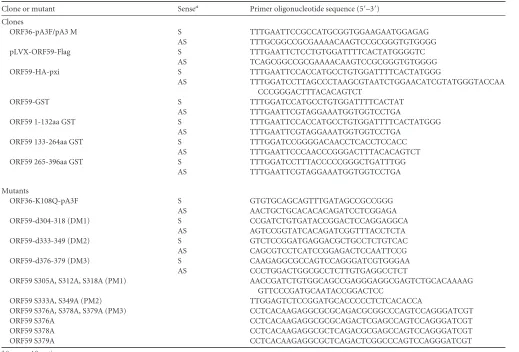
KSHV kinase (ORF36) binds to ORF59 between aa 1 and 264
and phosphorylates between aa 265 and 396.
Previous studies
have indicated that ORF59 is a phosphoprotein, but the
signifi-cance of phosphorylation was not determined (
36
). Although
de-tectable binding between a kinase and its substrate may not be
necessary for phosphorylation to occur, we determined that
ORF36 did in fact bind to ORF59 (
Fig. 1A
, fourth lane). This
binding was specific, as the vector control lane did not show any
detectable levels of ORF59 (
Fig. 1A
, ORF59-Flag lane). In order to
determine the domain of ORF59 interaction, an
in vitro
binding
assay with ORF59-GST and three ORF59-GST-fused segments,
segments from amino acids (aa) 1 to 132, aa 133 to 264, and aa 265
to 396 (
Fig. 1B
and
C
), was performed and identified that ORF36
binds between aa 133 and 264 with a higher affinity and between
aa 1 and 264 at a slightly lower affinity (
Fig. 1D
; compare lanes 6
and 7). As a control, we used
in vitro
-translated luciferase protein,
which showed no binding to ORF59-GST, and the GST without
the fusion protein confirmed the specificity of binding between
ORF36 and ORF59 (
Fig. 1D
, lane 3). Furthermore,
in vitro
analysis
also confirmed that no other proteins were necessary for the
bind-ing between ORF36 and ORF59.
In order to determine the site of phosphorylation, we used
ORF59-GST-tagged segments for a kinase assay with ORF36 (viral
kinase)-immunoprecipitated ORF36-Flag expressed in 293T cells.
ORF36-Flag and Flag vector immunoprecipitates were washed
with RIPA buffer containing higher salt (300 mM NaCl) to ensure
on November 7, 2019 by guest
http://jvi.asm.org/
that no additional protein carried over for the kinase assay, as the
cellular kinases may also be pulled down, which may convolute
the results. Immunoprecipitated ORF36-Flag and Flag vector
were used for kinase assay with ORF59-GST and GST-tagged
ORF59 fragments (aa 1 to 132, aa 133 to 264, aa 265 to 396) (
Fig.
2A
). Our results show that ORF59 is heavily phosphorylated by
ORF36 (
Fig. 2B
, lane 3). Phosphorylation of ORF59 fragments
identified that the site of phosphorylation lies between aa 265 and
FIG 1KSHV kinase (ORF36) binds to ORF59 between aa 1 and 264. (A) Binding between full-length ORF59 and ORF36 was established by cotransfecting ORF36-Flag and ORF59-HA plasmids and immunoprecipitating with anti-Flag antibody. The Western blot shows specific binding between ORF36 and ORF59, as the vector did not precipitate any ORF59. (B) Schematic of ORF59 GST-tagged segments used forin vitrobinding assay. NLS, nuclear localization signal. (C) GST-fused full-length ORF59 and ORF59 segments were expressed inE. colias stated in Materials and Methods and visualized on a Coomassie gel. Red asterisks, each of the purified proteins. (D) In order to identify the site of interaction, full-length ORF59 as well as its GST-fused segments (aa 1 to 132, aa 133 to 264, aa 265 to 396) were used for anin vitrobinding assay within vitro-translated [35S]methionine-labeled ORF36. As a control, luciferase was translatedin vitroand didnot bind to either GST or ORF59-GST. Radioactively labeled ORF36 was detected with ORF59-GST as well as the segments from aa 1 to 132 and aa 133 to 264, binding with a higher affinity to the latter segment.
FIG 2ORF36 phosphorylates ORF59 between aa 265 and 396. (A) Coomassie-stained gel of GST-ORF59 and segments used in the kinase assay. Lanes 1 and 3, ORF59-GST; lanes 4 to 6, ORF59-GST segments from aa 1 to 132, aa 133 to 264, and aa 265 to 396, respectively; lane 2, GST protein used in the assay. (B) Autoradiography image of thein vitrokinase assay performed on ORF59 and its GST-fused segments using immunoprecipitated ORF36-Flag as the kinase and the Flag vector as a control for kinase. Lane 1, GST-ORF59 as a negative control showing no phosphorylation when ORF36 was not present; lane 3, ORF59-GST phosphorylation when ORF36 kinase was present (asterisk). Among the GST-ORF59 domains, the region from aa 265 to 396 (lane 6) was phosphorylated by ORF36 (red asterisk). Lanes 2 to 6, ORF36 autophosphorylation, which is essential for its kinase activity.
on November 7, 2019 by guest
http://jvi.asm.org/
[image:5.585.112.473.69.306.2] [image:5.585.135.451.478.668.2]396 (
Fig. 2B
, lane 6). Since ORF36 requires autophosphorylation
for its kinase activity, detection of ORF36 phosphorylation in all
the lanes confirmed the presence of active kinase (
Fig. 2B
, lanes 2
to 6).
As a negative control, we performed a kinase assay on
ORF59-GST with a Flag vector-immunoprecipitated complex as well as
ORF36 with GST as a substrate. Consequently, we detected no
phosphorylation of ORF59-GST when assayed with the Flag
vec-tor (
Fig. 2B
, lane 1), and although there was autophosphorylation
of ORF36, the absence of GST phosphorylation confirmed the
specificity of the assay (
Fig. 2A
and
B
, lane 2).
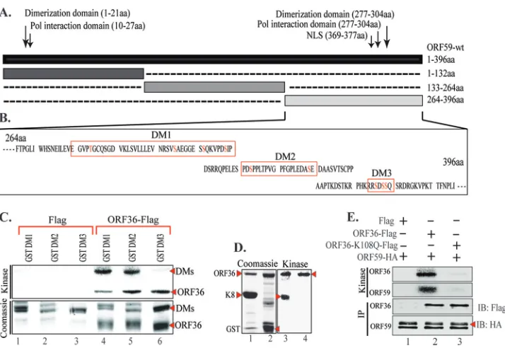
ORF36 phosphorylated ORF59 at Ser between aa 265 and
396.
In order to identify the probable phosphorylation sites on
ORF59 between aa 265 and 396, we used a program, KinasePhos
(
http://kinasephos.mbc.nctu.edu.tw/
; National Chiao Tang
Uni-versity, Taiwan), to determine the phosphorylation sites. The
pro-gram predicted one threonine and several serine residues to be the
potential sites of phosphorylation (
Fig. 3A
). We grouped these
residues to make three ORF59 deletion mutants (DM1, DM2,
DM3) (
Fig. 3B
). The sequences in
Fig. 3B
outlined with a red box
denote the sequences deleted from full-length ORF59. ORF59
de-letion mutants were constructed by sequential PCR, and the
pres-ence of deletions was confirmed by sequpres-ence analysis. These
dele-tion mutants were cloned in frame with GST to produce GST
fusion proteins.
GST and GST-tagged ORF59, DM1, DM2, and DM3 proteins
were purified and used for
in vitro
kinase assay.
Immunoprecipi-tated ORF36-Flag and Flag vector were washed with RIPA buffer
with 300 mM NaCl to remove any additional carryover proteins.
Consequently, the results of the kinase assay showed that ORF36
failed to phosphorylate ORF59 DM3 (
Fig. 3C
, lane 6) but not
DM1 or DM2 (
Fig. 3C
, lanes 4 and 5). Autophosphorylation of
ORF36 in this assay confirms the presence of active kinase in all
three lanes (
Fig. 3C
, lanes 4 to 6). The phosphorylation of these
truncation mutants by ORF36 was specific, as the
immunopre-cipitate with the Flag vector control was unable to phosphorylate
these mutants (
Fig. 3C
, lanes 1 to 3). This suggested that ORF36
phosphorylates ORF59 at residue Ser376, Ser378, or Ser379.
For a positive control, we used K8, a known substrate of
ORF36, to test the activity of the kinase used in this assay, and as
seen in
Fig. 3D
, lane 3, ORF36 was able to autophosphorylate as
well as phosphorylate K8. As a negative control, GST was used as a
substrate with ORF36, which showed autophosphorylation,
indi-cating active kinase; however, no signal for GST phosphorylation
was detected, suggesting the specificity of the assay (
Fig. 3D
, lane
4). We also tested the specificity of phosphorylation by ORF36 by
using a kd mutant of ORF36 (K108Q). The kd mutant of ORF36
was generated by a two-step approach similar to the approach
used to generate ORF59 deletion mutants. Kinase assay with
ORF36 K108Q showed that ORF59 was specifically
phosphory-FIG 3ORF36 phosphorylates ORF59 between aa 376 and 379. (A) Results of KinasePhos program analysis identifying possible phosphorylation sites on ORF59 between aa 364 and 396. The sites were grouped as indicated by the red boxes in panel B to generate three ORF59 deletion mutants (DMs). (B) Schematic of ORF59 deletion mutants used for mapping of the sites of phosphorylation, and the deleted sequences are marked with red boxes. Potential phosphorylation sites (S/T) are shown as red text. (C)In vitrokinase assay done with ORF59 GST-fused deletion mutants. (Top) Autoradiography image of ORF59 DM1, DM2, and DM3 with control Flag vector (lanes 1 to 3) and with immunoprecipitated ORF36-Flag (lanes 4 to 6). As seen in lane 6, ORF59 DM3 was not phosphorylated by ORF36, which thus identified aa 376 to 379 to be the specific site of phosphorylation by ORF36. Autophosphorylation of ORF36 (required for its kinase activity) is indicated in lanes 4 to 6. (Bottom) The proteins used in the kinase assay. Lanes 1 through 3, ORF59-GST DM1, DM2, and DM3 with Flag, respectively; lanes 4 to 6, DM1, DM2, and DM3 with ORF36, respectively. (D) A positive control was included with a known ORF36 substrate, K8. (Left) Coomassie-stained gel of K8 and GST; (right) kinase assay showing autophosphorylation of K8 (lane 3) but not GST (lane 4). Autophosphorylation of ORF36 (active kinase) is indicated by an arrowhead (lanes 3 and 4). (E) As a control, ORF59 was used for anin vitrokinase assay with ORF36-Flag and the kd mutant ORF36 (K108Q). The results showed specific phosphorylation of ORF59 with ORF36-Flag (lane 2) but not with the Flag vector (lane 1). Lane 3, the kd mutant of ORF36 was unable to phosphorylate ORF59 (ORF59) or itself (ORF36), suggesting the specificity of ORF59 phosphorylation by ORF36 kinase.on November 7, 2019 by guest
http://jvi.asm.org/
[image:6.585.114.471.63.309.2]lated by the viral kinase, as the kd mutant was unable to
phosphor-ylate ORF59 (
Fig. 3E
, lane 2). Abolition of kinase activity in the kd
mutant was evidenced by the lack of autophosphorylation of
ORF36 as well (
Fig. 3E
, lane 3).
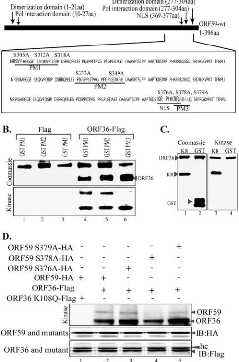
Moreover, we were concerned that a large deletion in ORF59
deletion mutants may have an effect on the protein conformation
and its tertiary structure; therefore, we generated ORF59 point
mutants (PM1, PM2, PM3) to verify our findings and conduct
further experiments. The point mutants, where each individual
serine residue was mutated to an alanine, were generated using a
QuikChange Lightning Multi site-directed mutagenesis kit (
Fig.
4B
). As with the ORF59 deletion mutants, ORF59 PM1, PM2, and
PM3 were used for
in vitro
kinase assay with immunoprecipitated
ORF36 or just the Flag vector as a control. As expected, ORF59
PM3 was not phosphorylated by ORF36, while ORF59 PM1 and
PM2 were phosphorylated by ORF36 (
Fig. 4B
, bottom; compare
lane 6 with lanes 4 and 5). ORF36 autophosphorylation is shown
in
Fig. 4B
as a control of kinase activity. As a control, K8 was
phosphorylated by ORF36, as demonstrated earlier, and GST
lacked a signal, as expected (
Fig. 4C
, lanes 3 and 4).
In an attempt to determine the exact residue of PM3
phosphor-ylated by ORF36, we generated single-amino-acid alanine
substi-tution mutants of ORF59 at residues 376, 378, and 379. Wild-type
ORF59 (
Fig. 4D
, lane 2) and its single-amino-acid alanine
substi-tution mutants (
Fig. 4D
, lanes 3 to 5) were subjected to kinase
assay with ORF36. Detection of phosphorylation showed that
ORF59 containing Ser378 mutated to alanine (
Fig. 4D
, lane 4) had
significantly reduced levels of phosphorylation, suggesting that
residue 378 is the primary site of ORF36-mediated
phosphoryla-tion. The levels of ORF59 and its substitution mutants as well as
the kinase, ORF36, were similar in all the lanes (
Fig. 4D
,
immu-noblot with HA [IB: HA] and IB: Flag, respectively), suggesting
that the ablation of phosphorylation was due to the lack of a
phos-phorylation site in the Ser378-to-alanine mutant (
Fig. 4D
, lane 4).
The specificity of ORF59 phosphorylation was once again
con-firmed, since the kd mutant of ORF36 did not phosphorylate
ORF59 (
Fig. 4D
, lane 1).
The ORF59 point mutant lacks the ability to bind to RTA.
Considering the functions of ORF59 and how the absence of
phos-phorylation may affect the virus, we evaluated the binding
be-tween ORF59 and its point mutants with the already known
bind-ing partners (
31
,
32
). First, we determined the binding of ORF59
PM1, PM2, and PM3 to ORF36 by coimmunoprecipitation
(CoIP) assays (
Fig. 5A
). Our data determined that the binding of
ORF36 to ORF59 containing substitution mutations was
unaf-fected, as all three point mutants (PM1, PM2, PM3) precipitated
ORF36, a result comparable to that for wt ORF59 (
Fig. 5A
, IB:
Myc; compare lane 7 with lanes 8 to 10). The vector control did
not precipitate ORF36, confirming the specificity of their
interac-tion (
Fig. 5A
, lane 6). We further determined whether the kd
mu-tant of ORF36 was capable of binding to ORF59 by performing
coimmunoprecipitation. Our data showed significantly reduced
levels of ORF59 coprecipitation for kd ORF36 compared to that
for active ORF36 (
Fig. 5B
, IB: HA; compare lane 8 with lane 9).
Additionally, we further analyzed the binding of ORF36 to
single-alanine-substitution mutant PM3, and no effect on its binding to
ORF36 was found, as expected (
Fig. 5B
, IB: HA, lanes 9 to 12).
Due to the proximity of these point mutations to ORF9, the
viral polymerase binding domain, we wanted to determine if these
mutations in ORF59 affected its ability to bind to or colocalize
FIG 4ORF36 phosphorylates ORF59 at Ser376, Ser378, and Ser379. (A) Sche-matic of ORF59 point mutants (PM1, PM2, PM3) used in the identification of specific phosphorylation targets by ORF36 kinase. The serine residues identi-fied in the ProsKinase program as potential phosphorylation targets are marked in red. The serine residues mutated to alanines are marked in green. (B)In vitrokinase assay with ORF59 GST-fused point mutants. (Bottom) Autoradiography image of ORF59 PM1, PM2, and PM3 with the control Flag vector (lanes 1 to 3) and with immunoprecipitated ORF36-Flag (lanes 4 to 6). ORF59 PM3 was not phosphorylated by ORF36, which thus identified Ser376, Ser378, and Ser379 to be the specific sites of ORF36 phosphorylation. Lanes 4 to 6 show that autophosphorylation of ORF36 is essential for its kinase func-tion. (Top) Proteins used in the kinase assay. Lanes 1 to 3, ORF59-GST PM1, PM2, and PM3 with Flag, respectively; lanes 4 to 6, PM1, PM2, and PM3 with ORF36 kinase, respectively. Lanes 4 to 6 of the Coomassie panel show the levels of ORF36 used for thein vitrokinase assay. (C) K8 was used as a positive control for ORF36 phosphorylation. (Left) Coomassie-stained gel of K8 (sub-strate) and GST, as a control; (right) phosphorylation of K8 as well as auto-phosphorylation of ORF36 but no auto-phosphorylation of just GST. (D) ORF59 with serine 378 mutated to alanine lacks ORF36-mediated phosphorylation. Mutants with a single amino acid mutation at Ser376A (lane 3), Ser378A (lane 4), and Ser379A (lane 5) were immunoprecipitated and subjected to the kinase assay with ORF36. ORF59 with Ser378 mutated to A showed almost no phos-phorylation (lane 4). The Ser379A mutant (lane 5) also had slightly reduced phosphorylation compared to that of Ser376A (lane 3) and wt ORF59 (lane 2). The kd mutant (ORF36 K108Q) was unable to phosphorylate ORF59 (lane 1). Immunoprecipitated ORF59 and its mutants were detected by anti-HA West-ern blotting (IB: HA). ORF36 and mutant were detected by anti-Flag WestWest-ern blotting (IB: Flag). hc, heavy chain.
on November 7, 2019 by guest
http://jvi.asm.org/
[image:7.585.300.542.76.442.2]with the viral polymerase. We cotransfected ORF9-Flag plasmid
with HA vector or HA-tagged ORF59 and its point mutants. The
CoIP was done with anti-HA antibody to precipitate ORF59 and
its mutants.
Figure 5C
shows that ORF9 was efficiently
coimmu-noprecipitated with ORF59 and its point mutants (
Fig. 5C
, IB:
Flag, lanes 7 to 10). A lack of ORF9 precipitation with the HA
empty vector confirms the specificity of interactions of these
pro-teins (
Fig. 5C
, IB: Flag, lane 6). These data indicate that
phosphor-ylation does not affect the ability of ORF59 to bind to the viral
DNA polymerase. Additionally, the introduction of single alanine
substitutions at the serines of PM3 (Ser376, Ser378, and Ser379)
did not affect its binding to ORF9, as expected (data not shown).
Viral polymerase translocates into the nucleus along with ORF59;
therefore, we determined the localization of ORF9 with alanine
mutants of ORF59. Our colocalization data showed that ORF9
efficiently translocated to the nucleus with wt ORF59 as well as the
points mutants (data not shown), confirming that ORF59 was still
able to recruit viral polymerase and transport it into the nucleus.
Furthermore, we wanted to determine whether mutation of
Ser376, Ser378, or Ser379 to alanine has any effect on the ability
of ORF59 to dimerize. To this end, we cotransfected Flag-tagged
ORF59 with HA-tagged ORF59 (
Fig. 5D
, lane 2) as a positive
control, Flag-tagged ORF59 (ORF59-pLVX-Flag) with
HA-tagged point mutant 3 (PM3-HA) (
Fig. 5D
, lane 3), or Flag-tagged
PM3 with HA-tagged PM3 (
Fig. 5D
, lane 4) into 293T cells to
FIG 5ORF59 S376A, S378A, and S379A kinase mutants do not bind to RTA ororiLyt. (A) Binding between ORF36 and ORF59 point mutants was deter-mined by CoIP assay. The ORF36-Myc plasmid was cotransfected with either
empty pLVX vector, pLVX-ORF59-Flag, pLVX-PM1-Flag, pLVX-PM2-Flag, or pLVX-PM3-Flag. Flag immunoprecipitation complexes were resolved on a Western blot and probed with anti-Myc and anti-Flag antibodies. Lanes 1 to 5, inputs; lanes 7 to 10, immunoprecipitated ORF59 and its point mutants pre-cipitating with ORF36-Myc. (B) ORF36 binding with single-amino-acid-sub-stitution mutants of ORF59. HA-tagged ORF59 was coexpressed with Flag vector (lane 1), kd ORF36 (lane 2), or ORF36 kinase (lane 3). Immunoprecipi-tation with anti-Flag antibody to precipitate ORF36 coprecipitated ORF59 (IB: HA, lane 9). The kd mutant of ORF36 showed significantly reduced coprecipi-tation of ORF59 (lane 8), suggesting that phosphorylation was important for the binding. Single-amino-acid substitution mutants of ORF59 with muta-tions at Ser376 (lane 10), Ser378 (lane 11), and Ser379 (lane 12) showed bind-ing comparable to that of wt ORF59. (C) Bindbind-ing between ORF9 and ORF59 point mutants was determined by cotransfecting ORF9-Flag with either the HA vector, ORF59-HA, PM1-HA, PM2-HA, or PM3-HA. Anti-HA immuno-precipitation complexes were resolved on an SDS-polyacrylamide gel and probed with anti-Flag and anti-HA antibodies. Lanes 1 to 5, inputs; lanes 7 to 10, immunoprecipitated ORF59. Coprecipitation of ORF9 showed binding comparable to that of wt ORF59 as well as that of its point mutants. (D) The serine residue mutated to alanine in PM3 does not affect ORF59 dimerization. A GFP-Flag-tagged lentivirus vector with PM3-HA, pLVX-ORF59-Flag with ORF59-HA, pLVX-ORF59-Flag with PM3-HA, or pLVX-PM3-Flag with PM3-HA was transfected into 293T cells and immunoprecipitated with anti-Flag antibody. The immunoprecipitated complex was resolved on an SDS-polyacrylamide gel and stained with anti-Flag and anti-HA antibodies. Lanes 1 to 4, inputs; lanes 6 to 8, dimerization was not affected by the S376A, S378A, or S379A mutation. (E) Binding between RTA and ORF59 point mutants deter-mined by CoIP. The RTA-expressing plasmid was cotransfected with empty pLVX vector, pLVX-ORF59-Flag, pLVX-PM1-Flag, pLVX-PM2-Flag, or pLVX-PM3-Flag. The anti-Flag IP complex was resolved on an SDS-polyacryl-amide gel and probed with anti-RTA and anti-Flag antibodies. Lanes 1 to 5, inputs; lanes 7 to 10, immunoprecipitated ORF59. The blot shows binding of RTA to PM1 (lane 3) and PM2 (lane 9) but not to PM3 (lane 10). (F) RTA binding to single-amino-acid-substitution mutants of ORF59. RTA-Flag was cotransfected with either wt ORF59 (lane 2) or its single-amino-acid alanine substitution mutant with a mutation at Ser376 (lane 3), Ser378 (lane 4), or Ser379 (lane 5). Immunoprecipitation of RTA with anti-Flag antibody effi-ciently coprecipitated wt ORF59 (lane 7) but showed reduced precipitation of the Ser378 (lane 9) and Ser379 (lane 10) mutants.
on November 7, 2019 by guest
http://jvi.asm.org/
[image:8.585.47.281.69.703.2]immunoprecipitate with anti-Flag antibody. If dimerization
oc-curred, the Flag-tagged protein would be able to bring down its
HA-tagged counterpart. We demonstrated that ORF59 was able to
dimerize with PM3 at a level similar to that with wt ORF59 (
Fig.
5D
, lanes 6 and 7). We also showed that PM3 dimerizes with itself
at relatively the same level as it does with wt ORF59, which further
verifies that dimerization was not affected by phosphorylation at
serine residue 376, 378, or 379 (
Fig. 5D
, lane 8). The vector control
did not precipitate ORF59 PM3, confirming the specificity of the
association identified above (
Fig. 5D
, lane 5).
As previously stated, the interaction between ORF59 and RTA
is critical for the function of ORF59 as a processivity factor (
31
).
We determined the binding of ORF59 point mutants and RTA by
cotransfecting an RTA expression plasmid with GFP-Flag-tagged
vector, ORF59, or ORF59 PM1, PM2, or PM3 pLVX-Flag.
Immu-noprecipitation of ORF59 and its point mutants showed that PM1
and PM2 were still able to bind to RTA (
Fig. 5E
, lanes 8 and 9),
while the PM3 mutants containing the S376A, S378A, and S379A
mutations were unable to bind to RTA (
Fig. 5E
, IB: RTA; compare
lane 10 with lanes 8 and 9). In an attempt to determine the exact
residues of PM3 responsible for binding to RTA, we used ORF59
containing single-amino-acid alanine substitutions at serine
resi-dues 376, 378, and 379. Immunoprecipitation of RTA showed
efficient coprecipitation of wt ORF59 as well as ORF59 with the
Ser376A mutant (
Fig. 5F
, IB: HA, lanes 7 and 8). Interestingly,
mutation of the Ser378 residue to alanine significantly reduced the
binding of RTA to ORF59 (
Fig. 5F
, IB: HA, lane 9). Since Ser379A
also showed slightly reduced binding to RTA (
Fig. 5F
, IB: HA;
compare lane 10 with lanes 8 and 9), we suggest that both serines
at residues 378 and 379 play critical roles in binding to RTA.
Point mutants of ORF59 do not affect its localization with
ORF36 but do affect its localization with RTA.
Since the binding
of ORF36 to ORF59 was unaffected even after the replacement of
the serines between aa 376 and 379, we wanted to determine their
localization in the cellular compartment by immunolocalization
assay. As expected, ORF59 and its point mutants (PM1 to PM3)
localized to the nucleus in a similar fashion as ORF36, with the
majority of the spot colocalizing to the same nuclear
compart-ments determined by yellow signals in the overlay panels (
Fig. 6A
).
We further determined the localization of RTA with ORF59 and
its point mutants by cotransfecting GFP-fused ORF59 along with
RTA expression plasmids, which were detected by RTA
anti-body. The yellow signals in the overlay panels show the
localiza-FIG 6The ORF59 point mutant can associate with ORF36 but not with RTA. (A) 293L cells were transfected with GFP-fused ORF59 and its point mutants (PM1 to PM3) along with Ds-Red-fused ORF36. The cells were fixed, perme-abilized, stained with TO-PRO-3 and nuclear stain, and imaged with a laser
confocal microscope. The colocalization of these two proteins, depicted as yellow signals (Overlay), in wt ORF59 as well as the point mutants is shown. (B) For determining the nuclear localization of ORF59 and RTA, GFP-fused ORF59 and its point mutants (PM1 to PM3) were cotransfected with RTA-expressing plasmid. Cells were fixed, permeabilized, and stained with mouse anti-RTA primary antibody. We used Alexa Fluor 594 (red) to visualize RTA localization. The overlay of red and green (yellow) indicates colocalization of RTA with ORF59, PM1, and PM2 but not with PM3. (C) Localization of RTA with ORF59 and its point mutants in BAC36⌬ORF59 cells. BAC36⌬ORF59 was transfected into 293L cells expressing either wtORF59 or point mutants, followed by selection for a pure population. Cells were induced to undergo lytic reactivation, followed by fixing and permeabilizing so that they could be stained with anti-ORF59 and anti-RTA antibodies. Alexa Fluor 594 was used for the detection of ORF59, and Alexa Fluor 647 was used for the detection of the RTA protein. The overlay panels show colocalization, detected by the yel-low signal, in wt ORF59 and PM1 and PM2 but that the colocalization was significantly reduced in PM3. DIC, differential inference contrast.
on November 7, 2019 by guest
http://jvi.asm.org/
[image:9.585.43.283.66.685.2]tion of RTA and ORF59 in the same nuclear compartments (
Fig.
6B
). PM1 and PM2 showed colocalization almost similar to that of
wt ORF59, but PM3, which had Ser376, Ser378, and Ser379
mu-tated to alanines, showed the least yellow signal in the overlay
panel, suggesting that the association of PM1 and PM2 is
depen-dent on these serines (
Fig. 6B
). These data corroborate the
immu-noprecipitation data and are significant, as they show that
ORF36-mediated phosphorylation of ORF59 plays an important role in
their interaction.
Additionally, we confirmed the association of RTA with
ORF59 and its point mutants in BAC36
⌬
ORF59-containing 293L
cells complemented with either wt ORF59 or the point mutants.
Induced cells were subjected to the detection of ORF59 and RTA
using specific antibodies. Localization of ORF59 with Alexa Fluor
594 and RTA with Alexa Fluor 647 showed yellow colocalizing
signals in wt ORF59 as well as in PM1 and PM2 (
Fig. 6C
).
How-ever, BAC36
⌬
ORF59 complemented with PM3 showed the
few-est yellow signals, confirming the results presented above and the
requirement of serine for its association with RTA.
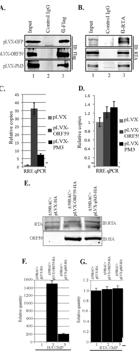
ORF59 PM3 lacks the ability to bind to
ori
Lyt.
Since the
bind-ing of ORF59 PM3 to RTA was severely affected, we wanted to
determine whether this mutant was also affected in its ability to get
recruited to the
ori
Lyt chromatin, which is required for
processiv-ity function. We performed a chromatin immunoprecipitation
assay in an overexpression system after transfection of 293L cells
with RTA-GFP or
ori
Lyt (8088sc) containing the plasmid
pLVX-GFP-Flag vector, pLVX-GFP-ORF59-Flag, or
pLVX-GFP-PM3-Flag. Sonicated chromatins were immunoprecipitated with
anti-Flag, anti-RTA, and IgG-matched control antibodies. A fraction
of the immunoprecipitated chromatin was analyzed in a Western
blot to ensure that these antibodies were efficiently precipitating
chromatins (
Fig. 7A
and
B
). DNA extracted from the
immuno-precipitated chromatin was used for quantitative PCR (qPCR)
with primers targeted to amplify the RTA response element (RRE)
of the
ori
Lyt sequence. The results, analyzed using the
⌬⌬
C
Tmethod, showed a dramatic decrease in the ability of ORF59 to
bind to
ori
Lyt when Ser376, Ser378, and Ser379 (PM3) of ORF59
were mutated to alanine (
Fig. 7C
; compare the black bar with the
dark gray bar). Since ORF59 binds to RRE through RTA, we also
quantified the binding of RTA in this overexpression ChIP.
Bind-ing of RTA to RRE was comparable (range, 1- to 1.5-fold) in all
three samples, suggesting that RTA binding was unaffected by
PM3 and the reduction in PM3 binding to
ori
Lyt was solely due its
inability to associate with RTA (
Fig. 7D
). These experiments were
performed three independent times, and all of them showed
al-most similar fold reductions in the binding of PM3 to
ori
Lyt.
We also confirmed the recruitment of ORF59 and PM3 on
FIG 7(A) ORF59 PM3 was defective in binding tooriLyt chromatin. 293L cells were transfected with RTA-GFP, the 8088sc-expressing plasmid (which containsoriLyt), and either the GFP-Flag-lentivirus vector, pLVX-ORF59-Flag, or pLVX-PM3-Flag. ChIP was done with Flag and RTA anti-bodies. Fractions of the precipitated proteins were resolved on an SDS-poly-acrylamide gel to check for the immunoprecipitated chromatin with anti-Flag
(ORF59) (A) or anti-RTA (B) antibodies. (C) Chromatin-boundoriLyt DNA with ORF59 and PM3 was analyzed by quantitative real-time PCR using RRE primers. The results were analyzed using the⌬⌬CTmethod and are presented as the fold change compared to the result for the vector control (pLVX). A significant decrease of PM3 binding tooriLyt compared to that for wt ORF59 was determined. (D) Relative binding of RTA tooriLyt chromatin was rela-tively unaffected. (E) Recruitment of ORF59 PM3 tooriLyt chromatin was affected in KSHV BAC36. BAC36⌬ORF59 transfected into 293L cells express-ing wt ORF59, PM3, or empty vector (pLVX-HA) was induced for lytic reac-tivation. (E) Expression of RTA postinduction and detection of ORF59 with anti-HA Western blotting.⌬59BAC36, BAC36⌬ORF59, (F) Detection of chromatin-boundoriLyt with wt ORF59 and the PM3 mutant. (G) Relative number of copies of RTA-boundoriLyt.
on November 7, 2019 by guest
http://jvi.asm.org/
[image:10.585.45.281.62.658.2]ori
Lyt chromatin in BAC36
⌬
ORF59 stable cells complemented
with the respective proteins. Cells were induced to undergo lytic
reactivation by a treatment with sodium butyrate and TPA.
Induc-tion of lytic reactivaInduc-tion was confirmed by detecInduc-tion of RTA
pro-tein in a Western blot (
Fig. 7E
). Expression of ORF59 and PM3
was also detected using anti-HA Western blotting with cells
un-dergoing lytic reactivation (
Fig. 7E
). Chromatin was
immunopre-cipitated with anti-HA and anti-RTA antibodies, and the relative
number of copies of
ori
Lyt bound to these two proteins was
deter-mined using a vector control as a reference (
Fig. 7F
and
G
, lanes 1).
The relative number of copies of
ori
Lyt determined by RRE
quan-titation showed specific binding of ORF59 to
ori
Lyt chromatin,
which was significantly reduced in the PM3 mutant (
Fig. 7F
, lane
1). The levels of RTA binding to
ori
Lyt chromatin were unaffected
in the vector control as well as the PM3 mutant, a finding which
was similar to the overexpression ChIP data (
Fig. 7D
and
G
).
KSHV BAC36
⌬
ORF59 reconstituted with ORF59 PM3 is
de-fective in virion production.
In order to determine the effect of
the ORF59 S376A, S378A, and S379A (PM3) point mutations on
the life cycle of KSHV, we used lentivirus vectors, ORF59, or
ORF59 PM3 to reconstitute the ORF59 deletion in a BAC36
⌬ORF59 BACmid. The BACmid has been proven to be a useful
tool for determining the role of individual viral genes or
muta-tions in the gene in the context of the viral genome (
42
).
BAC36
⌬
ORF59 BACmid was generated by site-directed
ho-mologous recombination using a Kan cassette and FLIP
recombi-nation (
Fig. 8A
), as previously stated (
39
,
40
). The site of Kan
cassette insertion was verified by digesting the intermediate
BACmid with PstI and analyzing it via Southern blotting using a
Kan probe. The resulting fragment containing the Kan cassette
yielded a fragment of the expected size (5,883 bp) with the Kan
probe (
Fig. 8B
). FLIP recombinase was then activated to remove
the Kan cassette, leaving one FRT site (5
=
-GAAGTTCCTATTCT
CTAGAAAGTATAGGAACTTC-3
=
) at the mutation site.
Al-though the BACmid contains the entire ORF59 sequence, the
in-sertion of the remaining FRT site interrupted the ORF59 open
reading frame (ORF) due to a stop codon and thus blocked ORF59
expression. Furthermore, the absence of the Kan cassette was
ver-ified with a Southern blot probing for the Kan cassette. The
junc-tion sequence with the FRT inserjunc-tion was PCR amplified and
se-quenced to confirm the FRT insertion. The remainder of ORF59
was detected with an ORF59 probe in a Southern blot to further
validate the clone (
Fig. 8B
).
In order to reconstitute BAC36
⌬ORF59, we first transduced
293L cells with the lentivirus pLVX vector pLVX-ORF59-HA or
pLVX-ORF59-PM3-HA. Following a puromycin selection for
transduced cells, we used the Metafectene Pro reagent to transfect
them with the BAC36
⌬ORF59 BACmid. A dual selection with
puromycin and hygromycin ensured that all surviving cells
con-tained the lentivirus and BAC36
⌬ORF59. In addition, by use of a
GFP cassette in the BAC backbone, we were able to monitor the
presence of BAC by GFP fluorescence (data not shown). We also
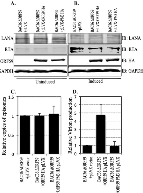
verified the stable maintenance of the BACmid with anti-LANA
and anti-RTA Western blots as well as the expression of ORF59
and PM3 with HA-probed Western blots (
Fig. 9A
and
B
).
Approximately 5
⫻
10
6cells with BAC36-⌬ORF59-pLVX,
BAC36-
⌬
ORF59-pLVX-ORF59-HA, or BAC36-
⌬
ORF59-pLVX
PM3-HA were collected for a modified Hirt’s extraction to
com-pare the number of copies of the KSHV genome latently
main-tained in these cells. The purified DNA was used for qPCR with
LANA (ORF73) primers, which determined that the genome copy
number in the latently persisting genome was almost identical
between these three cell lines (
Fig. 9C
).
In order to determine whether PM3, which lacks the ability to
bind to RTA, has any effect on viral DNA replication and, thus,
virion production, we compared the numbers of virions produced
from BAC36
⌬
ORF59 reconstituted with either wt or PM3
ORF59. The same numbers of cells were induced with 20 ng of
12-
O
-tetradecanoylphorbol-13-acetate (TPA) per ml and 1.5 mM
sodium butyrate (Sigma-Aldrich, St. Louis, MO) for 5 days. The
supernatant was collected to determine the relative number of
copies of virions produced from these cells by extracting the DNA
from the virions released into the culture supernatants. Purified
DNA was used for qPCR with LANA (ORF73) primers, and the
relative amounts of virions produced were plotted using a vector
control (BAC36-
⌬
ORF59-pLVX) as a reference. The results
showed a significant reduction in the amounts of virions
pro-duced from the cells complemented with ORF59 mutated at
Ser376, Ser378, and Ser379 (PM3) (
Fig. 9D
). Induction of lytic
reactivation by sodium butyrate and TPA was confirmed with
BAC36-containing 293L cells, which showed an approximately
9-fold increase in the numbers of virions after induction of lytic
DNA replication. Since the virion production levels of
PM3-re-constituted BAC36
⌬
ORF59 was similar to that of empty
vector-reconstituted BAC36
⌬
ORF59, we conclude that the function of
ORF59 is dependent on the phosphorylation of the Ser376,
Ser378, or Ser379 residue by the viral kinase (ORF36).
DISCUSSION
Kaposi’s sarcoma-associated herpesvirus is closely associated with
multiple human malignancies, including Kaposi’s sarcoma (KS),
primary effusion lymphomas (PELs), and multicentric
Castle-man’s disease (MCD). KSHV establishes a lifelong latency
follow-ing a primary infection with the help of viral and cellular factors.
During latency, viral replication is dependent on the cellular
rep-lication machinery. Upon reactivation, most of the viral genes are
expressed, including those necessary and sufficient for KSHV lytic
replication (ORF9, ORF6, ORF40/41, ORF44, ORF56, ORF59,
and ORF50) (
24
). Interestingly, most of the cells in KSHV tumors
are latent, and only a subpopulation (approximately 2 to 5% of
latent cells) undergoes spontaneous lytic reactivation to produce
virions which are implicated in tumorigenesis (
10
,
11
).
Furthermore, similar to other herpesviruses, the replication
and transcription activator (RTA) is accepted as one of the most
important proteins required for lytic reactivation (
43
). RTA
binds to the C/EBP
␣
and the RTA response element (RRE)
binding motifs within the origin of lytic replication (
ori
Lyt)
and as a result activates lytic DNA replication through
tran-scriptional activation as well as recruitment of additional
fac-tors (
6
–
8
). RTA recruits the viral processivity factor ORF59 to
ori
Lyt, where it acts as an accessory factor or sliding clamp,
stabilizing the binding between viral polymerase (ORF9) and
the DNA. ORF59 forms a homodimer in the cytoplasm, binds
to ORF9 directly, and specifically recruits it to the nucleus,
which is crucial for long-chain DNA synthesis (
32
,
36
,
44
,
45
).
Rossetto et al. have shown that ORF59’s binding to the C/EBP␣
binding motif within
ori
Lyt is necessary for its function and is
dependent on its binding to RTA (
31
). Furthermore, the
pro-cessivity function of ORF59 is dependent upon its interaction
on November 7, 2019 by guest
http://jvi.asm.org/
with RTA; consequently, the function of the viral polymerase is
also dependent upon this interaction (
31
).
Although the structures of processivity factors among
dif-ferent herpesviruses are variable, their functions are fairly
con-served. The human cytomegalovirus (hCMV) processivity
fac-tor, ppUL44, is phosphorylated by viral Ser/Thr kinase
(ppUL97), which modulates its ability to localize to the nucleus
(
33
). BMRF1, the Epstein-Barr virus (EBV) processivity factor, is
phosphorylated by the BGLF4 viral kinase within a hinge
region-like domain known to be important for inducing conformational
changes (
34
). Consequently, phosphorylation by BGLF4 enhances
the transactivation activity of Zta (RTA homolog) and the
syner-FIG 8BAC36⌬ORF59 was generated with Red recombination. (A) General schematic of the Red recombination method used to generate the⌬ORF59 BACmid. A PCR-amplified Kan cassette containing homologous ends to ORF59 was transformed into EL350 bacteria containing the BAC36 KSHV genome. FLIP recombinase was then activated in the bacterial culture to remove the Kan cassette using flanking FRT sequences. The final product was sequenced to show the remaining FRT sequence, which contains a stop codon, which thus blocked ORF59 synthesis. nt, nucleotides; AA seq, amino acid sequence. (B) The intermediate clone was screened by Southern blotting, with a band at 5,883 bp expected to be seen when the BACmid was digested with PstI and probed with the Kan probe. As shown, the wild type and the final BACmid did not contain a Kan cassette. Since the ORF59 sequence was not deleted but was interrupted, the presence of the ORF59 sequence in the BACmid was verified with an ORF59 probe.on November 7, 2019 by guest
http://jvi.asm.org/
[image:12.585.136.447.63.561.2]gistic activation of lytic replication at
ori
Lyt (
35
). Similarly, ORF59
is a phosphoprotein which is phosphorylated by the KSHV Ser/Thr
kinase, ORF36, but the consequences of phosphorylation on DNA
processivity or virion production were not evaluated (
36
).
Phosphorylation of viral processivity factors is an important
modification among human herpesviruses. Our aim was to
iden-tify the ORF36 target sites on the KSHV processivity factor
(ORF59) and the possible effects of phosphorylation on viral
pro-duction. In this study, residues Ser376, Ser378, and Ser379 were
identified as the major ORF36 kinase targets on ORF59 through
use of the KinasePhos program (
http://kinasephos.mbc.nctu.edu
.tw/
) (
Fig. 3A
). We mapped the binding region of ORF36 to
ORF59 between aa 133 and 264 on ORF59 (
Fig. 1
), which did not
correspond with the identified phosphorylation sites, Ser376,
Ser378, and Ser379. Nonetheless, as indicated by the ORF59
crys-tal structure, these regions are in close proximity and may thus
allow ORF36 to phosphorylate ORF59 at the C terminus, which
harbors the identified phosphorylation sites (
46
). In fact,
detect-able binding between kinase and its substrate is not required for
phosphorylation to take place. For instance, no binding between
the EBV processivity factor (BMRF1) and viral kinase (BGLF4)
was detected; however, phosphorylation of BMRF1 by BGLF4 was
revealed to be necessary for the initiation of lytic replication at
ori
Lyt (
35
).
Due to the close proximity of the identified phosphorylation
sites with the ORF59 dimerization domain, the ORF9-interacting
domain, and nuclear localization signals, we investigated the
con-sequences that these mutations could have on lytic DNA
replica-tion by evaluating the effects of S376A, S378A, and S379A (ORF59
PM3) on known functions of ORF59. Binding between ORF9,
viral polymerase, and ORF59 is crucial for DNA replication;
there-fore, we determined that binding between these two proteins and
translocation of ORF9 to the nucleus was not affected by the
mu-tation of phosphorylation sites of PM3 (Ser376, Ser378, and
Ser379) (
Fig. 5C
). The binding between ORF59 and ORF9 is
de-pendent upon the ability of ORF59 to dimerize; therefore, we
expected that phosphorylation might not have any effect on
dimerization of ORF59 (
47
). Nevertheless, we confirmed that,
in-deed, the lack of phosphorylation did not affect the dimerization
of ORF59 (
Fig. 5C
).
Notably, upon examination of binding between ORF59 point
mutants and RTA, we determined that the S376A, S378A, and
S379A mutations of ORF59 PM3 abolished its binding to RTA
(
Fig. 5E
). Fine mapping of the residues required for their
interac-tion suggested that both Ser378 and Ser379 are cooperatively
re-quired for binding to RTA and mutants with both of these
muta-tions showed reduced binding. Previous data suggested that
dimerization of ORF59 is not necessary for its interaction with
RTA; therefore, it is not surprising that despite ORF59 PM3’s
ability to dimerize, it was unable to bind to RTA. Interestingly, a
similar interaction was observed between BMRF1, EBV
processiv-ity factor, and BZLF1, which plays an important role in EBV lytic
replication (
48
). These findings are significant because the
re-duced binding between PM3 and RTA hampered the recruitment
of ORF59 to
ori
Lyt, which could subsequently affect the synthesis
of viral DNA.
Moreover, our chromatin immunoprecipitation assay and
re-constituted BAC36
⌬
ORF59 demonstrated the downstream
ef-fects of the S376A, S378A, and S379A ORF59 mutations on
ORF59 recruitment to
ori
Lyt chromatin and synthesis of viral
DNA. Although the replacement of ORF59 Ser376, Ser378, and
Ser379 with alanines had no effect on its ability to form a
ho-modimer or to bind to viral polymerase (ORF9), the mutations
hindered its ability to bind to RTA, which raised the question of
whether ORF59 PM3 is able to bind to
ori
Lyt and promote DNA
synthesis. We used ChIP to compare the efficiency of ORF59 and
its PM3 binding to
ori
Lyt in cells transfected with an
ori
Lyt
plas-mid in the presence of RTA as well as in cells maintaining the
KSHV genome. The results showed a significant decrease in
ORF59 PM3 binding at
ori
Lyt compared with that of the wt (
Fig.
7C
and
F
). The mutations further proved to have deleterious
ef-FIG 9The ORF59 PM3 mutant of KSHV is defective in virion production. (Aand B) Cells transfected with BAC36⌬ORF59 into either empty lentivirus vector, wt ORF59-expressing cells, or PM3-expressing cells were tested for LANA-, RTA-, and HA-tagged ORF59 or PM3 expression. Uninduced cells were shown to express LANA (IB: LANA) and ORF59 and PM3 (IB: HA) but to express very little RTA (IB: RTA). The results for GAPDH (IB: GAPDH) are presented to show equal loading. (B) Cells were induced with TPA and sodium butyrate to induce reactivation, which was confirmed by the detection of RTA (IB: RTA) All these cell lines showed similar levels of LANA RTA expressions. (C) DNA was extracted from uninduced cells to examine the latently main-tained genome copies in these cell lines. LANA (ORF73) primers were used to calculate the KSHV genome copy number using the⌬⌬CTmethod of quanti-tation. The relative number of copies of episomally maintained genomes was plotted by using a vector control as a reference. The results showed mainte-nance of almost identical numbers of viral episomes in these cell lines. (D) Cells were induced with 20 ng TPA and 1.5 mM sodium butyrate for 5 days, after which the supernatant was collected and virion DNA was extracted. The relative numbers of virions produced were determined by real-time qPCR, which showed a significant decrease in virion production in ORF59 with mu-tated Ser376, Ser378, and Ser379 (PM3) compared to that in cells with wt ORF59.