Copyright © 2000, American Society for Microbiology. All Rights Reserved.
Nitric Oxide Synthesis Enhances Human Immunodeficiency
Virus Replication in Primary Human Macrophages
DONATIENNE BLOND, HERVE´ RAOUL, ROGER L
EGRAND,*
ANDDOMINIQUE DORMONT
Service de Neurovirologie, Commissariat a` l’Energie Atomique, DSV/DRM, CRSSA, Institut Paris-Sud sur les
Cytokines, Fontenay aux Roses, France
Received 13 March 2000/Accepted 10 July 2000
Macrophages are suspected to play a major role in human immunodeficiency virus (HIV) infection
patho-genesis, not only by their contribution to virus dissemination and persistence in the host but also through the
dysregulation of immune functions. The production of NO, a highly reactive free radical, is thought to act as
an important component of the host immune response in several viral infections. The aim of this study was to
evaluate the effects of HIV type 1 (HIV-1) Ba-L replication on inducible nitric oxide synthase (iNOS) mRNA
expression in primary cultures of human monocyte-derived macrophages (MDM) and then examine the effects
of NO production on the level of HIV-1 replication. Significant induction of the iNOS gene was observed in
cultured MDM concomitantly with the peak of virus replication. However, this induction was not accompanied
by a measurable production of NO, suggesting a weak synthesis of NO. Surprisingly, exposure to low
concen-trations of a NO-generating compound (sodium nitroprusside) and
L-arginine, the natural substrate of iNOS,results in a significant increase in HIV replication. Accordingly, reduction of
L-arginine bioavailability afteraddition of arginase to the medium significantly reduced HIV replication. The specific involvement of NO was
further demonstrated by a dose-dependent inhibition of viral replication that was observed in infected
mac-rophages exposed to
N
G-monomethyl
L-arginine andN
G-nitro-L-arginine methyl ester (L-NAME), two
inhibi-tors of the iNOS. Moreover, an excess of
L-arginine reversed the addition of L-NAME, confirming that anarginine-dependent mechanism is involved. Finally, inhibitory effects of hemoglobin which can trap free NO in
culture supernatants and in biological fluids in vivo confirmed that endogenously produced NO could interfere
with HIV replication in human macrophages.
The macrophage represents one of the major target cell for
human immunodeficiency virus (HIV) infection and is likely to
play a major role in persistence and tissue dissemination of this
virus (35, 39). Macrophage immune functions are also altered
by HIV type 1 (HIV-1) infection: (i) synthesis of inflammatory
cytokines is dysregulated (8, 29, 94); (ii) in vitro-infected
monocyte-derived macrophages (MDM) have decreased
abil-ity to act as accessory cells for T-lymphocyte proliferation (28);
and (iii) the production of free radicals, such as hydrogen
peroxide (H
2O
2), superoxide anion (O
2⫺), and hydroxyl
radi-cals (HO
䡠) (13, 74, 84), is impaired and may therefore
facili-tate the development of opportunistic intracellular pathogens.
Among free radicals, NO is of particular interest. This
mole-cule is generated by nitric oxide synthase (NOS) from
L-argi-nine and rapidly reacts in vivo with oxygen to form nitrite and
nitrate, its two stable end products (72, 76, 90). Three distinct
isoforms of NOS have been described. Two are constitutive
and mainly found in endothelial and neuronal cells (32, 53);
the third, inducible NOS (iNOS), was originally described in
murine macrophage (95). NO mediates numerous
physiologi-cal functions and is known to be implicated in several
immu-nological disorders. Besides its participation to the relaxation
of blood vessels and glutamate-induced neurotoxicity (11, 12,
49), the production of NO represents an important component
of the host immune response against viral infections (20, 52,
67, 69) including retroviruses (Friend leukemia virus) (2).
An-tiviral effects occur through its microbiostatic and microbicidal
activity, and probably also through its proinflammatory and
immunoregulatory properties (33, 50, 71).
In some cases, the production of NO during infectious diseases
may also be deleterious. This may be particularly true in HIV
infection, where NO may contribute to AIDS pathogenesis:
sig-nificant increases in nitrite and nitrate (the two stable end
prod-ucts of NO) concentrations were evidenced in peripheral blood
mononuclear cells (PBMC), polymorphonuclear leukocytes, and
sera of patients with AIDS, especially in individuals with
neuro-logical disorders and pulmonary disease caused by intracellular
opportunistic pathogens (30, 92; D. Torre, G. Ferrario, G.
Bonetta, C. Zeroli, M. Giola, and G. P. Fioli, Abstr. 10th Int.
Conf. AIDS, Int. Conf. STD, abstr. PAO114, 1996). HIV-related
neurological disorders could in part be attributed to excessive
production of NO. Indeed, high concentrations of NO could be
obtained in vitro (i) after direct interactions between viral
com-ponents and neuronal cells, since gp120-induced injury in primary
neuronal cultures involves NO (23, 24, 26), and (ii) after HIV-1
infection of macrophages infiltrating the brain tissue (55). Direct
evidence for the presence and distribution of iNOS has been
reported in human pulmonary tissue (54, 82) and in the central
nervous system of HIV-infected patients, especially in areas of
acute and chronic inflammation (1). The production of NO by
human monocytes/macrophages could result from the induction
of iNOS expression by the proinflammatory cytokines (21, 65, 77,
78, 85). Synthesis of these cytokines is induced in vivo and in vitro
in response to HIV-1 infection (42, 89) and may directly regulate
iNOS expression (15, 83). However, we cannot exclude that
HIV-1 can also directly interact with iNOS expression in
mono-cytes/macrophages. Indeed, viral regulatory proteins such as Tat
may directly enhance the transcription of the iNOS.
Interestingly, Groeneveld et al. have shown that levels of
nitrate in the serum are positively correlated with plasma and
* Corresponding author. Mailing address: Service de
Neurovirolo-gie, Commissariat a` l’Energie Atomique, DSV/DRM/CRSSA, Institut
Paris-Sud sur les Cytokines, B.P. 6, 92265 Fontenay aux Roses Cedex,
France. Phone: 33 1 46 54 73 74. Fax: 33 1 46 54 77 26. E-mail: legrand
@dsvidf.cea.fr.
8904
on November 9, 2019 by guest
http://jvi.asm.org/
cell-associated virus loads, suggesting that HIV could induce
NO synthesis in vivo (46). In the simian immunodeficiency
virus/macaque model, significantly increased concentrations of
NO
2⫺and NO
3⫺were measured in plasma during primary
infection, coincident with viremia peaks, and in the absence of
opportunistic infections (10).
The objectives of this study were to (i) evaluate the effects of
HIV-1 infection on iNOS mRNA expression and NO
produc-tion in cultured human MDM, (ii) assess whether the
endog-enous NO release interfere with HIV replication in cultured
MDM, and (iii) study the mechanisms of NO regulation in
response to MDM infection with primary HIV-1 isolates.
MATERIALS AND METHODS
Isolation and characterization of monocytes.Fresh human PBMC were ob-tained from healthy HIV-1-seronegative donors after centrifugation of heparin-ized venous blood over Ficoll-Hypaque gradients. Monocytes were isolated from PBMC by centrifugal elutriation (Beckman J2-21/ME centrifuge, JE-5 rotor; Beckman Instruments, Gagny, France) as previously described by Figdor et al. (31). Purified monocytes were cultivated at the concentration of 2⫻105cells/ml in 48-well microtiter plates and progressively allowed to differentiate into mac-rophages for 7 days in a 5% CO2atmosphere at 37°C. The culture medium was constituted of RPMI 1640 medium (Boehringer Mannheim, Mannheim, Ger-many) supplemented with 10% heat-inactivated fetal calf serum (Boehringer Mannheim), 2 mM glutamine (Boehringer Mannheim), and 0.2M antibiotics (penicillin-streptomycin-neomycin [Life Technologies, Inc., Berlin, Germany]). Immunophenotyping of the monocyte fraction was performed by standard fluo-rescence-activated cell sorter analysis as previously described (10), using a FAC-Scan Plus cytofluorometer and LYSIS II software (Becton Dickinson, Mountain View, Calif.). Cellular purity was greater than 97%; these cells were negative for expression of CD3 (CD3 Leu4, immunoglobulin G1 [IgG1]; Becton Dickinson); and CD19 (CD19-fluorescein isothiocyanate [FITC], IgG1; Immunotech, Mar-seille, France) but positive for CD14 (CD14-FITC, IgG1; Becton Dickinson) and CD64 (CD64-FITC, IgG1; Medarex, West Lebanon, N.H.) expression (81.6 and 88.5%, respectively).
Infection of MDM and detection of virus replication.MDM were infected with macrophagetropic HIV-1 reference strain Ba-L, a generous gift from A.-M. Auber-tin (Strasbourg, France). This strain, initially obtained from a primary culture of postmortem lung tissue from an infant who died from AIDS (35, 36), replicates well in human macrophage cultures. HIV-1 Ba-L was grown to high titers in phytohem-agglutinin-stimulated human cord blood mononuclear cells. The cell-free superna-tant was clarified at 10,000⫻gfor 5 min and ultracentrifuged at 360,000⫻g (Beckman TL100; Beckman Instruments) for 10 min. The viral pellet was resus-pended in phosphate-buffered saline. Virus stock was titered on cord blood lympho-cytes in a 96-well microplate assay as measured by endpoint dilution. The 50% tissue culture infectious dose (TCID50) was determined by the Karber’s formula (47). The virus stock used was endotoxin free, as assessed by the limulus amebocyte lysate assay (Sigma, St. Louis, Mo.). MDM were infected with HIV-1 Ba-L at 105TCID
50/ 106cells. Twenty-four hours after onset of HIV-1 infection, MDM were washed with phosphate-buffered saline (Boehringer Mannheim) to remove excess virus. MDM were then treated or not with various reagents at the desired concentration. Medium and reagents were replaced every 3 or 4 days. Cells were maintained in culture for 4 weeks after infection. Culture supernatants were kept frozen at⫺20°C before HIV replication measurement. For each tested compound, morphology and viability of culture MDM were evaluated by microscope examination and trypan blue exclusion dye.
HIV replication was assessed by reverse transcriptase (RT) activity measure-ment in the culture supernatants. RT activity was determined as previously described by Rey et al. (86). Briefly, the culture supernatants were ultracentri-fuged for 5 min at 360,000⫻g(Beckman TL100), and viral pellets were lysed in 20l of NTE (10 mM NaCl, 10 mM Tris [pH 7.8], 1 mM EDTA) containing 0.1% Triton X-100. Ten microliters of viral lysate was added to a reaction mixture containing 5 mM MgCl2, 1 mM dithiothreitol, 2.5 mg of poly(rA)-oligo(dT) per ml as a template-primer, and [methyl-3H]TTP (4 pmol/ml; TRK 354, 1.1 TBq/mmol; Amersham Life Science, Buckinghamshire, England). The mixture was incubated for 1 h at 37°C, placed on nitrocellulose filters, and extensively washed. Filters were dried for 20 min at 80°C. RT activity was quantified by measuring the level of incorporated [3H]TTP (results are expressed as counts per minute per hour per milliliter). It is of note that the RT activity measured in supernatants of HIV-infected MDM followed similar kinetics as reported previously (19, 64, 66).
Two types of representation were used to characterize HIV replication in human MDM: (i) kinetic curves of HIV-1 replication indicating the mean of three independent culture wells and (ii) the sum of the RT activities, for an individual well, of each time point of medium exchange (every 2 or 3 days). Three wells were monitored simultaneously; results are presented as the mean for the three wells⫾standard deviation (SD).
Statistical analysis was performed using the nonparametric Mann-WhitneyU
test (Statview 4.5; Abacus Concepts, Berkeley, Calif.). Differences between treated and untreated HIV-infected cultures were considered significant ifPwas
⬍0.05.
RNA extraction.At different culture time points, infected and noninfected MDM were scraped off and lysed in guanidinium isothiocyanate solution. Total cellular RNA was extracted as described by Chomczynski and Sacchi (18) by a phenol-chloroform method, precipitated in the presence of isopropanol at
⫺20°C overnight, and then washed twice in 75% ethanol. Total RNA extracted was resuspended in sterilized distilled water. The RNA concentration was de-termined by the absorbancy at 260 nm.
Quantification of iNOS mRNA expression by RT-PCR.RT-PCR was per-formed using 106freshly isolated MDM for the quantification of mRNA expres-sion. Total RNA was subjected to first-strand cDNA synthesis for 1 h at 42°C in a 30-l reaction volume containing 0.25 M Tris-HCl (pH 8.3), 0.375 M KCl, 15 mM MgCl2, 30 U of recombinant RNase inhibitor (Clontech, Palo Alto, Calif.), 30M each deoxynucleoside triphosphate, 0.3g of oligo(dT)12–18(Sigma), and 150 U of Moloney murine leukemia virus RT (GIBCO-BRL, Grand Island, N.Y.). After completion of first-strand synthesis, the reaction mixture was diluted to 160l. Five microliters of this dilution was used for each PCR. The PCR mixture (in a volume of 50l) contained a 10M each deoxynucleoside triphos-phate, 100 ng of each specific primer, buffer as supplied by manufacturer, and 0.5 U ofTaq polymerase (ATGC Biotechnologie, Noisy le Grand, France). Se-quences of primers specific for glyceraldehyde-3-phosphate dehydrogenase (GAPDH) (9, 17, 93) and iNOS (85) were as follows: iNOS-5⬘, 5⬘-TCCGAGG CAAACAGCACATTCA; iNOS-3⬘, 5⬘-GGGTTGGGGGTGTGGTGATG; GAPDH-5⬘, 5⬘-ACCACCATGGAGAAGGCTGG; and GAPDH-3⬘, 5⬘-CTAA GTGTAGCCCAGGATGC. All primers crossed introns to avoid amplification of potentially contaminant genomic DNA. However, the absence of DNA con-taminants was controlled by DNase treatment before RT-PCR amplification. PCR was performed in an Omnigene thermocycler (Ce´ra-labo, Aubervilliers, France). The cycle program for GAPDH amplification included denaturing at 94°C for 45 s, annealing at 60°C for 2 min, and extension at 72°C for 1 min, for a total of 32 cycles. The iNOS cycle program included denaturing at 94°C for 45 s, annealing at 60°C for 45 s, and extension at 72°C for 2 min, for a total of 40 cycles. The optimal number of PCR cycles was determined by using a variable number of cycles to identify a linear range of amplification for each transcript. Eight-microliter aliquots of amplification mixtures were electrophoresed on a 1.5% agarose gel, and PCR products were detected by ethidium bromide stain-ing. The intensity of the signal was quantified using NIH Image 1.52 software (developed by Wayne Rasband, National Institutes of Health, Bethesda, Md.). Results were expressed, as previously published (16, 17), as the ratio of the signal obtained for each tested mRNA to the signal obtained for GAPDH mRNA.
Nucleotide sequence determination.PCR specificity was confirmed by deter-mination of nucleotide sequence. PCR products were purified with a USB US 70995 reagent pack (Amersham Life Science, Cleveland, Ohio), using exonucle-ase I and shrimp alkaline phosphatexonucle-ase, before sequencing with a Dye Terminator Cycle Sequencing kit (Perkin-Elmer). The product was then loaded onto a 6% polyacrylamide gel in an automated laser fluorescent DNA sequencer (ABI model 377; Perkin-Elmer). Direct cycle sequencing was done withTaqDNA polymerase and antisense iNOS primers. DNA sequences were aligned and analyzed using the Sequed program (Applied Biosystems). We confirmed that mRNA encoding the iNOS had been amplified and that PCR products had no significant similarity to human neuronal constitutive NOS and endothelial con-stitutive NOS (ncNOS and ecNOS, respectively).
NO colorimetric assay.At various time points of culture, supernatants were harvested and NO accumulation was assessed by a colorimetric assay using the Griess reaction (56). Nitrite (NO2⫺) measurement was used as an indicator of NO production (the RPMI 1640 medium was free of nitrite, according to the manufacturer’s data) and biological fluids. Briefly, 100l of culture supernatant was added to 100l of Griess reagent, made of a 1/1 mixture of 1% (wt/vol) sulfanilamide and 0.5% (wt/vol)N-(1-naphthyl)ethylenediamine dihydrochloride (Sigma) in 30% acetic acid, in each well of a 96-well plate. Reactions were performed in triplicate at room temperature for 10 min. Chromophore absor-bancy was then measured at 550 nm in a microplate reader (Bio-TEK Instru-ments model EL 311; OSI, Paris, France). Nitrite concentration was evaluated by comparison with a sodium nitrite or nitrate standard curve (Sigma). The lower limit of the method for nitrite or nitrate concentration determination is 250 nM. Materials and reagents.L-Arginine was obtained from Sigma. Sodium nitrite,
sodium nitroprusside (SNP), potassium ferricyanide (KPC), S-nitroso-N -acetylpenicillamine (SNAP), andN-acetylpenicillamine were purchased from Sigma. The iNOS inhibitorsNG-monomethylL-arginine (L-NMMA) andNG -nitro-L-arginine methyl ester (L-NAME) were obtained from Cayman Chemical
Company (Ann Arbor, Mich.).NG-monomethylD-arginine (D-NMMA) was pro-vided by Cayman, andD-arginine was from Sigma. Arginase and bovine
hemo-globin (Hb) were obtained from ICN Pharmaceuticals (Orsay, France).
RESULTS
Effect of HIV-1 Ba-L replication on iNOS mRNA
transcrip-tion and NO
2ⴚproduction in human MDM cultures.
Human
monocytes obtained from healthy HIV-seronegative donors by
on November 9, 2019 by guest
http://jvi.asm.org/
centrifugal elutriation were allowed to differentiate into
MDM, without granulocyte-macrophage colony-stimulating
factor stimulation, for 7 days before infection with HIV-1
Ba-L. The RT activity measured in culture supernatants
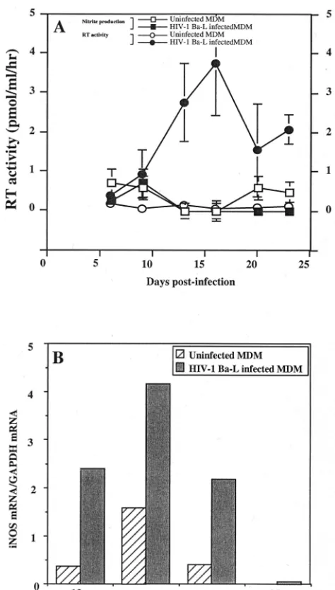
peaked between days 15 and 21 (Fig. 1). As observed in Fig. 1,
a significant induction of iNOS gene expression occurred at the
time of viral replication peak (Fig. 1B). However, this was not
associated with the production of detectable amounts of
ni-trites in culture supernatants (Fig. 1A).
Effects of NO-generating compounds on HIV-1 replication
in MDM.
As reported by Bukrinsky et al. (14, 15), low
expres-sion of iNOS may lead to low production of nitrites remaining
undetectable by the Griess assay, the sensitivity of which is
approximately 250 nM. We therefore designed an
experimen-tal approach to determine whether low levels of NO released
in culture supernatants could modulate HIV replication in
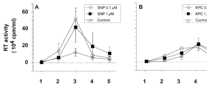
MDM. HIV-1 Ba-L-infected MDM were treated with low
con-centrations of NO donors such as SNP, which is a classically
used NO-generating compound (6, 37, 61, 70, 88). Doses of
SNP that we used were reported by others to be efficient in
human cell cultures (27, 60, 70) and appeared to be suitable for
the in vivo situation, consistent with the levels of NO detected
in plasma of asymptomatic seropositive patients (5, 62, 92). We
verified that in our model, NO
2⫺was generated from SNP in
a dose-dependent manner (data not shown). A sequential and
time-dependent release of nitrite was consistently obtained at
the dose of 10
M. Therefore, 24 h after HIV infection, MDM
were treated with SNP at doses ranging from 0.1 to 10
M.
Concentration in the culture medium was maintained constant
throughout the postinfection period. As control, HIV-1
Ba-L-infected MDM were treated with identical concentrations of
KPC, a compound that is very similar in chemical structure to
SNP but does not generate free NO in culture supernatants.
Unexpectedly, treatment at lower SNP concentrations, 0.1
and 1
M, resulted in a significant increase of viral replication
in MDM cultures (Fig. 2A). This was confirmed in cultures of
MDM obtained from three out of the four tested donors (Fig.
3). At a higher concentration of SNP, 10
M, HIV-1
replica-tion in MDM was inhibited (Fig. 3) as previously reported (68).
NO generated by SNP in culture medium is naturally and
rapidly reduced in nitrite (NO
2⫺). We verified that NO
2⫺was
not, by itself, responsible for the increased replication of
HIV-1 by treating MDM with NaNO
2with doses ranging
be-tween 0.1 and 10
M (data not shown).
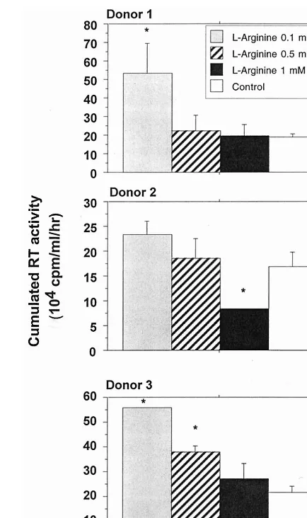
L-Arginine causes a dose-dependent enhancement of HIV-1
replication in human macrophages.
L-Arginine is the natural
substrate of iNOS. In this experiment, MDM were cultured in
the presence of different doses of
L-arginine, ranging from 0.1
to 1 mM, previously reported by Belenky et al. (7) to modulate
NO synthesis in cultured macrophages. These doses did not
appear to affect MDM viability (data not shown). Low
concen-trations of
L-arginine (0.1 and 0.5 nM) appeared to enhance
replication of HIV-1 Ba-L in MDM; this was statistically
sig-nificant for two different donors tested. Addition of 1 mM
L-arginine had no marked effect on viral replication (Fig. 4).
As a reciprocal control, arginase was used to deplete
L-arginine in the culture medium. This enzyme has potential
implication in AIDS pathogenesis since abnormal
concentra-tions could be identified in patient fluids. A dose-dependent
inhibition of HIV replication was observed in the MDM
cul-tures maintained in the presence of arginase (1 to 20 U/ml)
(Fig. 5) with no significant decrease of cellular viability. At the
dose of 10 U/ml, arginase significantly reduces the replication
of HIV (
P
⬍
0.05).
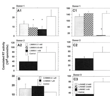
Effect of iNOS inhibitors on HIV-1 replication in human
macrophages.
Two specific competitive inhibitors of iNOS,
[image:3.612.53.290.71.488.2]L
-NMMA and
L-NAME, represent powerful tools to confirm
that endogenous production of NO modulates viral replication
(25, 41, 43, 51). Twenty-four hours after HIV infection, MDM
were treated with one of the two inhibitors at doses ranging
from 0.1 to 1 mM. These components were maintained at
constant concentrations in the culture medium throughout the
postinfection period.
L-NMMA significantly reduced, in a
dose-dependent manner, the replication of HIV-1 Ba-L (Fig.
6A). Moreover, treatment of infected MDM with equal molar
concentrations of
D-NMMA, an inactive enantiomer of
L-NMMA, had no significant effect on HIV-1 replication (Fig.
FIG. 1. Effects of HIV-1 Ba-L replication on iNOS mRNA transcription and NO2⫺production in human MDM cultures. (A)F, level of HIV replication determined by the measure of RT activity in culture supernatants (mean of three independent culture wells⫾SD);E, RT activity in uninfected controls (mean of
three independent culture wells⫾SD);■, production of nitrites in culture supernatants of infected macrophages (mean of three independent culture wells⫾SD);䊐, nitrite production in uninfected control cultures (mean of three independent culture wells⫾SD). (B) Expression of iNOS mRNA in the same culture of infected, or uninfected macrophages, determined as the ratio of the signal obtained for tested mRNA to the signal obtained for GAPDH mRNA (mean of two independent culture wells). Bars represent the mean of two inde-pendent measures.
on November 9, 2019 by guest
http://jvi.asm.org/
6B). Similarly, 1 mM
L-NAME substantially reduced the level
of HIV-1 Ba-L in MDM of three out of the four tested donors
(Fig. 6C and D). The addition of
L-arginine reversed the effects
of
L-NAME in a dose-dependent manner (Fig. 7), confirming
that an arginine-dependent mechanism is involved in the
mod-ulation of HIV replication in MDM.
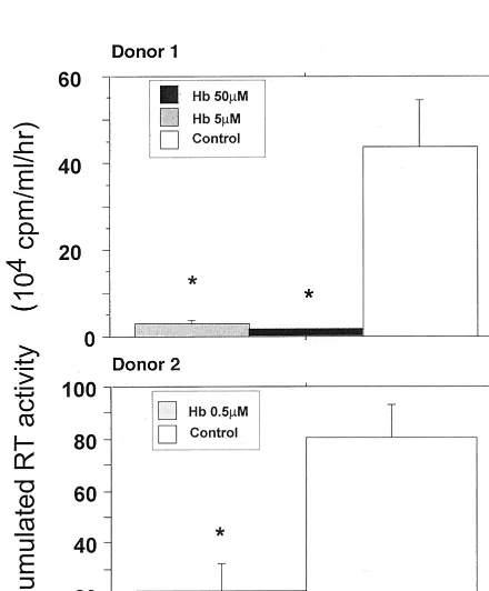
Effects of a NO scavenger, Hb, on HIV-1 replication in
MDM.
NO is a potent local messenger molecule capable of
rapid migration from cell to cell, exerting its effect in both
autocrine and paracrine manners (72). The paracrine mode is
the major mechanism of NO activity (57). The biological
ac-tivity of NO could be abolished in vivo by Hb, which oxidizes
NO to nitrate (48). Zinetti et al. have reported that monocyte
hyperactivation by lipopolysaccharide could be affected by Hb
and
L-NMMA, through the modulation of the NO-dependent
release of tumor necrosis factor alpha (TNF-
␣
) (97). Addition
of Hb in culture supernatants mainly affects extracellular NO
activity (91) by trapping NO produced in culture medium (40).
We observed that 0.5
M Hb is able to significantly decrease
the viral replication in HIV-1 Ba-L-infected MDM (Fig. 8). At
that concentration, no toxic effects of Hb on cultured MDM
have been observed. This confirms the data reported by others
(14, 70). In our culture system, Hb concentrations above 200
M are needed to affect MDM viability (data not shown).
DISCUSSION
The aim of this study was to determine the relationships
between the production of NO and modulation of HIV-1
rep-lication in human macrophages. We first observed that iNOS
gene expression is induced in primary human macrophage
cultures during infection in vitro with the macrophagetropic
HIV-1 Ba-L. Conflicting reports have been published
regard-ing the expression of an iNOS gene in human macrophages;
nevertheless, the increase that we observed confirms the
re-sults found by Bukrinsky et al. in HIV-infected cultures (15).
Despite a significant induction of iNOS expression, we did not
succeed, as others have previously reported (15), in detecting
any nitrite production in culture supernatants. This
observa-tion is nevertheless in agreement with results of Padgett and
Pruett (79), who also detected no nitrite production after
ac-tivation of human macrophages with lipopolysaccharide,
gamma interferon, phorbol myristate acetate, or opsonized
zymosan. However, we cannot exclude that this discrepancy
may also be attributable in part to the low sensitivity of the
Griess reaction, which is estimated to be approximately 250
nM (15, 73, 83).
In vivo, there is convincing evidence that human
macro-phages may synthesize detectable NO during HIV infection. In
plasma, the nitrite and nitrate concentrations correlate with
levels of neopterin, a marker of activation of mononuclear
phagocytes (34). Increased production of NO was evidenced in
PBMC of AIDS patients, in particular in individuals with
op-portunistic infections. Asymptomatic seropositive patients
ex-hibited low production of NO (
⬍
1
M).
The detection of iNOS expression that we observed, without
any substantial accumulation of nitrite, suggested that NO
could be released at low levels after HIV-1 infection. We
therefore investigated the direct effects of low concentrations
of NO on HIV-1 replication in MDM. The release of this
unstable free radical in culture was obtained by using SNP, an
exogenous NO donor. Interestingly, treatment of MDM with
low doses of SNP enhanced viral replication, indicating that
NO may interfere with viral replication mechanisms. This
in-teraction could be directly mediated by NO but may also result
from indirect mechanisms related, for instance, to macrophage
activation (14, 15, 60). Indeed, very low doses of NO (
⬍
1
M)
could enhance soluble guanylate cyclase and GTPase activities,
two markers of macrophage activation.
The observation that the modulation of NO synthesis by
infected macrophages, using
L-arginine, iNOS inhibitors, Hb,
and arginase, modulates in the same direction the replication
of HIV in MDM argues for a pivotal role of NO in the
induc-tion of this phenomenon. The specific involvement of the iNOS
pathway in our experimental model was further demonstrated
by the dose-dependent inhibition of viral replication in the
presence of specific NOS inhibitors.
In summary, the effects of
L-NMMA,
L-NAME, and arginase
on the infected macrophage cultures show that NO can
influ-ence the viral replication through an inducible
L-arginine-de-pendent pathway, which is in accordance with previous report
(14). While it is possible that human macrophages may be
stimulated to produce reactive nitrogen intermediate by
cyto-kines and/or pathogens, our results confirm that the specific
L-arginine-dependent mechanism could be also modulated in
turn, by HIV replication in human macrophage cultures as
described elsewhere (25, 38). This is potentially important,
since it contrasts sharply with the well-established
antimicro-bial and antiviral properties of NO. The unusual low
produc-FIG. 2. Effects of NO-generating compounds on HIV-1 replication in MDM. Purified monocytes from PBMC were cultured for 7 days in 24-well microtiter plates and then infected with HIV-1 Ba-L. Culture supernatant was completed removed every 2 or 3 days and replaced by fresh culture medium. HIV replication in macrophages was determined by the mean level of RT (⫾SD) activity in culture supernatants of three independent wells. Thexaxis indicates the time points of medium exchange. (A) Replication of HIV in infected macrophages treated or not with SNP; (B) effect of the KPC control on HIV replication in macrophages.
on November 9, 2019 by guest
http://jvi.asm.org/
[image:4.612.121.478.71.220.2]tion of NO by HIV-infected human monocytes could probably
explain the lack of antiviral activity. However, these low
con-centrations could be sufficient to affect the biology of MDM,
resulting in enhanced HIV-1 replication.
The molecular mechanisms involved in the induction of NO
production in HIV-infected MDM remain unclear. We can
postulate that iNOS mRNA expression may result from direct
[image:5.612.321.538.70.436.2]interactions between virus and resident macrophages.
Pi-etraforte et al. (83) reported that recombinant HIV envelope
glycoprotein gp120 stimulates a very low production of NO by
human MDM. Enhanced replication of HIV may also involve
the activation of NF-
B, which is a cellular component
regu-lating HIV replication and also the expression of several
cyto-kines (4, 75). Indeed, NO induces the production of TNF-
␣
,
which may in turn activate viral replication in MDM (63, 64).
Previous findings concerning the modulation of NF-
B
activa-tion by NO are controversial. An early study indicates that
chemical NO donors (SNP and SNAP) are able to activate
NF-
B in human peripheral blood mononuclear cells. Lander
et al. (61) have reported that production of nitric oxide radicals
activates the NF-
B transcription factor in doses within the
range of those used in our experiments (68, 80). In our
exper-iments, partial inhibition of HIV needs 10- to 100-times-higher
concentrations of SNP to decrease activation of NF-
B.
How-ever, such high NO concentrations do not reflect the
produc-tion of NO observed in vivo in human fluids (5, 30, 92, 96;
Torre et al., Abstr. 10th Int. Conf. AIDS, 1996).
FIG. 3. Effects of the NO-generating compound SNP on HIV replication in macrophages obtained from four different donors. HIV replication is repre-sented by the sum, at the end of the culture of an individual well, of the RT activities of each time point of medium exchange (every 2 or 3 days). Bars indicate the mean (⫾SD) of RT activity of three independent wells. Statistical analysis was performed using the nonparametric Mann-WhitneyUtest.ⴱ, sig-nificant difference between treated and untreated HIV-infected cultures (P⬍
0.05).
FIG. 4. Effects ofL-arginine on HIV replication in macrophages obtained
from three different donors. HIV replication is represented by the sum, at the end of the culture of an individual well, of the RT activities of each time point of medium exchange (every 2 or 3 days). Bars indicate the mean (⫾SD) of RT activity of three independent wells. Statistical analysis was performed using the nonparametric Mann-WhitneyUtest.ⴱ, significant difference between treated and untreated HIV-infected cultures (P⬍0.05).
on November 9, 2019 by guest
http://jvi.asm.org/
[image:5.612.79.283.74.561.2]Primary targets of reactive nitrogen oxide species may be
different in cells submitted to low (
⬍
1
M) or steady-state
concentrations of NO. An explanation for these conflicting
results might be related to the different fluxes of NO used in
these experiments. Indeed, Lander et al. found that
micromo-lar or submicromomicromo-lar concentrations of pharmacological
sources of NO were sufficient to activate NF-
B via an
en-hancement of GTPase activity (58–60). In other reports,
inhib-itory concentrations of NO donors were frequently 100 times
higher than micromolar amounts shown to activate NF-
B per
se (68, 81). Therefore, it may be that low amounts of NO would
activate NF-
B, whereas high fluxes of NO would be
inhibi-tory. This hypothesis is reinforced by biphasic effects of NO on
GTPase activity, which is inhibited by high concentrations of
NO (58).
In the same way, the tendency to decrease HIV-1 replication
that was observed with higher concentrations of
L-arginine (1
mM) could be due to a negative feedback exerted by NO on
NOS activity as previously described (3, 45, 87).
AIDS is associated with activation of the immune system.
Correlations of nitrite and nitrate with the immune activation
markers (sTNFR 55 and TNFR 75) and neopterin in
HIV-1-infected patients (96) suggest that endogenous cytokines, like
TNF-
␣
, could activate inflammatory cells (44). This additional
priming could be sufficient to amplify the induction of iNOS
and increase NO production by the infected macrophage (10).
The increased production of cytokines and NO may in turn
contribute to the immunopathogenesis of HIV disease both by
enhancing HIV replication and by direct effects on target
tis-sues, such as the brain and lung.
[image:6.612.63.282.72.190.2]The impact of NO production on HIV-1 infection is still
difficult to predict. Our results suggest that NO, in vivo, may
FIG. 5. Effects of arginase on HIV replication in macrophages. HIV repli-cation is represented by the sum, at the end of the culture of an individual well, of the RT activities of each time point of medium exchange (every 2 or 3 days). Bars indicate the mean (⫾SD) of RT activity of three independent wells. Sta-tistical analysis was performed using the nonparametric Mann-WhitneyUtest.ⴱ, significant difference between treated and untreated HIV-infected cultures (P⬍
0.05).
FIG. 6. Effects of iNOS inhibitors on HIV-1 replication in human macrophages obtained from different donors. (A) Effect of the inhibitorL-NMMA; (B) effect of D-NMMA, used as inactive control; (C) effect of the inhibitorL-NAME. HIV replication is represented by the sum, at the end of the culture of an individual well, of
the RT activities of each time point of medium exchange (every 2 or 3 days). Bars indicate the mean (⫾SD) of RT activity of three independent wells. Statistical analysis was performed using the nonparametric Mann-WhitneyUtest.ⴱ, significant difference between treated and untreated HIV-infected cultures (P⬍0.05).
on November 9, 2019 by guest
http://jvi.asm.org/
[image:6.612.111.492.358.689.2]favor virus replication in MDM rather than exert an efficient
antiviral activity. Nevertheless, considerable controversy
re-mains regarding the ability of human macrophages to generate
biologically significant amounts of NO, and it is not clearly
established whether an elevated nitrite level in serum or tissues
of HIV-infected patients is a cause, effect, or epiphenomenon
of HIV-1 infection. However, Torre et al. have shown that
HIV-1 stimulates NO production by human macrophages and
that the NO concentration is increased in the sera of patients
with AIDS, especially in those with neurological disorders and
pulmonary disease caused by intracellular opportunistic
patho-gens (Torre et al., Abstr. 10th Int. Conf. AIDS, 1996).
More-over, Groeneveld et al. have demonstrated that serum nitrate
in such patients correlates positively with viral load, strongly
suggesting that our in vitro observations may be relevant for in
vivo situations and thus should be considered with special
attention for the design of new therapeutic strategies (46). In
addition, the significant increased concentrations of NO
2⫺and
NO
3⫺that we previously observed in plasma in the macaque
model during primary simian immunodeficiency virus infection
(10) seems to be closely related to active virus production.
Peaks of NO
3⫺in plasma and p27 antigenemia were detected
simultaneously in the absence of any opportunistic infections,
suggesting that NO production may therefore contribute to
virus-induced pathogenesis as early as the first days following
infection.
ACKNOWLEDGMENTS
We thank L. Minghetti for helpful discussions and for critical
read-ing of the manuscript. We acknowledge the Centre de Cytaphe´re`se de
l’Hoˆpital Saint-Louis (Paris, France) for technical assistance. We
thank Dominique Marce´ for technical support.
This work was supported by the Agence Nationale de Recherches
sur le SIDA (ANRS; Paris, France), Centre de Recherches du Service
de Sante´ des Arme´es (CRSSA; La Tronche, France), Commissariat a`
l’Energie Atomique (CEA; Fontenay aux Roses, France), and Institut
Paris-Sud sur les Cytokines (IPSC) and Tous ensemble contre le SIDA
(SIDACTION) (Paris, France).
REFERENCES
1.Adamson, D. C., B. Wildemann, M. Sasaki, J. D. Glass, J. C. McArthur, V. I. Christov, T. D. Dawson, and V. L. Dawson.1996. Immunologic NO synthase: elevation in severe AIDS dementia and induction by HIV-1 gp41. Science 274:1917–1921.
2.Akarid, K., M. Sinet, B. Desforges, and M. A. Gougerot-Pocidalo.1995. Inhibitory effect of nitric oxide on the replication of a murine retrovirus in vitro and in vivo. J. Virol.69:7001–7005.
3.Assreuy, J., F. Q. Cunha, F. Y. Liew, and S. Moncada.1993. Feedback inhibition of nitric oxide synthase activity by nitric oxide. Br. J. Pharmacol. 108:833–837.
4.Bachelerie, F., J. Alcami, F. Arenzana-Seisdedos, and J. L. Virelizier.1991. HIV enhancer activity perpetuated by NFB induction on infection of mono-cytes. Nature350:709–712.
5.Baldeweg, T., S. Sooranna, I. Das, J. Catalan, and B. Gazzard.1996. Serum nitrite concentration suggests a role for nitric oxide in AIDS. AIDS10:451– 452.
6.Bates, J. N., M. T. Baker, R. J. Guerra, and D. G. Harrison.1991. Nitric oxide generation from nitroprusside by vascular tissue. Evidence that reduc-tion of the nitroprusside anion and cyanide loss are required. Biochem. Pharmacol.42:S157.
7.Belenky, S. N., R. A. Robbins, and I. Rubinstein.1993. Nitric oxide synthase inhibitors attenuate human monocyte chemotaxis in vitro. J. Leukoc. Biol. 49:380–389.
8.Bender, B. L., B. L. Davidson, R. Kline, C. Brown, and T. C. Quinn.1988. Role of the mononuclear phagocyte system in the immunopathogenesis of human immunodeficiency virus infection and the acquired immunodefi-ciency syndrome. Rev. Infect. Dis.10:1142–1154.
9.Benveniste, O., B. Vaslin, R. Le Grand, A. Che´ret, F. Matheux, F. The´odoro, M. P. Granage, and D. Dormont.1996. Comparative interleukin (IL)-2/ interferon (IFN)-␥and IL-4/IL-10 responses during acute infection of ma-caques inoculated with attenuatednef-truncated or pathogenic SIVmac251 virus. Proc. Natl. Acad. Sci. USA93:3658–3663.
10. Blond, D., A. Che´ret, H. Raoul, R. Le Grand, P. Caufour, F. The´odoro, and D. Dormont.1998. Nitric oxide synthesis during acute SIVmac251 infection of macaques. Res. Virol.149:75–86.
11. Bredt, D. S., C. E. Glatt, P. M. Hwang, M. Fotuhi, T. M. Dawson, and S. H. Snyder.1991. Nitric oxide synthase protein and mRNA are discretely local-ized in neuronal populations of mammalian CNS together with diaphorase. Neuron7:615–624.
12. Bredt, D. S., P. M. Hwang, and S. H. Snyder.1990. Localization of nitric oxide synthase indicating a neural role for nitric oxide. Nature347:768–770. 13. Buhl, R.1994. Imbalance between oxidants and antioxidants in the lungs of
HIV-seropositive individuals. Chem. Biol. Interact.91:147–158.
[image:7.612.55.293.73.203.2]14. Bukrinsky, M., H. Schmidtmayerova, G. Zybarth, L. Dubrovsky, B. Sherry, FIG. 7. Reversion of the effect of the inhibitorL-NAME on HIV-1
replica-tion in human macrophages by the addireplica-tion of arginine. HIV replicareplica-tion is represented by the sum, at the end of the culture of an individual well, of the RT activities of each time point of medium exchange (every 2 or 3 days). Bars indicate the mean (⫾SD) of RT activity of three independent wells. Statistical analysis was performed using the nonparametric Mann-WhitneyUtest.ⴱ, sig-nificant differences between treated and untreated HIV-infected cultures (P⬍
0.05).
FIG. 8. Effects of the NO scavenger Hb on HIV-1 replication in MDM in macrophages obtained from two different donors. HIV replication is represented by the sum, at the end of the culture of an individual well, of the RT activities of each time point of medium exchange (every 2 or 3 days). Bars indicate the mean (⫾SD) of RT activity of three independent wells. Statistical analysis was per-formed using the nonparametric Mann-WhitneyUtest.ⴱ, significant differences between treated and untreated HIV-infected cultures (P⬍0.05).
on November 9, 2019 by guest
http://jvi.asm.org/
[image:7.612.63.283.400.666.2]and G. Enikolopov.1996. A critical of nitric oxide in human immunodefi-ciency virus type 1-induced hyperresponsiveness of cultured monocytes. Mol. Med.2:460–468.
15.Bukrinsky, M. I., H. S. L. M. Nottet, H. Schmidtmayerova, L. Dubrovsky, C. R. Flanagan, M. E. Mullins, S. A. Lipton, and H. E. Gendelman.1995. Regulation of nitric oxide synthase activity in human immunodeficiency virus type 1 (HIV-1)-infected monocytes: implications for HIV-associated neuro-logical disease. J. Exp. Med.181:735–745.
16. Che´ret, A., P. Caufour, R. Le Grand, F. The´odoro, F. Boussin, B. Vaslin, and D. Dormont.1997. Macrophage inflammatory protein-1␣mRNA expression in mononuclear cells from different tissues during acute simian immunode-ficiency virus strain mac251 infection of macaques. AIDS11:257–258. 17. Che´ret, A., R. Le Grand, P. Caufour, N. Dereudre-Bosquet, F. Matheux, O.
Neildez, F. The´odoro, N. Maestrali, O. Benveniste, B. Vaslin, and D. Dor-mont.1996. Cytokines mRNA expression in mononuclear cells from differ-ent tissues during acute SIVmac251 infection of macaques. AIDS Res. Hum. Retroviruses12:1263–1272.
18. Chomczynski, P., and N. Sacchi.1987. Single-step method of RNA isolation by acid guanidium thiocyanate-phenol-chloroform extraction. Ann. Bio-chem.162:156–159.
19. Clayette, P., N. Dereuddre-Bosquet, M. Martin, P. Fretier, and D. Dormont. 1997. Effects of RP55778, a tumor necrosis factor alpha synthesis inhibitor, on antiviral activity of dideoxynucleosides. Antimicrob. Agents Chemother. 41:875–877.
20. Croen, K. D.1993. Evidence for antiviral effect of nitric oxide. Inhibition of herpes simplex virus type 1 replication. J. Clin. Investig.91:2446–2452. 21. Curran, R. D., T. R. Billiar, D. J. Stuehr, J. B. Ochoa, B. G. Harbrecht, S. G.
Flint, and R. L. Simmons.1990. Multiple cytokines are required to induce hepatocyte nitric oxide production and inhibit total protein synthesis. Ann. Surg.212:462–471.
22. Currie, G. A.1978. Activated macrophages kill tumour cells by releasing arginase. Nature273:758–759.
23. Dawson, V. L., T. M. Dawson, D. A. Bartley, G. R. Uhl, and S. H. Snyder. 1993. Mechanisms of nitric oxide-mediated neurotoxicity in primary brain cultures. J. Neurosci.13:2651–2661.
24. Dawson, V. L., T. M. Dawson, E. D. London, D. S. Bredt, and S. H. Snyder. 1991. Nitric oxide mediates glutamate neurotoxicity in primary cortical cul-tures. Proc. Natl. Acad. Sci. USA88:6368–6371.
25. Denis, M. 1991. Interferon-gamma-treated murine macrophages inhibit growth of tubercle bacilli via the generation of reactive nitrogen intermedi-ates. Cell Immunol.132:150–157.
26. Dreyer, E. B., K. Kaiser, J. T. Offerman, and S. A. Lipton.1990. HIV-1 coat protein neurotoxicity prevented by calcium channel antagonists. Science 248:364–367.
27. Efron, D. T., S. J. Kirk, M. C. Regan, H. L. Wasserkrug, and A. Barbul.1991. Nitric oxide generation fromL-arginine is required for optimal human
pe-ripheral blood lymphocyte DNA synthesis. Surgery110:327–334. 28.Ennen, J., I. Seipp, S. G. Norley, and R. Kurth.1990. Decreased accessory
cell function of macrophages after infection with human immunodeficiency virus type 1 in vitro. Eur. J. Immunol.20:2451–2456.
29.Estevez, M. E., I. J. Ballart, R. A. Diez, N. Planes, C. Scaglione, and L. Sen. 1986. Early defect of phagocytic cell function in subjects at risk for acquired immunodeficiency syndrome. Scand. J. Immunol.24:215–222.
30.Evans, T. G., K. Rasmussen, G. Wiebke, and J. B. Hibbs.1994. Nitric oxide synthesis in patients with advanced HIV infection. Clin. Exp. Immunol. 97:83–86.
31.Figdor, C. G., J. M. M. Leemans, W. S. Bont, and J. E. Vries.1983. Theory and practice of centrifugal elutriation (CE). Factors influencing the separa-tion of human blood cells. Cell. Biophys.5:105–118.
32.Forstermann, U., L. D. Gorsky, J. S. Pollock, K. Ishii, H. H. H. W. Schmidt, M. Heller, and F. Murad.1990. Hormone-induced biosynthesis of endothe-lium-derived relaxing factor/nitric-like material in NIE-115 neuroblastoma cells requires calcium and calmodulin. Mol. Pharmacol.38:7–15. 33. Fu, Y., and E. P. Blankehorn.1992. Nitric oxide-induced antimitogenic
effects in high and low responder rat strains. J. Immunol.148:2217–2223. 34. Fuchs, D., A. Hausen, G. Reibnegger, E. R. Werner, P. Dierich, and H.
Watcher.1988. Neopterin as a marker for activated cell-mediated-immunity: application in HIV infection. Immunol. Today369:150–155.
35. Gartner, S., P. Markovits, D. M. Markovitz, M. H. Kaplan, R. C. Gallo, and M. Popovic.1986. The role of mononuclear phagocytes in HTLV-III/LAV infection. Science233:215–219.
36. Gartner, S., D. M. Markovitz, R. F. Markovitz, P. Betts, and M. Popovic. 1988. Virus isolation from and identification of HTLV-III/LAV-producing cells in brain tissue from a patient with AIDS. JAMA256:2365–2371. 37. Gartwright, J. E., G. Whitley, and A. P. Johnstone.1997. Endothelial cell
adhesion molecule expression and lymphocyte adhesion to endothelial cells: effect of nitric oxide. Exp. Cell Res.235:431–434.
38. Gazzinelli, R. T., I. P. Oswald, S. Hieny, S. L. James, and A. Sher.1992. The microbicidal activity of interferon-gamma-treated macrophages against Trypanosoma cruzi involves anL-arginine-dependent, nitrogen
oxide-medi-ated mechanism inhibitable by interleukin-10 and transforming growth fac-tor-beta. Eur. J. Immunol.22:2501–2506.
39. Gendelman, H. E., J. M. Orenstein, L. M. Baca, B. Weiser, H. Burger, D. C. Kalter, and M. S. Meltzer.1989. The macrophage in the persistence and pathogenous of HIV infection. AIDS3:475–495.
40. Goretski, J., and T. C. Hollocher.1988. Trapping of nitric oxide produced during dentrification by extracellular hemoglobin. J. Biol. Chem.263:2316– 2323.
41. Granger, D. L., J. B. J. Hibbs, J. R. Perfect, and D. I. Durack.1991. Specific amino acid (L-arginine) requirement for the microbiostatic activity of murine
macrophages. J. Clin. Investig.81:1129–1136.
42. Graziosi, C., K. R. Gantt, M. Vaccarezza, J. F. Demarest, M. Daucher, M. S. Saag, G. M. Shaw, T. C. Quinn, O. J. Cohen, C. C. Welbon, G. Pantaleo, and A. S. Fauci.1996. Kinetics of cytokine expression during primary human immunodeficiency virus type 1 infection. Proc. Natl. Acad. Sci. USA93: 4386–4391.
43. Green, S. J., R. M. Crawford, J. T. Hockmeyer, M. S. Meltzer, and C. A. Nacy.1990. Leishmania major amastigotes initiate theL-arginine-dependent
killing mechanism in IFN-␥stimulated macrophages by induction of tumor necrosis factor-␣. J. Immunol.145:4290–4297.
44. Grimaldi, L. M. E., G. V. Martino, D. M. Franciotta, R. Brustia, A. Cast-agna, R. Pristera, and A. Lazzarin.1991. Elevated alpha-tumor necrosis factor levels in spinal fluid from HIV-1-infected patients with central nervous system involvement. Ann. Neurol.29:21–25.
45. Griscavage, J. M., N. E. Rogers, M. P. Sherman, and L. J. Ignarro.1993. Inducible nitric oxide synthase from a rat alveolar macrophage cell line inhibited by nitric oxide. J. Immunol.151:6329–6337.
46. Groeneveld, P. H., F. P. Kroon, P. H. Nibbering, S. M. Bruisten, P. van Swieten, and R. van Furth.1996. Increased production of nitric oxide cor-relates with viral load and activation of mononuclear phagocytes in HIV-infected patients. Scand. J. Infect. Dis.28:341–345.
47. Harada, S., Y. Koyanagi, and N. Yamamoto.1985. Infection of HTLV-IIIB/ LAV in HTLV-I-carrying cells MT2 and MT4 and application in a plate assay. Science229:563–566.
48. Haussmann, N., and J. Werringloer.1985. Nitric oxide and nitrite formation during degradation ofN-nitrosoamines. Naunyn Schmiedbergs Arch. Phar-makol.329:R21.
49. Ignarro, L. J., R. E. Burns, G. M. Buga, and K. S. Wood.1987. Endothelium-derived relaxing factor from pulmonary artery and vein possesses pharma-cologic and chemical properties identical to those of nitric oxide radical. Circ. Res.61:866–879.
50. Isobe, K., and I. Nakashima.1992. Feedback suppression of staphylococcal enterotoxin-stimulated T-lymphocyte proliferation by macrophages through inductive nitric oxide synthesis. Infect. Immun.60:4832–4837.
51. James, S. L., and J. Glaven.1989. Macrophage cytotoxicity against schisto-somula ofSchistosoma mansoniinvolves arginine-dependent production of reactive nitrogen intermediates. J. Immunol.143:4208–4212.
52. Karupiah, G., Q. Xie, R. M. L. Buller, C. Nathan, C. Duarte, and J. D. MacMicking.1993. Inhibition of viral replication by interferon-gamma in-duced nitric oxide synthase. Science261:1445–1448.
53. Knowles, R. G., M. Palacios, R. M. J. Palmer, and S. Moncada.1989. Formation of nitric oxide fromL-arginine in the central nervous system: a
transduction mechanism for stimulation of guanylate cyclase. Proc. Natl. Acad. Sci. USA86:5159–5162.
54. Kobzick, L., D. S. Bredt, C. J. Lowenstein, J. Drazen, B. Gaston, D. Sugar-baker, and J. S. Stamler.1993. Nitric oxide synthase in human and rat lung: immunocytochemical and histochemical localization. Am. J. Respir. Cell. Mol. Biol.9:371–377.
55. Koening, S., H. E. Gendelman, J. M. Orenstein, M. C. Dal-Canto, G. H. Pezeshkpour, M. Yungbluth, F. Janotta, A. Aksamit, M. A. Martin, and A. S. Fauci.1986. Detection of AIDS virus in macrophages in brain tissue from AIDS patients with encephalopathy. Science233:1089–1093.
56. Kolb, J. P., N. Paul-Eugene, C. Damais, K. Yamaoka, J. C. Drapier, and B. Dugas.1994. Interleukin-4 stimulates cGMP production by IFN-␥-activated human monocytes. J. Biol. Chem.269:9811–9816.
57. Lancaster, J. R.1994. Stimulation of the diffusion and reaction of endog-enously produced nitric oxide. Proc. Natl. Acad. Sci. USA91:8137–8141. 58. Lander, H. M., D. P. Hajjar, B. L. Hempstead, U. A. Mirza, B. T. Chait, S.
Campbell, and L. A. Quilliam.1997. A molecular redox switch on p21(ras)-structural basis for the nitric oxide-p21(ras) interaction. J. Biol. Chem.272: 4323–4326.
59. Lander, H. M., J. S. Ogiste, S. F. A. Pearce, R. Levi, and A. Novogrodsky. 1995. Nitric oxide-stimulated guanine nucleotide exchange on p21ras. J. Biol. Chem.270:7017–7020.
60. Lander, H. M., P. Sehajpal, D. M. Levine, and A. Novogrodsky.1993. Acti-vation of human peripheral blood mononuclear cells by nitric oxide-gener-ating compounds. J. Immunol.150:1509–1516.
61. Lander, H. M., P. K. Sehajpal, and A. Novogrosky.1993. Nitric oxide sig-naling: a possible role for G proteins. J. Immunol.151:7182–7187. 62. Lane, T., M. J. Buchmeier, D. D. Wtry, and S. Fox.1996. Expression of
inflammatory cytokines and inducible nitric oxide synthase in brains of SIV-infected rhesus monkeys: applications to HIV-induced central nervous sys-tem disease. Mol. Med.2:27–37.
63. Le Naour, R., P. Clayette, Y. Henin, A. Mabondzo, H. Raoul, A. Bousseau,
on November 9, 2019 by guest
http://jvi.asm.org/
and D. Dormont.1994. Infection of human macrophages with endogenous tumor necrosis factor-␣(TNF-␣)-independent human immunodeficiency vi-rus type 1 isolate is unresponsive to the TNF-␣synthesis inhibitor RP55778. J. Gen. Virol.75:1379–1388.
64. Le Naour, R., H. Raoul, A. Mabondzo, Y. Henin, A. Bousseau, and D. Dormont.1994. Treatment of human monocyte-derived macrophages with a TNF-␣synthesis inhibitor prior to HIV-1 infection: consequences on cyto-kine production and viral replication. Res. Virol.145:199–207.
65. Lorsbach, R. B., W. J. Murphy, C. J. Lowenstein, S. H. Snyder, and S. W. Russel.1993. Expression of the nitric oxide synthase gene in mouse macro-phages activated for tumor cell killing. J. Biol. Chem.268:1908–1913. 66. Mabondzo, A., F. Boussin, H. Raoul, R. Le Naour, G. Gras, B. Vaslin, J.
Bartholeyns, J.-L. Romet-Lemonne, and D. Dormont.1994. Antibody-de-pendent cellular cytotoxicity and neutralization of human immunodeficiency virus type 1 by high affinity cross-linking of gp41 to human macrophage Fc IgG receptor using bispecific antibody. J. Gen. Virol.75:1451–1456. 67. Mannick, J. B.1995. The antiviral role of nitric oxide. Res. Immunol.146:
693–697.
68. Matthews, J. R., C. H. Botting, M. Panico, H. R. Morris, and R. T. Hay.1996. Inhibition of NF-B DNA binding by nitric oxide. Nucleic Acids Res.14: 2236–2242.
69. Melkova, Z., and M. Esteban.1995. Inhibition of vaccinia virus DNA repli-cation by inducible expression of nitric oxide synthase. J. Immunol.155: 5711–5718.
70. Merryman, P. F., R. M. Clancy, X. Y. He, and S. B. Abramson.1993. Modulation of human T cell responses by nitric oxide and its derivative, S-nitrosoglutathione. Arthritis Rheum.36:1414–1422.
71. Mills, C. D.1991. Molecular basis of suppressor macrophages: arginine metabolism via the nitric oxide synthetase pathway. J. Immunol.146:2719– 2723.
72. Moncada, S., R. M. Palmer, and E. A. Higgs.1991. Nitric oxide: physiology, pathophysiology, and pharmacology. Pharmacol. Rev.43:109–142. 73. Mordvintcev, P., A. Mulsch, R. Busse, and A. Vanin.1991. On-line detection
of nitric oxide formation in liquid aqueous phase by electron paramagnetic resonance spectroscopy. Anal. Biochem.199:142–146.
74. Murray, H. W.1988. Survival of intracellular pathogens within human mono-nuclear phagocytes. Semin. Hematol.25:101–111.
75. Nabel, G., and D. Baltimore.1987. An inducible transcription factor acti-vates expression of human immunodeficiency virus in T cells. Nature326: 711–713.
76. Nathan, C. 1992. Nitric oxide a secretory product of mammalian cells. FASEB J.6:3051–3064.
77. Nussler, A., M. DiSilvio, T. Billiar, R. Hoffman, D. Geller, R. Selby, J. Madariaga, and R. L. Simmons.1992. Stimulation of the nitric oxide syn-thase pathway in human hepatocytes by cytokines and endotoxin. J. Exp. Med.176:261–264.
78. Nussler, A. K., D. A. Geller, M. A. Sweetland, M. DiSilvio, T. R. Billiar, J. B. Madriaga, R. L. Simmons, and J. R. Lancaster.1993. Induction of nitric oxide synthesis and its reactions in cultured human hepatocytes and stimu-lated with cytokines plus LPS. Biochem. Biophys. Res. Commun.194:826– 835.
79. Padgett, E. L., and S. B. Pruett.1992. Evaluation of nitrite production by human monocyte-derived macrophages. Biochem. Biophys. Res. Commun. 186:775–781.
80. Peng, H. B., P. Libby, and J. K. Liao.1995. Induction and stabilization of IB␣by nitric oxide mediates inhibition of NF-B. J. Biol. Chem.270:14214– 14219.
81. Peng, H. B., T. B. Rajavaschisth, P. Libby, and J. K. Liao.1995. Nitric oxide inhibits macrophage-colony stimulating factor gene transcription in vascular endothelial cells. J. Biol. Chem.270:17050–17055.
82. Persoons, J. H. A., K. Schornagel, F. F. H. Tilders, J. de Vente, F. Berken-bosch, and G. Kraal.1996. Alveolar macrophages autoregulate IL-1 and IL-6 production by endogenous nitric oxide. Am. J. Respir. Cell Mol. Biol. 14:272–278.
83. Pietraforte, D., E. Tritarelli, U. Testa, and M. Minetti.1994. Gp 120 HIV envelope glycoprotein increases the production of nitric oxide in human monocyte-derived macrophages. J. Leukoc. Biol.55:175–182.
84. Raoul, H., R. Le Naour, D. Blond, and D. Dormont.1998. HIV-1 infection of human macrophages induces an up-regulation of manganese superoxide dismutase gene that may protect cells from death. AIDS Res. Hum. Retro-viruses14:427–434.
85. Reiling, N., A. J. Ulmer, M. Duchrow, M. Ernst, H. D. Flad, and S. Haus-childt.1994. Nitric oxide synthase: mRNA expression of different isoforms in human monocytes/macrophages. Eur. J. Immunol.24:1941–1944. 86. Rey, M., D. Dormont, F. Barre´-Sinoussi, L. Montagnier, and J. C.
Cher-mann.1984. Characterization of the RNA-dependent DNA polymerase of a new human lymphotropic retrovirus (lymphadenopathy-associated virus). Biochem. Biophys. Res. Commun.121:126–133.
87. Sheffler, L. A., D. A. Wink, G. Melillo, and G. W. Cox.1995. Exogenous nitric oxide regulate IFN-␥plus lipopolysaccharide-induced nitric oxide synthase expression in mouse macrophages. J. Immunol.155:886–894.
88. Shoker, A. S., H. Yang, M. A. Muramit, H. Jamil, A. Al-Ghoul, and K. Okasha.1997. Analysis of thein vitroof exogenous nitric oxide on human lymphocytes. Mol. Cell. Biochem.171:75–83.
89. Sinicco, A., A. Biglino, M. Sciandra, B. Forno, A. M. Pollono, R. Raiteri, and P. Gioannini.1993. Cytokine network and acute primary HIV-1 infection. AIDS7:1167–1172.
90. Stamler, J. S., D. J. Singel, and J. Loscalzo.1992. Biochemestry of nitric oxide and its redox-activated forms. Science258:1896–1902.
91. Stuehr, D. J., and C. F. Nathan.1989. Nitric oxide. A macrophage product responsible for cytostasis and respiratory inhibition in tumor target cells. J. Exp. Med.169:1543–1555.
92. Torre, D., G. Ferrario, G. Bonetta, F. Speranza, and C. Zeroli.1995. Pro-duction of nitric oxide from peripheral blood mononuclear cells and poly-morphonuclear leukocytes of patients with HIV-1 infection. AIDS1995:979– 980.
93. Villinger, F., D. Hunt, A. Mayne, M. Vuchetich, H. Findley, and A. A. Ansari. 1993. Qualitative and quantitative studies of cytokines synthesized and se-creted by non-human primate peripheral blood mononuclear cells. Cytokine 5:469–479.
94. Wright, S. C., C. U. Jewett, R. Mitsuyasu, and B. Bonavida.1988. Sponta-neous cytotoxicity and tumor necrosis factor production by peripheral blood monocytes from AIDS patients. J. Immunol.141:99.
95. Xie, Q. W., H. J. Cho, J. Calaycay, R. A. Mumford, K. M. Swiderek, T. D. Lee, A. Ding, T. Troso, and C. Nathan.1992. Cloning and characterization of inducible nitric oxide synthase from mouse macrophages. Science256:225– 228.
96. Zangerle, R., D. Fuchs, G. Reibnegger, G. Werner-Felmayer, H. Gallati, H. Wachter, and E. R. Werner.1995. Serum nitrite plus nitrate in infection with human immunodeficiency virus type-1. Immunobiology193:59–70. 97. Zinetti, M., G. Fantuzzi, R. Delgado, E. Di Santo, P. Chezzi, and M. Fratelli.
1995. Endogenous nitric oxide production by monocytic cells regulates LPS-induced TNF production. Eur. Cytokine Netw.6:45–48.
on November 9, 2019 by guest
http://jvi.asm.org/