Copyright © 1998, American Society for Microbiology
Expression of CCR5 Increases during Monocyte Differentiation
and Directly Mediates Macrophage Susceptibility to Infection
by Human Immunodeficiency Virus Type 1
DANIEL L. TUTTLE,1JEFFREY K. HARRISON,2CYNTHIA ANDERS,1JOHN W. SLEASMAN,1,3
ANDMAUREEN M. GOODENOW1,3*
Department of Pathology, Immunology, and Laboratory Medicine,1Department of Pharmacology and Therapeutics,2
and Division of Immunology and Infectious Diseases, Department of Pediatrics,3University of Florida
College of Medicine, Gainesville, Florida 32610
Received 25 November 1997/Accepted 3 March 1998
The stage of differentiation and the lineage of CD41cells profoundly affect their susceptibility to infection
by human immunodeficiency virus type 1 (HIV-1). While CD41T lymphocytes in patients are readily
suscep-tible to HIV-1 infection, peripheral blood monocytes are relatively resistant during acute or early infection, even though monocytes also express CD4 and viral strains with macrophage (M)-tropic phenotypes predom-inate. CCR5, the main coreceptor for M-tropic viruses, clearly contributes to the ability of CD41T cells to be
infected. To determine whether low levels of CCR5 expression account for the block in infection of monocytes, we examined primary monocyte lineage cells during differentiation. Culturing of blood monocytes for 5 days led to an increase in the mean number of CCR5-positive cells from <20% of monocytes to >80% of monocyte-derived macrophages (MDM). Levels of CCR5 expression per monocyte were generally lower than those on MDM, perhaps below a minimum threshold level necessary for efficient infection. Productive infection may be restricted to the small subset of monocytes that express relatively high levels of CCR5. Steady-state CCR5 mRNA levels also increased four- to fivefold during MDM differentiation. Infection of MDM by M-tropic HIV-1JRFLresulted in >10-fold-higher levels of p24, and MDM harbored >30-fold more HIV-1 DNA copies
than monocytes. In the presence of the CCR5-specific monoclonal antibody (MAb) 2D7, virus production and cellular levels of HIV-1 DNA were decreased by >80% in MDM, indicating a block in viral entry. There was a direct association between levels of CCR5 and differentiation of monocytes to macrophages. Levels of CCR5 were related to monocyte resistance and macrophage susceptibility to infection because infection by the M-tropic strain HIV-1JRFLcould be blocked by MAb 2D7. These results provide direct evidence that CCR5
functions as a coreceptor for HIV-1 infection of primary macrophages.
Tissue macrophages are a major target for human immuno-deficiency virus type 1 (HIV-1) infection (19, 45). Chronically infected macrophages could provide a reservoir of HIV-1 that persists in patients whose CD41T-cell viral burdens have been drastically reduced by highly active antiretroviral combination therapies which include protease inhibitors (30). Monocyte-derived macrophages (MDM) are not killed by HIV-1 infec-tion but produce virus for as long as several weeks in cultures. In contrast, activated CD41T cells are highly sensitive to the cytopathic effects of HIV-1. MDM have the potential to act as long-lived reservoirs for HIV-1 and to disseminate the virus to other tissues (30, 45, 46). Elucidation of the mechanism of macrophage infection is essential to the design of effective therapeutic strategies that not only block viable virus produc-tion by short-lived T cells but also prevent new infecproduc-tions of long-lived tissue macrophages.
The stage of differentiation and the lineage of CD41cells profoundly affect their susceptibility to infection by HIV-1. For example, memory CD41T cells, which express CD45RO and differentiate from naive CD41T cells in a postthymic antigen-dependent process, are infected to greater levels than naive CD45RA-expressing CD41 T cells (36, 43). During acute stages of pediatric infection, as many as 1 to 10% of peripheral
blood CD41T cells can harbor proviral forms of HIV-1 (3). In contrast, cells of the monocyte lineage in peripheral blood are rarely infected during acute or early infection (3, 25, 26, 37), even though monocytes express CD4 and viral strains with macrophage (M)-tropic phenotypes predominate (15, 31, 52). Differentiation of monocytes to macrophages in tissues such as brain or lung can result in susceptibility to infection. Mono-cytes in cultures must also undergo differentiation to macro-phages to become maximally susceptible to productive infec-tion by M-tropic viruses (20, 32, 45). Levels of CD4 expression in differentiated macrophages are lower than those in blood monocytes, indicating that monocyte resistance and macro-phage susceptibility to infection involve some factor(s) other than absolute levels of CD4 (46).
The chemokine receptor CCR5 serves as a coreceptor for M-tropic HIV-1 entry into CD4-expressing T lymphocytes (4, 9, 11, 17, 18, 24) and is a critical factor in HIV-1 pathogenesis. Individuals deficient in CCR5 and peripheral blood mononu-clear cells (PBMC) derived from these subjects show high levels of resistance to HIV-1 infection (16, 21, 24, 27, 34). CCR5 is differentially expressed on naive and memory T cells (8). Low levels of CCR5 on CD45RA T cells and high levels on CD45RO T cells appear to be associated with differential levels of infection in these CD41T-cell subsets. Infection of CD41T cells by M-tropic strains of HIV-1 is antagonized by the che-mokine peptides RANTES, MIP-1a, and MIP-1b, which are natural ligands of CCR5 (5, 13, 17).
Although CCR5 clearly contributes to the susceptibility of
* Corresponding author. Mailing address: Box 100275, 1600 SW Archer Rd., University of Florida College of Medicine, Gainesville, FL 32610. Phone: (352) 392-3429. Fax: (352) 392-0481. E-mail: goodenow [email protected].
4962
on November 9, 2019 by guest
http://jvi.asm.org/
CD41 T lymphocytes to infection, the evidence that CCR5 participates in infection of CD41macrophages by M-tropic viruses is less convincing. For example, some blood monocytes and tissue macrophages express CCR5 (8, 50), and deletion of CCR5 renders MDM resistant to infection by viruses that require CCR5 (9), supporting a role for CCR5 in macrophage infection. However, the data are contradictory with regard to the ability of CCR5 ligands to inhibit infection of MDM, rais-ing the possibility that macrophages are infected by a CCR5-independent mechanism or that cell type influences interac-tions among CCR5, its ligands, and the HIV-1 envelope glycoprotein (9, 35, 49).
Chemokine receptor-dependent HIV-1 infection can be in-hibited not only by ligands for receptors but also by monoclo-nal antibodies (MAb) (49). We used 2D7, a MAb that recog-nizes an epitope in the second extracellular loop of CCR5 (49), to assess the role of CCR5 in HIV-1 infection of monocytes and macrophages. We found (i) that there is a direct associa-tion between levels of CCR5 and differentiaassocia-tion of monocytes to macrophages, (ii) that levels of CCR5 are related to mac-rophage susceptibility to infection, and (iii) that macmac-rophage infection by M-tropic strain HIV-1JRFLcan be blocked by 2D7. Our results provide direct evidence that CCR5 functions as a coreceptor for HIV-1 infection of macrophages.
MATERIALS AND METHODS
MDM cultures.PBMC were obtained by Lymphoprep (Sigma) density gradi-ent cgradi-entrifugation of commercial leukocyte preparations from HIV-1-negative donors. Monocytes were enriched from the PBMC fraction by 42.55% Percoll density gradient centrifugation, adherence to plastic for 1 h in RPMI 1640 (Gibco-BRL) plus 20% human AB serum that was negative for human immu-nodeficiency virus and hepatitis B virus antibodies (Sigma), and extensive rinsing in RPMI 1640 without serum (14). Monocyte preparations in which 85 to 90% of the CD4-positive cells were also CD14 positive were cultured at approximately 53105cells per well in 24-well plates (Falcon 3047) containing differentiation medium (RPMI 1640 plus 20% human AB serum and supplemented with 1 ng of recombinant human granulocyte-macrophage colony-stimulating factor [Gibco-BRL] per ml, 100 U of penicillin per ml, and 100mg of streptomycin per ml). Monocytes differentiated after 5 days in culture into macrophages (MDM); the macrophages were evaluated by morphological criteria, esterase staining, and flow cytometry analysis, which indicated,3% contamination with T cells. MDM preparations are routinely characterized for susceptibility to infection by M-tropic HIV-1JRFLand resistance to infection by T-cell-line-tropic HIV-1LAIor HIV-1MN.
Virus infection.HIV-1JRFL, which uses the chemokine receptor CCR5 as a cofactor, was used in all experiments (9, 23). A stock of HIV-1JRFLwas produced from infected PBMC supernatants, which were filtered through 0.45-m m-pore-size sterile membranes, and stored in aliquots at280°C. The infectivity titer was quantified by a p24 antigen assay (Coulter) based upon a 50% tissue culture infective dose in a terminal dilution assay with MDM. The same stock of virus was used in all experiments. The HIV-1JRFLstock was also titrated on HOS-CD4.CCR5 cells (human osteosarcoma cells stably expressing CD4 and CCR5; NIH AIDS Research and Reference Reagent Program); the amount of virus used to infect primary monocytes or MDM was in the range of inoculation that maximally infected this cell line. Monocytes and MDM were infected with 15 50% tissue culture infective doses of virus in a medium volume of 0.25 ml at 37°C for 12 to 14 h. Excess virus was removed by three washes with phosphate-buffered saline. HIV-1 production was assessed by measuring the amount of soluble p24 antigen released into the supernatant by duplicate cultures. For experiments involving blocking of infection by antibodies, cells were treated for 30 min with various antibody concentrations prior to virus inoculation at 37°C. The stock CCR5-specific MAb 2D7 concentration was 392mg/ml, and dilutions were made from this stock.
Analysis of chemokine receptor mRNA.Total RNA was isolated from mono-cytes and MDM with RNeasy Kits (Qiagen), treated with DNase I (Gibco-BRL), and processed with the RNeasy RNA Clean-up protocol to remove residual deoxyribonucleotides prior to quantitation by spectrophotometry. Cyclophilin, which is expressed constitutively in human leukocytes and serves as a standard to normalize input template (6, 33), or CCR5 cDNAs were synthesized with specific downstream primers (6, 24), 0.2 or 1.0mg of total RNA for cyclophilin or CCR5, respectively, by use of the Superscript Preamplification System (Gibco-BRL). Amplifications were performed in a 9600 GeneAmp PCR system thermocycler (Perkin-Elmer Cetus) by use of an initial denaturation cycle at 94°C for 5 min, 30 cycles of amplification (1 min at 94°C, 1 min at 60°C, and 1 min at 72°C for each cycle), and a final extension cycle at 72°C for 10 min. For each RNA preparation,
cyclophilin and CCR5 were amplified in control reactions without reverse tran-scriptase to demonstrate the absence of DNA contamination. Products were separated by electrophoresis on ethidium bromide-stained 1% agarose gels. The image was stored digitally with a charge-coupled device camera system (29) and quantified with SIGMAGEL software (Jandel Scientific). Regression analysis was performed to determine the linear range in which the amount of product was proportional to the amount of template (39, 40). Regression coefficients for the linear range of cyclophilin amplification were always.0.97. Relative amounts of cyclophilin and CCR5 mRNAs were determined from double log graphs.
Analysis of HIV-1 DNA.High-molecular-weight genomic DNA was isolated from monocytes or MDM at 5 and 10 days postinfection. Cell monolayers were washed in phosphate-buffered saline and lysed directly in wells containing 200ml of K buffer (10 mM Tris-HCl [pH 8.3], 50 mM KCl, 2.5 mM MgCl2, 0.1 mg of gelatin per ml, 0.45% Nonidet P-40, 0.45% Tween 20, 60mg of proteinase K per ml). Lysates were incubated at 50°C for 1 h and at 95°C for 5 min and then stored at 4°C.
Aliquots of cell lysates were amplified in 50-ml reaction mixtures containing 13PCR Buffer II (Perkin-Elmer), 1.75 mM MgCl2, 0.2 mM each deoxyribo-nucleoside triphosphate, 1 U of Taq DNA polymerase (Perkin-Elmer), and 0.1
mM each env region primer (1). Reactions were carried out for an initial dena-turation cycle at 94°C for 5 min, amplification at 94°C for 30 s, 60°C for 1 min, and 72°C for 2 min for 35 cycles, and a final extension cycle at 72°C for 10 min. These conditions yield products in the linear range of amplification, as deter-mined by titration of 8E5 cell DNA (1). Intersample DNA amounts were stan-dardized in reactions with primers specific for humanb-actin (2). Products were analyzed by hybridization (2, 41). For calculation of cell numbers and estimation of numbers of HIV-1 DNA copies in infected cells, serial dilutions of DNA from 8E5 cells, which contain one complete proviral copy per cell, were amplified with
b-actin and env primers, and the resulting products were hybridized for use as DNA standards (47).
Flow cytometry analysis.PBMC and MDM were prepared for two-color flow cytometry as described previously (42), except that a final concentration of 2% human serum was added to all staining and wash buffers. MDM were harvested by scraping cells gently from culture dishes. Cells were stained with MAb for CCR5 (5C7 and 2D7) or CXCR4 (12G5), which were obtained from the NIH AIDS Research and Reference Reagent Program, and a fluorescein isothiocya-nate-conjugated goat anti-mouse MAb. Isotype-matched control MAb for im-munoglobulin G1 (IgG1) and IgG2a and phycoerythrin-conjugated MAb di-rected against CD4 (Leu-3a), CD14 (Leu-M3), or CD3 (Leu-4) were purchased from Becton Dickinson. Cell fluorescence was measured with a FACScan (Bec-ton Dickinson) flow cytometer that was routinely standardized by use of Auto-comp according to manufacturer’s protocols. The percentage of cells that were positive was determined by the cumulative subtraction method (38). Histograms of staining by the isotype-matched control antibody and the specific antibody were superimposed, and the channel number at the point of intersection of the two histograms was determined. The percentage of positive cells was determined by subtraction of the integrated area above that channel marker for the negative control antibody from that for the specific antibody.
Statistical analysis.Statistical significance for differences in mean levels of specific mRNAs was assessed by one-way analysis of variance and Dunnett’s test (SigmaStat). For comparison of chemokine receptor fluorescence-activated cell sorter (FACS) analyses, differences in p24 production, and viral DNA amplifi-cation in monocytes and MDM, statistical significance was determined with paired Student’s t tests.
RESULTS
Susceptibility of monocytes and MDM to HIV-1 infection.
To evaluate the differential susceptibility of blood monocytes and MDM to HIV-1 infection, monocytes from six indepen-dent donors, as well as macrophages derived from the mono-cytes, were inoculated with HIV-1JRFL. Levels of p24 antigen in culture supernatants were assayed at 5 and 10 days postin-fection (Fig. 1). At 5 days postinpostin-fection, mean levels of p24 produced by monocytes were 243 (6129) pg per ml. In con-trast, infected MDM produced mean p24 levels of 9,288 (63,824) pg per ml. By 10 days postinfection, after multiple rounds of virus replication had occurred, infected monocytes produced 2,019 (6982) pg of p24 per ml, while MDM pro-duced 24,130 (65,945) pg of p24 per ml (P,0.05). Monocytes were permissive for low levels of virus replication, although differentiation for 5 days prior to infection enhanced virus production by as much as 100-fold. Absolute amounts of p24 production by infected monocytes or MDM varied depending upon individual donors. To account for this variation, p24 levels produced by MDM relative to levels produced by mono-cytes were calculated for each individual. By this calculation,
on November 9, 2019 by guest
http://jvi.asm.org/
mean levels of virus produced when MDM were infected were about 13-fold higher than those produced when monocytes were infected (P,0.05).
To determine whether cultured monocytes might harbor viral DNA that was not expressed as p24 in culture superna-tants, HIV-1 env region sequences were amplified from mono-cyte and MDM DNAs that were isolated 5 days postinfection. DNA quantities were standardized by amplification withb -ac-tin primers so that similar cell equivalents of monocyte and MDM DNAs were analyzed. At 5 days postinfection, 20- to 30-fold differences in HIV-1 DNA copies were found between monocytes and MDM. The levels of infected cells in either the monocyte or the MDM populations were generally related to differences in virus production by monocytes and MDM. Re-sults indicated that monocytes were less susceptible than dif-ferentiated macrophages to virus infection by an M-tropic virus that uses CCR5 as a coreceptor.
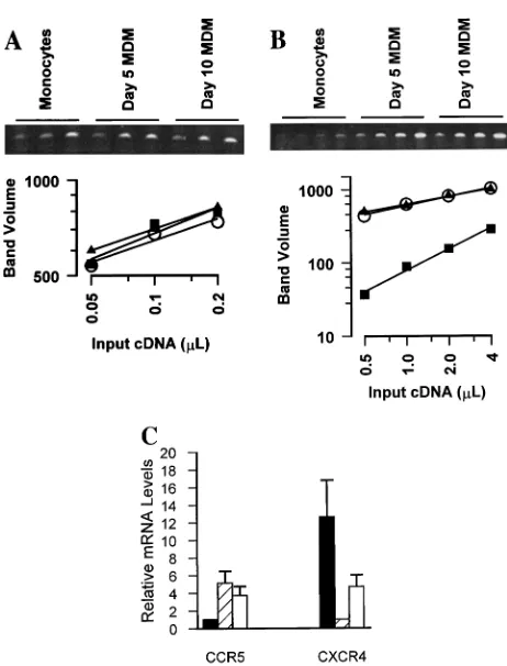
HIV-1 coreceptor mRNA levels in blood monocytes and MDM. One mechanism that could account for the different susceptibilities of monocytes and MDM to HIV-1 infection is the level of CCR5 expression. To determine whether CCR5 was modulated during the differentiation of monocytes to mac-rophages, steady-state levels of chemokine receptor mRNAs in monocytes and MDM from each individual were assessed. RNA amounts were first standardized by cyclophilin amplifi-cation (Fig. 2A), and then levels of CCR5 mRNA from differ-entiated MDM were related to CCR5 mRNA levels in mono-cytes for one individual (Fig. 2B). Monomono-cytes expressed CCR5 mRNA at levels that were about eightfold lower than the levels expressed by MDM after 5 days in culture (Fig. 2B). Steady-state levels of CCR5 expression by differentiated macrophages were stable between 5 and 10 days in culture.
To determine the range of changes in CCR5 expression among individuals, monocytes and MDM from five indepen-dent donors were evaluated. The mean increase in CCR5 mRNA levels was calculated for the group of individuals (Fig. 2C). Mean levels of CCR5 mRNA were about fivefold higher in differentiated macrophages than in peripheral blood mono-cytes (P , 0.05). Between 5 and 10 days in culture, CCR5 mRNA levels in MDM decreased but remained significantly higher than levels of CCR5 mRNA expressed by monocytes (P , 0.05). Conversely, mean steady state levels of CXCR4
mRNA were 12-fold higher in monocytes than in MDM cul-tured for 5 days (P,0.05) (Fig. 2C).
To verify that enrichment of monocytes by Percoll and ad-herence did not affect CCR5 or CXCR4 expression and to rule out a contribution by residual T cells to levels of chemokine receptor expression, monocytes were selected by an alternative method with immunomagnetic beads and an anti-CD14 MAb prior to RNA extraction. This method results in monocyte preparations that contain fewer than 1% T cells (2, 44). We found that steady-state levels of CCR5 and CXCR4 mRNAs in monocytes enriched by immunoselection were indistinguish-able from expression by monocytes enriched by adherence (data not shown). In addition, immunoselected monocytes reg-ulated CCR5 and CXCR4 levels during differentiation to the same degrees as monocytes isolated by adherence. Conse-quently, levels of CCR5 and CXCR4 mRNAs were unper-turbed by monocyte enrichment from peripheral blood and reflected expression by monocytes rather than residual T cells which might be present. CCR5 mRNA expression was low in monocytes and increased during differentiation, while high lev-FIG. 1. HIV-1 infection of monocytes and MDM. Freshly isolated peripheral
[image:3.612.311.542.62.365.2]blood monocytes and MDM cultured in duplicate for 5 days were infected with HIV-1JRFL. p24 antigen levels in culture supernatants were determined for monocytes (squares) and MDM (circles) 5 and 10 days after infection. Values are the mean6standard error of the mean for cells obtained from six uninfected donors.
FIG. 2. Differentiation of monocytes to macrophages modulates coreceptor mRNA. Total RNA was extracted from blood monocytes or MDM after differ-entiation for 5 to 10 days. Relative levels of mRNAs were evaluated after synthesis of specific cDNA and amplification by a PCR titration method. Am-plification of RNA carried out without reverse transcriptase never produced a product. (A) Twofold serial dilutions of cyclophilin cDNA were amplified (ethidium-stained agarose gel) and quantified (squares, monocytes; triangles, day-5 MDM; circles, day-10 MDM) to demonstrate that equal amounts of total RNA were introduced into each cDNA reaction mixture. (B) Twofold serial dilutions of CCR5 cDNA were amplified from monocytes and MDM from one individual (symbols are as in panel A). (C) CCR5 and CXCR4 mRNAs from five individuals were titrated and quantified. Mean (6standard error of the mean) levels of CCR5 in MDM after culturing for 5 days (hatched bar) or 10 days (open bar) are reported relative to levels in monocytes (solid bar). CXCR4 levels in monocytes (solid bar) and MDM cultured for 10 days (open bar) are reported relative to expression by MDM after culturing for 5 days (hatched bar).
on November 9, 2019 by guest
http://jvi.asm.org/
[image:3.612.102.232.67.214.2]els of CXCR4 expression by monocytes decreased during the course of differentiation into macrophages.
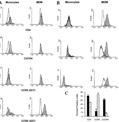
Cell surface protein expression of HIV-1 receptors differs between blood monocytes and MDM.To determine whether cell surface protein levels of chemokine receptors were also modulated during differentiation, monocytes and MDM from one individual were analyzed for CD14, CD4, CCR5, and CXCR4 levels by two-color flow cytometry. CD4 or CXCR4 was detected on approximately 60 or 80% of CD14-expressing monocytes, respectively (Fig. 3A, left panels). In contrast, only about 11 to 38% of CD14-positive peripheral blood monocytes also expressed CCR5, depending on the anti-CCR5 MAb used in the analysis. MAb 5C7, which recognizes an epitope in the N-terminal domain, consistently detected a lower percentage
of positive cells than an MAb (2D7) that recognizes an epitope in the second extracellular loop (49).
[image:4.612.91.508.69.497.2]Monocytes from the same individual were cultured in dif-ferentiation medium and, after 5 days, the resulting MDM were analyzed. Differentiation induced a modest decline in surface CD4 expression to about 50% of the CD14-positive MDM (Fig. 3A, right panel), while differentiation of mono-cytes to MDM had a dramatic impact on chemokine receptor levels. CXCR4 was reduced from expression on 80% of mono-cytes to essentially undetectable levels on MDM. In contrast, CCR5 was detected on virtually all CD14-positive MDM (.90%) when MAb 2D7 was used (Fig. 3A, right panel). N-terminal domain MAb 5C7 detected only a slight upregula-tion (10%) of surface CCR5 expression.
FIG. 3. Surface expression of HIV-1 receptors on monocytes and MDM. Freshly isolated PBMC and MDM cultured for 5 days were stained with MAb specific for CD4, CXCR4 (MAb 12G5), or CCR5 (MAb 2D7 and 5C7) and analyzed by two-color flow cytometry. Cells were gated on CD14 for analysis, and percentages of positive cells were calculated as described in Materials and Methods. (A) Cells obtained from subject 1. Histograms of monocytes (left panels) and MDM (right panels) for specific antibody staining (shaded histograms) are shown relative to histograms for isotype-matched, nonspecific antibody staining (open histograms). Fluorescence intensity is displayed on the x axis. (B) Monocytes and MDM from three additional individuals were stained for CD14 and CCR5 with MAb 2D7 and reported as in panel A. (C) Mean (6standard error of the mean) percentages of positive cells for peripheral blood monocytes (solid bars) and MDM after 5 days of differentiation (hatched bars) from four individuals are shown.
on November 9, 2019 by guest
http://jvi.asm.org/
Significant individual variation in CCR5 expression, ranging from 0 to 38% positive, was revealed by analysis of monocytes from three additional individuals (Fig. 3B). However, MDM tended to display similar numbers of CCR5-positive cells fol-lowing 5 days in culture (69 to 97%). The number of CCR5-positive MDM after 5 days in culture was independent of the levels displayed on a given individual’s monocytes.
To estimate differences in relative CCR5 expression per cell between monocytes and MDM, the ratios of peak fluorescence intensity of positively stained cells to that of control stained cells were calculated (38). The mean ratio of peak fluorescence intensity of positively stained cells to that of negatively stained cells in monocytes was 1.6, while that in MDM was 4.8, indi-cating greater per-cell CCR5 expression by MDM than by monocytes (Fig. 3B).
CD14-positive monocytes and MDM from a total of four different individuals were evaluated for CD4 and CCR5 ex-pression, and mean values for the group were calculated (Fig. 3C). CD4 expression declined overall from 76% (64%) of CD14-positive monocytes to 51% (60.6%) of CD14-positive MDM, a small but significant reduction (P,0.05) (Fig. 3C). During the same period of differentiation, mean surface ex-pression of CCR5 (MAb 2D7) increased approximately four-fold, from 19% (range, 0 to 38%) of peripheral blood mono-cytes to 81% (range, 69 to 97%) of MDM (P,0.01).
In contrast to the increased levels of surface CCR5 on dif-ferentiated macrophages, CXCR4 levels declined by sixfold. Approximately 62% (64%) of monocytes but only about 10% (61.5%) of MDM displayed levels of surface CXCR4 that
were detectable by MAb 12G5 (P,0.01). These results dem-onstrate that differentiation of monocytes into macrophages is accompanied by increased expression of CCR5 and downregu-lation of CXCR4. Changes in CCR5 or CXCR4 protein levels reflect steady-state expression of mRNAs for each of the che-mokine receptors.
A CCR5-specific MAb blocks HIV-1 infection of MDM.We wanted to demonstrate directly that increased expression of CCR5 on MDM was responsible for increased MDM suscep-tibility to HIV-1 infection. Although the CCR5 ligands MIP-1a, MIP-1b, and RANTES can block infection of PBMC by CCR5-dependent viruses, these chemokines can be inadequate to inhibit infection of MDM by viruses that depend upon CCR5 as a coreceptor (4, 12, 18, 28, 35; data not shown). Accordingly, we assessed the ability of the CCR5-specific MAb 2D7 to inhibit MDM infection by HIV-1JRFL.
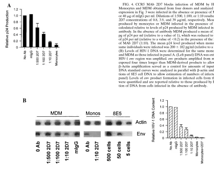
[image:5.612.64.504.64.415.2]Monocytes and MDM from the same four individuals whose cells were analyzed by FACS were incubated either in the absence or in the presence of various amounts of MAb 2D7 for 30 min prior to infection with HIV-1JRFL. To account for variation in the ability of MDM from different individuals to be infected and produce virus, supernatant p24 production in the presence of antibody is shown relative to p24 production in the absence of antibody for each individual (normalized to a value of 1; Fig. 4A). Mean p24 production by MDM was inhibited by MAb 2D7 in a concentration-dependent manner. Concentra-tions of 2D7 as low as 0.8mg per ml of culture (1:500) reduced productive infection by 38% (614%). The highest concentra-tion of 2D7 (1:10, or 39mg/ml of culture) produced a mean p24 FIG. 4. CCR5 MAb 2D7 blocks infection of MDM by HIV-1JRFL. (A) Monocytes and MDM obtained from four donors and analyzed for coreceptor expression in Fig. 3 were infected in the absence or presence of MAb (Ab) 2D7 or 40mg of mIgG per ml. Dilutions of 1:500, 1:100, or 1:10 resulted in final MAb 2D7 concentrations of 0.8, 3.9, and 39mg/ml, respectively. Mean levels of p24 produced by monocytes or MDM infected in the presence of antibody were calculated relative to levels of p24 produced by MDM infected in the absence of antibody. In the absence of antibody MDM produced a mean of 2,939 (61,129) pg of p24 per ml (relative to a value of 1), which was reduced to 421 (6105) pg of p24 per ml (relative to a value of,0.2) in the presence of the largest amount of MAb 2D7 (1:10). The mean p24 level produced when monocytes from the same individuals were infected was 2006102 pg/ml (relative to a value of,0.1). (B) Levels of HIV-1 DNA were determined for the same monocytes (Monos) and MDM as those infected in panel A. (Left panel) DNA was extracted, and the HIV-1 env region was amplified. env products amplified from monocytes were exposed four times longer than MDM-derived products to allow visualization. b-Actin amplification served as a control for amounts of input DNA. HIV-1 DNA standard curves were analyzed in parallel withb-actin and env amplifica-tions of 8E5 cell DNA to allow estimation of numbers of infected cells. (Right panel) Levels of env product formation in infected cells from the four donors were quantified and are reported relative to those produced by PCR amplifica-tion of DNA from cells infected in the absence of antibody.
on November 9, 2019 by guest
http://jvi.asm.org/
decrease of 84% (62.7%) compared to p24 production in MDM infected without prior antibody blocking (P , 0.001) (Fig. 4A). The effect of the CCR5-specific antibody on virus production was specific because treatment with 40mg of con-trol mouse IgG (mIgG) per ml prior to infection of MDM caused no suppression of p24 production (Fig. 4A).
To demonstrate that MAb 2D7 blocked virus infection, cel-lular DNA from the same monocytes and MDM infected in the presence or absence of MAb 2D7 was extracted 5 days postin-fection and amplified withb-actin and env primers. By com-parison of amplification products from infected-cell DNA sam-ples to standard curves of amplified 8E5 cell DNA, cell numbers, based on b-actin amplification, and numbers of HIV-1 DNA copies, based on env amplification, were esti-mated. Viral DNA in MDM declined with increasing levels of MAb 2D7 (Fig. 4B). In the absence of antibody against CCR5 or in the presence of an irrelevant antibody (mIgG), approxi-mately 72,500 HIV-1 copies were present per 105MDM (Fig. 4B, left panel). At the maximum amount of antibody, approx-imately 3,200 HIV-1 copies per 105MDM were detected. Low levels of monocyte infection by HIV-1 were also reduced by 2D7 from about 2,100 copies per 105MDM without antibody to fewer than 1,000 (below the level of detection) when mono-cytes were treated with 2D7 prior to infection (Fig. 4B, left panel).
To account for individual variations among monocytes ob-tained from four individuals, HIV-1 DNA quantities are ex-pressed relative to those in cells infected in the absence of antibody pretreatment (Fig. 4B, right panel). The mean level of infection of MDM without antibody treatment was 63,000 HIV-1 copies per 105 cells (set at a relative value of 1); this value was reduced by more than 60% in the presence of as little as 800 ng (1:500 dilution) of MAb 2D7 per ml of culture. HIV-1 DNA levels were reduced by more than 90% when MDM were treated with the highest concentration of MAb 2D7 (1:10) prior to infection. These data indicate that MAb 2D7 inhibits productive infection and viral DNA formation by HIV-1JRFLin monocytes and MDM by blocking CCR5-depen-dent entry.
DISCUSSION
Our experiments were designed specifically to understand the role of CCR5 in infection of cells of the monocyte lineage by viruses that rely on CCR5 as a coreceptor. Monocytes and macrophages express CD4, the primary receptor for HIV-1 infection. However, monocytes in peripheral blood from pa-tients who harbor M-tropic, non-syncytium-inducing viruses do not serve as significant reservoirs of HIV-1 infection (3, 25, 26, 37), and freshly isolated peripheral blood monocytes are poorly susceptible to productive infection by M tropic HIV-1 strains (45).
We provide direct evidence that the differentiation of mono-cytes to macrophages produces a significant increase in the number of cells expressing CCR5. The number of CCR5-ex-pressing cells detected on monocytes and MDM by FACS was higher with MAb 2D7 than with MAb 5C7, presumably reflect-ing the specificities of the antibodies. MAb 2D7 recognizes an epitope in the second extracellular loop of CCR5, while MAb 5C7 recognizes an epitope in the N-terminal domain (49). In fact, extremely variant results for proportions of CCR5-posi-tive monocytes, depending on the antibody used, have been reported (28a, 50, 51).
A direct relationship between levels of mRNA and surface expression of CCR5 or CXCR4 during differentiation indicates that chemokine receptors themselves are modulated and rules
out the possibility that epitopes on receptors recognized by MAb 2D7 or 12G5 are merely altered or modulated. Immu-nomagnetic beads, which are used to select monocytes from peripheral blood, activate phagocytosis (2). However, the ac-tivation of phagocytosis was insufficient to affect CCR5 or CXCR4 levels on monocytes, supporting the conclusion that monocyte differentiation is essential for changes in chemokine receptor levels.
The patterns of CCR5 and CXCR4 expression on blood monocytes and macrophages are consistent with the physiolog-ical function(s) of the receptors. A natural ligand for CXCR4, SDF-1, has a role in early events in immune surveillance (7), so that elevated levels of CXCR4 on monocytes may initiate the differentiation of circulating monocytes into macrophages in vivo in response to SDF-1. In contrast, ligands for CCR5 (RANTES, MIP-1a, and MIP-1b) are produced as part of the inflammatory response in tissues and may involve later events in macrophage development (48). CCR5 expression is not re-stricted to macrophages that differentiate in cultures but is found as well on tissue macrophages (50). Reciprocal differ-ences in patterns of expression of CCR5 and CXCR4 on monocytes and MDM indicate that CCR5 and CXCR4 can serve as differentiation markers for cells of the monocyte-macrophage lineage.
The critical question that we addressed is the role of CCR5 in infection of macrophages by M-tropic HIV-1. There are conflicting results regarding the ability of ligands for CCR5 to block infection of MDM by CCR5-dependent viruses (4, 12, 18, 28, 35), raising the possibility that virus entry into phages is independent of CCR5. However, blocking macro-phage infection with MAb 2D7 is a significant result from our studies which provides direct evidence that virus infection of macrophages can be CCR5 receptor mediated. If infection occurred by some other mechanism, such as Fc receptors, irrelevant antibodies should have diminished infection. In fact, irrelevant antibodies were ineffective in diminishing MDM in-fection by CCR5-dependent virus.
The inability of CCR5 agonists to block macrophage infec-tion while effectively inhibiting infecinfec-tion of T lymphocytes may reflect cell-specific differences in CCR5 or in the interactions among CCR5, its various ligands, and HIV-1 envelope glyco-protein. It is possible that CCR5 expressed by MDM has a faster turnover or a lower stability, although the demonstrated effectiveness of a single treatment with MAb 2D7 prior to infection indicates a relatively low rate of turnover of CCR5 on the surface of macrophages. Chemokine peptides could be less stable in MDM cultures either because of the human serum component or because of secretions by MDM into the media. Cell differences might also involve an altered affinity of ligands for CCR5 due to MDM-specific, CCR5-associated membrane or intracellular proteins that participate in signal transduction. The proportion of peripheral blood monocytes expressing CCR5 varied among individuals to an extent similar to that observed for CCR5 expression on lymphocytes from individu-als homozygous for wild-type alleles of CCR5 (50). Even though CCR5 levels on blood monocytes varied, differentiation into macrophages always involved significant increases in num-bers of cells expressing CCR5. Macrophages from different donors expressed similar amounts of CCR5 independent of the proportion of CCR5-positive monocytes. However, macro-phages differed in their ability to produce virus, suggesting that other cellular factors influence virus replication in macro-phages (10).
Low-level infection of monocytes in our experiments could reflect the high titers of virus used or the long inoculation periods, during which CCR5 may be upregulated. Populations
on November 9, 2019 by guest
http://jvi.asm.org/
of monocytes included some CCR5-positive cells. Levels of CCR5 expression per monocyte were generally lower than those on MDM, perhaps below a minimum threshold level necessary for efficient infection. Productive infection may be restricted to the small subset of monocytes that express rela-tively high levels of CCR5. The fact that MAb 2D7 blocked infection of monocytes indicates that infection of these cells also can occur via CCR5. Our data demonstrate that 2D7 blocks HIV-1JRFLentry into both monocytes and MDM, indi-cating that CCR5 is the principal coreceptor used by HIV-1JRFLto gain access into these CD4-positive cells. However, entry into macrophages by some primary HIV-1 isolates may require cofactors other than CCR5 (10a; unpublished results). Results from our experiments provide a molecular explana-tion for the infrequent infecexplana-tion of peripheral blood monocytes during acute or early infection by HIV-1, when high levels of M-tropic viruses can be found in peripheral blood lymphocytes and in plasma. Conversely, the presence of high levels of CXCR4 on monocytes provides a feasible molecular basis for the observation that blood monocytes can be infected in late-stage disease, when T-cell-line-tropic, CXCR4-requiring vi-ruses predominate (22). Infection of tissue macrophages most likely occurs by association with T lymphocytes trafficking through tissues. If tissue macrophages can serve as a long-lived reservoir of viruses that are resistant to or sequestered from antiviral therapies, novel strategies are needed to protect mac-rophages from infection and to target the delivery of drugs to eliminate them as reservoirs of infection.
ACKNOWLEDGMENTS
The following reagents were obtained through the AIDS Research and Reference Reagent Program, AIDS Program, NIAID: CXCR4 monoclonal antibody (12G5) from James Hoxie, CCR5 MAb (2D7 and 5C7) from LeukoSite, Inc., and HIV-1JRFLfrom Irvin Chen.
This work was supported by PHS awards HD32259, HL58005, and AI28571. D.L.T. is an Elizabeth Glaser Pediatric AIDS Foundation Scholar and was supported by grant T32 CA09126.
REFERENCES
1. Aleixo, L. F., M. M. Goodenow, and J. W. Sleasman. 1997. Zidovudine administered to women infected with human immunodeficiency virus type 1 and to their neonates reduces pediatric infection independent of an effect on levels of maternal virus. J. Pediatr. 130:906–914.
2. Aleixo, L. F., M. M. Goodenow, and J. W. Sleasman. 1995. Molecular analysis of highly enriched populations of T-cell-depleted monocytes. Clin. Diagn. Lab. Immunol. 2:733–739.
3. Aleixo, L. F., and M. M. Goodenow. Unpublished data.
4. Alkhatib, G., C. Combadiere, C. C. Broder, Y. Feng, P. E. Kennedy, P. M.
Murphy, and E. A. Berger.1996. CC CKR5: a RANTES, MIP-1breceptor as a fusion cofactor for macrophage-tropic HIV-1. Science 272:1955–1958. 5. Arenzana-Seisdedos, F., J.-L. Virelizier, D. Rousset, I. Clark-Lewis, P.
Loetscher, B. Moser, and M. Baggiolini.1996. HIV blocked by chemokine antagonist. Nature 383:400.
6. Bergsma, D., C. Eder, M. Gross, H. Kersten, D. Sylvester, E. Appelbaum, D.
Cusimano, G. Livi, M. McLaughlin, K. Kasyan, T. Porter, C. Silverman, D. Dunnington, A. Hand, W. Prichett, M. Bossard, M. Brandt, and M. Levy.
1991. The cyclophilin multigene family of peptidyl-prolyl isomerases. Char-acterization of three separate human isoforms. J. Biol. Chem. 266:23204– 23214.
7. Bleul, C. C., R. C. Fuhlbrigge, J. M. Casasnovas, A. Aiuti, and T. A.
Springer.1996. A highly efficacious lymphocyte chemoattractant, stromal cell-derived factor 1 (SDF-1). J. Exp. Med. 184:799–802.
8. Bleul, C. C., L. Wu, J. A. Hoxie, T. A. Springer, and C. R. Mackay. 1997. The HIV coreceptors CXCR4 and CCR5 are differentially expressed and regu-lated on human T lymphocytes. Proc. Natl. Acad. Sci. USA 94:1925–1930. 9. Broder, C. C., and R. G. Collman. 1997. Chemokine receptors and HIV. J.
Leukocyte Biol. 62:20–29.
10. Chang, J., H. M. Naif, S. Li, J. S. Sullivan, C. M. Randle, and A. L.
Cunningham.1996. Twin studies demonstrate a host cell genetic effect on productive human immunodeficiency virus infection of human monocytes and macrophages in vitro. J. Virol. 70:7792–7803.
10a.Cheng-Mayer, C., R. Liu, N. R. Landau, and L. Stamatatos. 1997. Macro-phage tropism of human immunodeficiency virus type 1 and utilization of the
CC-CKR5 coreceptor. J. Virol. 71:1657–1661.
11. Choe, H., M. Farzan, Y. Sun, N. Sullivan, B. Rollins, P. D. Ponath, L. Wu,
C. R. Mackay, G. LaRosa, W. Newman, N. Gerard, C. Gerard, and J. Sodroski. 1996. Theb-chemokine receptors CCR3 and CCR5 facilitate infection by primary HIV-1 isolates. Cell 85:1135–1148.
12. Cocchi, F., A. L. DeVico, A. Garzino-Demo, A. Cara, R. C. Gallo, and P.
Lusso.1995. Identification of RANTES, MIP-1a, and MIP-1bas the major HIV suppressive factors produced by CD81T cells. Science 270:1811–1815.
13. Cocchi, F., A. L. DeVico, A. Garzino-Demo, S. K. Arya, R. C. Gallo, and P.
Lusso.1996. The V3 domain of the HIV-1 gp120 envelope glycoprotein is critical for chemokine-mediated blockade of infection. Nat. Med. 2:1244– 1247.
14. Collota, S., G. Peri, A. Villa, and A. Mantovani. 1984. Rapid killing of actinomycin D-treated tumor cells by human mononuclear cells. J. Immunol.
132:936.
15. Connor, R. I., K. E. Sheridan, D. Ceradini, S. Choe, and N. R. Landau. 1997. Change in coreceptor use correlates with disease progression in HIV-1-infected individuals. J. Exp. Med. 185:621–628.
16. Dean, M., M. Carrington, C. Winkler, G. A. Huttley, M. W. Smith, R.
Allikmets, J. J. Goedert, S. P. Buchbinder, E. Vittinghof, E. Gomperts, S. Donfield, D. Vlahov, R. Kaslow, A. Saah, C. Rinaldo, R. Detels, and S. J. O’Brien.1996. Genetic restriction of HIV-1 infection and progression to AIDS by a deletion allele of the CKR5 structural gene. Science 273:1856– 1862.
17. Deng, H., R. Liu, W. Ellmeier, S. Choe, D. Unutmaz, M. Burkhart, P. Di
Marzio, S. Marmon, R. E. Sutton, C. M. Hill, C. B. Davis, S. C. Peiper, T. J. Schall, D. Littman, and N. R. Landau. 1996. Identification of a major co-receptor for primary isolates of HIV-1. Nature 381:661–666.
18. Dragic, T., V. Litwin, G. P. Allaway, S. R. Martin, Y. Huang, K. A.
Na-gashima, C. Cayanan, P. J. Maddon, R. A. Koup, J. P. Moore, and W. A. Paxton.1996. HIV-1 entry into CD41cells is mediated by the chemokine
receptor CC-CKR-5. Nature 381:667–673.
19. Dunne, A. L., H. Siregar, J. Mills, and S. Crowe. 1994. HIV replication in chronically infected macrophages is not inhibited by the Tat inhibitors Ro-5-3335 and Ro-24-7429. J. Leukocyte Biol. 56:369–373.
20. Hollinger, F. B., J. W. Bremer, L. E. Myers, J. W. Gold, and D. L. McQuay. 1992. Standardization of sensitive human immunodeficiency virus coculture procedures and establishment of a multicenter quality assurance program for the AIDS Clinical Trials Group. J. Clin. Microbiol. 30:1787–1794. 21. Huang, Y., W. A. Paxton, S. M. Wolinsky, A. U. Neumann, L. Zhang, T. He,
S. Kang, D. Ceradini, Z. Jin, K. Yazdanbakhsh, K. Kunstman, D. Erickson, E. Dragon, N. R. Landau, J. Phair, D. D. Ho, and R. A. Koup.1996. The role of a mutant CCR5 allele in HIV-1 transmission and disease progression. Nat. Med. 2:1240–1243.
22. Innocenti-Francillard, P., K. Brengel, C. Guillon, F. Mallet, P. Morand, R.
Gruters, and J.-M. Seigneurin.1994. Blood monocytes infected in vivo by HIV-1 variants with a syncytium-inducing phenotype. AIDS Res. Hum. Ret-roviruses 10:683–690.
23. Koyanagi, Y., W. A. O’Brien, J. Q. Zhao, D. W. Golde, J. C. Gasson, and I. S.
Chen.1988. Cytokines alter production of HIV-1 from primary mononuclear phagocytes. Science 241:1673–1675.
24. Liu, R., W. A. Paxton, S. Choe, D. Ceradini, S. R. Martin, R. Horuk, M. E.
MacDonald, H. Stuhlman, R. A. Koup, and N. R. Landau.1996. Homozy-gous defect in HIV-1 coreceptor accounts for resistance of some multiply-exposed individuals to HIV-1 infection. Cell 86:367–377.
25. McElrath, M. J., J. E. Pruett, and Z. A. Cohn. 1989. Mononuclear phago-cytes of blood and bone marrow: comparative roles as viral reservoirs in human immunodeficiency virus type 1 infections. Proc. Natl. Acad. Sci. USA
86:675–679.
26. McIlroy, D., B. Autran, R. Cheymier, S. Wain-Hobson, J.-P. Claulel, E.
Oksenhendler, P. Debre, and A. Hosmalin.1995. Infection frequency of dendritic cells and CD41T lymphocytes in spleens of human
immunodefi-ciency virus-positive patients. J. Virol. 69:4737–4745.
27. Michael, N. L., G. Chang, L. G. Louie, J. R. Mascola, D. Dondero, D. L. Birx,
and H. W. Sheppard.1997. The role of viral phenotype and CCR-5 gene defects in HIV-1 transmission and disease progression. Nat. Med. 3:338–340. 28. Moriuchi, M., H. Moriuchi, C. Combadiere, P. M. Murphy, and A. S. Fauci. 1996. CD81T-cell-derived factor(s), but notb-chemokines RANTES,
MIP-1a, and MIP-1b, suppress HIV-1 replication in monocyte/macrophages. Proc. Natl. Acad. Sci. USA 93:15341–15345.
28a.Naif, H. M., S. Li, M. Alali, A. Sloane, L. Wu, M. Kelly, G. Lynch, A. Lloyd,
and A. L. Cunningham.1998. CCR5 expression correlates with susceptibility of maturing monocytes to human immunodeficiency virus type 1 infection. J. Virol. 72:830–836.
29. Nakayama, H., H. Yokoi, and J. Fujita. 1992. Quantification of mRNA by non-radioactive RT-PCR and CCD imaging system. Nucleic Acids Res.
20:4939–4943.
30. Perelson, A. S., P. Essunger, Y. Cao, M. Vesanen, A. Hurley, K. Saksela, M.
Markowitz, and D. D. Ho.1997. Decay characteristics of HIV-1 infected compartments during combination therapy. Nature 387:189–191. 31. Reinhardt, P. P., B. Reinhardt, J. L. Lathey, and S. A. Spector. 1995. Human
cord blood mononuclear cells are preferentially infected by
on November 9, 2019 by guest
http://jvi.asm.org/
inducing, macrophage-tropic human immunodeficiency virus type 1 isolates. J. Clin. Microbiol. 33:292–297.
32. Rich, E. A., I. Chen, J. A. Zack, M. L. Leonard, and W. A. O’Brien. 1992. Increased susceptibility of differentiated mononuclear phagocytes to produc-tive infection with human immunodeficiency virus-1 (HIV-1). J. Clin. Invest.
89:176–183.
33. Ryffel, B., G. Woerly, B. Greiner, B. Haendler, and M. J. Mihatsch. 1991. Distribution of the cyclosporine binding protein cyclophilin in human tissues. Immunology 72:399–404.
34. Samson, M., F. Libert, B. J. Doranz, J. Rucker, C. Liesnard, C.-M. Farber,
S. Saragosti, C. Lapoumeroulie, J. Conaux, C. Foceille, G. Muyldermans, C. Verhostede, G. Burtonboy, M. Georges, T. Imai, S. Rana, Y. Yi, R. J. Smyth, R. G. Collman, R. W. Doms, G. Vassart, and M. Parmentier.1996. Resis-tance to HIV-1 infection in caucasian individuals bearing mutant alleles of the CCR-5 chemokine receptor gene. Nature 382:722–725.
35. Schmidtmayerova, H., B. Sherry, and M. Burkinsky. 1996. Chemokines and HIV replication. Nature 382:767.
36. Schnittman, S. M., H. C. Lane, J. Greenhouse, J. S. Justement, M. Baseler,
and A. S. Fauci.1990. Preferential infection of CD41memory T cells by
human immunodeficiency virus type 1: evidence for a role in the selective T-cell functional defects observed in infected individuals. Proc. Natl. Acad. Sci. USA 87:6058–6062.
37. Schuitemaker, H., N. A. Kootstra, M. Koppelman, M. Bruisten, H. G.
Huis-man, M. Tersmette, and F. Miedema.1992. Proliferation-dependent HIV-1 infection of monocytes occurs during differentiation into macrophages. J. Clin. Invest. 89:1154–1160.
38. Shapiro, H. M. 1995. Cumulative (Overton) subtraction, p. 197. In Practical flow cytometry, 3rd ed. Wiley-Liss, Inc., New York, N.Y.
39. Siebert, P. D. 1993. In Quantitative RT-PCR. Clontech Laboratories, Palo Alto, Calif.
40. Singer-Sam, J., M. O. Robinson, A. R. Bellve, M. I. Simon, and A. D. Riggs. 1990. Measurement by quantitative PCR of changes in HPRT, PGK-1, PGK-2, APRT, MTase, and Zfy gene transcripts during mouse spermato-genesis. Nucleic Acids Res. 18:1255–1259.
41. Sleasman, J. W., T. O. Harville, G. B. White, J. F. George, D. J. Barrett, and
M. M. Goodenow. 1994. Arrested rearrangements of T cell receptor Vb
genes in thymocytes from children with X-linked severe combined immuno-deficiency disease. J. Immunol. 153:442–448.
42. Sleasman, J. W., M. Henderson, and D. J. Barrett. 1991. Con A-induced
suppressor cell function depends on the activation of the CD41CD45RA
inducer T cell subpopulation. Cell. Immunol. 133:367–378.
43. Sleasman, J. W., L. F. Aleixo, A. Morton, S. Skoda-Smith, and M. M.
Goodenow.1996. CD41memory T cells are the predominant population of
HIV-1 infected lymphocytes in neonates and children. AIDS 10:1477–1484. 44. Sleasman, J. W., B. H. Leon, L. F. Aleixo, M. Rojas, and M. M. Goodenow. 1997. Immunomagnetic selection of purified monocyte and lymphocyte pop-ulations from peripheral blood mononuclear cells following cryopreserva-tion. Clin. Diagn. Lab. Immunol. 4:653–658.
45. Sonza, S., A. Maerz, N. Deacon, J. Meanger, J. Mills, and S. Crowe. 1996. Human immunodeficiency virus type 1 replication is blocked prior to reverse transcription and integration in freshly isolated peripheral blood monocytes. J. Virol. 70:3863–3869.
46. Sonza, S. A., A. Maerz, S. Uren, A. Violo, S. Hunter, W. Boyle, and S. Crowe. 1995. Susceptibility of human monocytes to HIV type-1 infection in vitro is not dependent on their level of CD4 expression. AIDS Res. Hum. Retrovi-ruses 11:769–776.
47. Spina, C. A., J. C. Guatelli, and D. D. Richman. 1995. Establishment of a stable, inducible form of human immunodeficiency virus type 1 DNA in quiescent CD4 lymphocytes in vitro. J. Virol. 69:2977–2988.
48. Wong, M., E. D. Silverman, and E. N. Fish. 1997. Evidence for RANTES, MCP-1, and MIP-1bexpression in Kawasaki disease. J. Rheumatol. 24:1179– 1185.
49. Wu, L., G. LaRosa, N. Kassam, C. J. Gordon, H. Heath, N. Ruffing, H. Chen,
J. Humblias, M. Samson, M. Parmentier, J. P. Moore, and C. R. MacKay.
1997. Interaction of chemokine receptor CCR5 with its ligands: multiple domains for HIV-1 gp120 binding and a single domain for chemokine bind-ing. J. Exp. Med. 186:1373–1381.
50. Wu, L., W. A. Paxton, N. Kassam, N. Ruffing, J. B. Rottman, N. Sullivan, H.
Choe, J. Sodroski, W. Newman, R. A. Koup, and C. R. MacKay.1997. CCR5 levels and expression pattern correlate with infectability by macrophage-tropic HIV-1 in vitro. J. Exp. Med. 185:1681–1691.
51. Zaitseva, M., A. Blauvelt, S. Lee, C. K. Lapham, V. Klaus-Kovtun, H.
Mostowski, J. Manischewitz, and H. Golding.1997. Expression and function of CCR5 and CXCR4 on human Langerhans cells and macrophages: impli-cations for HIV primary infection. Nat. Med. 3:1369–1375.
52. Zhu, T., H. Mo, N. Wang, D. S. Nam, Y. Cao, R. A. Koup, and D. D. Ho. 1993. Genotypic and phenotypic characterization of HIV-1 patients with primary infection. Science 261:1179–1181.