DISSERTATION ON
A STUDY ON EPISTAXIS
Submitted in partial fulfillment of the requirements for
M.S. DEGREE BRANCH-IV OTORHINOLARYNGOLOGY
of
THE TAMILNADU DR. M.G.R. MEDICAL UNIVERSITY
UPGRADED INSTITUTE OF OTORHINOLARYNGOLOGY
MADRAS MEDICAL COLLEGE
CHENNAI – 600 003.
CERTIFICATE
This is to certify that this dissertation entitled
“A STUDY
ON EPISTAXIS”
submitted byDr. R. NANMULLAI,
appearing forM.S. ENT.
Branch IV Degree examination in March 2010 is a bonafide record of work done by him under my direct guidance and supervision in partial fulfillment of regulations of the Tamil Nadu Dr. M.G.R. Medical University, Chennai. I forward this to the Tamil Nadu Dr. M.G.R. Medical University, Chennai, India.DEAN,
Madras Medical College, Government General Hospital,
Chennai – 600 003.
DIRECTOR & PROFESSOR,
Upgraded Institute of Otorhinolaryngology, Madras Medical College,
ACKNOWLEDGEMENT
I would like to express my sincere gratitude to
Prof J MOHANASUNDARAM M.D, DNB,Ph D , The DEAN, Madras Medical College, and Government General Hospital chennai, for having permitted me to use the hospital material in this study.
I am immensely grateful to Prof. K. BALAKUMAR, M.S., D.L.O.,
The Director and Professor, Upgraded Institute of Otorhinolaryngology, for his valuable suggestions, encouragement and help in conducting this study.
I am immensely thankful to Prof. A. MURALEEDHARAN, M.S., D.L.O., Professor, Upgraded Institute of Otorhinolaryngology, for his valuable guidance in conducting this study.
I am greatly indebted to Prof. JACINTH.C.CORNELIUS, M.S., D.L.O., Professor, Upgraded Institute of Otorhinolaryngology, who encouraged and helped me throughout this study.
I am greatly thankful to Prof. G.GANANATHAN, M.S., D.L.O.,
Professor, Upgraded Institute of Otorhinolaryngology for helping me in this study.
I express my sincere thanks to all the Assistant Professors, for their thoughtful guidance throughout the work.
I thank all the paramedical staff and other staff of the Upgraded Institute of Otorhinolaryngology for all their help and cooperation in conducting this study.
I thank all my colleagues and friends for their constant encouragement and valuable criticism.
Last but not least, I express my gratitude for the generosity shown by all the patients who participated in this study.
CONTENTS
Chapter No
TITLE Page NO
1 INTRODUCTION 1
2 AIMS OF THE STUDY 2
3 REVIEW OF LITERATURE 3
4 MATERIAL AND METHODS 43
5 OBSERVATIONS 45
6 DISCUSSION 57
7 SUMMARY 62
8 CONCLUSION 64
9 BIBLIOGRAPHY 65
10 PROFORMA 70
11 ABBREVIATIONS 78
12 MASTER CHART 79
INTRODUCTION
Epistaxis by definition is bleeding through the nose and is one of the most common and most difficult emergencies to treat. About 60% of people experience the episode atleast once in life time, with less then 10% of these requiring medical attention18,19.
Most episodes are minor in nature and affected persons don't seek medical attention. In rare cases there may be massive epistaxis also. It gives much fear and anxiety of that of tumour and other non treatable conditions to the general population.
Epistaxis can have an anterior or posterior source and can be from septum or lateral nasal wall. A brief and pertinent history and physical examination generally determine the cause of bleeding.
Both systemic and local factors play a role. Hospital admission should be considered for patients with significant comorbid conditions or complications of blood loss.
AIMS OF THE STUDY
1. To find out the most common aetiology
2. Prevalence of epistaxis in each sex / age groups.
3. Relation and correlation of nasal anatomical variations with respect to epistaxis.
REVIEW OF LITERATURE
HISTORY OF EPISTAXIS
Epi-from above.
Staxis – drop by drop of fluid.
Bleeding through the nose.
Bleeding from the nose has been known from time
immemorial. It has been mentioned in medical literature as early as 5th century B.C.
• HIPPOCRATES 5th century was the person first to note that pressure
on the alae nasi was an effective method of controlling nose bleeds. He only first used cold fomentation and nasal packing to arrest bleeding1.
• ALI IBN RABBAN – AL-TABIRI (AD 850) – in “The paradise of
Wisdom” has written that “The complaint of nose bleeding is due to swelling of a vein and its rupture or perhaps a reduction in the force which confines the blood within”1.
• MORGAGNI (1769) recognized 'The extremely turgid blood vessels
by posterior nostril into the fauces'1.
• MORGAGNI drew his inspiration from his teacher “VALSALVA”
and for this reason little's area is called “LOCUS VALSALVAE”1. VALSALVA – Thought that the nasal bleeding was arterial in origin, and he stopped the bleeding by 'syringing the nose with cold water and applied spirit of wine to constrict the mouths of swollen arteries1.
• MAHOMED (1880 – 81) described relation of hypertension with
epistaxis in old age1. He stated that 'the frequency with which severe
epistaxis occurs in old people with high arterial pressure is striking and for them very fortunate for if their noses did not bleed their brains would1.
• LITTLE (1879) in 'Hospital Gazette' – described the caudal end of
septum as the site of bleeding1.
• KIESSEL BACH (1880) described similar observations, as LITTLE1. • PILZ OF BRESSLAU (1868) was the first to tie common carotid
artery for epistaxis1.
• SEIFFERT (1928) first described ligation of internal maxillary artery
via transantral route for epistaxis1.
• GOOD YEAR (1937) – first to tie anterior ethmoidal artery for
epistaxis.
diagnosing epistaxis but also treated epistaxis by SPHENOPALATINE ARTERY LIGATION using endoscope6.
• LANGER AND TERRY – demonstrated ANTERIOR ETHMOIDAL
VASCULAR ANATOMY OF NOSE
LAST (1978) CLASSIFICATION OF BLOOD SUPPLY OF NOSE3 TWO SYSTEMS OF ARTERIES SUPPLIES THE NOSE.
•Internal carotid artery.
•External carotid artery. Internal carotid artery supplies nose via,
•Anterior ethmoidal artery. •Posterior ethmoidal artery.
External carotid artery supplies nose via,
•Sphenopalatine artery – Artery of Epistaxis.
•Greater palatine artery.
•Superior labial artery.
INTERNAL CAROTID SYSTEM
Anterior ethmoidal artery
•Arises from the ophthalmic branch of the internal carotid artery.
•2.4 cm posterior to anterior lacrimal crest.
•Supplies anterosuperior quadrant of the nasal septum and the lateral nasal wall.
Posterior ethmoidal artery
•Arises from the ophthalmic branch of the internal carotid artery.
•3.6 cm posterior to anterior lacrimal crest.
•Supplies posterosuperior quadrant of the septum and lateral wall of the nose.
•Dominant vessel of nose in embryonic life.
•If anterior ethmoid is absent, may arise directly from circle of Willis.
•Smaller than the anterior ethmoidal and absent in 20 – 30% of
individuals11,22.
EXTERNAL CAROTID SYSTEM
Sphenopalatine artery
•Artery of epistaxis – coronary artery of nose. •Main arterial supply of nose.
•Arises from the 3rd part of the internal maxillary branch of the
external carotid artery.
•Supplies posterior part of the lateral nasal wall and posteroinferior part of the septum.
•Comes out as 3 or more branches from sphenopalatine foramen into nasal cavity.
•Arises from the 3rd part of the internal maxillary branch of the
external carotid artery.
•Supplies anteroinferior quadrant of the nasal septum and lateral wall of the nose.
Superior labial artery
• Branch of the facial artery of the external carotid artery.
SUMMARY OF VASCULAR SUPPLY OF NASAL CAVITY
External carotid artery
Superior labial artery
Facial artery Lateral nasal artery
Ascending palatine artery
Maxillary artery Greater palatine artery
Sphenopalatine artery Lateral nasal
branch
Posterior septal branch
Internal carotid artery
Anterior ethmoidal artery
ARTERIAL SUPPLY – LATERAL WALL OF NOSE
MICROVASCULAR ANATOMY OF NOSE
Knowledge of the micro circulation of the nasal cavity is essential for the best management of epistaxis. But there are lot of anatomical variations in the microvascular anatomy of the nose particularly, the branching of the sphenopalatine artery – the artery of epistaxis5.
Sphenopalatine artery is the most important arterial supply to nasal cavity (septum and lateral nasal wall). It enters through the sphenopalatine foramen which lies just inferior to the horizontal attachment of the middle turbinate and may be damaged in excessive enlargement of a middle meatal antrostomy. It divides into posterior septal and posterior lateral rami.
Posterior lateral division gives the inferior and middle turbinate arteries which runs in bony tunnels within the turbinates with reduced likelihood of that can be involved in epistaxis9,10. In the inferior meatus, the
sphenopalatine branch dips below the level of the palate to re-emerge anteriorly, leaving the central portion of the meatus relatively avascular.
anteroinferiorly in the mucoperichondrium.
The greater palatine artery enters the nasal cavity via the incisive canal and supplies the anteroinferior part of the septum and part of the lateral nasal wall adjacent to palate.
Nasal septal rami of the superior labial branch of the facial artery supplies most anterior part of the septum and contributes to kiesselbach's plexus. Lateral nasal artery, branch of the facial artery supplies vestibule and ascending palatine artery of the facial artery supplies a small area of the nasal cavity.
Anterior ethmoidal artery after its origin from the ophthalmic artery in the orbit passes between superior oblique and medial rectus muscle to the anterior ethmoidal canal and traverses the ethmoid and nasal cavities. It terminates in the region of the ethmoid fovea in a meningeal branch and a larger branch to the nasal roof, olfactory cleft and superior turbinate. It reaches the nasal cavity through the cribroethmoidal foramen and the cribriform plate and divides into the anterior nasal artery with superior, lateral and medial nasal branches, as well as a posterior branch. Anterior ethmoidal artery supplies anterosuperior part of the septum and the lateral nasal wall.
ethmoidal artery and is present in only 80% of individuals11. It passes above
the superior oblique muscle to enter the posterior ethmoidal foramen situated 5mm anterior to the optic canal and 10 to 15mm behind the anterior ethmoidal foramen. It divides into a terminal meningeal branch and a branch to the posterosuperior nasal cavity (supplies posterosuperior portion of the nasal septum and the lateral nasal wall), olfactory sulcus and sphenoethmoidal recess.
There is considerable overlap between the internal and external carotid arterial systems on each side and between right and left side which may complicate attempts at arterial ligation in the management of epistaxis. Compensatory anastomotic flow via the facial arteries is thought to explain rebleeding which may occur following ligation or embolization.
There is a sinusoid system in the nasal submucosa under autonomic control on the most anterior septum and on the septum adjacent to the inferior turbinate and on the inferior and middle turbinates. This anterior septal tubercle was first described by Morgagni.
SURGICAL ANATOMY OF THE SPHENOPALATINE
FORAMEN
• Portal for the major arterial supply of the nasal cavity.
• Laterally lies the pterygopalatine space.
• Transmits the sphenopalatine artery, vein and the nasal palatine nerve.
• Key to the procedure of Endoscopic Sphenopalatine Artery Ligation (ESPAL).
SPECIAL AREAS OF BLEEDING
Arterial bleeding
LITTLE'S AREA – James Little (1879)8
(or)
Kiesselbach's plexus (1880)8
(or) Locus valsalvae
Caudal end of septum is the site where anastomosis of branches of the
,
Superior labial artery
,
Nasopalatine artery
,
Greater palatine artery
Anterior ethmoidal artery and
.
Coronary artery of nose takes place
Majority of bleeding occurs from here in children.
Bleeding from Little's area is more common because:
•Little's area is more vascular and it is covered by thin mucosa, so it is easily susceptible to injuries, particularly nose picking.
air flow is more rapid and directed towards septum. Rapid dry air causes inhibition of normal mucociliary action and in turn causes crust formation and bleeding.
Venous bleeding
1.Common in young persons.
2.Arises from vein which lies immediately behind the columella at the anterior edge of Little's area.
3.Mostly from retrocolumellar vein.
4.It runs vertically downwards and crosses the floor of the nose obliquely before joining the plexus on lateral wall of nose.
Haemorrhagic nodules
•Padgham and Parham (1993)20.
•Consists of an aneurysmal dilatation of an unusually sited
muscular artery with evidence of hypertensive changes in the wall and thrombus and haemorrhage in adjacent connective tissue.
Woodruff's plexus
•Woodruff (1949)25.•Naso-nasopharyngeal plexus.
lateral wall of the inferior meatus posteriorly.
•Appears to originate from the posterior pharyngeal wall.
•Venous in orgin.
Deviated Nasal Septum and Septal spur
Very few individuals have a perfectly straight septum. Most deviated nasal septums are asymptomatic.
The septal deformities may be classified into: 1. Deviations.
Deviations are smooth deflections involving bony or
cartilaginous portions or both. They are 'C' shaped or 'S' shaped.
2. Spurs.
Sharp angulations in septum. 3. Dislocations.
Displacement of septal cartilage into nasal cavity.
which states that when there is a flow of gas through a constriction lateral pressure drops which will, in turn, predispose to mucosal oedema in the affected area, thus further increasing the obstruction and leads to infection
and epistaxis.
Septal turbinate
The septal turbinate represents an area (often visible on CT ) of engorged vascular nasal mucosa on the septum. It may be unilateral or bilateral and can be a source of profound epistaxis. Its location may explain why a submucous resection cures some cases of septal epistaxis.
OTHER SITES OF BLEEDING
High in anterior part of septum.Brown's area – posterior part of middle turbinate. Inferior turbinates.
Lateral nasal wall. Paranasal sinuses. Nasopharynx.
VASCULAR PHYSIOLOGY OF NOSE
The dynamics of the nasal circulation depend to a large extent on the presence of ARTERIOARTERIAL ANASTOMOSES between the various arteries which contribute to the vascular supply of the nose. The branches of the anterior and posterior ethmoidal arteries join in a series of arcades in the upper one-third of the nose and the branches of the sphenopalatine artery anastomoses with those of the ethmoidal arteries above the level of the middle turbinate. Opposing heads of pressure meet in the anastomoses with a sharp interface between the two, which can be displaced by dropping the pressure in one or other of the opposing systems1.
Shaheen (1967) demonstrated, by means of dye injection into the carotid vessels of live humans, that the dispersion of dye in the nasal mucous membrane could be affected by dropping the pressure in the system not being injected10. For instance, dye injected into the internal carotid artery
The importance of possible ANASTOMOSES ACROSS THE MIDLINE also must not be overlooked, either at the nasopharyngeal end or between the two anterior ethmoidal arteries at the crista galli. These observations could well explain the many documented reports of failed ligation in which surgeons assumed, probably incorrectly, that they had tied the wrong vessel simply because bleeding had not stopped after ligation1.
VASCULAR PATHOLOGY OF NOSE
Examination of the medium and smaller nasal arteries of persons dying in middle and old age has shown that these are subject to a progressive replacement of the muscle tissue in the tunica media by collagen (Shaheen, 1967)10. This change varies from interstitial fibrosis to almost complete
replacement of the muscle by scar tissue. It seems that persons giving a history of epistaxis exhibit the more severe changes, but this is not to say that these changes are necessarily responsible for vessel rupture. They could, however, account for the lengthy duration of arterial haemorrhages, presumably because of a failure of the vessel to contract down in the absence of sufficient muscle in the tunica media1.
It is also apparent that larger vessels of the calibre of the maxillary artery are prone to calcification (Monckeberg's sclerosis)1. The resulting
lack of elasticity could be contributed to the pathogenesis of small vessel rupture by the creation of a local systolic hypertension.
The precise mechanism of bleeding is thought to be a dissecting aneurysm of the nasopalatine artery or one of its branches, but the factors initiating this process have, so far, not been identified.
vein, and this could possibly signify an area of vessel wall weakening, perhaps as a result of localized ischaemia and or trauma.
CLASSIFICATION OF EPISTAXIS
Structured clinical classification30Primary No proven causal factor.
Secondary Proven causal factor.
Childhood < 16 Years.
Adult > 16 Years.
Anterior Bleeding point anterior to piriform aperture.
Posterior Bleeding point posterior to piriform aperture.
• Anterior epistaxis: Bleeding from a source anterior to the plane of the piriform aperture. This includes bleeding from the anterior septum and rare bleeds from the vestibular skin and mucocutaneous junction.
• Posterior epistaxis: Bleeding from a vessel situated posterior to the piriform aperture. This allows further subdivision into lateral wall, septal and nasal floor bleeding.
• Pearson(1983)42 considered posterior epistaxis when bleeding point
could not be located despite examination with a headlight, vasoconstrictors and suction, which is a clinically useful definition.
SYSTEM FOR NAMING BLOOD-CLOTTING FACTORS
41aFactor VI is not a separate entity and has been dropped.
Names I II III IV Calcium V VII VIII IX
X Stuart-power factor XI XII XIII HMW-K PL Factora Fibrinogen Prothrombin Thromboplastin
Proaccelerin, labile factor, accelerator globulin Proconvertin, SPCA, stable factor
Antihemophilic factor (AHF), antihemophilic factor A, Antihemophilic globulin (AHG)
Plasma thromboplastic component (PTC), christmas factor, Antihemophilic factor B
Plasma thromboplastin antecedent (PTA), antihemophilic factor C Hageman factor, glass factor
Fibrin-stabilizing factor, Laki-Lorand factor
High-molecular-weight kininogen, Fitzgerald factor Pre-Ka Prekallikrein, Fletcher factor
Ka Kallikrein
RESPONSE TO INJURY
41 Injury to wall ofblood vessel
Contraction Collagen Tissue
thromboplastin
Platelet Activation of
reactions coagulation
Loose platelet Thrombin Aggregation
Limiting reactions Summary of reactions involved in hemostasis.
The dashed arrow indicates inhibition.
Injury when occurs initiates a series of events that lead to the formation of a clot (hemostasis) which seals off the damaged region that prevents further blood loss.
Initial event is constriction of the vessel and formation of a temporary hemostatic plug of platelets.
Temporary hemostatic
plug
Definitive hemostatic
CLOTTING MECHANISM
40Endothelial cell
Protein C Activated protein C (APC)
+ Protein S
VIIIa Inactive VIIIa Va Inactive Va
Inactivates inhibitor of
tissue plasminogen activator (t – PA)
Plasminogen Plasmin
Thrombin t-PA, u-PA Lyses fibrin
The fibrinolytic system and its regulation by protein C41
Anticlotting mechanism Thrombomodulin
AETIOLOGY OF EPISTAXIS
COMMONEST CAUSE BEING IDIOPATHIC. Classified into1,7:
•Local Cause •Systemic Cause
Local Congenital
Unilateral choanal atresia Meningocoele
Encephalocoele Glioma
Acquired
Infective
Acute
Viral Bacterial Fungal
Chronic Specific
Leprosy
Rhinoscleroma
Non specific
Ozaena Inflammatory
Rhinosinusitis (allergic / vasomotor) Nasal polyposis
Trauma
Iatrogenic Facial trauma Foreign body Surgery Idiopathic
Little's area,
Superior part of nose, Middle meatus,
Woodruff's plexus Neoplastic
Benign
Others.
Malignant
Squamous cell carcinoma, Adenocarcinoma,
Adenoid cystic carcinoma, Olfactory neuroblastoma, Lymphoma.
Drug-induced
Rhinitis medicamentosa (topical decongestants / cocaine) Inhalants
Tobacco, Cannabis, Heroin, Chrome, Mercury, Phosphorus, Wood dust.
Systemic
● Coagulopathies
■ Inherited
Coagulation factor deficiencies,
i.e. factor VIII (haemophilia A,B) and factor IX deficiency.
■ Acquired
Anticoagulants, Liver disease,
Vitamin K deficiency
Disseminated intravascular coagulation (DIC) Acquired inhibitor.
● Platelet disorders
■ Thrombocytopenia
Congenital
Acquired
Marrow failure, Aplasia,
Drugs, Infiltration,
DIC,
Hypersplenism, Massive blood loss.
■ Platelet dysfunction
congenital
Von Willebrand's disease Bernard Soulier syndrome Glanzmann's thrombasthenia Acquired
Myeloproliferative disease Leukaemia
Uraemia
Dysparaproteinaemias Drugs : Aspirin, NSAID's Acquired storage pool disease i.e. bypass.
● Blood vessel disorders
Congenital
Osteogenesis imperfecta
Amyloid Vasculitis
Vitamin C deficiency
● Hyperfibrinolysis
Congenital
α2 antiplasmin deficiency
Acquired
Malignancy DIC
Fibrinolytic therapy i.e. streptokinase. 2.Drugs
Aspirin
Anticoagulants Chloramphenicol Methotrexate
Immunosuppressives Alcohol
Dipyridamole 3. Neoplasms
4. Idiopathic
Sarcoidosis
Wegener's granuloma Lethal midline granuloma 5. Others
Liver failure Hypothyroidism
Cardiovascular conditions that increase venous pressure (Congestive heart failure, mitral valve stenosis)
Hypertension (unproven relationship)14
EPISTAXIS TRAY
SUPPLIES AND EQUIPMENT FOR THE EVALUATION
AND TREATMENT OF EPISTAXIS IN THE URGENT
CARE SETTING
1. Personal protective supplies Mask
Gown
Eye protection • Wall suction and tubing
• Frazier tip suctions, size 10 and 12 French • Headlight
• Nasal speculum • Bayonet forceps
• Cotton or neurosurgical cottonoid sponges • Silver nitrate cautery sticks
• Packing materials
Surgicel (Johnson & Johnson) • Suction cautery unit
• 0- and 30- degree rigid nasal endoscopes with light source • Optional loupe magnification
• Vasoconstrictors and anesthetics
4% or 10% cocaine (combination anesthetic / decongestant) 0.5% to 1% phenylephrine
2% topical lidocaine solution
1% injection lidocaine with 1:100,000 epinephrine • Antibiotic ointment (without neomycin)
• Tongue blades
• Two 10-cc syringes with 18- and 27- gauge needles
• Epistaxis tray helps to attend cases immediately, to take emergency measures and to treat the patients effectively.
INVESTIGATIONS OF EPISTAXIS PATIENT
Depends upon provisional diagnosis.
1. Complete Haemogram. 2. Blood Grouping and Typing. 3. Bleeding Profile.
4. Diagnostic Nasal Endoscopy.
5. Serum Biochemistry (Urea / Sugar / Electrolytes). 6. Urine Analysis.
7. Liver Function Test.
8. X-ray Paranasal Sinuses – Water's View.
9. CT Paranasal Sinuses and Brain – Plain and Contrast. 10.Electro Cardiography.
11.Chest X-ray PA View / CT scan.
TREATMENT OF EPISTAXIS
Depends on cause, site and severity of bleeding
TREATMENT CONSISTS OF
General treatment.
Local treatment to stop bleeding.
Treatment of specific cause of bleeding. Arterial ligation – rarely.
GENERAL TREATMENT
Reassurance Bed rest Sedation
Hospitalisation
Treatment of Shock – Resuscitation
1. Fluid replacement – crystalloids, colloids.
2. Blood Transfusion – if packed cell volume below 25%. Antibiotic – for prevention of secondary infection.
LOCAL TREATMENT TO STOP BLEEDING
1. Temporary pressure over the alae nasi by pinching the nose, while sitting upright.
2. Anterior Nasal Packing :
1. Vaseline Gauze 2. Gelfoam
3. Merocel, etc.,
3. Postnasal Packing if necessary 1. Gauze
2. Foley's catheter 3. Balloon packs, etc.,
4. Cauterization of bleeding point
Chemical - 1% Silver nitrate
- 40% Trichloracetic acid - Copper sulfate
- Carbolic acid, etc., Diathermy
5. Direct Therapies
Bipolar diathermy Chemical cautery
Endoscopy control – bipolar diathermy
6. Indirect Therapies
Nasal packing – ribbon gauze with petroleum jelly or
bismuth iodoform paraffin paste
(BIPP)
Merocel and kaltostat – special tampons Balloon catheters
Hot water Irrigation15 – water at 50 degree C
Systemic medical therapy – Tranexamic acid
– Epsilon Amino Caproic Acid (EACA)
7. Surgical Management
▪ Posterior packing;
▪ Ligation techniques;
▪ Septal surgery techniques;
▪ Embolization techniques;
TREATMENT OF SPECIFIC CAUSE
The specific cause of bleeding must be identified and treated promptly.
a) MEDICAL – MOSTLY FOR SYSTEMIC CAUSES b) SURGICAL – MOSTLY FOR LOCAL CAUSES ARTERIAL LIGATION
For severe epistaxis where causes are not known.
DIRECT APPROACH
External carotid artery. Internal maxillary artery.
Anterior ethmoidal artery can be ligated – As adjuvant procedure or in cases of confirmed ethmoidal bleeding (Ethmoidal #, Iatrogenic tear).
ENDOSCOPIC APPROACH
Sphenopalatine artery ligation27,29.
Anterior ethmoidal artery can be ligated38.
ENDOSCOPIC MANAGEMENT OF EPISTAXIS CONSISTS OF
1. Endoscopic cauterization. 2. Endoscopic septal surgery. 3. Endoscopic sinus surgery.
4. Endoscopic removal of mass, foreign body. 5. Endoscopic ligation of vessels.
Management Protocols for acute epistaxis
Severe→ Cauterize/AnteriorPack→Ligation/Embolization
→ Moderate → Cauterize / Anterior Pack
Mild→Cauterize/Medical Therapy→Anterior Pack(for
failures)
Severe → Anterior / Posterior pack → Ligation /
Embolization
→ Moderate → Endoscopic Cautery
Mild→ Medical therapy→Endoscopic Cautery (for failures)
Conservative treatment
Medical - For mild, recurrent without active bleeding
– Minimize or eliminate the initiating or exacerbating factors, limiting trauma.
– Nasal hydration.
– Hematological assessment.
– Hippocratic technique – advice to pinch nostril.
Management protocols for chronic or recurrent epistaxis
23Anterior Medical Therapy Cautery Anterior Pack
Ligation
Posterior Medical Therapy Endoscopic Cautery Ligation
Endoscopy → Medical Therapy → CT Scan → Anterior Pack → Posterior Pack → Ligation
Bleeding
Source Evident
Examination
MANAGEMENT STRATEGY FOR ADULT PRIMARY
EPISTAXIS
23 Resuscitation Initial examination Vessel NOT located Vessel located EndoscopyVessel NOT located
Direct therapy . .
e g bipolar
Indirect therapy . .
e g anterior packs
Bleeding controlled * Packs 48 hours
* ,
minimum Direct same day discharge
Continued bleeding
* Posterior packs * Septal surgery
* Ligation ESPAL ( ) * Angiography andContinued bleeding
embolization
*Repeat above steps
* Check for secondary
TREATMENT ALGORITHM FOR HEREDITARY
HAEMORRHAGIC TELANGIECTASIA
16.
Recurrent epistaxis in HHT
Blood transfusions
Severe Moderate
Mild
No blood transfusions
Coagulating laser
. .
e g argon
Nasal closure Septodermoplasty
Hormones
MATERIAL AND METHODS
Study design – Prospective
Study period – July 2007 to September 2009
Study place – Upgraded institute of otorhinolaryngology, Government General Hospital, Madras Medical College, Chennai.
Patients above 12 years who reported with history of bleeding through the nose were examined and full head and neck examination done after elaborating detailed history.
• Examined to identify whether anterior or posterior bleeding and to identify the bleeding source.
• If bleeding was severe, anterior nasal packing and posterior nasal packing if necessary was done and all patients were admitted in the ward.
• Complete blood investigations and other relevant investigations like DNE, X-Ray, PNS/Chest, CTPNS, ECG etc done.
• For the patients for whom no active bleeding or source identified at the time of examination were advised relevant investigations, treatment and followup.
General Hospital and Madras Medical College, Chennai.
Inclusion Criteria
1. Age group above 12 years
2. History of bleeding through the nose
Exclusion Criteria
1. Age below 12 years
2. Patients who are not willing for study
No attempt was made to assess the nose of bleeding patients in severe epistaxis as immediate care was the primary requirement.
Traumatic epistaxis with poly trauma was excluded to give attention to immediate care of the patient to rule out and treat injury to other vital structures.
Patients with life threatening emergencies (ie myocardial infarction etc.)
Endoscopes : 0 and 30 degree, 4 mm rigid nasal endoscopes (Serwel and storz ) camera and light source and necessary instruments.
Anaesthesia : 4% Xylocaine topical and decongestant
OBSERVATIONS
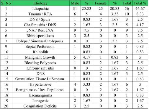
Table 1
AETIOLOGY OF EPISTAXIS
DNS – Deviated nasal septum.JNA – Juvenile nasopharyngeal angiofibroma
The most common cause in this study is idiopathic (46.67 %).
Other causes noted are trauma(8.33%), DNS with Spur and sinusitis(10.84%), polyps(5%), benign(1.76%) and malignant(5%) growth. Hypertension, rhinosporidiosis, hematological causes are noted.
Haemangioma, rhinolith, septal perforation and iatrogenic causes have also been noted.
JNA has been noted exclusively in adolescent males.
S. No Etiology Male % Female % Total Total %
1 Idiopathic 31 25.83 25 20.83 56 46.67
2 Trauma / RTA 6 5 4 3.33 10 8.33
3 1 0.83 2 1.67 3 2.5
4 2 1.67 3 2.5 5 4.17
5 JNA / Rec. JNA 9 7.5 0 0 9 7.5
6 3 2.5 0 0 3 2.5
7 0 0 3 2.5 3 2.5
9 1 0.83 0 0 1 0.83
10 1 0.83 0 0 1 0.83
11 Malignant Growth 5 4.17 1 0.83 6 5
12 1 0.83 2 1.67 3 2.5
13 Chronic sinusitis 0 0 2 1.67 2 1.67
14 DNS 1 0.83 2 1.67 3 2.5
15 Granulation Tissue Lt Septum 1 0.83 0 0 1 0.83
16 Hypertensive 4 3.33 2 1.67 6 5
17 0 0 2 1.67 2 1.67
18 1 0.83 0 0 1 0.83
19 2 1.67 0 0 2 1.67
20 Coagulation Deficits 3 2.5 0 0 3 2.5
DNS / Spurr Chr.Sinusitis / DNS
Rhinosporidiosis Polyps / Sinonasal Polyposis
Septal Perforation Rhinolith Bleeding Polyposis
Benign mass / Inv. Papilloma Haemangioma
Idiopathic Trauma / RTA DNS / Spur Chr.Sinusitis / DNS JNA / Rec. JNA Rhinosporidiosis Polyps / Sinonasal Polyposis Septal Perforation Rhinolith Malignant Growth Bleeding Polyposis Chronic sinusitis DNS Granulation Tissue Septum Hypertensive Benign mass / Inv. Papilloma Haemangioma Iatrogenic Coagulation Deficits
0 10 20 30 40 50 60
56 10 3 5 9 3 3 1 1 6 3 2 3 1 6 2 1 2 3
AETIOLOGY OF EPISTAXIS
No. of Patients
D
ia
gn
os
AETIOLOGY OF EPISTAXIS
[image:54.595.152.473.255.635.2]MALE AND FEMALE (%)
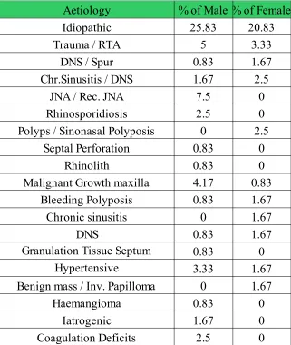
Table 2
Aetiology % of Male % of Female
Idiopathic 25.83 20.83
Trauma / RTA 5 3.33
DNS / Spur 0.83 1.67
1.67 2.5
JNA / Rec. JNA 7.5 0
2.5 0
0 2.5
0.83 0
0.83 0
Malignant Growth maxilla 4.17 0.83
0.83 1.67
Chronic sinusitis 0 1.67
DNS 0.83 1.67
Granulation Tissue Septum 0.83 0
Hypertensive 3.33 1.67
0 1.67
0.83 0
1.67 0
Coagulation Deficits 2.5 0
Chr.Sinusitis / DNS
Rhinosporidiosis Polyps / Sinonasal Polyposis
Septal Perforation Rhinolith
Bleeding Polyposis
Benign mass / Inv. Papilloma Haemangioma
Aetiology of Epistaxis
Idiopathic Trauma / RTA DNS / Spur Chr.Sinusitis / DNS JNA / Rec. JNA Rhinosporidiosis Polyps / Sinonasal Polyposis Septal Perforation Rhinolith Malignant Growth Bleeding Polyposis Chronic sinusitis DNS Granulation Tissue Septum Hypertensive Benign mass / Inv. Papilloma Haemangioma Iatrogenic Coagulation Deficits
0 5 10 15 20 25 30
25.83 5 0.83 1.67 7.5 2.5 0 0.83 0.83 4.17 0.83 0 0.83 0.83 3.33 0 0.83 1.67 2.5 20.83 3.33 1.67 2.5 0 0 2.5 0 0 0.83 1.67 1.67 1.67 0 1.67 1.67 0 0 0
% of Male % of Female
% of Patients
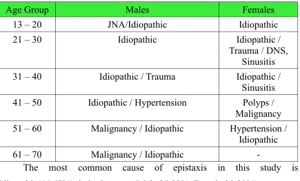
COMMONEST CAUSES IN EACH AGE GROUPS
Table 3
Age Group Males Females
13 – 20 JNA/Idiopathic Idiopathic
21 – 30 Idiopathic Idiopathic /
Trauma / DNS, Sinusitis
31 – 40 Idiopathic / Trauma Idiopathic /
Sinusitis
41 – 50 Idiopathic / Hypertension Polyps /
Malignancy 51 – 60 Malignancy / Idiopathic Hypertension /
Idiopathic
61 – 70 Malignancy / Idiopathic
-The most common cause of epistaxis in this study is idiopathic(46.67%), in both sexes. (Male 25.83%, Female 20.83%)
Other common causes in this study are trauma (8.33%), DNS with chronic rhinosinusitis and DNS with spur (10.84%) and polyps (5%).
JNA is exclusively noted in second decade males, malignancy (5%) and hypertension has been noted in older age group.
DISTRIBUTION-AGE GROUP
Table 4
• The symptom of nose bleeding is more commonly noted in this study in 21 to 30 (27.5 %), 13 to 20 (23.33 %) age groups.
• The next most frequently involved group is 51 to 60 (16.67 %), 41 to 50 (15 % ) age group.
• JNA is exclusively noted in second decade adolescent males.
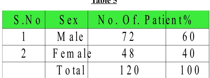
DISTRIBUTION-SEX
Table 5
S e x
N o . O f. P a tie n t %
1
M a le
7 2
6 0
2
F e m a le
4 8
4 0
T o ta l
1 2 0
1 0 0
S .N o
S. No Male Female Total %
1 13 - 20 17 11 28 23.33
2 21 - 30 17 16 33 27.5
3 31 - 40 10 7 17 14.17
4 41 - 50 13 5 18 15
5 51 - 60 11 9 20 16.67
6 61 - 70 4 0 4 3.33
13 - 20
21 - 30
31 - 40
41 - 50
51 - 60
61 - 70
0 2 4 6 8 10 12 14 16 18
17 17 10 13 11 4 11 16 7 5 9 0 DISTRIBUTION-AGE GROUP
NUMBER OF PATIENTS
Male Female 0
10 20 30 40 50 60 70 80
72
48 60
40
DISTRIBUTION - SEX
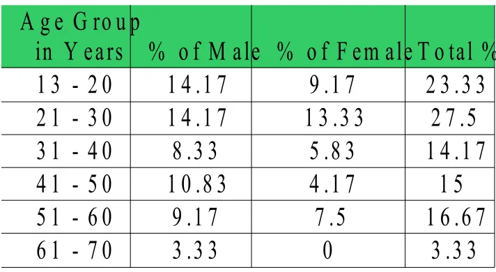
DISTRIBUTION-SEX - AGE GROUP
Table 6
• Males are more commonly affected in this study(60%).
• Males are commonly affected more in almost all age groups.
• JNA is exclusively noted in adolescent males in this study.
% o f M ale % o f F em ale T o tal %
1 3 - 2 0
1 4 .1 7
9 .1 7
2 3 .3 3
2 1 - 3 0
1 4 .1 7
1 3 .3 3
2 7 .5
3 1 - 4 0
8 .3 3
5 .8 3
1 4 .1 7
4 1 - 5 0
1 0 .8 3
4 .1 7
1 5
5 1 - 6 0
9 .1 7
7 .5
1 6 .6 7
6 1 - 7 0
3 .3 3
0
3 .3 3
ANATOMICAL VARIATIONS ASSOCIATED WITH
EPISTAXIS
Table 7
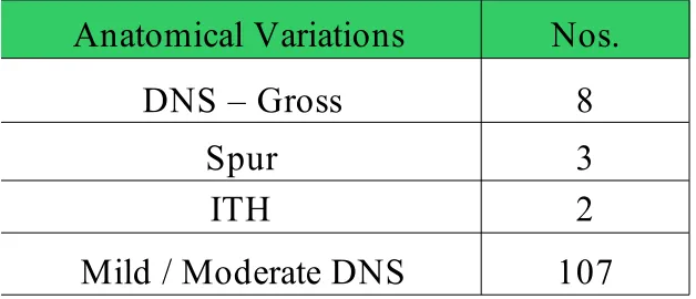
Very few individuals have a perfectly straight septum. Mostly deviated nasal
septums are asymptomatic.
Septal abnormalities are common. Between 1 and 80% of the population have a significant deviation37. The perceived association between epistaxis
and septal abnormalities could be coincidental.
Anatomical Variations
Nos.
DNS – Gross
8
Spur
3
ITH
2
8 3 2
107
ANATOMICAL VARIATIONS ASSOCIATED WITH EPISTAXIS
DNS Gross Spur ITH
ANTERIOR NASAL PACKING
ANTERIOR NASAL BLEEDING
LITTLE'S AREA CONGESTION
SEPTAL PERFORATION
DNS WITH SPUR
RHINOSPORIDIOSIS
INVERTED
PAPILLOMA
DNS WITH SINUSITIS
DNS WITH
JNA PATIENT WITH ANTEIOR
NASAL PACKING
AXIAL- CT SCAN
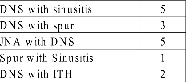
MULTIPLE FINDINGS IN SINGLE PATIENT
Table 8
commonly noted finding is deviated nasal septum
Septal abnormalities are common. Between 1 and 80% of the population have a significant deviation. The perceived association between epistaxis and septal abnormalities could be coincidental.
BLEEDING DUE TO SYSTEMIC DISEASES
Table 9
Although hypertension has long been considered as a cause of epistaxis no causal relationship has been proved14.
Hypertension can cause posterior bleeding and can cause severe bleeding in
D N S w ith sin u sitis
5
D N S w ith sp u r
3
JN A w ith D N S
5
S p ur w ith S in u sitis
1
D N S w ith IT H
2
C au se
N o s
H y p erten sio n
6
older age group.
IATROGENIC CAUSES
Table 10
Settled with conservative management with anterior nasal packing
SYMPTOMATOLOGY
Table 11
SYMPTOMS
NOs
Bleeding through
nose
120
Nasal block
50
Head ache
39
Others
15
All the patients complained bleeding through nose of varying severity. Fifty patients had nasal block also with epistaxis. Thirty nine of the patients reported head ache with epistaxis.
Some patients had symptoms of allergy reporting sneezing and running nose. One JNA patient had mild proptosis with swelling of cheek.
Two of the malignant growth maxilla patients also reported mild swilling of cheek.
C au se
N o s
1
1
P o st S ep tal co rrectio n
NATURE OF BLEEDING
Table 12Anterior Bleeding
75
Posterior Bleeding
33
Ant + Post
Bleeding
12
Most of the patients had anterior nasal bleeding (75). Thirty three patients had posterior bleeding and twelve patients had both anterior and posterior bleeding.
In anterior epistaxis bleeding was from septum in 57 patients and bleeding was from lateral nasal wall in 18 patients.
In posterior epistaxis 12 cases had bleeding from septum and 21 cases had bleeding form lateral nasal wall.
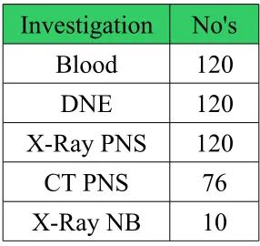
[image:71.595.250.393.615.750.2]INVESTIGATION
Table 13
Investigation No's
Blood 120
DNE 120
X-Ray PNS 120
CT PNS 76
SYMPTOMATOLOGY
NATURE OF BLEEDING
Bleeding Nasal block Head ache Others
0 20 40 60 80 100 120 140
120
50
39
15
7575
33 33 1212
Anterior Bleed-ing
Posterior Bleed-ing
Basic blood investigation was done to all the patients (Complete blood count, bleeding time, clotting time, blood urea, sugar, serum creatinine, serum electrolytes, blood grouping and typing). Complete bleeding profile investigation was done according to history, clinical suspicion and clinical diagnosis and based on other investigations.
Diagnostic nasal endoscopy was done to all the patients to identify the source of bleeding after removal of nasal packing if done prior.
X-ray PNS-water's view was taken to all the patients as a basic investigation. For those patients who had history of trauma X-ray nasal bone was also taken.
CT PNS was done to 76 patients and CT angiogram was done to one JNA patient.
TREATMENT
Table 14
Nasal Packing No's Anterior Packing 85 Ant + Post Packing 14 Observation 21
INVESTIGATIONS
TREATMENT
85
14 21
Anterior Packing Ant + Post Pack-ing
Observation
Blood DNE CTPNS X-Ray PNS X-Ray NB
0 20 40 60 80 100 120 140
120
120 120120
76 76
120 120
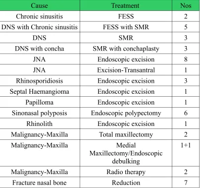
TREATMENT OF RELATED CAUSE
Table 15
Cause Treatment Nos
Chronic sinusitis FESS 2
DNS with Chronic sinusitis FESS with SMR 5
DNS SMR 3
DNS with concha SMR with conchaplasty 3
JNA Endoscopic excision 8
JNA Excision-Transantral 1
Rhinosporidiosis Endoscopic excision 3
Septal Haemangioma Endoscopic excision 1
Papilloma Endoscopic excision 1
Sinonasal polyposis Endoscopic polypectomy 6
Rhinolith Endoscopic excision 1
Malignancy-Maxilla Total maxillectomy 2
Malignancy-Maxilla Medial
Maxillectomy/Endoscopic debulking
1+1
Malignancy-Maxilla Radio therapy 2
[image:76.595.86.490.242.621.2]DISCUSSION
Epistaxis- bleeding through the nose is one of the most common and most difficult emergencies to treat. About 60% of people experience the episode atleast once in life time with less than 10% of these requiring medical attention.
Most episodes are minor in nature but in some cases there could be massive bleeding. Epistaxis can be from anterior or posterior source and it can be from septum or lateral nasal wall. Both systemic and local factors play a role.
In the present study, 120 cases were studied from the out patient department and wards of the upgraded institute of otorhinolaryngology of madras medical college and government general hospital Chennai.
Aetiology :- In this study most cases were due to idiopathic cause (46.67%). Our study correlates with
Pollice PA and Yoder MG17.- Epistaxis.
Tan LKS and Calhoun KH33- Epistaxis.
Pond F and sizeland A34- Epistaxis.
Often no cause for the bleeding is identified.
Shaheen (1987)21 has stated that majority of cases of epistaxis are idiopathic and this study also proves this.
Age group Distribution :- In the present study more commonly affected age group are 21 to 30, 13 to 20.
Next commonly affected age group is 51 to 60 and then 41 to 50
Shaheen showed in his study of age distribution of epistaxis an increase in frequency between ages 15 – 25 years and 45 – 65 years and this present study more or less correlates with that.
JNA occurs exclusively in adolescent males and this study also proves this
Sex Distribution :- In the present study males are commonly affected than females and this study more or less correlates with studies of
Juselius H. Epistaxis13
after 13 to 30 years age group
Padgham N Epistaxis12 :- Study show male preponderance.
Pollice PA and Yoder MG17 and Rubin Grandis J, et al32:- Epistaxis appears to occur more often in males than in females.
Tomkinson A and other26Males are slightly more affected than females.
In the present study most cases were anterior nasal bleeds and were managed conservatively with anterior nasal packing24.
Juselius H. Epistaxis13, Emanuel JM Epistaxis2, O Donnell M Robertson
G, McGarry GW35:- Most cases are easily manageable anterior nasal bleeds.
Surgical management were needed in 3 patients to control nasal bleeding.
Kotecha B, Fowler S, Harkness P, Walmsley J, Brown P, Topham J36:- Fewer than 10% of hospitalized patients require a general anaesthetic procedure to secure haemostasis.
Mild to moderate septal deviation were noted in most of the patients without any significant symptoms.
Investigations :- Basic blood investigation and diagnostic nasal endoscopy were done to all patients.
X-Ray nasal bone was taken for the patients with history of trauma to nose when nasal bone fracture was suspected clinically.
X-ray PNS was taken to all patients as a basic investigation to rule out any
gross pathology.
CTPNS was taken to patients with clinical suspicion of malignancy, older age group patients, adolescent males with suspicion of JNA, patients of chronic sinusitis and to other required patients. MRI was taken when necessary.
ECG, chest X-ray, test for bleeding profile were taken to all relevant cases. In older age groups radiological investigation is needed to rule out any malignancy.
Spontaneous, profuse bleeding in adolescent males requires nasal endoscopy, CT, MRI PNS to rule out JNA
Treatment :- Most cases were treated conservatively with anterior nasal packing(85 patients), some cases whom were without active bleeding during admission were observed(21 patients) and some cases were treated with anterior and posterior nasal packing(14 patients).
done to one patient. Endoscopic sphenopalatine artery ligation was done to one patient.
Septal heamangioma and bleeding polyposis were treated endoscopically. Other conditions like DNS, chronic sinusitis, rhinosporidiosis, nasal bone fracture, JNA, benign and malignant conditions were treated accordingly. The endoscopy helps to identify bleeding points and to treat them effectively4,6.
SUMMARY
Epistaxis- bleeding through the nose is one of the most common and most difficult emergencies to treat. About 60% of people experience the episode atleast once in life time with less than 10% of these requiring medical attention.
Aims of this study is to find out the most common cause, early identification of the cause and thereby early management, prevalence in various age groups and sex and correlation of anatomical variations with respect to epistaxis.
In the present study, 120 cases were studied from the OPD and wards of the UIORL of MMC and GGH Chennai.
The commonest cause of epistaxis in both sexes was found to be idiopathic (46.67%). Other causes are trauma(8.33%), DNS with chronic sinusitis with spur (10.84%) and polyps (5%) malignancy (5%) . JNA is exclusively seen in adolescent males (5%). In older age group hypertension has to be considered and should be treated effectively.
Males (60%) were commonly affected than females (40%) in the present study.
(23.33%), 51 to 60 years (16.67%) and 41 to 50 years (15%).
Idiopathic as the commonest cause, males more commonly affected and age group involvement in the present study more or less correlates with other studies.
Most of the cases were anterior nasal bleed (62.5%) and were treated conservatively with anterior nasal packing.
Mild to moderate septal deviations was noted in most of the cases . These patients were not having any significant symptoms.
The incidence of recurrent epistaxis decreased significantly as most cases were diagnosed and treated immediately.
In older age groups radiological investigation was needed to rule out any malignancy.
Spontaneous, profuse bleeding in adolescent males requires nasal endoscopy, CT, MRI PNS to rule out JNA. Regular follow up was needed in post operative JNA cases rhinosporidiosis and in malignancy.
Moderate to severe epistaxis requires immediate packing and commonest problem in post packing nose is mucosal oedema and mucosal injury which may alter or mask the original pathology.
CONCLUSION
• Idiopathic is the most common cause of epistaxis in this present study ()46.67%.
• Other causes are trauma (8.33%), DNS with spurs and sinusitis (10.84%) and JNA in adolescent males.
• Males are commonly affected than females (60%).
• The most common age group affected is 13 to 30 years (50.83%) and 41 to 60 years (31.67%).
• Most cases are treated by conservative measures.
In older age groups radiological investigation is needed to rule out any malignancy.
BIBLIOGRAPHY
1. J.C Watkinson. Epistaxis. Rhinology 6th edition 1997 p. 4/18/1-p.
4/18/8.
2. Jane M.Emanuel,Epistaxis, Otolaryngology and Head & Neek Surgery, third edition, 1998. Vol.2. p.852-865
3. John K.S Woo, surgery for epistaxis, Head and neck operative surgery, Ch.36, p.256-262
4. Bingham B and Dingle A.F., 1991. Endoscopic management of severe epistaxis – Journal of otolaryngology 20 : p. 442 – 443
5. Emmanuel babies, Sylvian Moreau, March Gaulliet de Rug, Pierre delams, Andre Valdazo. Arnaud Bequignon, Anatomic variations of arteries of nasal fossa, otolaryngology & Head and Neck surgery. Vol 128. No.2
6. Marcus MJ. Nasal endoscopic control of epistaxis. A preliminary evaluation otolargyngoloy and Head & Neck surgery. 102 : 273, 1990. 7. Morgagni JP. The seats and causes of diseases. Vol. 1. Alabama:
Gryphon editions Ltd., 1983: 312 – 54 (first published in 1761)
8. Mackenzie D. Little's area or the locus Kiesselbachii. Journal of Laryngology. 1914; 1: 21 – 2
lateral nasal wall and their relation to turbinates and sinuses. Journal of Laryngology and Otology. 1935; 8: 569 – 93.
10.Shaheen OH. Epistaxis in the middle aged and elderly. Thesis for the master of surgery in the university of London, 1967.
11.Caliot PH Plessis JL, Midy D.Poirier M, Ha JC. The intraorbital arrangement of the anterior and posterior ethmoidal formina. Surgical radiologic anatomy. 1995;17:29- 33
12.Padgham N. Epistaxis: anatomical and clinical correlates. Journal of Laryngology and otology. 1990; 104: 308 – 11.
13.Juselius H. Epistaxis: a clinical study of 1724 patients. Journal of Laryngology and otology. 1974; 88: 317 – 27
14.Lubianca-Neto JF, Bredemeier M, Carvahal EF Arruda CA, Estrella E, Pletsch A et al. A study of the association between epistaxis and the severty of hypertension. American Journal of Rhinology. 1998; 12: 269 – 72.
15.Stangerup SE, Dommerby H, Siim C, Kemp L, Stage J. New modification of hot-water irrigation in the treatment of posterior epistaxis. Archives of Otolaryngology-Head and Neck surgery 1999; 125: 686 – 90
Rhinology. 1999; 13: 319 – 22.
17.Pollice PA and Yoder MG. Epistaxis: a retrospective review of hospitalized patients. Otolaryngol. Head Neck Surg. 1997; 117: 49-53 18.Petruson B. Epistaxis. A clinical study with special reference to
fibrinolysis. Acta Otolaryngol. Suppl. 1974; 317: 1 – 73
19.Schaitkin B, Strauss M and Houck JR. Epistaxis: Medical versus surgical therapy: A comparison of efficacy, complications, and economic considerations. Laryngoscope 1987; 97: 1392 – 1396.
20.PADGHAM N.P. and PARHAM D.M. (1993) Haemorrhagic nasal nodules. Clinical otolaryngology 18, 118-120.
21.SHAHEEN O.H (1987) Epistaxis. Rhinology 5th edition London
Butterworths pp 272 – 282
22.Kirchner JA, Yanagisawa E, Crelin ES: Surgical anatomy of the ethmoidal arteries, Arch Otolaryngol 74: 40, 1961.
23.Marks SC: Nasal and sinus surgery. Philadelphia, Saunders, 2000, p 451
24.McGarry GW, Epistaxis MD thesis University of Glasgow 1996
25.WOODRUFF G.H. (1949) Cardiovascular epistaxis and the naso-nasopharyngeal plexus. Laryngoscope 59, 1238 – 1247
27.Voegels RL and others: Endoscopic ligature of the sphenopalatine artery for severe posterior epistaxis, Otolaryngol Head neck surg 124: 464, 2001.
28.Asian journal of ear, nose and throat vol 3, no 4, october to december 2005. management of epistaxis, Corry J Kucik and Timothy Clenney. 29.BolgerWE, Borgie RC, Melder P. the role of the crista ethmoidalis in
endoscopic sphenopalatine artery ligation. American journal of Rhinology. 1999; 13: 81 – 6
30.Maran AGD, Lund VJ. (eds). Chapter 28. In: Clinical rhinology, New Yourk: Thieme Medical Publishers, 1990: 101 – 4
31.Stell PM. Epistaxis. Clinical Otolaryngology. 1977; 2: 263 – 73.
32.Rubin Grandis J, et al. The Management of Epistaxis 3rd Edition,
American Academy of Otolaryngology -Head and Neck Surgery Foundation, Alexandria Va 1999
33.Tan LKS and Calhoun K H. Epistaxis. Med. Clin. North Am. 1999; 83:43-56
34.Pond F and Sizeland A. Epistaxis. Strategies for management. Aus.Fam. Physician 2000; 29:933-938
36.Kotecha B, Fowler S, Harkness P, Walmsley J, Brown P, Topham J, Management of epistaxis: A national survey. Annals of the Royal collage of Surgeons of England. 1996; 78:444-6. Provides a snapshot of current practice.
37.Roblin DG, Eccles R. Review: What, if any, is the value of septal surgery? Clinical Otolaryngology2002; 27:77.
38.Woolfood TJ. Jones NS, Endoscopic ligation of anterior ethmoidal artery in treatment of epistaxis. Journal of Laryngology and Otology. 2000; 114:858-60
39.Weiss NS. Relation of high blood pressure to headache, epistaxis and selected other symptoms. The new England journal of Medicine. 1972; 287:631-3
40.Hemodynamic disorders, thrombosis and shock. Richard N. Mitchell and Ramzi. S. Cotran in pathologic basis of disease 6th edition.
41.Review of medical physiology. William F. Ganong MD 23rd edition
2007
PROFORMA
UPGRADED INSTITUTE OF OTORHINOLARYNGOLOGY
GOVERNMENT GENERAL HOSPITAL,
MADRAS MEDICAL COLLEGE,
CHENNAI – 3
Name Unit Age Sex
O.P. No, : I.P. No : Occupation
Address
Date of admission Date of endoscopy Date of surgery Date of discharge
Presenting Complaints
Bleeding through the nose Nasal block
Nasal discharg Headache Sneezing Loss of smell
H/O hematemesis, melena, hemoptysis H/O Ear, throat symptoms
Past History
H/o Previous episodes H/o Foreign body
H/o Drug intake (oral, topical) H/o Bleeding disorder
H/o Blood transfusion
H/o Systemic disorders – IHD, hypertension, diabetes, asthma H/o Previous surgery.
Personal History
H/o Smoking, H/o snuff use, H/o Alcohol, H/o Nose picking, H/o Metallic poisoning.
Family History
H/o Bleeding diathesis
GENERAL EXAMINATION
Pulse rate, Blood pressure, Anemia,
Jaundice, Cyanosis, Clubbing, Petechiae, Ecchymosis,
Generalized lymphadenopathy, Cardiovascular system,
ENT EXAMINATION
NOSE
External examination
External contour, Bony tenderness, Bony crepitus,
Columella retraction, Other findings.
Anterior Rhinoscopy
Vestibule, Little's area, Mucosa, Septum, Turbinates,
Posterior Rhinoscopy
Choana,
Posterior end of turbinates, Roof of nasopharynx, Eustachian tube orifice, Bleeding points,
Adenoids / mass, Other findings.
Ear :
Throat :
DIAGNOSTIC NASAL ENDOSCOPY
Right Left
Little's area
Turbinoseptal classification
Inferior matus
Woodruff's area Other findings
Inferior turbinate
Septum DNS
Spur
Middle turbinate
Right Left High septal deviation
Superior turbinate
Spheno ethmoid recess Apex of nasal fossa Choana
Nasopharynx
Eustachian tube orifice Roof of nasopharynx Fossa of Rosenmuller
OTHER INVESTIGATION
Complete haemogram, Packed cell volume, Blood grouping & typing, Bleeding profile,
Liver function tests, Blood urea / sugar, Chest X-ray,
ECG,
X-ray paranasal sinuses – Water's view,
CT PNS – coronal and axial cuts → 5mm → plain and contrast films, Angiogram / Brain,
Digital substraction angiogram, MRI – PNS / Brain / Angiogram
EXPERT OPINION
Haematologist opinionPhysician opinion
ABBREVIATIONS
DNS – Deviated Nasal Septum.
JNA – Juvenile Nasopharyngeal Angiofibroma Chr.Sinusitis – Chronic Sinusitis
ITH – Inferior Turbinate Hypertrophy DNE – Diagnostic Nasal Endoscopy
ESPAL – Endoscopic Sphenopalatine Artery Ligation PCV – Packed Cell Volume
EACA – Epsilon Amino Caproic Acid BIPP – Bismuth Iodoform Paraffin Paste Inv. Papilloma – Inverted Papilloma
FESS – Functional Endoscopic Sinus Surgery SMR – Sub Mucosal Resection
Endo. Excision – Endoscopic Excision ECA – External Carotid Artery
SI. No Name Age Sex Symptomatology Investigations Diagnosis Treatment Blood DNE Primary Related 91 Eswari 25 F 68191 + √ √ √ Trauma # N.B Observation Reduction 92 Priya 21 F 77301 + √ √ √ Trauma # N.B AN packing Reduction 93 Barathi 25 F 67667 + + + + √ √ √ √ AN packing
94 Lalitha 34 F 40971 + + + √ √ √ √ DNS / Bil.ITH Observation
95 Nagalakshmi 33 F 45154 + + √ √ √ DNS / Spur AN packing SMR 96 Vasantha 60 F 72230 + √ √ √ √ Idiopathic AN packing
97 Samakhani 49 F 80121 + + √ √ √ √ AN packing 98 Kanchana 24 F 61029 + + + √ √ √ √ AN packing
99 Priya 18 F 57968 + + √ √ √ DNS / Spur AN packing SMR 100 Radha 30 F 50066 + + + √ √ √ √ AN packing FESS / SMR 101 Kavitha 20 F 40903 + + + + √ √ √ √ Chr. Sinusitis Observation FESS 102 Suganthi 24 F 18253 + + + + √ √ √ √ Chr. Sinusitis AN packing FESS 103 Pushpavalli 38 F 55830 + + + √ √ √ √ AN packing
104 Bhuvaneswari 28 F 73556 + + + √ √ √ √ AN packing 105 Seethammal 54 F 74458 + + + √ √ √ √ Observation 106 Vennila 45 F 72706 + + + √ √ √ √ AN packing 107 Priya 18 F 50113 + √ √ √ Idiopathic AN packing 108 Nirmala 26 F 53612 + √ √ √ Idiopathic AN packing 109 Anjalai 25 F 55318 + √ √ √ Idiopathic AN packing 110 Mari 48 F 46068 + + + √ √ √ √ AN packing 111 Chithra 24 F 32733 + √ √ √ √ Idiopathic AN packing 112 Lakshmi 25 F 32746 + √ √ √ √ Idiopathic AN packing 113 Parimala 31 F 75226 + √ √ √ √ Idiopathic
114 Muniyammal 60 F 57044 + √ √ √ √ Idiopathic AN packing 115 Meera 20 F 84278 + √ √ √ Idiopathic AN packing 116 Gandimathi 50 F 13356 + √ √ √ √ Idiopathic AN packing 117 Umadevi 22 F 1548 + √ √ √ Idiopathic AN packing
118 Janath 27 F 56286 + √ √ √ Trauma # N.B AN packing Reduction 119 Rajakumari 45 F 53045 + + √ √ √ √ CA. Maxilla Observation Rediotheraphy 120
Mallika 50 F 58116 + + + √ √ √ √ Lt. Nasal mass AN packing Endo. Excision I.P No. /
O.P. No. Blee Ding Nasal Block Head Ache Oth Ers X-Ray PNS C.T PNS
DNS / Concha
Bullosa ConchaplastySMR / SMR / Turbinectomy REC. Inverted Papilloma DNS / Bil.Concha / Chr. Sinusitis
FESS / Conchaplasty
/ SMR DNS / Chr.
Sinusitis
DNS / Chr. Sinusitis
FESS / Septoplasty / Turbinoplasty Sinonasal
Polyposis PolypectomyEndo. AntraChoanal
Polyp Lt
Endo. Polypectomy Bil.Ethmoidal
Polyposis PolypectomyEndo.
Bleeding
Polyposis PolypectomyEndo.
AN + PN packing + Endo. Cauterization