0022-538X/05/$08.00⫹0 doi:10.1128/JVI.79.4.2199–2210.2005
Copyright © 2005, American Society for Microbiology. All Rights Reserved.
Kinetics of Human Immunodeficiency Virus Type 1 Decay following
Entry into Resting CD4
⫹
T Cells
Yan Zhou,
1Haili Zhang,
1Janet D. Siliciano,
1and Robert F. Siliciano
1,2*
Johns Hopkins University School of Medicine1and Howard Hughes Medical Institute,2Baltimore, Maryland
Received 8 July 2004/Accepted 1 October 2004
In untreated human immunodeficiency virus type 1 (HIV-1) infection, most viral genomes in resting CD4ⴙ T cells are not integrated into host chromosomes. This unintegrated virus provides an inducible latent reservoir because cellular activation permits integration, virus gene expression, and virus production. It remains controversial whether HIV-1 is stable in this preintegration state. Here, we monitored the fate of HIV-1 in resting CD4ⴙcells by using a green fluorescent protein (GFP) reporter virus carrying an X4 envelope. After virus entry into resting CD4ⴙT cells, both rescuable virus gene expression, visualized with GFP, and rescuable virion production, assessed by p24 release, decayed with a half-life of 2 days. In these cells, reverse transcription goes to completion over 2 to 3 days, and 50% of the viruses that have entered undergo functional decay before reverse transcription is complete. We distinguished two distinct but closely related factors contributing to loss of rescuable virus. First, some host cells undergo virus-induced apoptosis upon viral entry, thereby reducing the amount of rescuable virus. Second, decay processes directly affecting the virus both before and after the completion of reverse transcription contribute to the loss of rescuable virus. The functional half-life of full-length, integration-competent reverse transcripts is only 1 day. We propose that rapid intra-cellular decay processes compete with early steps in viral replication in infected CD4ⴙT cells. Decay processes dominate in resting CD4ⴙ T cells as a result of the slow kinetics of reverse transcription and blocks at subsequent steps. Therefore, the reservoir of unintegrated HIV-1 in recently infected resting CD4ⴙT cells is highly labile.
The outcome of human immunodeficiency virus type 1 (HIV-1) infection of CD4⫹ T cells differs depending on the activation state of the infected cell (34, 45). Activated CD4⫹T cells support rapid viral replication and produce most of the free virus found in the plasma (31). However, the majority of CD4⫹T cells are in a resting state. Understanding the fate of HIV-1 in resting CD4⫹T cells is thus an important issue. The predominant form of HIV-1 DNA in resting CD4⫹T cells is full-length, linear, and unintegrated (5, 8). When resting cells with unintegrated HIV-1 DNA are activated in vitro, at least some of extrachromosomal viral DNA integrates into the host cell genome, allowing virus gene expression and virus produc-tion. In this sense, resting CD4⫹ T cells with unintegrated HIV-1 DNA represent an inducible latent reservoir for the virus (5, 33, 45, 46).
Insight into this preintegration form of latency has come from molecular studies of resting CD4⫹T cells that have been infected with HIV-1 in vitro. Infection of truly quiescent CD4⫹ T cells is nonproductive as a result of blocks at several early steps in the viral life cycle. For viruses utilizing CCR5 as a coreceptor, entry is inefficient because the surface expression level of CCR5 is low on resting CD4⫹T cells (2, 27). Even if entry does occur, reverse transcription requires up to 3 days to complete (29, 33), presumably due to an insufficient supply of nucleotides (20, 45). In addition to the slow kinetics of reverse transcription, deletion of nucleotides from the ends of the
reverse-transcribed HIV-1 DNA has been observed in resting CD4⫹T cells but not in activated CD4⫹T cells (29). Another proposed block to the viral replication is at the subsequent step of nuclear import of the preintegration complex, as the major-ity of viral DNA in recently infected resting CD4⫹T cells is localized to the cytoplasmic compartment (4). There is some evidence that Nef and Tat can be produced from unintegrated viral DNA and that these factors can then enhance T-cell activation upon subsequent stimulation (43). These blocks on viral replication in resting CD4⫹T cells are removed when the cells are activated by antigen or subtle stimulatory signals from the microenvironment of the lymphoid tissues (13, 49), such as cytokines and soluble factors secreted by B cells (38, 41).
An important question concerning the fate of HIV-1 in resting CD4⫹T cells is whether HIV-1 is stable in the prein-tegration state. A longitudinal study of acute seroconverters with high viral loads demonstrated that after the initiation of treatment with potent antiretroviral drugs, there is a biphasic decay in the amount of virus that can be rescued from resting CD4⫹T cells by cellular activation, a result which suggests that the virus in the preintegration state is less stable than inte-grated provirus (3). However, the interpretation of the in vivo data is complicated by new rounds of infection of resting CD4⫹ T cells that are not completely blocked by antiviral drugs. In vitro studies have yielded inconsistent results. One report showed that HIV-1 cDNA is rapidly degraded, with a half-life of 1 day (45). Another study has found that linear extrachro-mosomal HIV-1 cDNA molecules are stable but that few main-tain the ability to integrate at the end of 2-week culture (35). Other investigators have demonstrated the establishment of a stable latent form of HIV-1 in acutely infected resting CD4⫹T
* Corresponding author. Mailing address: Department of Medicine, Johns Hopkins University School of Medicine, Ross 1049, 720 Rutland Ave., Baltimore, MD 21205. Phone: (410) 2958. Fax: (410) 955-0964. E-mail: [email protected].
2199
on November 8, 2019 by guest
http://jvi.asm.org/
cells (33). Thus, the fate of unintegrated HIV-1 DNA in in-fected resting CD4⫹T cells remains unclear.
In previous studies, we have used recombinant HIV-1 viri-ons pseudotyped with the G envelope protein from vesicular stomatitis virus (VSV) to study early events following HIV-1 entry into resting CD4⫹T cells. We found that the majority of the recombinant HIV-1 decays rapidly before the completion of reverse transcription (29). The functional half-life of the recombinant HIV-1 is 1 day. Although the usage of VSV-G facilitates the generation of high-titer recombinant virus stocks, differences between VSV-G-pseudotyped virus and wild-type HIV-1 may affect decay processes. HIV-1 envelope has recently been reported to induce intracellular signals that program resting CD4⫹T cells to support viral replication (9, 19). It remains unknown how these stimulatory signals affect the lability of HIV-1 in the preintegration state in resting CD4⫹T cells. Because the pseudotyped viruses do not bind to CD4 or chemokine receptors, these effects are not induced. Instead of direct fusion with plasma membrane, VSV-G-pseudotyped virions enter the host cell by endocytosis and then fuse with the endosomal compartment membrane (22). The subsequent uncoating step of VSV-G-pseudotyped virus is dif-ferent from that of wild-type HIV-1 in that it bypasses the requirement of Nef and the inhibition by cyclosporine (1, 7). In this study, we sought to understand the fate of HIV-1 in resting CD4⫹T cells by using a reporter virus carrying the wild-type HIV-1 envelope. Our data demonstrate that although HIV-1 can slowly complete reverse transcription in resting CD4⫹T cells, the virus decays due to infection-induced apoptosis and intracellular degradation before and after the completion of reverse transcription.
MATERIALS AND METHODS
Isolation of resting CD4ⴙT cells from uninfected individuals.Highly purified
resting CD4⫹T cells were isolated from peripheral blood mononuclear cells
(PBMC) of HIV-1-negative donors. PBMC were washed twice in wash medium (WM; phosphate-buffered saline [pH 7.2 to 7.4] supplemented with 2% fetal calf
serum, 0.1% glucose, 100 U of penicillin/ml, 100g of streptomycin/ml, and 12
mM HEPES [pH 7.2]) and then placed in culture medium (CM; RPMI 1640 with GlutaMAX supplemented with 10% heat-inactivated fetal calf serum, 20 U of
penicillin/ml, and 20g of streptomycin/ml). Adherent cells were removed by 3 h
of incubation in a 37°C incubator with 5% CO2. Isolation of resting CD4⫹T cells
was achieved by positive selection of CD4⫹T cells followed by negative selection
to remove activated CD4⫹T cells. PBMC were washed once with WM and then
resuspended in a small volume of ice-cold WM. Magnetic beads conjugated with a monoclonal anti-CD4 antibody (CD4 positive isolation kit; Dynal) were mixed
with PBMC in a cryovial with a 2:1 ratio of beads to CD4⫹T cells. Beads and
cells were incubated at 4°C with gentle rocking for 20 min. Cells in the super-natant were discarded. The beads with bound cells were gently washed six times
with WM. Bound cells were eluted from the beads by adding 100l of CM and
10l of DETACHaBEAD CD4 (Dynal) for every 107CD4⫹T cells and then
gently rolling for 15 min at room temperature. The eluted CD4⫹T cells were
collected and subsequent negative selection was carried out as described
previ-ously (16). Briefly, enriched CD4⫹T cells were incubated with monoclonal
antibodies against T-cell activation markers (CD69, CD25, and HLA-DR) as well as antibodies against markers on unwanted cell populations (CD8, CD19, CD16, and CD14). Cells that bound the monoclonal antibodies were removed by using magnetic beads coated with sheep anti-mouse immunoglobulin G antibody.
The purity of resting CD4⫹T cells was analyzed by flow cytometry. Typically,
99.9% of the resulting cell population expressed CD4, and 98% of the cells were negative for activation markers.
Virus preparation and infection.Viral vector NL4-3-GFP was used to make reporter virus that encodes the enhanced green fluorescence protein (GFP) in
place of HIV-1env(29). An integrase mutant reporter virus was constructed by
replacing the SalI-BamHI fragment of the D64N mutant HIV-1 LAI provirus
with that of NL4-3-GFP. Reporter virus particles coated with HIV-1 envelope
were generated by transfecting 30⫻106
293 T cells in a T150 flask with 20g
of viral vector and 10g of an X4 HIV-1 envelope expression vector by using
Lipofectamine 2000 (Invitrogen) according to the manufacturer’s instructions. The transfection media was replaced by CM 4 h later. Supernatant containing viral particles was collected 48 h after transfection. Cell debris was removed from
the supernatant by centrifugation at 470⫻gfor 5 min and subsequent filtration
through a 0.22-m-pore-size filter. The supernatant was concentrated by
ultra-centrifugation at 112,000⫻gat 4°C for 1.75 h. Concentrated virus was further
purified by spinning through a sucrose cushion. Twenty percent (wt/vol) sucrose was prepared in TNE buffer (20 mM Tris [pH 8.0], 150 mM NaCl, and 2 mM
EDTA) and filtered through a 0.22-m-pore-size filter. The sucrose was loaded
beneath the infectious supernatant in a cryovial with a 1:10 volume ratio of viral
supernatant to sucrose solution. The virus was pelleted at 16,000⫻gfor 2 h at
4°C. Aliquots were frozen at⫺80°C. The viral titer was calculated based on the
number of activated CD4⫹T cells expressing GFP 3 days after infection.
Infection of resting CD4⫹T cells was carried out by 2 h of spinoculation as
previously described (23), followed by 2 h of incubation at 37°C in the infectious supernatant. Cells were then washed three times with WM and cultured in CM
containing 5g of the fusion inhibitor T1249/ml (42) at 2⫻106
cells/ml in 24-well plates. The culture medium was changed daily with fresh CM and T1249.
In some experiments, infected cells were treated with 10M of the reverse
transcriptase inhibitor lamivudine (3TC; obtained from the NIH AIDS Research and Reference Reagent Bank) 4 h prior to activation. In some experiments, the integrase inhibitor L-731,988 (Merck) was added to the cells at a final
concen-tration of 40M at the time of activation. Fresh medium with antiviral drugs was
added to the cells daily after T-cell activation. When supernatants were to be assayed for the production of HIV-1 p24, antiviral drugs were replenished daily in the culture without changing the medium.
Activation of resting CD4ⴙT cells.Purified resting CD4⫹T cells were acti-vated with phytohemagglutinin (PHA; Murex) and irradiated allogeneic PBMC as previously described (29). Briefly, freshly isolated PBMC from HIV-1-sero-negative donors were inactivated by irradiation with 5,000 rads in a cesium source irradiator and then washed twice. To distinguish the irradiated PBMC
from infected CD4⫹T cells in flow cytometric analysis, the irradiated PBMC
were labeled with excitable dye PKH26 (Sigma) according to the manufacturer’s instructions. A 10-fold excess of PHK26-labeled irradiated PBMC was mixed
with 0.2⫻106
infected resting CD4⫹T cells in STCM (CM supplemented with
100 U of recombinant human interleukin 2/ml and 2% of the supernatant from
activated PBMC) containing 1g of PHA/ml in 96-well plates. The PHA was
removed the next day and the culture was maintained in STCM until analysis was
performed. The percentage of infected resting CD4⫹T cells that expressed GFP
upon cellular activation was determined by flow cytometric analysis as the per-centage of PHK26-negative cells expressing GFP 48 h after activation.
NERT and PCR analysis.Natural endogenous reverse transcription (NERT) stimulation was carried out as described previously (11). Concentrated virus was treated with CM containing 1 mM concentrations of deoxynucleoside
triphos-phates (dNTP; Sigma), 30M spermidine (pH 7.2; Sigma), and 2.5 mM MgCl2
at 37°C for 4 h before pelleting through a sucrose cushion at 16,000⫻gfor 2 h.
The virus was then treated with DNase I (Invitrogen) in the presence of 5 mM
MgCl2at 37°C for 2 h before the infection of resting CD4⫹T cells that had been
pretreated with 10M 3TC. DNA was extracted from the cells immediately after
infection and analyzed by PCR for the presence of early and late products of reverse transcription with previously described primers and PCR conditions (45). Briefly, the R-U5 region was detected with primer set M667 (nucleotides 495 to 516 of HIV-1 NL4-3)-AA55 (nucleotides 612 to 635 of HIV-1 NL4-3). The long terminal repeat (LTR)-Gag region was detected with primer set M667-M661 (nucleotides 672 to 694 of HIV-1 NL4-3). Primers that detected the albumin gene were used as an internal control (12).
Flow cytometric analysis.Induced expression of GFP was examined by flow cytometry 48 h after T-cell activation. Infected cells were fixed with 1% para-formaldehyde in PBS for 30 min on ice before analysis. Cells undergoing apo-ptosis were detected with V-PE and 7-amino-actinomycin D (7AAD) (Annexin V-PE apoptosis detection kit I; BD Pharmingen) according to the manufactur-er’s instructions. Flow cytometric analysis was performed by using FACSCalibur with CellQuest software (Becton Dickinson).
RESULTS
Decay of HIV-1 in infected resting CD4ⴙT cells.To study the fate of HIV-1 following entry into resting CD4⫹T cells, we isolated resting CD4⫹T cells from the blood of healthy donors
2200 ZHOU ET AL. J. VIROL.
on November 8, 2019 by guest
http://jvi.asm.org/
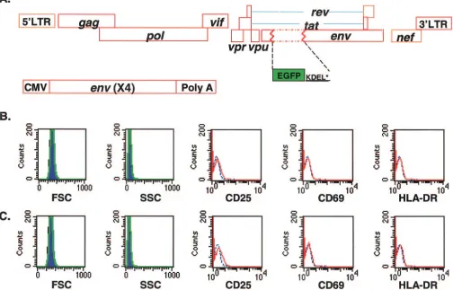
and infected the cells with a modified form of a previously described replication-incompetent HIV-1 reporter virus, NL4-3-GFP (29). This virus carries enhanced GFP in theenvopen reading frame. The GFP coding sequence is followed by a KDEL sequence, which serves to retain the protein in the endoplasmic reticulum, and a stop codon. In contrast to the VSV-G-pseudotyped vectors used in previous studies (29), this recombinant HIV-1 vector was pseudotyped with X4 HIV-1 envelope (Fig. 1A). Therefore, in this system entry occurs by the normal HIV-1 fusion pathway, and potential effects of envelope-mediated signaling on the subsequent course of in-fection are captured. Resting CD4⫹ T cells were cultured under conditions that allowed uninfected cells to maintain a quiescent phenotype, as evidenced by uniform small cell size, low granularity, and the absence of T-cell activation markers (CD25, CD69, and HLA-DR) (Fig. 1B). In addition, no cell cycle progression was detected in the cultured resting CD4⫹T cells by CFSE staining (data not shown). Infected CD4⫹T cells showed the same quiescent phenotype as uninfected CD4⫹T
cells with the exception of a low level of CD25 at day 4 in the culture (Fig. 1C). No accumulation of p24 was detected in the medium during the culture period (data not shown), indicating that the infection does not proceed to the stage of active viral gene expression in the infected resting CD4⫹T cells. In con-trast, virus production is readily detectable 1 day after infection of activated CD4⫹ T cells with the same preparation of pseudotyped virions (data not shown).
To address whether HIV-1 was functionally labile in infected resting CD4⫹T cells, the cells were activated at various times after infection in order to rescue virus gene expression (Fig. 2A). For activation, 0.2⫻106viable resting CD4⫹T cells were stimulated with PHA and irradiated allogeneic PBMC that had been prelabeled with red fluorescence dye PKH26. These ac-tivation conditions have been previously shown to induce 100% of resting CD4⫹T cells to undergo blast transformation and proliferate (18). To rule out the possible contribution of the decay of extracellular virus attached to the surface of the target cells, the viral entry inhibitor T1249 was added to
in-FIG. 1. In vitro model of acutely infected resting CD4⫹T cells. (A) Recombinant HIV-1 vector used for infection of resting CD4⫹T cells. Enhanced GFP was inserted in frame into the HIV-1envgene in place of nucleotides 6348 to 7251 to produce NL4-3-GFP as previously described. Translation is initiated at theenvATG and terminated by a stop codon after the endoplasmic reticulum retention signal KDEL at the C terminus of GFP. Pseudotyped virions carrying an X4 Env protein were generated by transfecting 293T cells with NL4-3-GFP and an expression vector for the X4 Env protein driven by a cytomegalovirus (CMV) promoter. (B) Phenotype of uninfected resting CD4⫹T cells. Forward (FSC) and side (SSC) scatter profiles were collected on day 1 (blue areas) and day 4 (green lines). The expression of activation markers was studied after cells were cultured for 4 days. Red lines, antibody staining; blue lines, isotype control. (C) Phenotype of resting CD4⫹ T cells infected with X4-pseudotyped NL4-3-GFP at a multiplicity of infection (MOI) of 1. Analysis of FSC, SSC, and activation marker expression was carried out as described for panel B.
on November 8, 2019 by guest
http://jvi.asm.org/
[image:3.585.45.549.81.404.2]2202 ZHOU ET AL. J. VIROL.
on November 8, 2019 by guest
http://jvi.asm.org/
fected CD4⫹T cells 4 h postinfection. In control experiments, we showed that when cells are infected in the presence of T1249 and subsequently activated, the expression of viral genes is completely inhibited (Fig. 2B). Thus, the presence of T1249 throughout the culture period and during mitogenic stimula-tion prevented activastimula-tion-induced viral gene expression from any attached virions, restricting the analysis to a cohort of viruses that had entered the resting CD4⫹T cells within the 4-h infection window.
When cells were activated early after exposure to the virus, a high level of GFP expression was induced within 48 h in up to 30% of the cells. In contrast, only a basal level of GFP was detected at day 3 postinfection when resting CD4⫹T cells were not stimulated (Fig. 2C). However, if infected CD4⫹T cells were cultured for prolonged periods before activation, a steady decline in the proportion of cells that could be induced to express GFP was observed (Fig. 2D). Accordingly, the ability of the infected cells to produce virus particles as measured by the release of p24 antigen into the supernatant decreased dra-matically over this time interval (Fig. 2E). Based on rescuable GFP expression, we calculated the half-life of HIV-1 in five independent experiments. The average half-life of HIV-1 in acutely infected resting CD4⫹T cells was 2.2⫾0.3 days.
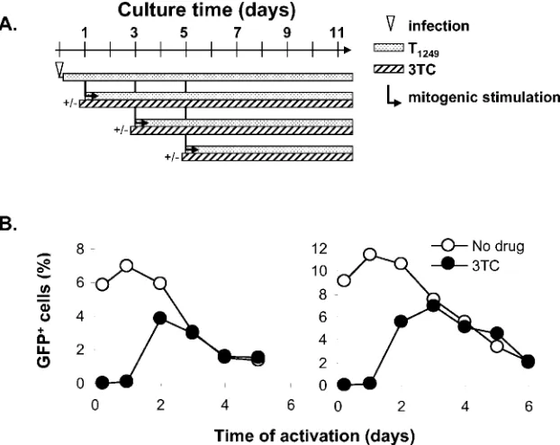
We next sought to understand the relationship between the observed virus decay and individual steps in viral replication in resting CD4⫹ T cells. Using a VSV-G-pseudotyped HIV-1 reporter virus system, we previously found that more than 85% of the virus decays before the completion of reverse transcrip-tion (29). In this study, we asked what fractranscrip-tion of virus decays before reverse transcription is completed when entry occurs through the normal fusion mechanism. This question is of particular importance because of evidence that the binding of HIV-1 envelope may trigger activation signals in resting CD4⫹ T cells. To assess whether reverse transcription was complete, infected resting CD4⫹ T cells were treated with the chain-terminating reverse transcriptase inhibitor lamivudine (3TC) before cellular activation (Fig. 3A). 3TC prevents virus gene expression in cells in which reverse transcription has not yet been completed. There was no detectable GFP expression when cells were activated in the presence of 3TC during the first 24 h after infection (Fig. 3B). As infected resting CD4⫹T cells were cultured for longer periods, induced GFP expression was detected in the presence of 3TC due to the completion of reverse transcription in some cells prior to 3TC treatment and cellular activation. The percentage of 3TC-treated cells
ex-pressing GFP upon activation peaked on day 2 or 3 after infection, indicating that it takes 2 to 3 days for HIV-1 to complete reverse transcription in some resting CD4⫹T cells. However, by that time, the percentage of 3TC-treated CD4⫹T cells expressing GFP was about half of the maximum percent-age of CD4⫹T cells that was induced to express GFP in the absence of 3TC. After 3 days, the decay curves for 3TC-treated and untreated cells were identical, reflecting the completion of reverse transcription in most of the cells. Therefore, approxi-mately 50% of HIV-1 decays before the completion of reverse transcription in resting CD4⫹T cells.
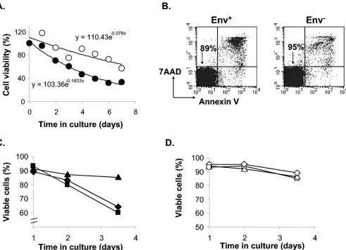
Infection-induced cell death contributes to virus decay.We next addressed the question whether the decay of rescuable virus is due to the death of host cells or the intrinsic instability of HIV-1 in the preintegration state. While mock-infected rest-ing CD4⫹ T cells (Fig. 4A) and uninfected resting CD4⫹T cells that were not centrifuged (data not shown) died slowly at similar rates due to the lack of survival signals, we found that infected cells died faster (Fig. 4A). To control for the possi-bility of toxic effects of components of the viral stock, we generated virions that were not coated with HIV-1 envelope protein by transfecting 293T cells with the NL4-3-GFP re-porter construct alone. Even in the absence of HIV-1 Env protein, HIV-1 Gag protein drives the assembly of virus par-ticles, which are released into the supernatant (24). Resting CD4⫹ T cells were spinoculated with either virions lacking HIV-1 Env or the infectious X4 Env-pseudotyped reporter virus. Both viral stocks were normalized based on levels of the HIV-1 Gag p24 antigen. Apoptotic cells were detected by annexin V and 7AAD staining. The percentage of apoptotic cells was higher in cells exposed to the X4-Env-pseudotyped reporter virus than in cells exposed to virions lacking HIV-1 Env (Fig. 4B). Therefore, the apoptosis of resting CD4⫹T cells was dependent upon interaction with Env-bearing virions and not other components of the virus stock or stress caused by centrifugal inoculation.
Interestingly, in these infections we detected infection-in-duced apoptosis 24 h after infection, earlier than the comple-tion of reverse transcripcomple-tion in most of the cells (Fig. 3B) and far earlier than the basal expression of GFP from noninte-grated viral DNA became detectable. This result suggested that apoptosis of infected, resting CD4⫹T cells was induced by early events in virus infection, prior to viral gene expression. Indeed, pretreating cells with reverse transcriptase inhibitor 3TC did not prevent apoptosis, indicating that reverse
tran-FIG. 2. Decay of HIV-1 in resting CD4⫹T cells. (A) Schematic representation of experimental strategy for studying HIV-1 decay in resting CD4⫹T cells. Resting CD4⫹T cells were infected, and 4 h later the cells were treated with the fusion inhibitor T1249 to block further entry. Cells were then cultured for the indicated times before they were activated with PHA and irradiated PBMC. (B) Inhibition of viral entry by T1249. Resting CD4⫹T cells were infected with the HIV-1 reporter virus in the absence (left panel) or presence (right panel) of T1249 and activated immediately after infection. GFP expression was examined 72 h later. (C) Activation-induced viral reporter gene expression. Infected resting cells were activated at day 1 postinfection (left panel), and GFP expression was examined 48 h later. Control cells were cultured for 4 days without activation (right panel). Mean fluorescence intensity for cells in the indicated gates is shown. (D) Loss of inducible GFP expression with time in culture. Resting CD4⫹T cells were infected with the HIV-1 reporter virus and cultured in the presence of 5g of T1249/ml as described above. At various times after infection, the cells were activated. The percentage of GFP-expressing cells was determined by fluorescence-activated cell sorter (FACS) analysis 48 h after activation. An exponential decay model was fitted to the data. y and x in the equations represent the same parameters indicated in the figure. The root mean square deviation (R2) is shown to indicate the fitness of the model. Results of three independent
experiments with CD4⫹T cells from different donors are presented. (E) Decay of rescuable viral production. Resting CD4⫹T cells were infected with the HIV-1 reporter virus. The supernatant was collected 7 or 14 days after activation for the p24 assay. Results of three independent experiments with CD4⫹T cells from different donors are presented.
on November 8, 2019 by guest
http://jvi.asm.org/
scription was not required for the induction of cell death (Fig. 4C). However, adding T1249 to the culture from the beginning of the infection prevented infection-induced apoptosis in rest-ing CD4⫹T cells, demonstrating that fusion and virus entry were required (Fig. 4C). The differential effects of 3TC and T1249 on infected cells did not result from drug toxicity, as neither drug affected the viability of cells that had been ex-posed to virions lacking HIV-1 Env (Fig. 4D). These data demonstrated that apoptosis of infected resting CD4⫹T cells occurs early upon viral entry and contributes to the loss of rescuable virus.
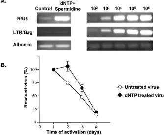
Intracellular degradation at early steps of the viral life cy-cle.The experiments described above indicated that the pref-erential death of infected cells may contribute to the loss of rescuable virus. However, the finding that significant decay occurs during the time when reverse transcription is being completed (Fig. 3) suggested that another component of the loss of rescuable virus might involve degradation of viral nu-cleic acids or critical protein components of the preintegration complex (29, 45, 46). With infection-induced apoptosis occur-ring simultaneously, it was difficult to test directly whether HIV-1 is susceptible to degradation in resting CD4⫹T cells that have not begun to undergo programmed cell death. It has been reported that free HIV-1 virions can be stimulated to undergo NERT, which not only yields nascent viral DNA but also alters the viral core structure (47, 48). We therefore tested whether stimulation of NERT with exogenous nucleotides would change the decay course of virus in infected resting CD4⫹T cells.
Equal amounts of reporter virus were pretreated with either
with 1 mM dNTP and 30M spermidine or culture medium alone. Treated and untreated viral stocks were used to infect equivalent numbers of resting CD4⫹T cells. To examine the effect of NERT, infected cells were collected immediately after spinoculation, and HIV-1 DNA was analyzed by PCR with previously described primer sets that detect early and late products of reverse transcription. As shown in Fig. 5A, NERT treatment resulted in more early reverse transcription products being detected with primers in the R and U5 regions, but no increase in late reverse transcription products was detected with primers in the LTR and Gag regions.
[image:6.585.137.448.69.316.2]We performed the same kinetic studies of virus decay on the two groups of cells infected with either dNTP-treated or un-treated virus. As shown in Fig. 5B, there was a delay in the decay of dNTP-treated virus between day 1 and day 2 postin-fection. Since cells infected with dNTP-treated virus died slightly faster than cells infected with the control virus (data not shown), the delay was not caused by different rates of apoptosis in two infected cultures. Rather, the initial delay may be due to the fact that early initiation of reverse transcription allowed dNTP-treated virus to survive intracellular degrada-tion during the first few days postinfecdegrada-tion. After the initial delay, the decay of dNTP-treated virus became similar to that of the untreated virus, suggesting that either host cell apoptosis becomes the dominant factor in the viral decay process or intracellular degradation also occurs in subsequent steps of the viral life cycle. The fact that the decay curve is different with dNTP-treated virus strongly supports the hypothesis that the observed decay of virus is not solely due to host cell apoptosis. Instead, these results are consistent with the conclusion that
FIG. 3. Decay of HIV-1 before the completion of reverse transcription. (A) Experimental strategy for measuring the kinetics of HIV-1 decay in relation to the completion of reverse transcription. In these experiments, 3TC was added 4 h prior to activation and kept in the activated culture to prevent virus gene expression in cells in which reverse transcription was not complete. (B) Decay of HIV-1 before the completion of reverse transcription. Resting CD4⫹T cells were infected with the X4 Env-pseudotyped NL4-3-GFP reporter virus at an MOI of 1 and cultured for the indicated times before being activated in the absence (open circles) or presence (filled circles) of 10M 3TC. The maximum percentage of GFP-positive cells observed following activation was plotted. Results of two independent experiments are presented.
2204 ZHOU ET AL. J. VIROL.
on November 8, 2019 by guest
http://jvi.asm.org/
HIV-1 is susceptible to degradation at early steps of viral life cycle in resting CD4⫹T cells.
Intracellular degradation of the integration-competent form of HIV-1.We next examined the fate of viruses that complete reverse transcription in resting CD4⫹T cells. For these viruses, the full-length linear cDNA remains in the cytoplasm and does not integrate due to a defect in nuclear import in resting cells (4). Previous studies by our laboratory demonstrated that in resting CD4⫹cells, but not in CD4⫹T lymphoblasts, a fraction of linear viral cDNAs develop deletions at the termini of the viral genome (29). These linear viral cDNAs with deletions are not capable of integrating into the host cell genome (15). To distinguish integration-competent HIV-1 from the other forms of viral cDNAs, we used the integrase inhibitor L-731,988 in an activation-dependent virus rescue system. L-731,988 binds to
[image:7.585.49.540.67.423.2]integrase in the preintegration complex and prevents strand transfer during the integration process by occupying the bind-ing site of the target DNA (14, 17). When added to infected CD4⫹T lymphoblasts at the time of infection, L-731,988 com-pletely inhibited virus particle production (Fig. 6A). The ex-pression of GFP was also strongly inhibited (Fig. 6B). How-ever, a low level of GFP expression was detected in a small fraction of infected cells, even in the presence of L-731,988 (Fig. 6B). This finding might reflect transcription from 2-LTR circles that are known to accumulate in infected cells treated with L-731,988 (17, 44). A low level of GFP expression was also observed when CD4⫹ T lymphoblasts were infected with a GFP reporter virus carrying a mutation (D64N) in the first catalytic residue of integrase (Fig. 6C, left panel). In contrast to NL-4-3-GFP virus, the integrase mutant virus expressed the
FIG. 4. Infection-induced apoptosis in resting CD4⫹T cells. (A) Differential survival of infected (filled circles) and mock-infected (open circles) resting CD4⫹T cells. Viable cells were counted by using trypan blue. An exponential decay model was fitted to the data. y and x in the equations represent the same parameters indicated in the figure. (B) Apoptosis induced in resting CD4⫹T cells by exposure to Env⫹virions. Resting CD4⫹ T cells were infected with equal numbers of virions with (left panel) or without (right panel) HIV-1 Env. Cell viability was examined by 7AAD and annexin V staining on day 1 postinfection. The percentages of viable cells are indicated in the diagrams. (C) Inhibition of infection-induced apoptosis by T1249 but not 3TC. Resting CD4⫹T cells were infected with the X4-pseudotyped NL4-3-GFP reporter virus in the absence of drugs (diamonds) or in the presence of 5g of T1249/ml (triangles) or 10M 3TC (squares). In the case of 3TC, cells were pretreated 12 h prior to infection to allow time for intracellular phosphorylation of the nucleoside analogue. The percentage of viable cells was determined by 7AAD and annexin V staining and FACS analysis. (D) Control infection with virions lacking Env protein demonstrating minimal toxicity of 3TC and T1249 on resting CD4⫹T cells. Resting CD4⫹T cells were inoculated with virions lacking HIV-1 Env in the absence of drugs (diamonds) or in the presence of 5g of T1249/ml (triangles) or 10M 3TC (squares) as described for panel C. Results shown are representative of those of three independent experiments.
on November 8, 2019 by guest
http://jvi.asm.org/
same level of GFP in the presence of L-731,988 (Fig. 6C, right panel). Therefore, L-731,988 reduces viral gene expression by blocking integration, allowing us to compare the activation-induced GFP expression from extrachromosomal viral DNA and integrated provirus.
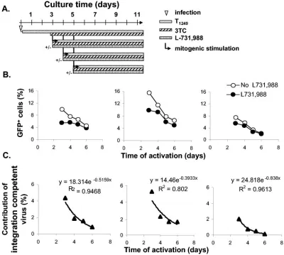
To measure the decay of full-length, integration-competent HIV-1 DNA, purified resting CD4⫹T cells were infected with the X4-Env-pseudotyped HIV-1 reporter virus. Four hours after infection, T1249 was added to block subsequent entry and synchronize the infection. To ensure that only virus that had completed reverse transcription in resting CD4⫹T cells was studied, the reverse transcriptase inhibitor 3TC was added to infected cultures 3 days after infection and maintained in all cultures from that point on. At different time points, infected cells were activated in the presence or the absence of L-731,988 (Fig. 7A). We observed that fewer cells were capable of expressing GFP upon cellular activation in the presence of L-731,988 than in the absence of L-731,988 (Fig. 7B). The difference reflected the existence of complete reverse tran-scripts that were competent for integration into the host ge-nome at the time mitogenic stimulation was provided. This difference diminished rapidly as infected resting CD4⫹T cells were cultured for prolonged periods (Fig. 7C). By day 6 postin-fection, the percentage of GFP-positive cells was very low
regardless of the L-731,988 treatment, suggesting that full-length linear HIV-1 cDNA capable of integration was no longer present. Residual induced GFP expression was proba-bly all from circular HIV-1 cDNAs (43, 44). The observed decay of integration-competent HIV-1 was independent of host cell apoptosis, since at each time point, the differences in induced GFP expression were caused by the block of integra-tion. Similar decay was observed when a more potent integrase inhibitor, C5, was used instead of L-731,988 (data not shown). Our data demonstrate that HIV-1 is still labile after comple-tion of reverse transcripcomple-tion in resting CD4⫹ T cells. The half-life of the integration-competent form of HIV-1 was de-termined to be 1.3⫾0.5 days (Fig. 7C).
DISCUSSION
[image:8.585.122.456.72.347.2]Resting CD4⫹ T cells harboring full-length, unintegrated linear viral DNA constitute an inducible latent reservoir for HIV-1 that is distinct from the reservoir of integrated virus (26). Previous studies have demonstrated that in comparison to viral replication in activated CD4⫹T cells, replication in rest-ing cells is reversibly blocked at multiple steps prior to inte-gration (28). The result is a state of preinteinte-gration latency which is important because it is the most common form of
FIG. 5. Delay of viral decay in resting CD4⫹T cells infected with nucleotide-treated virions. (A) Analysis of viral cDNA species generated through NERT. Resting CD4⫹T cells were infected with dNTP-treated or untreated virus preparations. Cells were collected immediately after spin-inoculation and nascent viral DNA was analyzed by PCR with primers that detect early reverse transcripts (R-U5), late reverse transcripts (LTR-Gag), or the cellular gene albumin. Copy number controls were set up by using the indicated numbers of copies of pNL4-3-GFP plasmid diluted into 6⫻104-cell equivalents of DNA from an HIV-1-negative donor. (B) Delay in viral decay caused by NERT. Resting CD4⫹T cells were
infected with dNTP-treated or untreated virus. At the indicated times, the same numbers of viable cells were activated in duplicate wells in a 96-well plate. The induced viral production was measured at day 7 postactivation by p24 assay. The amounts of rescued virus at various times were normalized to the amount of virus rescued by activation on the first day postinfection. Results shown are representative of those of two independent experiments.
2206 ZHOU ET AL. J. VIROL.
on November 8, 2019 by guest
http://jvi.asm.org/
infection in resting CD4⫹T cells in untreated individuals (5, 8). It remains controversial whether HIV-1 is stable in the preintegration state. The recent discovery that subtle stimula-tion of resting CD4⫹T cells allows HIV-1 to overcome these blocks further highlights the need for a more complete under-standing of the finely tuned host-virus interaction in resting CD4⫹T cells (13, 25, 38, 41, 49). In the study presented here, a novel HIV-1 Env-pseudotyped reporter virus was used to study the fate of HIV-1 in resting CD4⫹T cells in a synchro-nized single-round infection. This approach allowed us to de-fine a rapid multifactorial viral decay process in resting CD4⫹ T cells.
The functional decay of HIV-1 in resting CD4⫹ T cells cannot be observed directly because resting CD4⫹ T cells showed minimal virus gene expression or virus production without cellular activation. Therefore, decay was monitored in rescue experiments in which infected resting CD4⫹ T cells were cultured for various periods of time and then subjected to conditions that activated 100% of the cells. We first demon-strated that rescuable viral gene expression and virus produc-tion from infected resting CD4⫹T cells declines steadily after
[image:9.585.265.539.72.379.2]infection, with a half-life of 2 days. We also extended previous observations of slow reverse transcription in resting CD4⫹T cells by showing that accumulation of complete functional re-verse transcription products occurred over the first 2 or 3 days postinfection. By using synchronized infections, it was possible to show that approximately 50% of the viruses that enter rest-ing CD4⫹T cells decay before completing reverse transcrip-tion. Interestingly, the decay of rescuable gene expression in cells infected with the HIV-1 reporter virus carrying an X4 HIV-1 envelope was slightly slower than the previously re-ported decay of VSV-G-pseudotyped recombinant HIV-1, which has a half-life of 1 day (29). When cells were activated in the presence of 3TC 2 days after infection, there were more GFP-expressing cells in cultures infected with the reporter virus carrying an X4 HIV-1 envelope than in cultures infected with VSV-G-pseudotyped virus. These data demonstrated that HIV-1 replication in resting CD4⫹T cells is affected by viral entry routes. It is possible that signals conveyed through bind-ing of HIV-1 Env to CD4 and CXCR4 favor the completion of reverse transcription. However, despite T-cell stimulatory sig-nals triggered by HIV-1 Env, decay processes still prevent viral
FIG. 6. Effects of integration inhibitor L-731,988. (A) Complete inhibition of virus production by integrase inhibitor L-731,988. Activated CD4⫹ T cells were infected with the X4-Env-pseudotyped HIV-1 reporter virus in the absence (open circles) or the presence of 40M L-731,988 (closed circles). The amount of p24 antigen in the supernatant was measured by enzyme-linked immunosorbent assay. Results shown are representative of those of three independent experiments. (B) Low level of GFP expression in the presence of L-731,988. Activated CD4⫹T cells were infected with NL4-3-GFP virus in the absence (left panel) or presence (right panel) of 40M L-731,988. GFP expression was examined 72 h postinfection. Results shown are representative of those of four independent experiments. (C) L-731,988 has little effect on expression of GFP from extrachro-mosomal viral DNA. Activated CD4⫹T cells from the same donor as in described for panel B were infected with a HIV-1 reporter virus carrying a D64N mutation in integrase in the absence (left panel) or presence (right panel) of 40M L-731,988. GFP expression was examined 72 h postinfection. Results shown are representative of those of four independent experiments.
on November 8, 2019 by guest
http://jvi.asm.org/
replication in resting CD4⫹T cells unless the cells are acti-vated before irreversible decay occurs.
The observed decay of rescuable HIV-1 is partially caused by apoptosis of the infected resting CD4⫹T cells. While some studies reported that productive infection of CD4⫹T cells is required for HIV-1-induced apoptosis (10, 21, 39), our data showed that infected resting CD4⫹T cells can undergo apo-ptosis even when HIV-1 reverse transcription is blocked. Ap-optosis was prevented if viral fusion was blocked. No syncytia were observed in the infected cultures (data not shown). In-terestingly, it has been reported that virion-associated Vpr induces cell cycle arrest in the presence of reverse transcriptase inhibitor (30). Current efforts are directed at determining whether Vpr or other components of the virus particle causes apoptosis in resting CD4⫹T cells at this early stage of infection
and whether the apoptosis of infected resting CD4⫹T cells plays a significant role in CD4⫹-T-cell depletion in vivo.
[image:10.585.91.491.69.428.2]In addition to host cell apoptosis, we demonstrated that rescuable HIV-1 is also lost due to intracellular decay events affecting the virus. An initial study by Zack et al. showed that the early reverse transcription products decreased in resting CD4⫹ T cells, with a half-life of 1 day (45). The reduced number of early reverse transcripts was documented by real-time PCR in a recent study (37). However, apoptosis of in-fected cells could account for loss of viral DNA. In this study, we compared the decay processes of HIV-1 pretreated with dNTP with those of untreated virus in order to control for the contribution of apoptosis to the observed viral decay. At early time points after infection, viruses that had initiated reverse transcription before infection were more likely to be rescued
FIG. 7. Decay of integration-competent HIV-1 in resting CD4⫹T cells. (A) Experimental strategy for monitoring the functional decay of full-length, integration-competent viral DNA by blocking integration with L-731,988 upon T-cell activation. 3TC was added to resting CD4⫹T cells at day 3 postinfection so that only viruses that had completed reverse transcription were studied. Cells were then activated at the indicated times in the absence or presence of L-731,988. At 48 h after activation, cells were examined for rescued GFP expression. (B) Decay of rescuable GFP expression in the presence or absence of L-731,988. Resting CD4⫹T cells were infected with the reporter virus at an MOI of 1. T1249 was added 4 h after infection, and 3TC was added 3 days after infection. Both drugs were then maintained in all cultures for the rest of the experiment. At the indicated times, infected cells were activated in the presence (closed circles) or absence (open circles) of 40M L-731,988. The percentage of GFP-expressing cells was determined by FACS analysis 48 h after activation. Results of three independent experiments are presented. (C) Decay of integration-competent HIV-1. GFP expression from integrated virus upon activation was calculated by subtracting the percentage of GFP-positive cells induced in the presence of L-731,988 and 3TC from the percentage of GFP-GFP-positive cells induced in the presence of 3TC alone. An exponential decay model was fitted to the data. y and x in the equations represent the same parameters indicated in the figure. The root mean square deviation (R2) is shown to indicate the fitness of the model.
2208 ZHOU ET AL. J. VIROL.
on November 8, 2019 by guest
http://jvi.asm.org/
by cellular activation than viruses that did not, suggesting that degradation of HIV-1 in resting CD4⫹T cells starts early in the viral life cycle, probably during early reverse transcription. The same degradation process might be involved in TRIM5␣ -mediated restriction of HIV infection in rhesus macaques (36, 40).
Intracellular decay is not restricted to early reverse tran-scription in resting CD4⫹ T cells. We also determined the decay rate of the integration-competent form of HIV-1 that has completed reverse transcription. Results from previous studies have suggested the lability of full-length, linear viral DNA. Stevenson et al. reported that extrachromosomal HIV-1 DNA persisted in monocyte-depleted PBMC for 2 weeks postinfection, yet only a small fraction became integrated upon activation at the end of the 2-week culture (35). Nucleotide deletions of various lengths are found in the U3 terminus of linear HIV-1 DNA (29). However, there has not been a pre-vious kinetic study of the decay of integration-competent HIV-1 DNA because the classical PCR methods do not dis-tinguish between the integration-competent form and other forms of viral DNA. Instead, it was recently reported that long reverse transcripts accumulate stably in resting CD4⫹T cells (37). In this study, we monitored the loss of integration-depen-dent, inducible GFP expression and demonstrated that after the completion of reverse transcription, the integration-com-petent form of HIV-1 decays with a half-life of 1 day in resting CD4⫹T cells. The decay of complete reverse transcripts was not detected in our previous study using VSV-G-pseudotyped virus since very few cells harbor complete reverse transcripts in that model.
Degradation of either viral DNA or viral protein in the preintegration complex will lead to functional decay of virus in the preintegration state. The proteasome has been demon-strated to play a role in viral degradation. Inhibition of pro-teasome function in infected cell lines increases the production of proviral DNA by blocking proteasome-mediated degrada-tive processes that act on preintegration complexes containing largely or fully completed reverse transcription products (6, 32).
The study presented here provides new insight on the fate of HIV-1 in resting CD4⫹T cells and allows us to give a detailed picture of the interactions between HIV-1 and resting CD4⫹T cells. Following entry, HIV-1 induces some resting CD4⫹ T cells to undergo apoptosis. In the remaining cells, the virus becomes susceptible to cellular degradation during early re-verse transcription. About 50% of the viruses that enter resting CD4⫹ T cells complete reverse transcription. This process takes 2 or 3 days. After completion of reverse transcription, the viral DNA starts to lose the capacity to integrate, presumably because the U3 terminus is degraded. The functional half-life of full-length, integration-competent HIV-1 DNA is as short as 1 day. We propose that the competing processes of viral rep-lication and intracellular degradation, both before and after the completion of reverse transcription, occur in all infected CD4⫹ T cells. Whether HIV-1 can replicate or establish a stable state of postintegration latency in CD4⫹T cells is de-termined by how quickly the virus becomes integrated into the host genome, which depends on the activation state of T cells relative to the rate of decay. In unstimulated resting CD4⫹T cells, such as those found in peripheral blood, HIV-1 fails to
progress quickly to integration because of slow reverse tran-scription and blocks at subsequent steps, and the degradation process dominates. As a result, only a labile HIV-1 latent reservoir is formed when these cells are infected.
ACKNOWLEDGMENTS
This work was supported by NIH grant AI43222, by the Doris Duke Charitable Foundation, and by the Howard Hughes Medical Institute.
REFERENCES
1.Aiken, C.1997. Pseudotyping human immunodeficiency virus type 1 (HIV-1) by the glycoprotein of vesicular stomatitis virus targets HIV-1 entry to an endocytic pathway and suppresses both the requirement for Nef and the
sensitivity to cyclosporin A. J. Virol.71:5871–5877.
2.Berger, E. A., P. M. Murphy, and J. M. Farber.1999. Chemokine receptors as HIV-1 coreceptors: roles in viral entry, tropism, and disease. Annu. Rev.
Immunol.17:657–700.
3.Blankson, J. N., D. Finzi, T. C. Pierson, B. P. Sabundayo, K. Chadwick, J. B. Margolick, T. C. Quinn, and R. F. Siliciano.2000. Biphasic decay of latently
infected CD4⫹T cells in acute human immunodeficiency virus type 1
infec-tion. J. Infect. Dis.182:1636–1642.
4.Bukrinsky, M. I., N. Sharova, M. P. Dempsey, T. L. Stanwick, A. G. Bukrin-skaya, S. Haggerty, and M. Stevenson.1992. Active nuclear import of human immunodeficiency virus type 1 preintegration complexes. Proc. Natl. Acad.
Sci. USA89:6580–6584.
5.Bukrinsky, M. I., T. L. Stanwick, M. P. Dempsey, and M. Stevenson.1991. Quiescent T lymphocytes as an inducible virus reservoir in HIV-1 infection.
Science254:423–427.
6.Butler, S. L., E. P. Johnson, and F. D. Bushman.2002. Human immunode-ficiency virus cDNA metabolism: notable stability of two-long terminal
re-peat circles. J. Virol.76:3739–3747.
7.Chazal, N., G. Singer, C. Aiken, M. L. Hammarskjold, and D. Rekosh.2001. Human immunodeficiency virus type 1 particles pseudotyped with envelope proteins that fuse at low pH no longer require Nef for optimal infectivity.
J. Virol.75:4014–4018.
8.Chun, T. W., L. Carruth, D. Finzi, X. F. Shen, J. A. DiGiuseppe, H. Taylor, M. Hermankova, K. Chadwick, J. Margolick, T. C. Quinn, Y. H. Kuo, R. Brookmeyer, M. A. Zeiger, P. Barditch-Crovo, and R. F. Siliciano.1997. Quantification of latent tissue reservoirs and total body viral load in HIV-1
infection. Nature387:183–188.
9.Cicala, C., J. Arthos, S. M. Selig, G. Dennis, Jr., D. A. Hosack, D. Van Ryk, M. L. Spangler, T. D. Steenbeke, P. Khazanie, N. Gupta, J. Yang, M. Daucher, R. A. Lempicki, and A. S. Fauci.2002. HIV envelope induces a cascade of cell signals in non-proliferating target cells that favor virus
rep-lication. Proc. Natl. Acad. Sci. USA99:9380–9385.
10.Corbeil, J., and D. D. Richman.1995. Productive infection and subsequent interaction of CD4-gp120 at the cellular membrane is required for
HIV-induced apoptosis of CD4⫹T cells. J. Gen. Virol.76:681–690.
11.Dornadula, G., S. Yang, R. J. Pomerantz, and H. Zhang.2000. Partial rescue of the Vif-negative phenotype of mutant human immunodeficiency virus type 1 strains from nonpermissive cells by intravirion reverse transcription. J.
Vi-rol.74:2594–2602.
12.Douek, D. C., J. M. Brenchley, M. R. Betts, D. R. Ambrozak, B. J. Hill, Y. Okamoto, J. P. Casazza, J. Kuruppu, K. Kunstman, S. Wolinsky, Z. Gross-man, M. Dybul, A. Oxenius, D. A. Price, M. Connors, and R. A. Koup.2002.
HIV preferentially infects HIV-specific CD4⫹T cells. Nature417:95–98.
13.Eckstein, D. A., M. L. Penn, Y. D. Korin, D. D. Scripture-Adams, J. A. Zack, J. F. Kreisberg, M. Roederer, M. P. Sherman, P. S. Chin, and M. A. Gold-smith.2001. HIV-1 actively replicates in naive CD4⫹T cells residing within
human lymphoid tissues. Immunity15:671–682.
14.Espeseth, A. S., P. Felock, A. Wolfe, M. Witmer, J. Grobler, N. Anthony, M. Egbertson, J. Y. Melamed, S. Young, T. Hamill, J. L. Cole, and D. J. Hazuda.
2000. HIV-1 integrase inhibitors that compete with the target DNA substrate define a unique strand transfer conformation for integrase. Proc. Natl. Acad.
Sci. USA97:11244–11249.
15.Esposito, D., and R. Craigie.1998. Sequence specificity of viral end DNA binding by HIV-1 integrase reveals critical regions for protein-DNA
inter-action. EMBO J.17:5832–5843.
16.Finzi, D., M. Hermankova, T. Pierson, L. M. Carruth, C. Buck, R. E. Chaisson, T. C. Quinn, K. Chadwick, J. Margolick, R. Brookmeyer, J. Gal-lant, M. Markowitz, D. D. Ho, D. D. Richman, and R. F. Siliciano.1997. Identification of a reservoir for HIV-1 in patients on highly active
antiret-roviral therapy. Science278:1295–1300.
17.Hazuda, D. J., P. Felock, M. Witmer, A. Wolfe, K. Stillmock, J. A. Grobler, A. Espeseth, L. Gabryelski, W. Schleif, C. Blau, and M. D. Miller.2000. Inhibitors of strand transfer that prevent integration and inhibit HIV-1
replication in cells. Science287:646–650.
18.Hermankova, M., J. D. Siliciano, Y. Zhou, D. Monie, K. Chadwick, J. B. Margolick, T. C. Quinn, and R. F. Siliciano.2003. Analysis of human
on November 8, 2019 by guest
http://jvi.asm.org/
munodeficiency virus type 1 gene expression in latently infected resting
CD4⫹T lymphocytes in vivo. J. Virol.77:7383–7392.
19.Kinter, A. L., C. A. Umscheid, J. Arthos, C. Cicala, Y. Lin, R. Jackson, E. Donoghue, L. Ehler, J. Adelsberger, R. L. Rabin, and A. S. Fauci.2003. HIV
envelope induces virus expression from resting CD4⫹T cells isolated from
HIV-infected individuals in the absence of markers of cellular activation or
apoptosis. J. Immunol.170:2449–2455.
20.Korin, Y. D., and J. A. Zack.1999. Nonproductive human immunodeficiency
virus type 1 infection in nucleoside-treated G0lymphocytes. J. Virol.73:
6526–6532.
21.Leonard, R., D. Zagury, I. Desportes, J. Bernard, J. F. Zagury, and R. C. Gallo.1988. Cytopathic effect of human immunodeficiency virus in T4 cells is linked to the last stage of virus infection. Proc. Natl. Acad. Sci. USA
85:3570–3574.
22.Matlin, K. S., H. Reggio, A. Helenius, and K. Simons.1982. Pathway of
vesicular stomatitis virus entry leading to infection. J. Mol. Biol.156:609–
631.
23.O’Doherty, U., W. J. Swiggard, and M. H. Malim.2000. Human immuno-deficiency virus type 1 spinoculation enhances infection through virus
bind-ing. J. Virol.74:10074–10080.
24.Ono, A., D. Demirov, and E. O. Freed.2000. Relationship between human immunodeficiency virus type 1 Gag multimerization and membrane binding.
J. Virol.74:5142–5150.
25.Oswald-Richter, K., S. M. Grill, M. Leelawong, and D. Unutmaz.2004. HIV infection of primary human T cells is determined by tunable thresholds of T
cell activation. Eur. J. Immunol.34:1705–1714.
26.Persaud, D., Y. Zhou, J. M. Siliciano, and R. F. Siliciano.2003. Latency in human immunodeficiency virus type 1 infection: no easy answers. J. Virol.
77:1659–1665.
27.Pierson, T., T. L. Hoffman, J. Blankson, D. Finzi, K. Chadwick, J. B. Mar-golick, C. Buck, J. D. Siliciano, R. W. Doms, and R. F. Siliciano.2000. Characterization of chemokine receptor utilization of viruses in the latent
reservoir for human immunodeficiency virus type 1. J. Virol.74:7824–7833.
28.Pierson, T., J. McArthur, and R. F. Siliciano.2000. Reservoirs for HIV-1: mechanisms for viral persistence in the presence of antiviral immune
re-sponses and antiretroviral therapy. Annu. Rev. Immunol.18:665–708.
29.Pierson, T. C., Y. Zhou, T. L. Kieffer, C. T. Ruff, C. Buck, and R. F. Siliciano.
2002. Molecular characterization of preintegration latency in human
immu-nodeficiency virus type 1 infection. J. Virol.76:8518–8531.
30.Poon, B., K. Grovit-Ferbas, S. A. Stewart, and I. S. Chen.1998. Cell cycle arrest by Vpr in HIV-1 virions and insensitivity to antiretroviral agents.
Science281:266–269.
31.Pope, M., and A. T. Haase.2003. Transmission, acute HIV-1 infection and
the quest for strategies to prevent infection. Nat. Med.9:847–852.
32.Schwartz, O., V. Marechal, B. Friguet, F. Arenzana-Seisdedos, and J. M. Heard.1998. Antiviral activity of the proteasome on incoming human
im-munodeficiency virus type 1. J. Virol.72:3845–3850.
33.Spina, C. A., J. C. Guatelli, and D. D. Richman.1995. Establishment of a stable, inducible form of human immunodeficiency virus type 1 DNA in
quiescent CD4 lymphocytes in vitro. J. Virol.69:2977–2988.
34.Stevenson, M.2003. HIV-1 pathogenesis. Nat. Med.9:853–860.
35.Stevenson, M., T. L. Stanwick, M. P. Dempsey, and C. A. Lamonica.1990.
HIV-1 replication is controlled at the level of T cell activation and proviral
integration. EMBO J.9:1551–1560.
36.Stremlau, M., C. M. Owens, M. J. Perron, M. Kiessling, P. Autissier, and J. Sodroski.2004. The cytoplasmic body component TRIM5 alpha restricts
HIV-1 infection in Old World monkeys. Nature427:848–853.
37.Swiggard, W. J., U. O’Doherty, D. McGain, D. Jeyakumar, and M. H. Malim.
2004. Long HIV type 1 reverse transcripts can accumulate stably within
resting CD4⫹T cells while short ones are degraded. AIDS Res. Hum.
Retrovir.20:285–295.
38.Swingler, S., B. Brichacek, J. M. Jacque, C. Ulich, J. Zhou, and M.
Steven-son.2003. HIV-1 Nef intersects the macrophage CD40L signalling pathway
to promote resting-cell infection. Nature424:213–219.
39.Sylwester, A. W., J. C. Grivel, W. Fitzgerald, J. L. Rossio, J. D. Lifson, and L. B. Margolis.1998. CD4⫹T-lymphocyte depletion in human lymphoid tissue ex vivo is not induced by noninfectious human immunodeficiency virus
type 1 virions. J. Virol.72:9345–9347.
40.Turelli, P., V. Doucas, E. Craig, B. Mangeat, N. Klages, R. Evans, G. Kalpana, and D. Trono.2001. Cytoplasmic recruitment of INI1 and PML on incoming HIV preintegration complexes: interference with early steps of
viral replication. Mol. Cell7:1245–1254.
41.Unutmaz, D., V. N. KewalRamani, S. Marmon, and D. R. Littman.1999. Cytokine signals are sufficient for HIV-1 infection of resting human T
lym-phocytes. J. Exp. Med.189:1735–1746.
42.Wild, C. T., D. C. Shugars, T. K. Greenwell, C. B. McDanal, and T. J. Matthews.1994. Peptides corresponding to a predictive alpha-helical do-main of human immunodeficiency virus type 1 gp41 are potent inhibitors of
virus infection. Proc. Natl. Acad. Sci. USA91:9770–9774.
43.Wu, Y., and J. W. Marsh.2001. Selective transcription and modulation of
resting T cell activity by preintegrated HIV DNA. Science293:1503–1506.
44.Wu, Y., and J. W. Marsh.2003. Early transcription from nonintegrated DNA
in human immunodeficiency virus infection. J. Virol.77:10376–10382.
45.Zack, J. A., S. J. Arrigo, S. R. Weitsman, A. S. Go, A. Haislip, and I. S. Chen.
1990. HIV-1 entry into quiescent primary lymphocytes: molecular analysis
reveals a labile, latent viral structure. Cell61:213–222.
46.Zack, J. A., A. M. Haislip, P. Krogstad, and I. S. Chen.1992. Incompletely reverse-transcribed human immunodeficiency virus type 1 genomes in qui-escent cells can function as intermediates in the retroviral life cycle. J. Virol.
66:1717–1725.
47.Zhang, H., G. Dornadula, J. Orenstein, and R. J. Pomerantz.2000. Mor-phologic changes in human immunodeficiency virus type 1 virions secondary to intravirion reverse transcription: evidence indicating that reverse tran-scription may not take place within the intact viral core. J. Hum. Virol.
3:165–172.
48.Zhang, H., G. Dornadula, and R. J. Pomerantz.1996. Endogenous reverse transcription of human immunodeficiency virus type 1 in physiological mi-croenvironments: an important stage for viral infection of nondividing cells.
J. Virol.70:2809–2824.
49.Zhang, Z. Q., T. Schuler, M. Zupancic, S. Wietgrefe, K. A. Staskus, K. A. Reimann, T. A. Reinhart, M. Rogan, W. Cavert, C. J. Miller, R. S. Veazey, D. Notermans, S. Little, S. A. Danner, D. D. Richman, D. Havlir, J. Wong, H. L. Jordan, T. W. Schacker, P. Racz, K. Tenner-Racz, N. L. Letvin, S. Wolinsky, and A. T. Haase.1999. Sexual transmission and propagation of SIV and HIV
in resting and activated CD4⫹T cells. Science286:1353–1357.
2210 ZHOU ET AL. J. VIROL.