Abstract— Skull Stripping is an inevitable computer-assisted medical brain mage pre-processing mechanism thataims to segregate the non-designated regions, in order to extract the designated brain regions. Those resultant brain region images arefurther subjected to diagnostic analysis to discover the brain diseases/ disorders. This paper presents an efficient method named, Threshold and Morphological Operations based Segmentation, (TMOS) for skull stripping is, exclusively designed for MRI brain images (Magnetic Resonance Images). It has two phases: the first phase localizes the brain region using Region of Interest (RoI) and in the second phase, morphological operations are applied to precisely extract the brain region by stripping out the skull. The performance of TMOS is validated using Minimal Interval Resonance Image in Alzheimer Disease (MIRIAD) dataset. The accuracy of TMOS is visually confirmed.
Keywords—Skull Stripping, T1-Weighted MRI brain images, Otsu’s threshold, Region of Interest method, Morphological Operation.
I. INTRODUCTION
The fully/semi-automated brain image analysis aims to discover brain abnormalities/ disorders and other diagnosis. Skull stripping is an essential step in extracting the brain regions for morphometric studies of Magnetic Resonance Images (MRI). In this article, a new segmentation scheme for MRI termed as, Thresholded and Morphological Operations based Segmentation (TMOS) is proposed. The following sections are organized as: Section II presents the review of a few significant MRI segmentation methods; methodology of TMOS is elucidate in Section III; results of TMOS and the related discussions are given in Section IV and the concluding remarks of this research work are furnished in Section V.
II. RELATED WORK
Hundreds of MRI brain segmentation techniques are already available in the literature. The common principles and mechanisms designed and developed are reported in [1 - 6]. Shijin kumara et al., [7] describedabout a skull-stripping technique which is designed based on the principle of connected regions as well as the morphological image operations for MRI images. 2D region growing algorithm by Park et.al., [8]automatically selects two exclusive seed pixels for the brain region and its counterpart. This approach is apparently confirmed to effectively segment the images of poor illumination and homogeneities and corrupted images.
Revised Manuscript Received on April 12, 2019.
S.Naganandhini, Research Scholar, Department of Computer Science and Applications The Gandhigram Rural Institute (Deemed to be University) Gandhigram-624 302, Dindigul, Tamil Nadu, India. ([email protected])
P.Shanmugavadivu, Professor, Department of Computer Science and Applications, The Gandhigram Rural Institute (Deemed to be University), Gandhigram-624 302, Dindigul, Tamil Nadu, India. ([email protected])
Rosnizaet.al., [9] presented a technique for MRI brain image for brain segmentation. However, this method fails on the brain images with intensity homogeneity. Even though numerous techniques are reported in the studies of skull stripping, region-growing and mathematical morphology method are widely employed for segmentation.
Sudipta Roy et al., [10] developed an automatic method for Artifacts and skull removal of brain in MRI using computational geometry, wavelet decomposition and thresholding as intermediate steps.
Benson et. al., [11] proposed two computationally simple techniques for MRI,based on mathematical morphological operations for two pre-processing operations -brain image enhancement and skull stripping, respectively.
SajjadMohsinet. al., [12] proposed an improved fully automated skull stripping technique. It assures higher accuracy when compared to its competitive methods.
Shafaf Ibrahim et. al., [13] proposed Seed-Based Region Growing (SBRG) technique based on the consensusamong the radiologist and radiographers. It is concluded that this skull stripping method is quite efficient.
The proposed TMOS is designed, using Region of Interest (RoI) and morphological operations. The primary objective of TMOS is to accurately segment the brain image, with minimum computational and time complexity. It is important to underline that TMOS is free from under-segmentation as well as over under-segmentation. Hence, TMOS, is confirmed to be an ideal pre-processing tool in brain image processing and analysis.
III. THERSHOLDED AND MORPHOLOGICAL
OPERATIONS BASED SEGMENTATATION (TMOS)
The MRI brain segmentation by TMOS involves RoI and thresholding-based region selection and morphological operations based segmentation.
In TMOS, the input MRI is binarized using Otsu’s thresholding wherein the selection of threshold is automatically done using [14-16]. This proven global thresholding method iterates through all the possible threshold values in order to optimally separate the foreground from the background.
The process of brain segmentation is optimized with the automatic/semi-automatic selection of RoI. In TMOS, the RoI is chosen based on the elliptical mask using interactive tool.
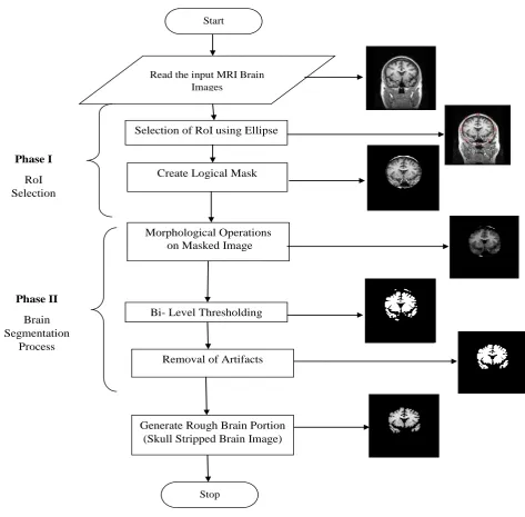
Based Brain Image Segmentation
Fig. 1 Flowchart of TMOS The masking operations with reference to the chosen
RoI, facilitates the selection of pixels confined within the boundary of RoI. The pixels bounded by the RoI are preserved while the remaining pixels are discarded using the ellipseConvolution filter,. This convolution operation enables to separate the RoI from MRI image, leaving out the remaining pixels (i.e) unwanted pixels. In general the morphological image operators viz.,erosion, dilation, opening and closing are applied on digital images separately or is combination, in order to refine the process of segmentation using structuring element of varying size, as required. The TMOS uses opening and closing for this purpose ofskull stripping on the brain image obtained out of RoI selection. The opening of A by B is computed as the erosion of A by B, which is suceeded by dilation. The
Morphological closing is similar to dilation in which the boundaries of foreground regions are based on the given structuring element. The closing of A by B is:
A•B = (AB)B (2)
E. Algorithm of TMOS
Input : Grayscale Image I(Normal/AD) Output: Extracted Brain Portion Step 1:Read the input MRI brain images
Step 2: Select the coordinates for RoI selection with ellipse
e=imellipse(gca[Xlim, Ylim, radius1, radius 2]) Step 3: Set colordraw RoIboundary
Setcolor(e,’new_color’);
Step 4: Add a new position call back to RoI object Read the input MRI Brain
Images
Selection of RoI using Ellipse
Morphological Operations on Masked Image
Bi- Level Thresholding
Removal of Artifacts Phase I
RoI Selection
Phase II Brain Segmentation
Process
Create Logical Mask
Generate Rough Brain Portion (Skull Stripped Brain Image)
Start
fcn=makeConstrainToRectFcn(‘imllipse’, get(gca, ‘XLim’), get(gca, ‘YLim’)
setPositionConstrainFcn(e,fcn) Step 6:Create Logical mask in the image
BW=createMask(e, h_im)
Step 7:Define Structuring Element (SE) and apply erosion
Se=stre1(‘disk’,2,0); XPX=imerode(XPX,se);
Step 8: With Structure inf apply Dilation Se1=strel(‘disk’,2,0);
Step 9: Apply Top-Hat filtering for refinement se3=strel('disk',30);
ft=imtophat(XPX1,se3);
Step 10: Apply morphological operations to separate the brain images using Opening and Closing function
se4 = strel('disk',3); s = imopen(ft,se); se = strel('disk',3); closeBW = imclose(s,se);
Step11: Segment brain image using Otsu threshold method
Thr=graythresh(ft);
Step 12: Convert the segmented image into binary image
bw1 = bwareaopen(BW,p,conn); Step 14: Segment the brain image
seggray=zeros(256, 256); fori=1:256
for j=1:256 if bw1(i,j)==1 seggray(i,j)=img(i,j); end
end end
seggray2=mat2gray(seggray); Step 15: Stop
IV. RESULTS AND DISCUSSION
In this study, normal and abnormal images of MIRIAD dataset
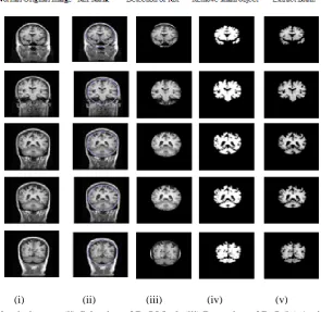
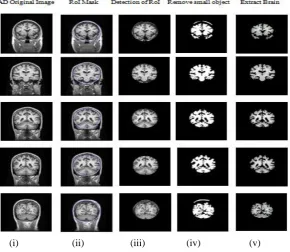
(http://www.ucl.ac.uk/drc/research/methods/miriad-scan-database) [19] are used to evaluate the performance of TMOS. It was tested on 120 images of MIRIAD. The visual perception of the segmented results validate the effectiveness of TMOS. The indicative results of TMOS for Normal Control and AD affected brain images are given in Fig. 2 & Fig. 3 respectively. The TMOS is implemented using MatlabR2013A.
Fig 2: (i) Normal MRI brain images (ii) Selection of RoI Mask (iii) Detection of RoI (iv) Artifacts Removed Brain images (v) Segmented by TMOS
[image:3.595.155.451.409.696.2]Fig. 3: (i) AD MRI brain images (ii) Selection of RoI Mask (iii) Detection of RoI (iv) Artifacts Removed Brain images (v) Segmented by TMOS
The sample images from the dataset and their corresponding segmented images are depicted in Fig. 2 & Fig.3. In Fig. 2, column 1 contains the Normal MRI Brain images, column 2 contains the Selection of RoI Mask, column 3 contains the Detection of RoI, column 4 contains the Artifacts removed brain images, and column 4 contains the segmented brain images by TMOS (Proposed). The respective columns is Fig.3 show the results on AD-affected MRI brain images.
V. CONCLUSION
A new technique TMOS for brain portion extraction is presented in this paper. It is designed using RoI selection and morphological operations for skull stripping. The developed method was tested on T1-weighted MRI brain images of MIRIAD (Minimal Interval Resonance Imaging in Alzheimer Disease) dataset. The visual perception of the skull stripped brain confirms the efficiency of TMOS. This method due to its accuracy finds application it is apparent that the devised TMOS accurately segments the brain regions from MRI brain images of two different datasets. The results of the TMOS can be directly used for higher degree of image processing
REFERENCES
1. Hahn H and Peitgen H.O, “The Skull Stripping problem in MRI solved by a Single 3D Watershed Transform”, in Proc. MICCAI. LNCS, Vol. 1935, pp. 134-143, 2000.
2. Ashburmer.J and FristonK.J, “Voxel Based Morphometry:
The Methods”, NeuroImage, Vol. 11, pp. 805-821, 2000.
3. Shattuck D.W, Sandor-Leahy S.R, Schaper K.A,
5. Lee J.M, Kim J.H, Kim I.Y, Kwon J.S and Kim S.I, “Evaluation of Automated and Semi-Stripping Algorithms: Similarity Index and Segmentation Error”, Computers in Biology and Medicine, vol. 33, no. 6, pp. 495-507, 2003.
6. Rex D.E, Shattuck D.W, Woods R.P, Narr K.L, Luders E,
Rehm K, Stolzner S.E, Rottenberg D.A and Toga A.W, “A meta-algorithm for brain extraction in MRI”, NeuroImage, Vol. 23, Pp. 625-637, 2004.
7. Shijin Kumar P.S and Dharun V.S, “An Efficient Skull Stripping Algorithm using Connected Regions and Morphological Operation”, APRN Journal of Engineering and Applied Science, Vol.11, No.7, pp. 4305-4309, 2017. 8. Park J.G and Lee C, “Skull Stripping based on Region
Growing for Magnetic Resonance Brain Images”, NeuroImage, Vol. 7, No. 5, pp. 1394-1407, 2009.
9. Rosniza R, Nursuriati J and Rozi M, “Skull Stripping Magnetic Resonance Images Brain Images: Region Growing versus Mathematical Morphology”, International Journal of Computer Information Systems and Industrial Management Applications, ISSN: 2150-7988, Vol.3, pp.150-158, 2011.
10. Sudipta Roy, Debnath Bhattacharyya, Samir K,
Bandyopadhyay and Tai-Hoon Kim, “Artifacts and Skull Stripping: An Application Towards the Pre-processing for Brain Abnormalities Detection from MRI”, International Journal of Control and Automation (IJCA), Science & Engineering Research Support Society, Vol.10, No.5, pp. 147-160, 2017.
11. Benson C.C and Leyish V.L, “Morphology Based
Enhancement and Skull Stripping of MRI Brain images”, International Conference on Intelligent Computing Applications, IEEE Proceedings, pp. 254-257, 2014. 12. SajjadMohsin, SadafSajjad, Zeeshan Malik, Abdul Hanan
Abdullah, “Efficient way of Skull Stripping in MRI to Detect Brain Tumor by Applying Morphological Operations, After Detection of false background”, International Journal of Information and Education
[image:4.595.160.451.60.312.2]Radiologists VS Radiographer’s Agreement”, Advanced Information Science and Service Sciences (AISS), Vol.9, No.2, pp.36-45, 2017.
14. Otsu, N, “A Threshold Selection Method from Gray-Level Histograms”,IEEE Transactions on System, Man andCybernatics Vol.9, No.1, pp.62-66. 1979.
15. Liao P.S, Chen T.S and Chung P.C, “A fast Algorithm forMulti-level Thresholding”, J. Inf. Sci. Eng., Vol. 107, No.5, pp. 713-727, 2001.
16. RafelC.Gonzalez, Richard Eugene Woods,SterenL.Eddins.
“Digital Image Processing using MATLAB”,
TataMcGraw-Hill Education, 2010.
17. Terol-Villalobos I.R, “Morphological image
enhancementand segmentation with analysis”, P. W. Hawkes, Ed. New York: Academic, pp. 207–273.2005. 18. Serra.J, Mathematical Morphology Vol. I. London, U.K.
Academic, 1982.
19. Reference this publication, describing the release of the data: Malone IB, Cash D, Ridgway GR, Macmanus DG, Ourselin S, Fox NC, Schott JM. MIRIAD-Public release of a multiple time point Alzheimer's MR imaging dataset. Neuroimage. 2012 Dec 28;70C:33-36