domain
.
White Rose Research Online URL for this paper:
http://eprints.whiterose.ac.uk/122608/
Version: Published Version
Article:
DiCara, DM, Chirgadze, DY, Pope, AR et al. (12 more authors) (2017) Characterization
and structural determination of a new anti-MET function-blocking antibody with binding
epitope distinct from the ligand binding domain. Scientific Reports, 7 (1). ARTN 9000.
ISSN 2045-2322
https://doi.org/10.1038/s41598-017-09460-2
[email protected]
https://eprints.whiterose.ac.uk/
Reuse
Unless indicated otherwise, fulltext items are protected by copyright with all rights reserved. The copyright
exception in section 29 of the Copyright, Designs and Patents Act 1988 allows the making of a single copy
solely for the purpose of non-commercial research or private study within the limits of fair dealing. The
publisher or other rights-holder may allow further reproduction and re-use of this version - refer to the White
Rose Research Online record for this item. Where records identify the publisher as the copyright holder,
users can verify any specific terms of use on the publisher’s website.
Takedown
If you consider content in White Rose Research Online to be in breach of UK law, please notify us by
Characterization and structural
determination of a new anti-MET
function-blocking antibody with
binding epitope distinct from the
ligand binding domain
Danielle M. DiCara
Dimitri Y
Chirgadze
Anthony R
Pope
Aneesh
Karatt-Vellatt
Anja
Winter
Peter
Slavny
Joop van den
Heuvel
Kothai
Parthiban
Jane
Holland
Len
C. Packman
Georgia
Mavria
Jens Ho mann Walter
Birchmeier
Ermanno Gherardi
&
John McCa erty
The growth and motility factor Hepatocyte Growth Factor Scatter Factor HGF SF and its receptor the product of the MET proto oncogene promote invasion and metastasis of tumor cells and have been
considered potential targets for cancer therapy. We generated a new Met-blocking antibody which binds outside the ligand binding site and determined the crystal structure of the Fab in complex with its target which identi es the binding site as the Met Ig domain The antibody A inhibited HGF SF induced cell migration and proliferation in vitro and inhibited growth of tumor xenografts
in vivo In biochemical assays A competes with both HGF SF and its truncated splice variant
NK for MET binding despite the location of the antibody epitope on a domain Ig not reported to bind NK or HGF SF Overlay of the Fab MET crystal structure with the InternalinB MET crystal structure shows that the A Fab comes into close proximity with the HGF SF binding SEMA domain when MET is in the compact InternalinB bound conformation but not when MET is in the
open conformation These ndings provide further support for the importance of the compact conformation of the MET extracellular domain and the relevance of this conformation to HGF SF binding and signaling.
A major challenge in the therapy of solid tumors, and notably of carcinomas that constitute over 85% of all human cancers, is the development of agents that inhibit metastasis, namely the growth of cells derived from the primary tumor at distant sites in the body. Metastasis is a multi-stage process in which cancer cells migrate into adjacent tissues (invasion), cross the wall of blood or lymphatic capillaries to be transported across the general circulation (intravasation), exit from the bloodstream into a secondary tissue/organ (extravasation) and inally give rise to
MRC Centre Hills Road Cambridge CB QH UK Department of Oncology University of Cambridge Cambridge Biomedical Campus Cambridge CB XZ UK Department of Biochemistry University of Cambridge Tennis Court Road Cambridge CB GA UK IONTAS Ltd Babraham Institute Babraham Cambridgeshire CB AT UK Helmholtz Zentrum für Infektionsforschung Inho enstraße Braunschweig Germany Max Delbrueck Center for Molecular Medicine MDC in the Helmholtz Association Berlin Germany Leeds Institute of Cancer and Pathology University of Leeds St James University Hospital Beckett Street Leeds LS TF UK Experimental Pharmacology Oncology Berlin Buch GmbH Robert Rössle Str Berlin Buch Germany Division of Immunology and General Pathology Department of Molecular Medicine via A Ferrata Pavia Italy Present address Genentech Inc South San Francisco USA Present address Faculty of Natural Sciences Keele University Sta ordshire ST BG UK Ermanno Gherardi and John McCa erty jointly supervised this work Correspondence and requests for materials should be addressed to E G email egherard unipv it or J M
(email: jmc iontas co uk) Received: 8 May 2017
Accepted: 25 July 2017
Published: xx xx xxxx
secondary tumors1. In principle, each of these stages may be targeted in therapy, and the ability to target multiple stages simultaneously is an attractive prospect.
he signaling system mediated by the growth and motility factor HGF/SF and its receptor MET, the tyros-ine kinase encoded by the MET proto-oncogene, has multiple and essential physiological roles in vertebrate embryogenesis, where it is required for normal development of the liver parenchyma and the labyrinth layer of the placenta2, 3 as well as distant migration of the myogenic precursor cells4. HGF/SF and MET have further and important physiological roles in postnatal life, where they control the regeneration ater injury of several epithelial organs including skin5 and liver6, 7. hey also exert, however, multiple and crucial roles in the early stages of metastasis of epithelial cancers by controlling (i) delamination of epithelial cells and the process of epithelial-mesenchymal transition crucial for long distance epithelial cell migration4, 8, (ii) degradation of the basement membrane and remodeling of the extracellular matrix via urokinase9 and matrix metalloproteinases10, (iii) integrin-dependent migration of cancer cells as a result of activation of focal adhesion kinase and paxillin11, (iv) formation of the pre-metastatic niche via tumor-derived exosomes12, (v) tumor lymphangiogenesis13, 14, a process essential for lymphatic metastasis and, (vi) haemangiogenesis15, 16. Further, there is growing evidence for a major role of HGF/SF and MET in the maintenance of cancer stem cells in colon17, breast18 and prostate19 car-cinomas, and accumulating reports of involvement of the MET-HGF/SF axis in cancer cell resistance to targeted therapies both in vitro20–23 and in cancer patients24–26.
he MET receptor is a single-pass transmembrane protein consisting of a large extracellular ectodomain, a transmembrane segment and an intracellular receptor tyrosine kinase domain27. he ectodomain is a heterodimer arising from furin cleavage of a single precursor chain and consists of an N-terminal 283 amino acid α-chain and the 625 extracellular amino acids of the β-chain. An N-terminal semaphorin homology domain (SEMA), which binds the ligand HGF/SF, is followed by a “stalk” comprising one cysteine rich (CR) domain and 4 immunoglobu-lin domains termed Ig1-Ig428. he MET ectodomain has been expressed recombinantly as a series of truncations containing the SEMA, CR and 0, 2 or 4 Ig domains, which we will refer to as MET567, MET741 and MET928, respectively. HGF/SF consists of an amino terminal domain, 4 kringle domains (K1 - K4) and a C terminal serine protease homology domain (SPH domain). he N terminal and irst kringle domain (NK1) of the HGF/SF ligand drive a high ainity interaction with MET29, and the SPH domain also binds MET30. he SEMA domain has been reported as necessary and suicient for HGF/SF binding28, although an interaction with the Ig3-Ig4 “lower stalk” region has also been reported31.
Here, we report the isolation of a new human, phage-derived monoclonal antibody to MET (107_A07) dis-playing potent receptor antagonistic activity, and we describe the activity of 107_A07 on cancer cells in vitro and in in vivo. We also report a crystal structure of the 107_A07 Fab in complex with a MET receptor fragment containing amino acids 519–740, which includes the CR, Ig1 and Ig2 domains but not the ligand-binding SEMA domain. hese data provide insights into the conformation of MET during ligand binding and cell signaling.
Materials and Methods
Protein production and puri cation
Soluble MET741 protein28 was produced from CHO Lec3.2.8.1 cells and puriied by ainity chromatography (NiNTA Superlow, Qiagen) followed by cation exchange (MonoS, GE Life Sciences). 107_A07 and D1.3 Fabs and IgG –formatted antibodies were produced by transfection of suspension HEK293F cells (Invitrogen) with Valproic Acid (Sigma) added to 4 mM following transfection. Fab proteins were puriied using ainity resins KappaSelect and/or GammaBind Plus (GE Life Sciences). IgG for-matted antibodies were puriied by Protein A ainity chromatography. For cell cycle analysis, 107_A07 and D1.3 Fabs were further puriied by gel iltration chromatography (Superdex 200 10/300 (GE Life Sciences). Unless stated otherwise, 7A2 scFv was produced in Pichia pastoris and puriied by Ni-NTA chromatography followed by gel iltration. Anti-MET antibody 5D5 sequences were obtained from US Patent No. 7,476,724 B2. Heavy and light chains were synthesized (GeneArt, hermo Fisher Scientiic) with restriction sites that allowed cloning into Fab vectors pBIOCAM1-3F and pBIOCAM3-3F, expressed in HEK293F cells and the Fab puriied by Ni-NTA chromatography.FAb PEGylation
Recombinant Fab were partially reduced with TCEP and PEGylated with maleimide-activated PEG (Sunbright ME-200 MAOB or Sunbright ME-200MA3, NOF Europe). PEG-Fab and free PEG were monitored by SDS-PAGE and barium chloride & iodine staining32.Isolation affinity maturation of functional MET blocking antibodies by phage dis
-play.
Biopanning with a scFv phage library33 was performed on solid-phase recombinant MET928 and light chain shuling performed on the output by cloning the resulting VH gene pool back into the original scFv phage library34. Biopanning with the chain-shuled library (109 clones) was performed with biotinylated MET928 insolution and streptavidin-coated dynabeads. Phage pools were cloned into expression vector35 and small-scale expressions performed in BL21 (DE3) bacteria in 96-well format. Approximately 960 supernatants were screened directly for inhibition of HGF/SF-induced scatter of BxPC-3 human pancreatic cancer cells. Ainity matura-tion was performed by diversiicamatura-tion of the CDR3 regions of 7A2 VH and VL using oligonucleotide-directed mutagenesis and stringent selection of the resulting phage library with biotinylated MET928 in solution.
In vitro
cell-based assays.
HGF/SF-induced cell migration across a porous membrane coated with 100 µg/without 0.9 µM 107_A07 FAb or 1 µM D1.3 FAb. Cells were trypsinised, ixed, stained with propidium iodide in
the presence of RNAse and analyzed by low cytometry according to standard procedures. In vitro angiogenesis assay was performed using the modiied co-culture assay as described previously36. Briely, ibroblast cells were seeded in gelatin-coated chamber slides. Human umbilical vein endothelial cells (HUVECs) were seeded on to the conluent ibroblasts and D1.3 and 107_A07 antibodies (200 nM) were added to the cells. he co-cultures were ixed and stained for CD31. Number of tubules was counted manually from 10 ields for each well and the ield area was measured using AngioSys 1.0 imaging sotware.
Tumor xenografts
NMRI nu/nu mice (Crl:NMRI-Foxn1nu) were obtained from Charles River Laboratories (Sulzfeld, Germany). U87MG cells were obtained from ECACC and identity checked at the DSMZ. Mice were injected subcutaneously with 107 U87MG human glioblastoma cells and antibody administered every 3–5 daysbetween day 7 and 33, when treatment stopped. Groups of 8 mice were treated with 107_A07 IgG at either 2 mg/ kg or 10 mg/kg. Control animals were treated with either PBS or D1.3 IgG at 10 mg/kg. All animal experiments were carried out in accordance with the United Kingdom coordinating committee on cancer research regulations for the welfare of animals and the German Animal Protection Law, and were also approved by the local responsi-ble authorities Landesamt für Gesundheit und Soziales Berlin (LAGeSo).
Surface Plasmon Resonance SPR studies
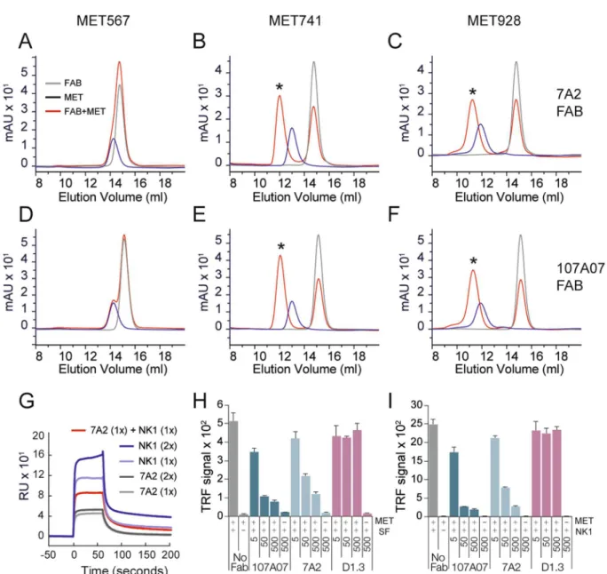
KD was determined using a Biacore instrument andrepresents the mean of three experiments using 107_A07 FAb puriied sequentially on both KappaSelect and GammaBind Plus chromatographic resins. For competition analysis by SPR, a CM5 chip coated with MET928 was exposed to: 7A2 scFv (134 nM, 268 nM), NK1 (238 nM, 476 nM) or a mixture of the two (134 nM 7A2 scFv, 238 nM NK1).
Competition analysis by ELISA
Fabs were mixed with 100 nM MET928 one hour before addition to microplates coated with HGF/SF or the fragment NK1. Ater one hour bound MET928 was detected with anti-5xHis (Qiagen) followed by DELFIA®
Eu-N1 rabbit anti-mouse-IgG. DELFIA®
enhancement solution was then added and signal quantitated using time-resolved luorescence with a Fusion instrument (Perkin Elmer).Complex formation analysis
Fab and MET proteins were co-incubated at a 3:2 molar ratio for 140 min-utes at room temperature in 25 mM Tris pH 7.4, 150 mM NaCl, centrifuged and analyzed by size exclusion chro-matography (Superdex 200 10/300).Co crystallization of MET Fab complex
Briefly, Fab 107_A07 and his-tagged MET741 were co-incubated, then digested with Pepsin and EndoHf deglycosidase. he remaining complex was puriied by Ni-NTA ainity resin and size exclusion chromatography, concentrated to 5.9 mg/ml and crystallized by the sitting-drop vapor difusion method at 19 °C.Data collection & model generation.
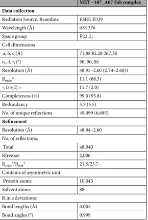
X-ray data collection experiments were performed at the European Synchrotron Radiation Facility (Grenoble France), beamline ID29. he crystals difracted to a maximum resolu-tion of 2.6 Å. he crystals contained two molecules of the MET/Fab complex in the asymmetric unit. he crystal structure was solved using the Molecular Replacement (MR) method. Ater several rounds of manual rebuilding and reinement the R/Rfree values reached 21.5% and 25.7% respectively. he coordinates of the structure havebeen deposited to the Protein Data Bank under accession ID 5LSP. Additional method information is provided in the Supplementary Information ile.
Additional Disclosures
D.D.C., A.P., J.M.C. and E.G. are co-inventors on a patent application relating to the antibodies described in this manuscript. D.D.C. is currently an employee of Genentech, Inc.Results
Generation and affinity maturation of a high affinity antibody blocking MET signal
-ing.
Recombinant antibodies recognizing MET928 were generated by phage display using two rounds of pan-ning on immobilized antigen. Binders were generated using a previously described human antibody phage display library of ~1010 clones formatted as single chain Fvs (scFvs)33. he selected population was ainity matured en masse by chain-shuling the selected population of VH genes34. A chain-shuled scFv library of 109 clones wasagonistic activity in scFv preparations was associated with larger, non-monomeric species (data not shown). Fab are much less likely than scFv to form higher-order species, and so we converted 7A2 to a Fab format.
Conversion of 7A2 to a Fab format diminished the agonistic activity as expected but also resulted in reduced ainity and potency, therefore an additional round of ainity maturation was conducted using oligonucleotide directed mutagenesis to create phage display libraries where the CDR3 region of the V gene heavy chain (VH) was diversiied. Mutagenic primers with contiguous blocks of 5–6 randomized amino acids using 3 overlapping prim-ers were used to randomize the 11 amino acids of the VH CDR3 and 9 amino acids from VL CDR3. Stringent selections were carried out using limiting amounts of biotinylated MET928 down to 10 pM. DNA from the output of the resulting selections was subcloned into a bacterial expression vector and 1152 clones were screened by ELISA and sequenced. 146 clones with unique sequences that were positive in ELISA were identiied for further study. VL mutants were not identiied, suggesting this library was inefective or was dominated by the parental sequence. he selected clones were expressed, puriied and the of-rates compared to the parental 7A2 clone using surface plasmon resonance (SPR). he top 8 clones showing the greatest improvement in of-rate were identiied and produced as Fab fragments. Sequence analysis revealed that the top group of clones came from VH CDR3 mutagenesis and all carried a Y105W mutation. In addition, 6/8 of these clones retained G106 and M107 despite the complete diversiication within this region of the selected clones. Based on ainity and potency rankings, the ainity-matured clone 107_A07 was selected for further biological and biochemical analysis. SPR experiments yielded a KD in the nanomolar range (3.2 ± 0.7 nM; mean ± SD, three experiments).
Antibody
A inhibits migration and proliferation of cancer cells and endothelial tubu
-logenesis.
HGF/SF-mediated activation of MET causes cell proliferation, cell migration, and angiogene-sis, all of which contribute to cancer progression38. We assessed 107_A07 Fab for inhibition of these processes. Inhibition of cell migration was compared with 7A2, the parental antibody of 107_A07, and as a control we also included a Fab-formatted version of 5D5.v1, the monoclonal antibody from which onartuzumab was derived37. 107_A07 Fab dose-dependently inhibited HGF/SF-induced cell migration of SKOV-3 human ovarian cancer cells (Fig. 1A) and U87MG human glioblastoma cells (Fig. 1B).A degree of agonism observed at higher concentrations with Fab formatted 107_A07 was lost following puri-ication of monomers by size exclusion chromatography, suggesting that the presence of low concentrations of multimeric material could drive agonism (Figure S2). To assess antibody activity on HGF/SF-induced cell pro-liferation, Fab were further puriied by size exclusion chromatography and serum-starved U87MG cells were incubated for 24 hours with HGF/SF in the presence of 107_A07 Fab or a control Fab (humanized anti-hen egg lysozyme, D1.339), followed by DNA content analysis by propidium iodide staining. U87MG cells express HGF/ SF40 and divide rapidly in culture, however addition of exogenous HGF/SF caused a further and measurable increase in DNA synthesis (Fig. 1C). Treatment with 107_A07 Fab but not control (D1.3) Fab reduced the per-centage of cells in S-phase and G2/M phase to basal levels, with a corresponding increase in cells in G1 (Fig. 1C). We also investigated PEGylation of the 107_A07 Fab, since PEGylation can be used to extend Fab pharma-cokinetics in vivo. PEGylation of 107_A07 Fab was achieved by covalent attachment of 20 kD polyethylene glycol (PEG) chains to the C-terminal cysteine residues41. he majority of unconjugated PEG was removed by size exclu-sion chromatography and ultrailtration. In SDS-PAGE, PEG-107_A07 Fab chains displayed reduced migration and insensitivity to reducing agents, consistent with removal of the inter-chain disulphide bond (Fig. S3). PEG-107_A07 Fab retained the ability to inhibit HGF/SF-induced cell migration and demonstrated essentially com-plete inhibition of cell migration at high concentrations (Fig. S4). In a ibroblast/endothelial cell in vitro model of angiogenesis in which ibroblast-derived HGF/SF induces human umbilical vein endothelial cell (HUVEC) tub-ulogenesis (a process that recapitulates in vitro the sprouting of new capillaries in vivo) (Fig. 1D), PEG-107_A07 Fab inhibited tubule formation (Fig. 1F and G).
A IgG inhibits the growth of human tumor xenografts
in vivo
.
he VH and VL domains of 107_A07 were expressed as an intact human IgG and the activity of 107_A07 IgG and control (D1.3) IgG were assessed in cell migration assays where agonism was found with 107_A07 IgG (Fig. S5). he peak of agonistic activity was again found to occur at low nanomolar concentration (as with the scFv, Fig. S1) with net antagonism found at higher concentrations (Fig. S5). 107_A07 IgG was administered over 26 days in a human U87MG glio-blastoma cell xenograt model and inhibited tumor growth very efectively for up to 70 days at 10 mg/kg 107_A07 IgG (Fig. 2). hus intact 107_A07 IgG inhibited tumor growth despite its bivalent format (Fig. 2), presumably because an IgG concentration was maintained where net antagonism was achieved.which pre-incubation of MET928 with either 7A2 or 107_A07 inhibited MET binding to both HGF/SF and NK1 (Fig. 3E and F).
Crystal structure of the
A and MET
A complex
In order to deine further the epitope and inhibitory mechanism of these antibodies, we determined the crystal structure of a complex formed by the 107_A07 Fab and a fragment of the MET ectodomain encompassing the small cysteine-rich domain and the upper stalk region (Ig domains 1 and 2). Initial crystallization experiments with complexes formed by the 107_ A07 Fab and MET741 failed to yield difraction grade crystals. We overcame this by exploiting a pepsin cleavage site (NGL|GCR) located in the linker connecting the SEMA and CR domains of MET, generating a receptor fragment containing the CR, Ig1 and Ig2 domains of MET, which contains MET residues 519–741. Crystals of the 107_A07 Fab – MET519–741 complex were obtained and difracted to a maximum resolution of 2.6 Å at theEuropean Synchrotron Radiation Facility (Beamline ID29, ESRF, Grenoble, France). he structure was solved by the Molecular Replacement method43 and reined to R
cryst and Rfree values of 21.5% and 25.7% respectively
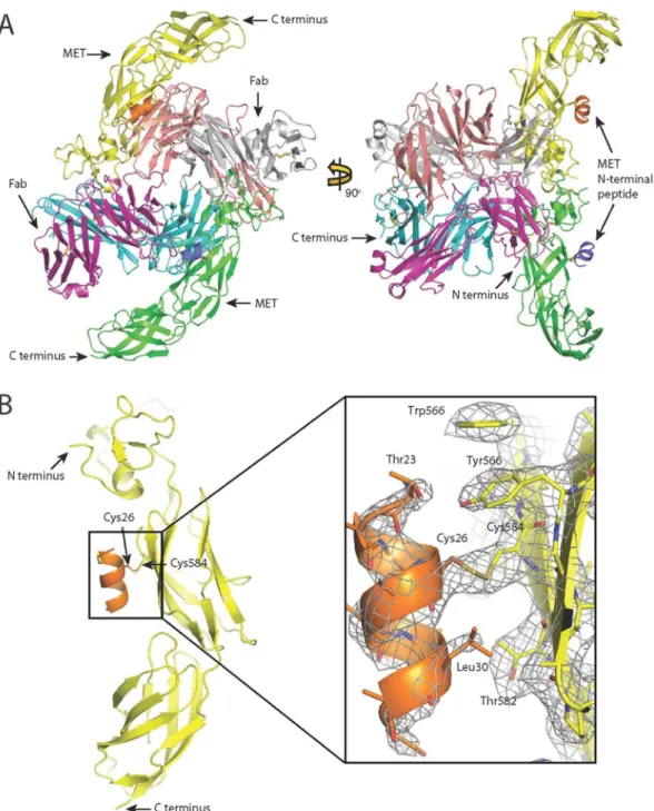
(Table 1). he asymmetric unit contained two 107_A07 - MET519–741 complexes (Fig. 4A) that, when superposed
on their MET-Ig1 domains, showed close alignment of the MET chain and VH and VL domains but approxi-mately 60° rotation of the CH1 and CL domains. Complex 1, containing MET chain A and Fab chains H and L (Fig. 5A) is described below because the antibody-receptor interactions seen within this complex are more com-plete than the ones seen in complex 2. he model contains MET residues 519–740. We also observed unexpected electron density adjacent to Cys584 in the Ig1 domain (Fig. 4B). MALDI analysis of the crystallization complex and subsequent MS/MS analysis identiied a peptide containing amino terminal residues of MET (residues 22–32, which include a single cysteine at position 26) attached to the complex by a disulphide bond. Together, these data indicate the presence of a disulphide bond between MET residues Cys26 and Cys584. Since MET519–741 was
puri-ied by enzymatic digestion of the MET741–107_A07 complex, it is uncertain whether the attachment occurred before or ater digestion.
he VH and VL domains of 107_A07 bind the Ig1 domain of MET (Fig. 5B) and bury 555 Å2 and 243 Å2
respectively. he VH domain binds a discontinuous epitope including the tip of the β-hairpin (R592, N593 and K595), residues connecting the β-hairpin and the β-strand C (K599 and K600) and the tip of β-strand C (R602) (Fig. 5B and Supplementary Table S1) and involves an extensive network of polar contacts and salt bridges involv-ing CDRs 1, 2 and 3 of the antibody heavy chain (Fig. 5C and Table S1).
he interactions of the VH domain with MET clarify the impact of the Y105W mutation that was found in each of the top 8 ainity matured clones. From the structure it is clear that the indole group of tryptophan creates an additional sidewall for the pocket formed by residues 99–105 of the heavy chain. his makes the pocket deeper and more complementary for the interactions with the antigen’s residue K599. he side chain of K599 is fully extended and inserts deep into the pocket, where the NH2 group creates an extensive hydrogen bonding network
with the carbonyl groups of the backbone (Fig. S6). In contrast, the VL domain binds a linear epitope (L612, T613, L614, S615 and E616, a sequence corresponding to β-strand D of canonical E set domains) and makes limited contacts with MET predominantly via L3 (Fig. 5C and Table S1).
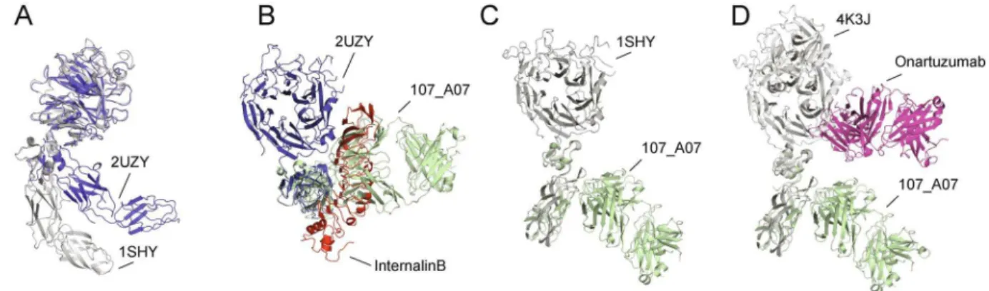
Within the 107_A07 Fab - MET complex, the conformation of the CR domain of MET relative to the Ig1-Ig2 fragment matches closely the conformation of the corresponding region of MET observed in the crystal structure of MET741 in complex with InlB321. InlB321 is a truncated fragment of InternalinB (InlB), a surface protein of Listeria monocytogenes that promotes binding and internalization of the bacterium through binding and acti-vation of the MET receptor44, 45 (PDB entry 2UZY). InlB321 binds with high ainity to a primary site in the Ig1 domain of MET with its leucine-rich repeat central domain. It also binds with lower ainity to a secondary site in the SEMA domain with its interdomain repeat. Binding to both sites is essential for receptor activation44, 45.
In contrast to InlB321, 107_A07 appears to contact only the MET Ig1 domain; however, the C-alpha atoms for the CR-Ig1-Ig2 structure of the InlB-MET741 superpose closely with the corresponding MET domains in the 107_A07 structure (r.m.s.d. is ~1.4 Å from the MET molecule in complex 1 and ~2.0 Å for the MET molecule in
complex 2 (Fig. 5D). Although extensive movements of the SEMA domain relative to the CR domain are enabled by the SEMA-CR domain linker30, 37, 44, 45, the structure of the MET CR-Ig1-Ig2 section reported here is rigid, due to extensive interactions between the top of Ig1 (notably the β-hairpin structure) and the CR domain (Fig. 5E), in addition to a second set of interactions between Ig1 and Ig2 (Fig. 5F).
Discussion
We have isolated and ainity matured a novel phage-derived anti-MET antibody, 7A2/107_A07, which competes for binding with both MET ligand HGF/SF and the HGF/SF fragment NK1. Antibody 7A2/107_A07 inhibited HGF/SF-induced cell migration and DNA synthesis in vitro, endothelial cell tubulogenesis in a co-culture assay and tumor growth in vivo in a xenograt model. Biochemical analysis and structural determination demonstrated that the antibody binds to the Ig1 domain, in contrast to HGF/SF, which binds to the SEMA domain.
Figure 3. 7A2 and 107_A07 bind within the Ig1-Ig2 domains of MET and compete with NK1 for MET binding. Panels A–F show size exclusion chromatography analysis of 6 µM Fab 7A2 (A–C) or 107_A07 (D–F) and 4 µM
he domain architectures of MET and its homologue RON difer from those of other receptor tyrosine kinases (RTK)46 and are evolutionary and structurally related to the plexins and their semaphorin ligands47. Cryo-EM and small angle X-ray scattering (SAXS) analysis of the soluble MET ectodomain42 and several crystal structures30, 37, 44, 45 have clearly established that the irst four extracellular domains of MET (the SEMA, CR and the irst two Ig domains) can adopt a more compact or a more extended conformation as a result of rotation and translation of the CR domain relative to the SEMA domain (Fig. 6A). he present study revealed, unexpectedly, that the CR-Ig1-Ig2 domains of MET form a rigid body and this indicates that the irst 741 residues of MET contain a single hinge, located between the SEMA and CR domains.
he rigid structure of the CR-Ig1-Ig2 fragment has profound implications for the mechanism of MET inhibi-tion by 107_A07. Crystal structures of soluble fragments of MET in complex with the SPH domain of HGF/SF30, 37 or InlB44, 45 have shown that the CR domain can adopt two main orientations relative to the SEMA resulting in a compact (PDB entry 2UZY) or extended (PDB entries 1SHY and 4K3J) arrangement of the SEMA, CR, Ig1 and Ig2 domains (Fig. 6A). he compact architecture prevails in solution as shown by SAXS experiments42 although the molecular mechanism for this apparent restriction in hinge lexibility is not known.
One other crystal structure of a MET-antibody complex is available (the Fab fragment of onartuzumab in complex with MET567 and the SPH domain of HGF/SF (4K3J)) and shows that onartuzumab binds an area of the SEMA domain that overlaps with the secondary binding site of InlB37. Onartuzumab competes for binding with both HGF/SF and NK137 and it has been concluded that the binding sites of HGF/SF, InlB and onartuzumab on the SEMA domain overlap in full or in part37. In contrast, 107_A07 binds an epitope on the Ig1 domain of MET that overlaps with the primary binding site of InlB, yet 107_A07 also competes with HGF/SF and NK1 for MET binding.
Structural alignment of the MET-InlB complex and the MET-107_A07 complex indicates that when MET is in the compact conformation, the binding sites for InlB on the MET SEMA and 107_A07 on the Ig1 domain are spa-tially proximate, despite their location on separate domains (Fig. 6B and Fig. S7). While an allosteric mechanism cannot be ruled out, steric competition between HGF/SF and 107_A07 when MET is in the compact conforma-tion would readily explain the observed biochemical competiconforma-tion between HGF/SF and 107_A07. he compact conformation of MET predominates in solution42 and occurs in the crystal structure of the InlB-MET741 com-plex44, 45. Our data further support a role for this conformation in HGF/SF-induced MET signaling.
Our data also demonstrate that the Ig1 residue Cys584 is capable of forming a disulphide bond external to the Ig1 domain. A putative disulphide linkage between Cys584 (Ig1 domain) and Cys26 (N-terminal) has been pos-tulated and discussed previously42, 44, 48 but to our knowledge this is the irst time that experimental evidence for
MET - 107_A07 Fab complex
Data collection
Radiation Source, Beamline ESRF, ID29 Wavelength (Å) 0.91376 Space group P22121
Cell dimensions
a, b, c (Å) 71.88 82.28 267.30
α, β, γ (°) 90, 90, 90
Resolution (Å) 48.95–2.60 (2.74–2.60)1 Rmeas2 11.1 (88.3)
<I/σ(I)> 11.7 (2.0) Completeness (%) 99.0 (95.8) Redundancy 5.5 (5.5) No. of unique relections 49,099 (6,685)
Reinement
Resolution (Å) 48.94–2.60 No. of relections:
Total 48,940
Rfree set 2,000 Rcryst3/Rfree4 21.5/25.7
Contents of asymmetric unit:
Protein atoms 10,043 Solvent atoms 88 R.m.s deviations:
Bond lengths (Å) 0.005 Bond angles (°) 0.999
Table 1. Crystallographic data collection and reinement statistics. 1he statistics shown in parentheses are
for the highest-resolution shell. 2R
meas= (Σhkl [N/(N-1)]1/2Σi |Ii(hkl)−Imean(hkl)|)/ΣhklΣi Ii(hkl), where N is redundancy. 3R
cryst=Σhkl ||Fobs(hkl)|−|Fcalc(hkl)||/Σhkl |Fobs(hkl)|. 4R free is the same as Rcryst for a random subset
this bond has been observed. Attachment of Cys584 to the N-terminal peptide of MET may have occurred before or ater proteolysis; whether this disulphide bond exists in intact, full-length MET remains to be determined. he possibility is intriguing as such a bond, if it exists, may go some way towards explaining why the compact arrange-ment of the SEMA, CR, Ig1 and Ig2 domains has been reported to dominate over the extended conformation in SAXS experiments42.
he pivotal roles of HGF/SF and MET in cancer progression and metastasis have led to considerable expecta-tion that agents blocking HGF/SF-MET signaling could have a strong impact in the therapy of metastatic tumors
Figure 5. Structure of the complex between 107_A07 and MET519-740. (A) Ribbon diagram of complex 1 of the 107_A07 - MET519-740 structure showing the MET519-740 molecule (green) and the 107_A07 Fab on the right (heavy chain cyan, light chain purple). (B,C) Residues involved in contacts between MET519-740 (B) and 107_A07 Fab (VH domain in cyan, VL domain in purple). he picture highlights the polar and charged nature of the extensive VH-MET contacts (see Table S1 for further details). (D) Superposition of the structures of the MET fragment containing residues 519–740 from the structure of the 107_A07-MET complex (chain A) and the InlB-MET structure (PDB accession 2UZY, chain B). (E and F) Contacts between the CR domain and the IG1 domain (E) of MET519-740 and between the bottom of IG1 domain and the top of the IG2 domain (F). hese two sets of contacts account for the rigid structure of the MET519-740 fragment of MET.
and led to a major efort in the design, synthesis and development of small molecule inhibitors of the MET kinase49 as well as blocking antibodies directed against the ligand HGF/SF50, 51 and the MET extracellular region37, 52–56. Several anti-MET antibodies progressed to clinical trials, including ABT-70057, LY287535858, ARGX-111 and onartuzumab. he development of a number of anti-MET antibodies with antagonistic activity has highlighted diverse mechanisms for receptor inhibition. For instance, onartuzumab37 and anti-MET CE-35562154 inhibit HGF/SF binding, MET activation and xenograt growth. Anti-MET LMH 8753 does not inhibit ligand binding but promotes receptor degradation and anti-MET F46 inhibits ligand binding and promotes receptor degradation59. An anti-MET nanobody (a single VH domain) inhibits MET activation and DNA synthesis in myeloma cells60 and a recent collection of llama anti-MET antibodies highlights multiple mechanisms of receptor inhibition56. hese multiple pathways of receptor inhibition relect the complex structural basis of MET signaling30, 44, 45. An anti-MET anticalin (PRS-110) with MET antagonistic activity has also been developed61. Interestingly, PRS-110 binds both to a loop in the SEMA domain in close proximity to the K1 binding site as well as the β-wing of the Ig1 of MET suggesting that the PRS-110 and 107_A07 epitopes may be closely related. While crystallographic analysis will be required in order to deine more accurately the epitopes of a number of other anti-MET antibodies under development for therapy, the structures of onartuzumab37 and 107_A07 (this work) in complex with MET ofer initial insights into mechanisms of MET inhibition. he unusual mechanism of MET inhibition by 107_A07, involving biochemical competition for ligand binding despite clear separation of binding footprints, contrasts with that of onartuzumab and illuminates the variety of potential mechanisms through which antibody-mediated MET inhibition can be achieved.
References
1. Valastyan, S. & Weinberg, R. A. Tumor metastasis: molecular insights and evolving paradigms. Cell147, 275–292 (2011). 2. Schmidt, C. et al. Scatter factor/hepatocyte growth factor is essential for liver development. Nature373, 699–702 (1995). 3. Uehara, Y. et al. Placental defect and embryonic lethality in mice lacking hepatocyte growth factor/scatter factor. Nature373,
702–705 (1995).
4. Bladt, F., Riethmacher, D., Isenmann, S., Aguzzi, A. & Birchmeier, C. Essential role for the c-met receptor in the migration of myogenic precursor cells into the limb bud. Nature376, 768–771 (1995).
5. Chmielowiec, J. et al. c-Met is essential for wound healing in the skin. J Cell Biol177, 151–162 (2007).
6. Borowiak, M. et al. Met provides essential signals for liver regeneration. Proc Natl Acad Sci USA101, 10608–10613 (2004). 7. Huh, C. G. et al. Hepatocyte growth factor/c-met signaling pathway is required for eicient liver regeneration and repair. Proc Natl
Acad Sci USA101, 4477–4482 (2004).
8. Stoker, M., Gherardi, E., Perryman, M. & Gray, J. Scatter factor is a ibroblast-derived modulator of epithelial cell mobility. Nature 327, 239–242 (1987).
9. Jefers, M., Rong, S. & Vande Woude, G. F. Enhanced tumorigenicity and invasion-metastasis by hepatocyte growth factor/scatter factor-met signalling in human cells concomitant with induction of the urokinase proteolysis network. Molecular and cellular biology16, 1115–1125 (1996).
10. Rosenthal, E. L. et al. Role of the plasminogen activator and matrix metalloproteinase systems in epidermal growth factor- and scatter factor-stimulated invasion of carcinoma cells. Cancer research58, 5221–5230 (1998).
11. Matsumoto, K., Matsumoto, K., Nakamura, T. & Kramer, R. H. Hepatocyte growth factor/scatter factor induces tyrosine phosphorylation of focal adhesion kinase (p125FAK) and promotes migration and invasion by oral squamous cell carcinoma cells.
he Journal of biological chemistry269, 31807–31813 (1994).
12. Peinado, H. et al. Melanoma exosomes educate bone marrow progenitor cells toward a pro-metastatic phenotype through MET.
Nature medicine18, 883–891 (2012).
13. Kajiya, K., Hirakawa, S., Ma, B., Drinnenberg, I. & Detmar, M. Hepatocyte growth factor promotes lymphatic vessel formation and function. EMBO J24, 2885–2895 (2005).
14. Jiang, W. G. et al. he potential lymphangiogenic efects of hepatocyte growth factor/scatter factor in vitro and in vivo. International journal of molecular medicine16, 723–728 (2005).
15. Sengupta, S. et al. Hepatocyte growth factor/scatter factor can induce angiogenesis independently of vascular endothelial growth factor. Arterioscler hromb Vasc Biol23, 69–75 (2003).
16. Zhang, Y. W., Su, Y., Volpert, O. V. & Vande Woude, G. F. Hepatocyte growth factor/scatter factor mediates angiogenesis through positive VEGF and negative thrombospondin 1 regulation. Proc Natl Acad Sci USA100, 12718–12723 (2003).
17. Vermeulen, L. et al. Wnt activity deines colon cancer stem cells and is regulated by the microenvironment. Nat Cell Biol12, 468–476 (2010).
18. Holland, J. D. et al. Combined Wnt/beta-catenin, Met, and CXCL12/CXCR4 signals characterize basal breast cancer and predict disease outcome. Cell reports5, 1214–1227 (2013).
19. Nishida, S. et al. Prostate cancer stem-like cells/cancer-initiating cells have an autocrine system of hepatocyte growth factor. Cancer science104, 431–436 (2013).
20. Straussman, R. et al. Tumour micro-environment elicits innate resistance to RAF inhibitors through HGF secretion. Nature487, 500–504 (2012).
21. Wilson, T. R. et al. Widespread potential for growth-factor-driven resistance to anticancer kinase inhibitors. Nature487, 505–509 (2012).
22. Johnson, J. et al. Genomic proiling of a Hepatocyte growth factor-dependent signature for MET-targeted therapy in glioblastoma. J Transl Med13, 306 (2015).
23. Corso, S. & Giordano, S. Cell-autonomous and non-cell-autonomous mechanisms of HGF/MET-driven resistance to targeted therapies: from basic research to a clinical perspective. Cancer Discov3, 978–992 (2013).
24. Engelman, J. A. et al. MET ampliication leads to geitinib resistance in lung cancer by activating ERBB3 signaling. Science316, 1039–1043 (2007).
25. Sequist, L. V. et al. Genotypic and histological evolution of lung cancers acquiring resistance to EGFR inhibitors. Sci Transl Med3, 75ra26 (2011).
26. Bean, J. et al. MET ampliication occurs with or without T790M mutations in EGFR mutant lung tumors with acquired resistance to geitinib or erlotinib. Proceedings of the National Academy of Sciences of the United States of America104, 20932–20937 (2007). 27. Park, M. et al. Sequence of MET protooncogene cDNA has features characteristic of the tyrosine kinase family of growth-factor
receptors. Proceedings of the National Academy of Sciences of the United States of America84, 6379–6383 (1987).
28. Gherardi, E. et al. Functional map and domain structure of MET, the product of the c-met protooncogene and receptor for hepatocyte growth factor/scatter factor. Proc Natl Acad Sci USA100, 12039–12044 (2003).
30. Stamos, J., Lazarus, R. A., Yao, X., Kirchhofer, D. & Wiesmann, C. Crystal structure of the HGF beta-chain in complex with the Sema domain of the Met receptor. he EMBO journal23, 2325–2335 (2004).
31. Basilico, C., Arnesano, A., Galluzzo, M., Comoglio, P. M. & Michieli, P. A high ainity hepatocyte growth factor-binding site in the immunoglobulin-like region of Met. he Journal of biological chemistry283, 21267–21277 (2008).
32. Kurfurst, M. M. Detection and molecular weight determination of polyethylene glycol-modiied hirudin by staining ater sodium dodecyl sulfate-polyacrylamide gel electrophoresis. Anal Biochem200, 244–248 (1992).
33. Schoield, D. J. et al. Application of phage display to high throughput antibody generation and characterization. Genome Biol8, R254 (2007).
34. Dyson, M. R. et al. Mapping protein interactions by combining antibody ainity maturation and mass spectrometry. Anal Biochem 417, 25–35 (2011).
35. Martin, C. D. et al. A simple vector system to improve performance and utilisation of recombinant antibodies. BMC Biotechnol6, 46 (2006).
36. Hetheridge, C., Mavria, G. & Mellor, H. Uses of the in vitro endothelial-ibroblast organotypic co-culture assay in angiogenesis research. Biochem Soc Trans39, 1597–1600 (2011).
37. Merchant, M. et al. Monovalent antibody design and mechanism of action of onartuzumab, a MET antagonist with anti-tumor activity as a therapeutic agent. Proc Natl Acad Sci USA110, E2987–2996 (2013).
38. Weidner, K. M., Sachs, M. & Birchmeier, W. he Met receptor tyrosine kinase transduces motility, proliferation, and morphogenic signals of scatter factor/hepatocyte growth factor in epithelial cells. he Journal of cell biology121, 145–154 (1993).
39. Foote, J. & Winter, G. Antibody framework residues afecting the conformation of the hypervariable loops. J Mol Biol224, 487–499 (1992).
40. Rosen, E. M. et al. Scatter factor expression and regulation in human glial tumors. Int J Cancer67, 248–255 (1996).
41. Humphreys, D. P. et al. Alternative antibody Fab’ fragment PEGylation strategies: combination of strong reducing agents, disruption of the interchain disulphide bond and disulphide engineering. Protein Eng Des Sel20, 227–234 (2007).
42. Gherardi, E. et al. Structural basis of hepatocyte growth factor/scatter factor and MET signalling. Proc Natl Acad Sci USA103, 4046–4051 (2006).
43. Navaza, J. AMoRe: an automated package for molecular replacement. Acta CrystA50, 157–163 (1994).
44. Ferraris, D. M., Gherardi, E., Di, Y., Heinz, D. W. & Niemann, H. H. Ligand-mediated dimerization of the Met receptor tyrosine kinase by the bacterial invasion protein InlB. J Mol Biol395, 522–532 (2010).
45. Niemann, H. H. et al. Structure of the human receptor tyrosine kinase met in complex with the Listeria invasion protein InlB. Cell 130, 235–246 (2007).
46. Blume-Jensen, P. & Hunter, T. Oncogenic kinase signalling. Nature411, 355–365 (2001).
47. Gherardi, E., Love, C. A., Esnouf, R. M. & Jones, E. Y. he sema domain. Curr Opin Struct Biol14, 669–678 (2004).
48. Chao, K. L., Tsai, I. W., Chen, C. & Herzberg, O. Crystal structure of the Sema-PSI extracellular domain of human RON receptor tyrosine kinase. PLoS One7, e41912 (2012).
49. Gherardi, E., Birchmeier, W., Birchmeier, C. & Woude, G. V. Targeting MET in cancer: rationale and progress. Nat Rev Cancer12, 89–103 (2012).
50. Burgess, T. et al. Fully Human Monoclonal Antibodies to Hepatocyte Growth Factor with herapeutic Potential against Hepatocyte Growth Factor/c-Met-Dependent Human Tumors. Cancer research66, 1721–1729 (2006).
51. Burgess, T. L. et al. Biochemical characterization of AMG 102: a neutralizing, fully human monoclonal antibody to human and nonhuman primate hepatocyte growth factor. Mol Cancer her9, 400–409 (2010).
52. Petrelli, A. et al. Ab-induced ectodomain shedding mediates hepatocyte growth factor receptor down-regulation and hampers biological activity. Proc Natl Acad Sci USA103, 5090–5095 (2006).
53. Greenall, S. A. et al. Non-agonistic bivalent antibodies that promote c-MET degradation and inhibit tumor growth and others speciic for tumor related c-MET. PLoS One7, e34658 (2012).
54. Michaud, N. R. et al. Biochemical and pharmacological characterization of human c-Met neutralizing monoclonal antibody CE-355621. mAbs4, 710–723 (2012).
55. Lee, J. M. et al. Cbl-independent degradation of Met: ways to avoid agonism of bivalent Met-targeting antibody. Oncogene33, 34–43 (2014).
56. Basilico, C. et al. Four individually druggable MET hotspots mediate HGF-driven tumor progression. The Journal of clinical investigation124, 3172–3186 (2014).
57. Gonzalez, A. et al. A novel antagonist anti-cMet antibody with antitumor activities targeting both dependent and ligand-independent c-Met receptors. International journal of cancer.139, 1851–1863 (2016).
58. Liu, L. et al. LY2875358, a neutralizing and internalizing anti-MET bivalent antibody, inhibits dependent and HGF-independent MET activation and tumor growth. Clinical Cancer Research20, 6059–6070 (2014).
59. Oh, Y. M. et al. A new anti-c-Met antibody selected by a mechanism-based dual-screening method: therapeutic potential in cancer.
Molecules and cells34, 523–529 (2012).
60. Slordahl, T. S. et al. Anti-c-MET Nanobody - A New Potential Drug in Multiple Myeloma Treatment. European journal of haematology (2013).
61. Olwill, S. A. et al. A highly potent and speciic MET therapeutic protein antagonist with both dependent and ligand-independent activity. Molecular cancer therapeutics12, 2459–2471 (2013).
62. DeLano, W. L. he PyMOL Molecular Graphics System. (DeLano Scientiic, 2002).
Acknowledgements
The crystallographic work was performed in the Crystallographic X-ray facility at the Department of Biochemistry, University of Cambridge. he large scale cultivation of CHO lec3.2.8.1 and production of MET741 was performed at the Helmholtz Protein Sample Production Facility, Braunschweig, Germany. his work was supported by grants from Cancer Research Technology (CRT) as well as a grant SFMET (Grant Agreement Number 201640) from the 7th EU Framework Programme (FP).
Author Contributions
D.D.C., D.Y.C., A.R.P., A.K.V., A.W., P.S., K.P. and L.C.P. designed and performed research and analyzed data. G.M. developed the angiogenesis assay. J.V.D.H., J.H., J.H., W.B., E.G. and J.M.C. directed and coordinated research and analyzed data. D.D.C., D.C., E.G. and J.M.C. wrote the manuscript.
Additional Information
Supplementary information accompanies this paper at doi:10.1038/s41598-017-09460-2
Publisher's note: Springer Nature remains neutral with regard to jurisdictional claims in published maps and institutional ailiations.
Open Access This article is licensed under a Creative Commons Attribution 4.0 International License, which permits use, sharing, adaptation, distribution and reproduction in any medium or format, as long as you give appropriate credit to the original author(s) and the source, provide a link to the Cre-ative Commons license, and indicate if changes were made. he images or other third party material in this article are included in the article’s Creative Commons license, unless indicated otherwise in a credit line to the material. If material is not included in the article’s Creative Commons license and your intended use is not per-mitted by statutory regulation or exceeds the perper-mitted use, you will need to obtain permission directly from the copyright holder. To view a copy of this license, visit http://creativecommons.org/licenses/by/4.0/.