A Complex of the Bacteriophage
T7 Primase-Helicase and DNA
Polymerase Directs Primer Utilization
The Harvard community has made this
article openly available.
Please share
how
this access benefits you. Your story matters
Citation
Kato, Masato, David N. Frick, Joonsoo Lee, Stanley Tabor, Charles
C. Richardson, and Tom Ellenberger. 2001. “A Complex of the
Bacteriophage T7 Primase-Helicase and DNA Polymerase Directs
Primer Utilization.” Journal of Biological Chemistry 276 (24): 21809–
20. https://doi.org/10.1074/jbc.m101470200.
Citable link
http://nrs.harvard.edu/urn-3:HUL.InstRepos:41483397
Terms of Use
This article was downloaded from Harvard University’s DASH
repository, and is made available under the terms and conditions
applicable to Other Posted Material, as set forth at
http://
A Complex of the Bacteriophage T7 Primase-Helicase and DNA
Polymerase Directs Primer Utilization*
Received for publication, February 15, 2001 Published, JBC Papers in Press, March 28, 2001, DOI 10.1074/jbc.M101470200
Masato Kato‡, David N. Frick, Joonsoo Lee, Stanley Tabor, Charles C. Richardson, and Tom Ellenberger§
From the Department of Biological Chemistry and Molecular Pharmacology, Harvard Medical School, Boston, Massachusetts 02115
The lagging strand of the replication fork is initially copied as short Okazaki fragments produced by the cou-pled activities of two template-dependent enzymes, a primase that synthesizes RNA primers and a DNA
po-lymerase that elongates them.Gene 4of bacteriophage
T7 encodes a bifunctional primase-helicase that assem-bles into a ring-shaped hexamer with both DNA unwind-ing and primer synthesis activities. The primase is also required for the utilization of RNA primers by T7 DNA polymerase. It is not known how many subunits of the primase-helicase hexamer participate directly in the priming of DNA synthesis. In order to determine the minimal requirements for RNA primer utilization by T7 DNA polymerase, we created an altered gene 4 protein that does not form functional hexamers and conse-quently lacks detectable DNA unwinding activity. Re-markably, this monomeric primase readily primes DNA synthesis by T7 DNA polymerase on single-stranded templates. The monomeric gene 4 protein forms a spe-cific and stable complex with T7 DNA polymerase and thereby delivers the RNA primer to the polymerase for the onset of DNA synthesis. These results show that a single subunit of the primase-helicase hexamer contains all of the residues required for primer synthesis and for utilization of primers by T7 DNA polymerase.
DNA replication is mediated by a complex of proteins that assembles at the replication fork and directs the coordinated synthesis of two DNA strands. Four proteins account for the major reactions occurring at the replication fork of bacterio-phage T7 as follows: the gene 5 DNA polymerase and its pro-cessivity factor,Escherichia colithioredoxin, the gene 2.5 sin-gle-stranded DNA-binding protein, and the gene 4 primase-helicase (1). The activities of these replication proteins are coordinated by their physical interactions during replication, which serve to couple the synthesis of the leading strand with that of the lagging strand (2). The primase-helicase is a fixture of the T7 replisome that directly contacts both the DNA polym-erase and the single-stranded DNA-binding protein (2– 6). A
C-terminal acidic segment of the primase-helicase is required for its interaction with T7 DNA polymerase (4). This stable protein-protein interaction could correspond to an interaction between the helicase bound to the lagging strand of the repli-cation fork and the polymerase on the leading strand.
As the replication fork moves along a DNA duplex, DNA primase periodically deposits short RNA primers at specific priming sequences on the lagging strand, triggering the syn-thesis of Okazaki fragments that are subsequently processed to form a continuous DNA strand (7, 8). In most DNA replication systems, a separate primase protein transiently interacts with the DNA helicase to initiate primer synthesis on the lagging strand. In E. coli, the strength of this interaction affects the frequency of priming and thereby sets the average length of Okazaki fragments (9). The primase and helicase activities of bacteriophage T7 are fused in a single polypeptide that assem-bles into a ring-shaped hexamer (10 –12). The bifunctional pri-mase-helicase unwinds DNA ahead of the replication fork, and it primes the discontinuous synthesis of the lagging strand of the replication fork. The short tetranucleotides synthesized by the primase domain of the primase-helicase are not extended by T7 DNA polymerase alone (13–15); they are elongated by the polymerase only if the primase-helicase is also present during primer extension. It is not known how many subunits of the hexameric primase-helicase directly participate in the priming of DNA synthesis, nor is it known how the primase-helicase stimulates primer utilization by T7 DNA polymerase. The pri-mase-helicase protein consists of an N-terminal primase do-main and C-terminal helicase dodo-main (12, 16, 17) that will separately catalyze tetraribonucleotide synthesis and DNA un-winding, respectively (16, 18, 19). However, the primase do-main alone does not support the extension of primers by T7 DNA polymerase (18). The dual requirement for the primase-helicase and the T7 DNA polymerase during RNA-primed syn-thesis of DNA suggests that these proteins associate in a com-plex that initiates the elongation of RNA primers synthesized by the primase (20).
The ring-shaped T7 primase-helicase catalyzes DNA un-winding by encircling one strand of DNA (12, 21) and moving in a 5⬘to 3⬘direction along one DNA strand while displacing the complementary strand (22–26). The vectorial movement of the protein on DNA is coupled to the hydrolysis of 2⬘ -deoxythymi-dine triphosphate (dTTP) (25, 27, 28), and thus, the helicase is a type of molecular motor. Crystal structures of the helicase domain of the primase-helicase (29, 30) revealed that the nu-cleotide-binding sites are located at the interfaces between subunits of the hexamer, where changes in the relative orien-tations of the subunits could influence the catalytic activities of the six potential active sites within the helicase. Three differ-ent relative oridiffer-entations of adjacdiffer-ent subunits are observed in
* This work was supported in part by grants from the National Institutes of Health (to T. E. and C. C. R.) and the Department of Energy (to S. T.). The costs of publication of this article were defrayed in part by the payment of page charges. This article must therefore be hereby marked “advertisement” in accordance with 18 U.S.C. Section 1734 solely to indicate this fact.
This manuscript is dedicated to the memory of Shenyuan Guo. ‡ Supported by the postdoctoral fellowships for research abroad from the Japan Society for the Promotion of Science.
§ To whom correspondence should be addressed: Dept. of Biological Chemistry and Molecular Pharmacology, Harvard Medical School, 240 Longwood Ave., Boston, MA 02115. Tel.: 0458; Fax: 617-432-3380; E-mail: [email protected].
© 2001 by The American Society for Biochemistry and Molecular Biology, Inc. Printed in U.S.A.
This paper is available on line at http://www.jbc.org
21809
by guest on October 5, 2019
http://www.jbc.org/
the hexameric helicase that was crystallized, and these differ-ent oridiffer-entations affect the nucleotide binding properties of in-dividual subunits (30). This conformational flexibility, together with previous biochemical and genetic data revealing the iden-tities of functionally important residues and the cooperative behaviors of nucleotide binding and hydrolysis by the hexam-eric helicase, are the basis for several proposed mechanisms of DNA unwinding (30, 31). In these models, nucleotide hydroly-sis is coupled to changes in protein conformation and DNA binding affinity that allow the protein to step along DNA, not unlike other motor proteins that travel along protein filaments in response to nucleotide hydrolysis (32, 33). Some aspects of the proposed mechanisms of DNA unwinding resemble those of the bind-change mechanism of rotary catalysis proposed for the mitochondrial F1-ATPase (34, 35).
There are currently few high resolution structures of pri-mases, and none with substrates bound (36 –38). The DNA binding and catalytic properties of the T7 primase are well characterized, making it an attractive candidate for structural analysis. Like the intact primase-helicase, a primase fragment (residues1–271)oftheT7primase-helicasecatalyzesthetemplate-dependent synthesis of RNA oligomers at specific priming sites as follows: 5⬘-(G/T)(G/T)GTC-3⬘(18, 39, 40), making predomi-nantly pppAC, pppACC(C/A), and pppACAC. The conserved 3⬘-C of the priming sites is required for primer synthesis, but it is not copied into the RNA products. The tetraribonucleotides synthesized by an isolated primase fragment are bona fide primers that can be extended by T7 DNA polymerase, provided the intact primase-helicase protein is added during primer extension (18). The primase domain fragment of the T7 pri-mase-helicase is monomeric even at very high protein concen-trations, and its failure to support primer utilization by T7 DNA polymerase suggested that several subunits of the hex-americ primase-helicase might cooperate to deliver tetranucle-otide primers to the polymerase. Such cooperation could occur either by recruiting the polymerase through protein-protein interactions or by preventing dissociation of the RNA primer from the DNA template by sequestering the primer in a stable protein-DNA complex. A long lived complex of the primase-helicase and M13 single-stranded DNA forms in the presence of ribonucleotide substrates for primer synthesis (13). This pri-mase-helicase-DNA complex most likely contains the ribonu-cleotide product of the primase annealed to DNA and ready for elongation by T7 DNA polymerase (14).
We wished to determine if a single subunit of the primase-helicase could provide all of the necessary DNA binding and protein contacts for priming synthesis of DNA by T7 DNA polymerase. In order to define the minimal requirements for primer utilization by T7 DNA polymerase, we have genetically engineered an altered primase-helicase that does not assemble into functional hexamers and therefore lacks DNA unwinding activity. We show that the monomeric primase physically as-sociates with T7 DNA polymerase in a protein-DNA complex that initiates RNA-primed synthesis of DNA.
EXPERIMENTAL PROCEDURES
Materials—The oligonucleotides used in these studies were synthe-sized on an Applied Biosystems model 394 DNA synthesizer and puri-fied by high performance liquid chromatography using an HQ20 anion exchange column (Applied Biosystems) or by gel electrophoresis using 20% acrylamide, 7Murea gels. The T7 primase-helicase and T7 DNA polymerase (a 1:1 complex of T7 gene 5 protein andE. colithioredoxin) were purified by published procedures (41, 42). A variant of T7 DNA polymerase with two amino acid substitutions in the active site of the 3⬘–5⬘-exonuclease (Asp-53Ala and Glu-73Ala; constructed by Stanley Tabor) was used for the assembly of stable protein-DNA com-plexes. This altered DNA polymerase has a wild-type level of DNA polymerase activity, but the exonuclease activity of the altered polym-erase is reduced by a factor of 106(43). We therefore refer to this
modified polymerase as exo⫺T7 DNA polymerase. The primase frag-ment (residues 1–271) of the T7 primase-helicase was purified as de-scribed previously (18).E. coliDH5␣was obtained from Life Technol-ogies, Inc., andE. coliBL21(DE3) was obtained from Novagen. M13 single-stranded DNA (ssDNA)1and restriction enzymes were obtained
from New England Biolabs. dNTPs were purchased from Promega Corp. ATP, CTP, and all radiolabeled materials were from Amersham Pharmacia Biotech. The diribonucleotide ApC was obtained from Sig-mao. The ribonucleoside triphosphate pppApC was prepared as de-scribed under “Exonuclease III Protection Assay” below.
Mutagenesis of the T7 Primase-Helicase—The mutant T7 gene 4 protein, gp4⌬D2D3, has the sequence SASASG substituted for residues 368 –382 of the primase-helicase, and consequently, it is nine residues shorter than the wild-type protein (Fig. 1). This amino acid substitution eliminates two␣-helices (helices D2 and D3) located at the subunit interface of the helicase domain of the primase-helicase (29, 30) and thus should prevent the formation of the hexamer. An expression plas-mid for gp4⌬D2D3 was constructed by cloning aBsaI-AflII fragment containing the desired modifications from the plasmid m4D1 (encoding a C-terminal fragment of the gene 4 protein, provided by Leo Guo, Harvard Medical School) into an expression plasmid for the full-length primase-helicase, pETgp4A⬘-A (obtained from David Frick, Harvard Medical School). The resulting plasmid, pGP4⌬D2D3, is based on the pET24a vector (Novagen Inc.), and it places the gene encoding the modified primase-helicase under control of a T7 promoter with a bind-ing site for the lac repressor, which in turn suppresses background expression of protein prior to induction. Codon 64 of the gp4⌬D2D3 coding sequence has been changed from methionine to a glycine codon in order to prevent internal initiation of translation at codon 64 (44).
Preparation of gp4⌬D2D3—A 5-ml overnight culture of E. coli
BL21(DE3) cells transformed with the pGP4⌬D2D3 expression plasmid was inoculated into 1 liter of Luria-Bertani (LB) medium containing 60
g/ml kanamycin and was shaken at 37 °C for about 6 h until the culture reached anA600of⬃2.0. The culture flasks were then chilled on
ice for 15 min; isopropyl--D-thiogalactopyranoside was added to a final concentration of 0.5 mM, and the induced cultures were incubated with shaking at 10 °C for an additional 18 h. The cells did not grow during incubation at 10 °C, yet the gp4⌬D2D3 protein was expressed as a mixture of soluble and insoluble protein. The soluble protein accounted for⬃10% of the total gp4⌬D2D3 produced. Protein expression at 22 or 37 °C resulted in higher levels of gp4⌬D2D3 expression, but all of the protein was insoluble in native buffers. After overnight induction at 10 °C, the cells (25 g wet weight) were collected by centrifugation and resuspended in 120 ml of lysis buffer (100 mMTris-HCl (pH 8.0), 500 mMNaCl, 1 mMEDTA, 1 mMDTT, and 0.1 mMphenylmethylsulfonyl fluoride). The suspended cells were lysed by sonication. The cell lysate was centrifuged at 49,000⫻gfor 40 min at 4 °C, and the supernatant (fraction I) was collected for purification of gp4⌬D2D3. About 90% of the gp4⌬D2D3 precipitated in the cell lysate, and no attempt was made to resuspend this insoluble material. 7.7 g of ammonium sulfate was added to 145 ml of fraction I (10% saturation), and the solution was centrifuged at 40,000 ⫻ g for 40 min at 4 °C. To the supernatant, ammonium sulfate (24.5 g, 40% saturation) was again added with stirring, and the solution was centrifuged as before. The resulting pellet containing the gp4⌬D2D3 protein (fraction II) was resuspended in 100 ml of Buffer A (20 mMTris-HCl (pH 8.0), 0.5 mMEDTA, and 0.5 mM DTT) and again centrifuged at 49,000⫻gfor 20 min at 4 °C to remove insoluble material. The clarified fraction II was loaded on a DEAE-Sepharose column (4.9 cm2⫻15 cm) and washed with 100 ml of Buffer
A. The DEAE column was then eluted with a 400-ml gradient of NaCl (0 – 400 mM) in Buffer A. The fractions containing gp4⌬D2D3 were identified by SDS-PAGE and Coomassie Blue G-250 staining and then pooled (72 ml, fraction III). Fraction III was diluted to 180 ml with Buffer A to decrease the salt concentration, and then it was applied in 2 equal aliquots to a Mono Q column (0.79 cm2⫻10 cm, Amersham
Pharmacia Biotech). The Mono Q column was washed with 20 ml of Buffer A and then eluted with a 120-ml gradient of NaCl (0 – 400 mM NaCl) in Buffer A. The fractions containing gp4⌬D2D3 were combined from both Mono Q separations (50 ml, fraction IV), diluted to 200 ml with Buffer A, and loaded onto two 5-ml Hi-Trap heparin-Sepharose columns (2 cm2⫻2.5 cm; Amersham Pharmacia Biotech) connected in
series. After washing the heparin-Sepharose with 50 ml of Buffer A, the
1The abbreviations used are: ssDNA, single-stranded DNA; DTT,
dithiothreitol; AMP-PCP, adenosine 5⬘-(,␥-methylene triphosphate); PAGE, polyacrylamide gel electrophoresis; BSA, bovine serum albumin; ddTMP, 2⬘,3⬘-dideoxythymidine.
by guest on October 5, 2019
http://www.jbc.org/
proteins were eluted with a 120-ml gradient of NaCl (0 – 400 mM) in Buffer A. The fractions containing gp4⌬D2D3 were combined, and the protein was precipitated by adding ammonium sulfate to 60% satura-tion and then resuspended in 2 ml of Buffer A (fracsatura-tion V). Fracsatura-tion V was loaded onto a Superdex 200 gel filtration column (5.3 cm2⫻60 cm;
Amersham Pharmacia Biotech) that had been equilibrated with Buffer A containing 100 mMNaCl. gp4⌬D2D3 eluted from the gel filtration column at the position of a 66-kDa protein standard, consistent with the monomeric protein. The purified fractions containing gp4⌬D2D3 were
combined (45 ml, fraction VI) and concentrated by ultrafiltration (Cen-triprep; Amicon Inc.) to a protein concentration of⬃20 mg/ml in Buffer A plus 100 mMNaCl. gp4⌬D2D3 purified from the soluble fraction of the cell lysate shows no signs of aggregation. The concentrated protein was used immediately or diluted 2-fold with glycerol (50% v/v final concen-tration) and stored at⫺20 °C. gp4⌬D2D3 prepared by this procedure is typically more than 95% pure, as judged by Coomassie Blue G-250 staining of the protein sample after SDS-PAGE. The purification procedure typically results in a yield of 2 mg of pure gp4⌬D2D3 per liter of culture. FIG. 1.Domain organization of T7 gene 4 protein.a,the modified region of the gp4⌬D2D3 primase is shown (boxed region) along with the corresponding sequence of the primase-helicase. A linker between the primase domain and the helicase domain (residues 245–272) is susceptible to proteolysis (19). The secondary structure of the helicase domain (29, 30) is shownabovethe conserved sequence motifs, withboxesdenoting
␣-helices andarrowsfor-strands. Helices D2 and D3 (residues 368 –382) within the helicase domain are replaced in gp4⌬D2D3 with the polar segment SASASG. The boundaries and conserved motifs of the primase and helicase domains are shown, as defined by Ilyinaet al. (55).b,two subunits of the hexamer are shown, coloredgreenandorange,respectively. The interface between subunits of the primase-helicase includes the active site of the helicase (with bound dATP) and the swapped helix A that packs against helices D1–D3 of the neighboring subunit (29, 30).
FIG. 2.Purification of the gp4⌬D2D3 primase.a,the purification scheme of the soluble gp4⌬D2D3 is described in detail under “Experimental Procedures” and is summarized by the gel analysis of the column fractions shown here.Lanes MW,molecular weight markers showing their molecular mass to theleftof the gel;Supernatant,the supernatant of the cell lysate;Pellet,the precipitate of the cell lysate;40% (NH4)2SO4, the
pellet of 40% ammonium sulfate precipitation;DEAE,the pool of fractions from the DEAE column;Mono Q,the pooled fractions after the Mono Q column;Heparin-Sepharose,the pool of fractions of the Hi-trap Heparin column;Superdex S200, the collected fractions from the gel filtration column.b,T7 primase-helicase and gp4⌬D2D3 were analyzed on a 15% native gel. The sample buffer contained 5 mM,␥-methylene ATP, and the electrophoresis buffer contained 1 mMATP to stabilize oligomers of the proteins (10, 11). The positions of molecular weight markers are indicated. During electrophoresis, the wild-type primase-helicase (10M) migrates with an apparent mass of 380 kDa (marked with anasterisk), consistent with the formation of a protein hexamer. A slowly migrating species is also evident, and it might consist of two hexamers stacked together. gp4⌬D2D3 (10M) predominantly migrates as a monomer (⬵70 kDa). Although several gp4⌬D2D3 oligomers are also present in the gel, no hexamers are detected.
by guest on October 5, 2019
http://www.jbc.org/
Nondenaturing Gel Electrophoresis—Native PAGE was performed in a 15% acrylamide Tris-glycine gel (Bio-Rad) with a Mini-PROTEAN II electrophoresis system (Bio-Rad). The electrophoresis buffer was 25 mM Tris-HCl, 190 mM glycine, 5 mM MgCl2, with 1 mM ATP added to
stabilize the hexameric form of the primase-helicase (10, 19). Protein samples (10Mmonomer) in 20 mMTris-HCl (pH 7.5), 10 mMDTT, 5 mM MgCl2, 5 mM , ␥-methylene ATP (AMP-PCP, to stabilize the
hexamer), and 40% glycerol were incubated on ice for 30 min before loading on the gel and electrophoresing the samples for 4 h at 150 V at an ambient temperature of 4 °C. The proteins were visualized after electrophoresis by staining the gel with Coomassie Blue G-250.
dTTP Hydrolysis—The dTTPase activity of the helicase domain of the primase-helicase is a sensitive indicator of hexamer formation (19) because the active site of the helicase is located at the interface between subunits of the hexamer (29). Hydrolysis of dTTP was monitored using thin layer chromatography as described previously (11). The 10-l reaction mixture contained 30 mMTris-HCl (pH 7.5), 10 mMMgCl2, 10
mMDTT, 5 mMdTTP (including 5Ci of [3H] dTTP), and the indicated concentrations of the wild-type primase-helicase or of gp4⌬D2D3. The hydrolysis reactions were incubated for 15 min at 30 °C and stopped by adding 5l of 500 mMEDTA. One microliter of the reaction was spotted on a cellulose polyethyleneimine plate (J. T. Baker Inc.), which was subsequently developed with 1Mformic acid and 0.8 MLiCl for 1 h. After drying the gel, the separated substrate and products were visu-alized by autoradiography and quantitated by densitometry.
Helicase Activity Assays—The helicase activity of the T7 primase-helicase was measured with two different assays, either by monitoring the enzymatic separation of two DNA strands (27) or by its ability to support DNA replication in a rolling circle DNA replication reaction (2). DNA strand separation activity was monitored as the dissociation of a 5⬘-32P-labeled 37-mer oligonucleotide (5⬘
-TCACGACGTTGTAAAAC-GACGGCCAGTTTTTTTTTTT-3⬘) annealed to M13 ssDNA (11). The oligo(dT) sequence at the 3⬘end of the oligonucleotide is not comple-mentary to M13 ssDNA, which allows the primase-helicase to load onto the M13 DNA at the junction between single-stranded and double-stranded regions. The helicase catalyzed strand separation in a 5⬘–3⬘
direction along the circular M13 DNA template. The 10-l reaction mixture contained 40 mMTris-HCl (pH 7.5), 10 mMMgCl2, 10 mMDTT,
50g/ml BSA, 5 mMdTTP, 20 ng/l DNA substrate (described above), and the indicated concentrations of the primase-helicase. The mixture was incubated for 15 min at 23 °C, and then the reaction was stopped by
adding 2l of 500 mMEDTA. The samples were loaded on the 15% nondenaturing polyacrylamide gel with TBE buffer (90 mMTris-HCl, 90 mMborate, 2 mMEDTA (pH 8.0)) and electrophoresed at 300 V for 2 h at an ambient temperature of 4 °C. The amounts of radiolabeled oligo-nucleotide remaining annealed to the substrate or in the dissociated product strand were visualized by autoradiography.
We also examined the ability of the primase-helicase to stimulate DNA synthesis by T7 DNA polymerase on a duplex DNA template in a reaction that requires DNA strand separation by the helicase (2). The DNA template for this assay was a circular 70-nucleotide DNA tem-plate annealed to an oligonucleotide with a 3⬘-hydroxyl annealed to the template and an unpaired 5⬘end that facilitates the loading of the primase-helicase onto DNA (Fig. 4,inset). The sequence of this sub-strate was designed to identify and quantitate specifically the leading strand synthesis that is linked to the DNA unwinding by measuring the incorporation of [␣-32P]dGMP into DNA. The DNA replication reaction
(25l) contained 40 mMTris-HCl (pH 7.5), 10 mMMgCl2, 10 mMDTT,
100g/ml BSA, 50 mMpotassium glutamate, 0.6 mMeach of dATP, dCTP, dGTP, and dTTP, with 333 mCi/mmol [␣-32P]dGTP, 100 n
MDNA template, 80 nMT7 DNA polymerase, and the indicated concentrations of the T7 primase-helicase or gp4⌬D2D3. The reaction mixture was incubated at 30 °C, and 4-l aliquots were removed at 1-min intervals and quenched immediately by the addition of EDTA. The amounts of DNA synthesized were measured by spotting the reaction aliquots onto DE81 filters, washing the filters 3 times with 0.3Mammonium formate (pH 8.0), and measuring the bound radioactivity by scintillation counting.
Oligoribonucleotide Synthesis—Oligoribonucleotides synthesized by the primase-helicase on a synthetic oligonucleotide (5⬘ -CAGT-GACGGGTCGTTTATCGTCGGC-3⬘; template 3 shown in Table IV) were analyzed by gel electrophoresis. The reaction mixture (10 l) contained 40 mMTris-HCl (pH 7.5), 10 mMMgCl2, 10 mMDTT, 100
g/ml BSA, 50 mMpotassium glutamate, 0.1 mM25-mer template, 1 mM each of [␥-32P]ATP and CTP, and 100 n
M T7 primase-helicase or gp4⌬D2D3. The reaction mixtures were incubated at 25 °C for 30 min and stopped by the addition of 2l of 98% formamide, 20 mMEDTA, 0.1% bromphenol blue, 0.1% xylene cyanol. The products were sepa-rated by electrophoresis in a 25% acrylamide gel containing 3Murea and visualized by autoradiography.
RNA-primed DNA Synthesis—The synthesis of tetraribonucleotides by the primase-helicase and their elongation by T7 DNA polymerase were measured as described previously (4). The reaction mixture (10l) contained 40 mMTris-HCl (pH 7.5), 10 mMMgCl2, 10 mMDTT, 100
mg/ml BSA, 50 mMpotassium glutamate, 25 ng/l M13 ssDNA, 1 mM each of ATP and CTP, 0.3 mMeach of dATP, dCTP, and dGTP, 0.3 mM FIG. 3.gp4⌬D2D3 lacks DNA unwinding activity.The DNA
un-winding activity of the primase-helicase was measured by the displace-ment of a radiolabeled 37-mer oligonucleotide annealed to M13 ssDNA. After incubation with gene 4 proteins, the substrates were separated by electrophoresis through a native gel (15%) and visualized by autora-diography.Lane 1shows the 5⬘-32P-labeled oligonucleotide alone, and lane 2shows the starting substrate with the oligonucleotide annealed to M13 ssDNA. The primase-helicase has significant DNA unwinding activity, assayed at several different protein concentrations in the pres-ence of dTTP (lanes 3and4; see “Experimental Procedures” for details). gp4⌬D2D3 lacks detectable DNA unwinding activity even at 10-fold higher protein concentration (lanes 5–7).
FIG. 4.gp4⌬D2D3 does not support synthesis of DNA catalyzed by T7 DNA polymerase on duplex templates.A rolling circle DNA replication reaction (2) was used to determine if gp4⌬D2D3 supports the replication of double-stranded DNA. The reaction contained 0.6 mM each of dATP, dCTP, dGTP, and dTTP with 333 mCi/mmol [␣-32P]dGTP, 100 n
MDNA template (seeinset), 80 nMT7 DNA polym-erase, and the indicated concentrations of the primase-helicase or gp4⌬D2D3. The incorporation of [␣-32P] dGMP into DNA was monitored
(see “Experimental Procedures” for the details). The primase-helicase functions in the efficient replication of a 70-nucleotide (70-nt) circular DNA, whereas gp4⌬D2D3 does not, apparently because it lacks DNA unwinding activity even within the context of the replication protein complex.
by guest on October 5, 2019
http://www.jbc.org/
[3H]dTTP, 100 n
M of exo⫺ T7 DNA polymerase, and the indicated concentrations of the primase-helicase or gp4⌬D2D3. The mixtures were incubated for 30 min at 37 °C, and then DNA synthesis was quantitated as the amount of radioactive dTMP incorporated into DNA using the DE81 filter binding described above.
Formation of a Priming Complex—We examined the stability of a protein-DNA complex consisting of gp4⌬D2D3 and T7 DNA polymerase bound to a primed DNA template using an exonuclease protection assay (20, 45). DNA complexed to the primase-helicase and the DNA polym-erase is protected from degradation by exonuclease III, whereas the DNA that dissociates from the complex is rapidly degraded. The assay consists of a reaction in which a radiolabeled primer is synthesized by the T7 primase and elongated by T7 DNA polymerase, followed by dilution of the reaction and the re-addition of one or both T7 protein(s) prior to the addition of a large molar excess of exonuclease III (refer to Fig. 6a). The conditions for synthesis of the radiolabeled primer are similar to those described above for RNA-primed synthesis of DNA. The 25-mer DNA template for this reaction, 5⬘ -CAGTGACGGGTCGTT-TATCGTCGGC-3⬘(template 3 in Table IV), includes a primase recog-nition site (underlined) for synthesis of the tetraribonucleotide pp-pACCC by the primase. The template sequence for synthesis of the tetraribonucleotide by the primase is followed by a sequence that allows primer extension by T7 DNA polymerase to be terminated site-specifi-cally, after the incorporation of 1– 4 deoxynucleotides. For example, a 6-mer primer strand (pppACCCdGddT) was synthesized in a 10-l reaction containing 40 mMTris-HCl (pH 7.5), 10 mMMgCl2, 10 mMDTT,
100g/ml BSA, 50 mMpotassium glutamate, 0.5 mMtemplate DNA, 1 mMeach of [␥-32P]ATP and CTP, 0.3 mMeach of dGTP and ddTTP, and 100 nMeach of gp4⌬D2D3 and exo⫺T7 DNA polymerase. The primer synthesis reaction was incubated at 25 °C for 30 min, and then it was diluted 100-fold before adding back one or both of the replication pro-teins (10 M) and challenging with exonuclease III. By diluting the initial reaction mixture to 1 nMprotein, we could then add back 10M gp4⌬D2D3 and/or 10MT7 DNA polymerase to determine if both proteins are required for the formation of a stable priming complex. A high concentration (10 mM) of the next 2⬘-deoxynucleotide matching the template was added to stabilize the 3⬘end of the primer in the polym-erase active site (20). The final reaction for exonuclease digestion (30l) consisted of 40 mMTris-HCl (pH 7.5), 10 mMMgCl2, 10 mMDTT, 100
g/ml BSA, 50 mMpotassium glutamate, 10 mMdCTP, 10 Meach gp4⌬D2D3 and/or exo⫺T7 DNA polymerase, and a 100-fold dilution of the initial primer synthesis reaction (final concentration of the template DNA was 5M). A 4-l aliquot was taken before the addition of exonu-clease III, and the sample was quenched by addition of 3l of formam-ide with bromphenol blue. This sample is regarded as the 0 min time
point of exonuclease III digestion. The nuclease digestion was initiated by adding exonuclease III to the reaction (4 units/l; New England Biolabs). The reaction was incubated at room temperature, and aliquots (4l) were removed at the indicated times and quenched with formam-ide/dye solution (3l). The radiolabeled products were separated by electrophoresis in a 25% acrylamide gel containing 3Murea and then visualized by autoradiography. Exonuclease III efficiently removes de-oxynucleotides from the 3⬘end of the radiolabeled primer strand of the unprotected DNA, leaving a tetraribonucleotide (pppACCC) that is resistant to further degradation. The time course of DNA dissociation from the priming complex was determined from the intensity ratio of the fully extended primer strand at each time point to the intensity at 0 min. Under these conditions the amount of exonuclease III was not completely saturating. The addition of 3-fold more exonuclease de-creased the apparent lifetime of the priming complex by about 10 –20%. The continued synthesis of tetraribonucleotides during exonuclease diges-tion presents another complicadiges-tion (described below) in determining the half-life of the priming complex. We have therefore operationally defined the apparent half-lives of the various priming complexes (Tables I–V) as the time of exonuclease digestion required to decrease the amount of extended primer strand to one-half of the amount present at 0 min.
To determine the effect of primer length on the stability of the priming complex, the compositions of 2⬘-deoxynucleotide(s) and 2⬘,3⬘ -dideoxynucleotide were adjusted in the primer synthesis reaction to create RNA-DNA products of the desired lengths. The appropriate dNTP (10 mM) complementary to the template nucleotide adjacent to the primer 3⬘end was then added to each protein-DNA complex prior to challenge with exonuclease III. For example, the complex with a 5-mer (pppACCCddG) primer strand was prepared with ATP, CTP, and ddGTP for the primer synthesis reaction, followed by the addition of dTTP for the assembly of the stable priming complex. DNA templates of different lengths (Table IV) were examined using similar methods.
To determine the contribution of the 5⬘-triphosphate moiety of the primer to the stability of the priming complex, primer synthesis was initiated with a preformed diribonucleotide triphosphate (pppAC), or an unphosphorylated diribonucleotide (AC), in a reaction that included [␣-32P]dGTP to radiolabel the primer strand. After primer synthesis,
exonuclease challenge was carried out as described above. pppAC was prepared by enzymatic synthesis using the primase fragment (residues 1–271) of the primase-helicase (18) and a short DNA template (5⬘ -GTCAA-3⬘). The reaction (2 ml) contained 40 mMTris-HCl (pH 7.5), 10 mMMgCl2, 10 mMDTT, 100g/ml BSA, 50 mMpotassium glutamate, 2
Mtemplate DNA, 2 mMeach ATP and CTP, and 1MT7 primase fragment. The reaction was incubated for 1 h at 37 °C, and the products were purified by reverse phase high performance chromatography FIG. 5.Synthesis of RNA primers and utilization for DNA synthesis.a,the primase activities of the primase-helicase and gp4⌬D2D3 mainly synthesize a tetraribonucleotide that can be utilized by T7 DNA polymerase and lesser amounts of a pppAC dinucleotide that cannot be elongated by the polymerase (15). The incorporation of [␥-32P]ATP into oligoribonucleotides by T7 primase was analyzed in reactions containing
0.1 mM25-mer template (template 3 in Table IV), 1 mMeach of [␥-32P]ATP and CTP, and 100 n
MT7 primase-helicase or gp4⌬D2D3. The products of this reaction were separated by electrophoresis in a 25% polyacrylamide gel containing 3Murea and visualized by autoradiography. Both primases produce similar amounts of the RNA products shown.b,the synthesis of tetraribonucleotides by the primase-helicase and their elongation by T7 DNA polymerase were carried in the reactions containing 25 ng/l M13 ssDNA, 1 mMeach of ATP and CTP, 0.3 mMeach of dATP, dCTP, and dGTP, 0.3 mM[3H]dTTP, 100 n
Mof exo⫺T7 DNA polymerase, and the indicated concentrations of the primase-helicase or gp4⌬D2D3. After incubation at 37 °C for 10 min, incorporation of [3H]dTTP into DNA was measured by scintillation counting. Like the wild-type primase-helicase,
gp4⌬D2D3 catalyzes the RNA-primed synthesis of DNA on a M13 ssDNA template. The gp4⌬D2D3 protein, which is predominantly monomeric and lacks the dTTPase activity associated with the primase-helicase, does not inhibit DNA synthesis at high concentrations, and it supports DNA synthesis at a rate that is about 1/10th the maximal rate of the wild-type primase-helicase in this assay.
by guest on October 5, 2019
http://www.jbc.org/
(Delta Pack C18 300 Å, Waters). The molecular mass of the purified pppAC was confirmed by matrix-assisted laser desorption ionization/time of flight mass spectrometry (theoretical mass, 812.4; measured mass, 813.2).
Isolation of a Protein Complex That Mediates Primer Utilization—
The protein-DNA complex containing the primase-helicase and T7 DNA polymerase was isolated by affinity capture on an immobilized DNA template (template 3 in Table IV) containing a 3⬘-biotin group bound to avidin-agarose beads. To form the immobilized complex, a 6-mer primer strand was enzymatically synthesized using the reaction conditions FIG. 6.Formation of a long lived
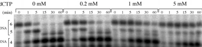
prim-ing complex. a, a stable complex of T7 DNA polymerase and gp4⌬D2D3 bound to a primed DNA template can be assembled un-der conditions that block extension of the primer strand by the polymerase. The pri-mase synthesizes a tetraribonucleotide (red) that can be extended by T7 DNA polymer-ase. DNA synthesis is terminated by the in-corporation of a 2⬘,3⬘-dideoxynucleotide. The primer synthesis reaction is then diluted 100-fold so that the priming complex can be assembled on the primed template using one or both T7 replication proteins. The protein-DNA complex protects the radiolabeled primer strand from degradation by exonu-clease III. The addition of dCTP, which binds to the polymerase active site, secures the polymerase to the 3⬘end of the primer strand (45). dCTP is not present during primer synthesis, so the polymerase and pri-mase readily dissociate from the primed DNA template (step 1), prior to assembly of the stable priming complex instep 2. The stability of this primer elongation complex is measured by challenging the protein-DNA complex with exonuclease III and monitor-ing the degradation of the radiolabeled primer strand as the complex dissociates. Exonuclease III removes the deoxynucleo-tides from the 3⬘end of the primer, leaving a tetraribonucleotide (pppACCC) that is re-sistant to further degradation.b,an autora-diograph showing the radiolabeled products of the primer extension reaction described in
a, after challenge with exonuclease III. The exonuclease challenge assays were carried out as described under “Experimental Pro-cedures,” and the degradation of the radio-labeled product (pppACCCdGddT) at the indicated times was analyzed by electro-phoresis in a 25% native gel and visual-ized by autoradiography. The lengths of the radiolabeled products are shown at theleft side of the figure, including the triribonucleotide (pppACC; labeled3) and the tetraribonucleotide (pppACCC; la-beled4) synthesized by the primase. The primer extension products resulting from the incorporation of deoxynucleotides by T7 DNA polymerase include a 5-mer (pp-pACCCdG) and a specifically terminated 6-mer product (pppACCCdGddT). In reac-tion 1, the addition of both T7 replication proteins (10Meach) and a bound nucle-otide substrate (dCTP) results in a very stable protein-DNA complex with a half-life of more than 60 min. Omitting any of these components (reactions 2–5) from the binding reaction results in the loss of pro-tection of the radiolabeled strand.c,the intensities of the 6-mer primer extension products of reactions 1–5 shown inbare plotted as a fraction of the starting mate-rial at 0 min for each reaction. The con-tinued synthesis of new primer strands with residual nucleotide substrate is a likely explanation for the initial increase in product abundance (reaction 1) during exposure to exonuclease III.
by guest on October 5, 2019
http://www.jbc.org/
described above, and then the reaction was diluted 10-fold into a bind-ing solution (10l) consisting of 40 mMTris-HCl (pH 7.5), 10 mMMgCl2,
10 mMdCTP, 10 mMDTT, 50 mMpotassium glutamate, 0.2% Tween 20, 3.5 l of NeutrAvidin-agarose resin (Pierce), and 10 M each of gp4⌬D2D3 and exo⫺T7 DNA polymerase (final concentration of the
template DNA was 10Min the binding reaction). The binding mixture was incubated on ice for 30 min, and the avidin resin was washed three times with 200l of wash buffer at 4 °C using a centrifugal filtration device (Nanosep MF; Pall Filtron). The wash buffer consisted of 40 mM Tris-HCl (pH 7.5), 10 mMMgCl2, 10 mMDTT, 50 mMpotassium
gluta-mate, 0.2% Tween 20, 200 mMNaCl, and 5% glycerol. The Tween 20 detergent was added to prevent nonspecific binding of proteins to the filtration membrane. 10 mMdCTP was added to the wash solution of complexes that were initially prepared in the presence of dCTP. The washed avidin beads were resuspended in 20l of SDS-PAGE sample buffer and centrifuged to elute the bound proteins. The eluted proteins were separated by SDS-PAGE and visualized by staining with Coomas-sie Blue G-250.
RESULTS
Construction of a Monomeric Primase-Helicase—In order to study the minimal requirements for primer utilization by T7 DNA polymerase, we constructed a monomeric variant of the T7 primase-helicase. Crystal structures of the helicase domain of the primase-helicase (29, 30) have revealed an interlocking arrangement of neighboring subunits within the hexamer. The N-terminal ␣-helix of the helicase domain (helix A; residues 272–281; Fig. 1b) extends from each subunit to pack against three helices of the adjacent subunit (helices D1–D3; residues 345–388) in a “helix swapping” arrangement (46). The hexamer is further stabilized by additional residues immediately N-terminal to helix A (19, 30) and several loops surrounding the nucleotide-binding site. Because the isolated primase domain is monomeric and shows no evidence of self-association even at high protein concentrations (19), it seemed likely that the main interactions stabilizing the primase-helicase hexamer were those observed in crystal structures of the helicase domain.
Our strategy to prevent oligomerization of the primase-heli-case was to eliminate two helices located at the subunit inter-face (helices D2 and D3; residues 368 –382), and to replace this segment with a short polar sequence (NH2-SASASG-COOH; Fig. 1a) that would be unlikely to stabilize the hexameric packing seen in the crystal structure (29, 30). The resulting 61-kDa protein, which we have named gp4⌬D2D3 to indicate the deletion of helices D2 and D3, lacks half the residues contributing to the helix swapping interaction that links adja-cent subunits of the primase-helicase. Although gp4⌬D2D3 is insoluble when expressed inE. coligrown at 37 °C, a signifi-cant amount of soluble protein can be obtained by inducing protein expression at 10 °C for 18 h (Fig. 2a; see “Experimental Procedures”). The gp4⌬D2D3 purified from the soluble fraction is well behaved and completely soluble at 25 mg/ml in buffer containing 100 mMNaCl. About 18 mg of soluble protein can be obtained from a 9-liter culture.
The oligomeric state of gp4⌬D2D3 was examined by native gel electrophoresis (Fig. 2b). 5 mM ,␥-methylene ATP and 1 mM ATP was included in the protein sample and in the gel running buffer, respectively, in order to favor the formation of oligomers (11, 47). Nucleotides bind at the interfaces between subunits of the primase-helicase (29, 30) and stabilize the hexameric form of the protein (10). The T7 primase-helicase has an apparent mass of⬃400 kDa, indicating that the hex-amer forms under these conditions (Fig. 2b). Variable amounts of a second, slowly migrating species are observed in current and previous experiments (11, 48). The apparent mass of this slowly migrating species is consistent with two hexamers as-sociated in the low ionic strength buffer used for native PAGE. In contrast to the wild-type primase-helicase, about 85% of the gp4⌬D2D3 is monomeric (⬵70 kDa) in the gel, and there is no detectable hexamer. However, trace amounts of several higher order oligomers of gp4⌬D2D3 are evident in the native gel, indicating some self-association into dimers and trimers.
gp4⌬D2D3 Lacks dTTPase and DNA Unwinding Activi-ties—Although the gp4⌬D2D3 is predominantly monomeric in solution, it was important to determine whether the mod-ified protein can assemble into functional hexamers on DNA. We therefore assayed gp4⌬D2D3 for its ability to hydrolyze dTTP and to unwind DNA, activities that depend upon the oligomerization of gp4. The hydrolysis of dTTP fuels the translocation of the primase-helicase along DNA in a 5⬘to 3⬘ direction, separating the strands of duplex DNA (22–26). The active site of the helicase consists of residues from two adja-cent subunits, and oligomerization is required for nucleotide hydrolysis (19). The addition of single-stranded DNA to the primase-helicase stimulates its rate of dTTP hydrolysis about 20-fold and promotes hexamer formation (11, 47). In contrast, gp4⌬D2D3 lacks detectable dTTPase activity in the presence or absence of M13 ssDNA (not shown), even at protein con-centrations as high as 50Musing conditions that support a high level of nucleotide hydrolysis activity by the wild-type primase-helicase. The results indicate that gp4⌬D2D3 does not form functional hexamers, even at protein concentrations that are much higher than those required to assemble a hexamer of the wild-type primase-helicase (Fig. 2b).
gp4⌬D2D3 also lacks DNA unwinding activity and thus is unable to assemble on DNA into functional hexamers (Fig. 3). The DNA unwinding activity of the wild-type primase-helicase readily displaces a radiolabeled oligonucleotide annealed to M13 ssDNA, whereas gp4⌬D2D3 is inactive, even at 100-fold higher protein concentrations (Fig. 3). Helicase activity was also assayed in a rolling circle DNA replication system in which DNA strand separation by the helicase is required for DNA synthesis (Fig. 4) (2). In the presence of the primase-helicase, T7 DNA polymerase incorporates radiolabeled nucleotides into DNA at a rate of 2–3 pmol/min. In contrast, gp4⌬D2D3 does not support DNA synthesis (incorporation of ⬍0.1 pmol/min) in this replication system even at 10 M gp4⌬D2D3, consistent with its lack of DNA unwinding activity. We could not exclude the possibility that gp4⌬D2D3 is inactive in this replication system because it fails to interact with the DNA or proteins of the replication fork (2, 49). We therefore examined whether or not gp4⌬D2D3 could inhibit the activity of wild-type primase-helicase in this reaction. When gp4⌬D2D3 (10 M) was mixed with the primase-helicase (60 nM) in the replication reaction, the rate of DNA synthesis was decreased to1⁄2–1⁄3of the wild-type rate (Fig. 4).
This indicates that gp4⌬D2D3 can interact with the T7 DNA rep-lication complex and interfere with DNA synthesis, presumably because the altered protein cannot unwind the DNA duplex.
gp4⌬D2D3 Primes DNA Synthesis by T7 DNA Polymerase— The synthesis of RNA primers by the primase-helicase and by
TABLE I
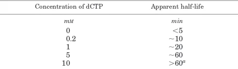
The apparent lifetime of the priming complex depends upon the concentration of the bound nucleotide substrate
The intensity of the fully extended primer strand at each time point (Figs. 6band 7) was quantitated by densitometry and normalized to the intensity at 0 min (Fig. 6c). The half-life of the protein-DNA complex is approximated by the time at which the normalized intensity was one-half the starting value at 0 min.
Concentration of dCTP Apparent half-life
mM min
0 ⬍5
0.2 ⬃10
1 ⬃20
5 ⬃60
10 ⬎60a
aThis value is estimated from Fig. 6c. The others are from Fig. 7.
by guest on October 5, 2019
http://www.jbc.org/
[image:8.605.51.296.125.197.2]gp4⌬D2D3 was examined in reactions with a 25-mer DNA template containing a primase recognition site (template 3 in Table IV). In the presence of [␥-32P]ATP and CTP, gp4⌬D2D3 synthesized the same amounts of di-, tri-, and tetraribonucle-otide products as the wild-type primase-helicase (Fig. 5a). The primase-helicase and gp4⌬D2D3 also synthesize a small amount of a pentaribonucleotide, presumably by misincorpo-rating AMP or CMP opposite a cytidine at this position of the template. The major product of RNA synthesis is a tetraribo-nucleotide that can be elongated by T7 DNA polymerase in the presence of dNTPs (see Fig. 6b).
The tetraribonucleotides synthesized by the T7 primase-he-licase can be extended by T7 DNA polymerase only in the presence of the primase-helicase (26, 28). Although a fragment spanning the N-terminal half of the primase-helicase correctly synthesizes tetraribonucleotides, the primase fragment does not support their utilization by T7 DNA polymerase (18). The primase fragment and gp4⌬D2D3 are both predominantly mo-nomeric proteins, and primer utilization by T7 DNA polymer-ase might require the presence of a hexameric primpolymer-ase. We tested this notion by measuring the amount of DNA synthesis catalyzed by T7 DNA polymerase in reactions containing the hexameric T7 primase-helicase or the predominantly mono-meric gp4⌬D2D3 protein. As expected, the wild-type primase-helicase supported the RNA-primed synthesis of DNA by T7 DNA polymerase in a reaction containing an M13 ssDNA tem-plate with ATP, CTP, and a mixture of all four dNTPs. The rate
of DNA synthesis increased with increasing concentrations of the primase-helicase protein until DNA synthesis was inhib-ited at very high protein concentrations (Fig. 5b). This inhibi-tion of DNA synthesis in the presence of a large molar excess of primase-helicase probably results from the depletion of dTTP in the DNA synthesis reaction through hydrolysis by the heli-case domain. Remarkably, gp4⌬D2D3 supports DNA synthesis at a rate approaching 1/10th the wild-type rate. High concen-trations of gp4⌬D2D3 do not inhibit DNA synthesis, a finding consistent with its lack of dTTPase activity. The lower efficiency of gp4⌬D2D3 in promoting primer utilization and DNA synthesis might be explained by its failure to rapidly locate priming sites on the M13 ssDNA because of the defective helicase domain, which is unable to actively translocate on DNA (11).
A Priming Complex of gp4⌬D2D3 and T7 DNA Polymerase— Although it is incapable of assembling into functional hexam-ers, gp4⌬D2D3 primes DNA synthesis catalyzed by T7 DNA polymerase. This finding suggests that a single subunit within the hexameric primase-helicase is sufficient to prime Okazaki fragment synthesis on the lagging strand of the replication fork. The requirement for the primase during extension of RNA primers further suggests that the T7 DNA polymerase engages short, naturally occurring primers only when they are bound to the primase protein. The primase might help initiate primer elongation by securing the RNA primer to the DNA template (3) and/or by recruiting the polymerase to the priming site through protein-protein interactions. A four amino acid loop located at the base of the thumb of T7 DNA polymerase is required for lagging strand synthesis (20). The selective loss of lagging strand synthesis upon deletion of the loop, together with its location near the polymerase active site, suggests that the loop might contact the RNA primer or interact with the primase during the elongation of a primer.
The association of the hexameric T7 primase with T7 DNA polymerase during extension of a tetraribonucleotide primer was previously demonstrated using an exonuclease challenge assay (20). We investigated the minimal requirements for primer utilization by T7 DNA polymerase, using the mono-meric gp4⌬D2D3 primase to synthesize and deliver tetraribo-nucleotide primers to the polymerase. A very stable protein-DNA complex containing gp4⌬D2D3 and T7 DNA polymerase
FIG. 7.Stabilization of the priming complex by the polymerase substrate dCTP.A high concentration of the next nucleotide (dCTP) matching the template sequence stabilizes the priming complex. The exonuclease III protection assay shown in Fig. 6 was used to monitor the formation of the priming complex in the presence of the nucleotide concentrations shown. Nucleotide concentrations higher than 5 mM(not shown) did not additionally increase the lifetime of the priming complex.
FIG. 8.Mismatched dNTPs can stabilize the priming complex.The stabilities of priming complexes assembled with the correct (dCTP) or mismatched nucleotide substrates were measured as described in the legend of Fig. 6, using 10 mMof each of the nucleotides listedabovethe gel. The extended primer strand was labeled by the incorporation of [␣-32P]dGMP into DNA, followed by chain termination with 2⬘,3⬘
-dideoxythymi-dine. The sequences of the extended primer strands are indicated on theleft sideof the figure.
TABLE II
The correct bound nucleotide, and some incorrect nucleotides, stabilize the priming complex
As described for the experiments shown in Table I, the intensities of the fully extended primer strands shown in Fig. 8 were quantitated by densitometry, and the half-lives were estimated for priming complexes formed in the presence of a nucleotide matching the DNA template (dCTP) or incorrect nucleotides.
Next incoming dNTP Apparent half-life
min
dCTP (correct) ⬎60
dTTP ⬃30
dATP ⬍5
dGTP ⬍5
by guest on October 5, 2019
http://www.jbc.org/
[image:9.605.131.474.52.132.2]is formed when DNA synthesis is halted by the incorporation of a 2⬘,3⬘-dideoxynucleotide during the initial cycles of de-oxynucleotide incorporation by the polymerase (Fig. 6a). The stability of the protein-DNA complex was determined by add-ing a high concentration of exonuclease III and monitoradd-ing the loss of the radiolabeled primer as DNA dissociates from the complex (Fig. 6a). Both gp4⌬D2D3 and T7 DNA polymerase are required for the formation of the stable complex with the nas-cent primer, suggesting that both proteins remain bound to DNA (compare the extent of protection forreaction 1with that ofreactions 3–5in Fig. 6b). If either protein is left out of the binding reaction, the primer is rapidly degraded to a tetraribo-nucleotide (labeled at the 5⬘ ␥-phosphate position), which re-sists further degradation by exonuclease III. A high concentra-tion of the substrate dCTP is also required to secure the primer in the active site of the polymerase (reaction 2, Fig. 6b) (20, 45). In the complete reaction, the radiolabeled primer DNA strand is very resistant to degradation by exonuclease III, and the protein-DNA complex dissociates slowly (Fig. 6c). The exposed 3⬘single-stranded end of the DNA template is a poor substrate for exonuclease III (50 –52), and it is degraded slowly (results not shown) in comparison to the recessed 3⬘end of the primer. A determination of the lifetime of the protein-DNA complex is complicated by an observed 2-fold increase in the amount of radiolabeled primer synthesized during the first 15 min of the exonuclease digestion (comparereaction 1withreactions 3–5, Fig. 6c). This increase is the result ofde novoprimer synthesis by the primase (10M) present during exonuclease digestion. Because of these complications, we operationally define the apparent half-life of the priming complex as the time when the amount of extended primer decreases to one-half the amount at 0 min. Theapparenthalf-lives of priming complexes measured in this way (Tables I–V) can then be compared to identify the experimental variables that affect complex stability. The prim-ing complexes containprim-ing the gp4⌬D2D3 monomer are almost as long lived as the priming complex containing the hexameric T7 primase-helicase (20).
A different type of interaction between gp4 and T7 DNA polymerase has been described (4) that requires an acidic seg-ment at the C terminus of the helicase domain. The analogous C-terminal region of gp4⌬D2D3 is not required for the forma-tion of the priming complex. A truncated version of gp4⌬D2D3 lacking this acidic segment forms priming complexes with T7 DNA polymerase that are as stable as those formed by the full-length gp4⌬D2D3 protein described above (not shown). The lack of involvement of the C-terminal segment of the helicase domain further implies that the priming complex is sustained by interactions between the primase domain of the primase-helicase and the polymerase. At very high protein concentra-tions (100Mprimase), the primase fragment (residues 1–271 (18)) of the primase-helicase lacking the helicase domain can function in the assembly of a short lived priming complex (half-life 1–2 min determined by exonuclease challenge; not shown). The added stability provided by the helicase domain of gp4⌬D2D3 might result from tighter interactions with the DNA template (40).
Several factors strongly influence the stability of the T7 priming complex. A high concentration of the incoming dCTP substrate is required to keep the polymerase bound to the primed DNA template (Fig. 7) (45). The priming complex be-comes more stable as the concentration of dCTP is increased, reaching maximal stability at 5–10 mM dCTP (Table I). Al-though dCTP is the correct incoming nucleotide specified by the template, the binding of several mismatched dNTP substrates in the polymerase active site measurably stabilizes the complex (Fig. 8). For these experiments, the polymerase incorporated [␣-32P]dGTP at the 3⬘ end of the tetraribonucleotide primer followed by the incorporation of ddTMP to terminate DNA synthesis. The addition of dCTP, which matches the next posi-tion of the template, produced a long lived priming complex, whereas the addition of dGTP resulted in a half-life of less than 5 min (Fig. 8 and Table II). The complex with dATP was initially degraded rapidly by the added exonuclease, but some of the radiolabeled primer was protected from degradation for more than 2 h. Surprisingly, a pairing of dTTP with the tem-plate guanosine was fairly effective at stabilizing the priming complex (Fig. 8). It is possible that the 10 mMdTTP present in the reaction permits re-synthesis of the extended primer fol-lowing the exonucleolytic removal of the 3⬘-ddTMP.
The length of the extended primer strand has a pronounced effect on the lifetime of the complex. The long lived complex described above contains an elongated primer consisting of 6 nucleotides (5⬘-pppACCCdGddT-3⬘) that is annealed to a 25-mer template (template 3 in Table IV). Similar priming com-plexes were prepared by halting primer elongation after the incorporation of 1– 4 deoxynucleotides by T7 DNA polymerase,
FIG. 9.The stability of the priming complex changes during elongation of the primer strand.The primer extension reaction (described in Fig. 6) was stopped at different positions of the DNA template by the incorporation of a chain-terminating 2⬘,3⬘-dideoxynucleotide. After the addition of gp4⌬D2D3, T7 DNA polymerase, and the appropriate dNTP, the stabilities of the resulting priming complexes were determined by exonuclease challenge. The complex containing the 6-mer primer extension product (pppACCCdGddT) is the most stable. Incorporation of fewer or more nucleotides prior to terminating synthesis resulted in faster dissociation of the protein-DNA complex (5-, 7-, and 8-mer complexes). The effect of primer strand length on the stability of the protein-DNA complex implies that specific contacts are made to the 5⬘and 3⬘ends of the primer strand within the complex.
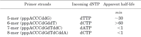
TABLE III
The stability of the priming complex depends upon the length of the primer strand in the complex
The intensities of the fully extended primer strands shown in Fig. 9 were analyzed by densitometry as described in Table I.
Primer strands Incoming dNTP Apparent half-life
min
5-mer (pppACCCddG) dTTP ⬃30
6-mer (pppACCCdGddT) dCTP ⬎60
7-mer (pppACCCdGdTddC) dATP ⬍1 8-mer (pppACCCdGdTdCddA) dCTP ⬍1
by guest on October 5, 2019
http://www.jbc.org/
[image:10.605.50.296.283.348.2]and the lifetimes of these complexes were determined by chal-lenge with exonuclease III. The complex with a single dideoxynucleotide appended to the tetraribonucleotide (the 5-mer complex in Fig. 9) is less stable than the 6-mer complex described above, and it has a half-life of about 30 min (Table III). The 6-mer complex is most stable, with a half-life of more than 60 min (Figs. 6band 9; Table III). Further elongation of the primer strand to create the 7- and 8-mer complexes incre-mentally decreases the stability of the priming complex (Fig. 9 and Table III). The destabilization of the priming complex with increasing length of the DNA product might indicate that in-teractions between the primase and polymerase are weakened as the polymerase moves away from the priming site during DNA synthesis. The elongating primer strand eventually be-comes effectively long enough that the primase is no longer needed for DNA synthesis.
The priming complexes described above were bound to a 25-mer DNA template (template 3 in Table IV). We also exam-ined DNA templates 19 –31 nucleotides in length with the same priming sequence in complexes containing the 6-mer elonga-tion product (pppACCCdGddT) and the bound nucleotide dCTP. All of these templates supported the synthesis of tet-raribonucleotides by gp4⌬D2D3 and their elongation by T7 DNA polymerase (data not shown). However, the stabilities of the stalled priming complexes depended upon template length (Table IV). The complex with the shortest template (a 19-mer, template 1 in Table IV) was least stable and had a half-life of less than 5 min. Addition of three bases to the 3⬘end of this sequence (template 2) significantly improved the stability of the priming complex (Table IV). A three-nucleotide extension to the 5⬘end of template 2 to create template 3 provided optimal stability. A template with three additional nucleotides at the 3⬘ end (template 4) produced an equally stable priming complex. However, the addition of nucleotides to the 5⬘end of the tem-plate (temtem-plates 5 and 6 in Table IV) destabilized the complex. The reason for this effect is not known.
The 5⬘-Triphosphate of the Primer Stabilizes the Priming Complex—The ribonucleotides synthesized by the T7 primase contain a 5⬘-triphosphate moiety from the ATP that initiates synthesis. The primase can also incorporate ATP analogs with chemically modified phosphate linkages at the 5⬘end of ribo-nucleotide products (39). This relaxed specificity suggests that the primase does not interact strongly with the 5⬘-triphosphate moiety during RNA synthesis. The unphosphorylated dinucle-otide AC is also efficiently extended by the primase to form a tetraribonucleotide (ACCC) that is utilized by T7 DNA polym-erase (14). Thus, the role of the 5⬘-triphosphate of the naturally occurring products of T7 primase is enigmatic. We examined whether or not the 5⬘-triphosphate contributes to the physical stability of priming complexes formed between gp4⌬D2D3 and T7 DNA polymerase in primer extension reactions initiated with pppAC or AC dinucleotides instead of ATP. Both dinucle-otides were readily extended by gp4⌬D2D3, and the resulting
tetraribonucleotide was elongated by T7 DNA polymerase in the presence dNTP and ddNTP substrates (Fig. 10). The prim-ing complex that was initiated with pppAC was as long lived as the complex initiated with ATP. In contrast, the priming com-plex initiated with the unphosphorylated AC was unstable, with a half-life of less than 5 min (Fig. 10 and Table V). These results show that the 5⬘-triphosphate of the primer strand contributes significantly to the stability of the priming com-plex, and they imply that the phosphate group plays a role in primer utilization.
Purification of the Priming Complex Using an Affinity-tagged Template—The exonuclease III protection assay de-scribed above provided indirect evidence that the T7 primase and the polymerase were stably associated in the priming complex. In order to observe the complex directly, we purified the complex by a biotin-avidin affinity capture procedure. The priming complex was assembled on a 3⬘-biotinylated template (template 3 in Table IV) coupled to avidin-agarose beads. The beads were washed to remove proteins bound nonspecifically, and the remaining proteins complexed to the template were eluted with SDS and visualized by SDS-PAGE (Fig. 11). In the complete priming reaction (lane 2) both gp4⌬D2D3 and T7 DNA polymerase remained associated with the template. If either the primer-template strand or the dCTP was left out of the reaction, no detectable protein remained associated with the immobilized template (lanes 3–5). When the correct incom-ing nucleotide (dCTP) was replaced with 10 mMdTTP, dATP, or dGTP a stable complex was not isolated (data not shown), providing further evidence of the specificity of protein interac-tions with the immobilized DNA template. Although a mis-matched dTTP provided some protection against exonuclease digestion (see above), the isolation of the priming complex requires the correct nucleotide substrate to be present during complex formation and in the wash buffer. The equal Coomas-sie Blue staining intensities of the gp4⌬D2D3 and T7 DNA polymerase eluted from the avidin-agarose beads (Fig. 11,lane 2) suggest that a 1:1 complex of primase and DNA polymerase is present in the stalled priming complex.
DISCUSSION
As the replication fork advances, RNA primers are periodi-cally synthesized by a primase on the lagging strand and ex-tended by a DNA polymerase to create Okazaki fragments several hundred to several thousand nucleotides in length (8). The priming sites used by prokaryotic replication systems are short sequences that are expected to occur by chance every several hundred nucleotides. However, not every potential priming site is used, and it has been suggested that a timing mechanism regulates the frequency of primer synthesis and hence the length of Okazaki fragments produced during repli-cation. InE. coli, the DnaG primase transiently associates with the replication machinery to prime Okazaki fragment synthe-sis, and then the primase dissociates from the replication
com-TABLE IV
DNA template requirements for the T7 priming complex
The apparent half-lives of priming complexes formed under the conditions described under “Experimental Procedures,” and using the DNA templates shown below, were estimated by densitometry (data not shown). See text for a complete description of these experiments.
Template Sequencea Apparent half-life
min
1 (19-mer) 3⬘-CTGCTATTTGCTGGGCAGT-5⬘ ⬍5
2 (22-mer) 3⬘-CTGCTATTTGCTGGGCAGTGAC-5⬘ ⬃20
3 (25-mer) 3⬘-CGGCTGCTATTTGCTGGGCAGTGAC-5⬘ ⬎60
4 (28-mer) 3⬘-TAACGGCTGCTATTTGCTGGGCAGTGAC-5⬘ ⬎60
5 (28-mer) 3⬘-CGGCTGCTATTTGCTGGGCAGTGACTTT-5⬘ ⬃20
6 (31-mer) 3⬘-CGGCTGCTATTTGCTGGGCAGTGACTTTTTT-5⬘ ⬃20
aThe primase recognition sites are denoted by bold letters. The common sequences are underlined.
by guest on October 5, 2019
http://www.jbc.org/
plex. The strength of this interaction between the primase and helicase might serve as a timing mechanism that controls the frequency of priming (9). The consolidation of primase and helicase functions in the bifunctional primase-helicase of bac-teriophage T7 raises questions about how many subunits of the hexamer participate in primer synthesis and utilization by T7 DNA polymerase, and how the frequency of these events is controlled during replication.
We have engineered an altered T7 primase-helicase (gp4⌬D2D3) that does not form functional hexamers, yet it primes DNA synthesis by T7 DNA polymerase (Fig. 5b). The
monomeric primase physically complexes with T7 DNA polym-erase during initiation of RNA primer extension. These results suggest that the primase complexed to the DNA template de-livers the newly synthesized tetraribonucleotide primer to the polymerase active site. Although gp4⌬D2D3 lacks two␣-helices (D2 and D3) that account for most of the buried surface of the subunit interface of the helicase domain (Fig. 1b) (29, 30), the modified protein has some residual tendency to oligomerize (Fig. 2b). The remaining interactions between subunits might be mediated by the N-terminal primase domain or by the loops surrounding the active site of the helicase (29, 30). Addition-ally, the linker region (residue 241–271) between the primase and helicase domain contributes to oligomerization of thegene 4 protein (19). gp4⌬D2D3 lacks the dTTPase and DNA unwinding activities associated with the hexameric primase-helicase, and it is predominantly monomeric, even at high protein concentrations (Fig. 2b). These results strongly suggest that a single protomer of the T7 primase-helicase can function in the utilization of tetrari-bonucleotide primers by T7 DNA polymerase.
gp4⌬D2D3 consists of both primase and helicase domains of the T7 gene 4 protein. A fragment of the primase-helicase spanning only the primase domain is a monomer, and it cata-lyzes the template-dependent synthesis of tetraribonucleotides at specific priming sites (18), as does gp4⌬D2D3. However, the minimal primase fragment does not efficiently function in primer utilization by T7 DNA polymerase. The additional hel-icase domain of gp4⌬D2D3 might support primer utilization by contributing DNA interactions that secure the nascent primer on the DNA template until it is elongated by T7 DNA polym-erase (40). The helicase domain might also participate in pro-tein-protein interactions with the polymerase. The primase-helicase interacts strongly with T7 DNA polymerase in the absence of DNA (3, 4, 19, 53). However, the priming complex (Ref. 20 and this work) is different from a previously described interaction between the primase-helicase and T7 DNA polym-erase that involves 17 residues at the C terminus of the heli-case domain (4). This protein-protein interaction is required to couple the synthesis of the lagging strand of the replication fork with synthesis of the leading strand (2). This interaction, which can be observed with the purified proteins bound to single-stranded DNA (4), might correspond to the interaction of the helicase on the lagging strand of the replication fork with the polymerase on the leading strand. In contrast, the interaction of the T7 primase with the DNA polymerase occurs under conditions specific for primer elongation. The formation of a stable complex requires a primed template and a nucleotide substrate bound to the polymerase (Figs. 6 and 11). The re-moval of the acidic segment from the C terminus of gp4⌬D2D3 does not interfere with the formation of a stable priming
com-FIG. 10.The 5ⴕ-triphosphate of the primer stabilizes the priming complex.Priming complexes were initiated with a phosphorylated dinucleotide (pppAC) or with the unphosphorylated dinucleotide (AC) and elongated by the primase and T7 DNA polymerase in the presence of [␣-32
P]dGTP to form radiolabeled products with the sequences shown in the figure. The stabilities of priming complexes initiated with either dinucleotide were compared with that of a standard complex in which primer synthesis was initiated with ATP (left sideof figure), using the exonuclease challenge assay described in Fig. 6. The unphosphorylated primer strand migrates more slowly than the phosphorylated strands. It is evident that priming complexes containing a 5⬘-triphosphate group on the primer strand (initiated with pppAC or ATP) are significantly more stable than the complex with the unphosphorylated primer.
TABLE V
The 5⬘-triphosphate of the primer contributes to stability of the priming complex
Primers synthesized from CTP and ATP or the diribonucleotides shown below were incorporated into priming complexes, and the appar-ent half-lives of these complexes were determined by exonuclease chal-lenge.
Precursor Apparent half-life
min
ATP, CTP ⬎60
pppAC, CTP ⬎60
AC, CTP ⬍5
FIG. 11. Isolation of a stable DNA complex containing both primase and polymerase. The priming complex described in the legend of Fig. 6 was purified using an immobilized DNA template that was attached to avidin-agarose beads through a 3⬘-biotin group. After washing the beads, the proteins were eluted with SDS and separated by electrophoresis on a 12% SDS-PAGE gel and then visualized by staining with Coomassie Blue. The priming complex (lane 2) contains both T7 DNA polymerase (the purified protein is shown in lane 6) and gp4⌬D2D3 (compare with pure protein inlane 7). The equal staining intensities of the proteins eluted from the priming complex suggest that T7 DNA polymerase and gp4⌬D2D3 are present in a 1:1 ratio. Neither protein alone stably interacts with the immobilized DNA (not shown). Immobilization of the proteins on the agarose beads requires dCTP (lane 3) and the DNA template (lanes 4and5), indicating that a specific complex forms on the primed DNA template.
by guest on October 5, 2019
http://www.jbc.org/