Open Access
Correspondence
Canine candidate genes for dilated cardiomyopathy: annotation of
and polymorphic markers for 14 genes
Anje C Wiersma*
1,2,3, Peter AJ Leegwater
3, Bernard A van Oost
4,
William E Ollier
2and Joanna Dukes-McEwan
1Address: 1Small Animal Teaching Hospital, University of Liverpool, Leahurst, Chester High Road, Neston, CH64 7TE, UK, 2Centre for Integrated
Genomic Medical Research, Division of Epidemiology and Health Sciences, The University of Manchester, Manchester, M13 9PT, UK, 3Department
of Clinical Sciences of Companion Animals, Faculty of Veterinary Medicine, Utrecht University, 3508 TD Utrecht, The Netherlands and 4American
University of the Caribbean, Department of Molecular Cell Biology, #1 University Drive at Jordan Road, Cupecoy, St.Maarten, Dutch Antilles
Email: Anje C Wiersma* - [email protected]; Peter AJ Leegwater - [email protected]; Bernard A van Oost - [email protected]; William E Ollier - [email protected]; Joanna Dukes-McEwan - [email protected]
* Corresponding author
Abstract
Background: Dilated cardiomyopathy is a myocardial disease occurring in humans and domestic animals and is characterized by dilatation of the left ventricle, reduced systolic function and increased sphericity of the left ventricle. Dilated cardiomyopathy has been observed in several, mostly large and giant, dog breeds, such as the Dobermann and the Great Dane. A number of genes have been identified, which are associated with dilated cardiomyopathy in the human, mouse and hamster. These genes mainly encode structural proteins of the cardiac myocyte.
Results: We present the annotation of, and marker development for, 14 of these genes of the dog genome, i.e. α-cardiac actin, caveolin 1, cysteine-rich protein 3, desmin, lamin A/C, LIM-domain binding factor 3, myosin heavy polypeptide 7, phospholamban, sarcoglycan δ, titin cap, α -tropomyosin, troponin I, troponin T and vinculin. A total of 33 Single Nucleotide Polymorphisms were identified for these canine genes and 11 polymorphic microsatellite repeats were developed.
Conclusion: The presented polymorphisms provide a tool to investigate the role of the corresponding genes in canine Dilated Cardiomyopathy by linkage analysis or association studies.
Background
Dilated cardiomyopathy (DCM) is a myocardial disease characterized by dilatation of the left ventricle, reduced systolic function and increased sphericity of the left ven-tricle. This disease has been described in different species and multiple genes have been found in the human [1], mouse [2] and hamster [3] causing DCM. These genes mainly encode cyto-skeletal components of the cardiac myocytes and can be divided into sarcomeric and extra-sarcomeric proteins. The identified extra-sarcomeric proteins
involved in DCM include α-cardiac actin, encoded by
ACTC [4], cysteine-rich protein 3 (CSRP3) [5], LIM-domain binding factor 3 (LDB3, also known as Cypher or
ZASP) [6], myosin heavy polypeptide 7 (MYH7) [7], titin cap (TCAP) [8], α-tropomyosin (TPM1), troponin I (TNNI3) [9], troponin T (TNNT2) [7], titin (TTN) [10] and vinculin (VCL) [11]. The extra-sarcomeric proteins implicated in DCM are encoded by the genes including caveolin 1 (CAV1) [2], desmin (DES) [12], lamin A/C (LMNA) [13], phospholamban (PLN) [14] and
sarcogly-Published: 19 October 2007
BMC Veterinary Research 2007, 3:28 doi:10.1186/1746-6148-3-28
Received: 9 March 2007 Accepted: 19 October 2007
This article is available from: http://www.biomedcentral.com/1746-6148/3/28
© 2007 Wiersma et al; licensee BioMed Central Ltd.
can δ (SGCD) [3]. The genes encoding all of the above proteins are located on the autosomal chromosomes. X-linked genes implicated in DCM include dystrophin (DYS) [15] and tafazzin (TAZ) [16]. In addition, mito-chondrial dysfunction and mitomito-chondrial DNA (mtDNA) mutations have been associated with maternally inherited DCM [17]. Furthermore, DCM has also been described with arrhythmias, with mutations in genes encoding sodium [18] and potassium channels [19].
DCM has been described in many different breeds of mostly giant and large dogs, including the Dobermann [20], Great Dane [21], Newfoundland [22] and Irish Wolfhound [23]. Clinical variation exists in the presenta-tion and progression of DCM between different dog breeds and breed specific variation has also been found in histological findings in DCM-affected hearts tissue [24]. Since clinical DCM may be a late onset disease, following a long pre-symptomatic course, dogs are often used for breeding before the disease becomes apparent [25]. So far, no causative mutation has been discovered in canine DCM. The phenotype of the adult onset forms of canine DCM in most breeds is consistent with a defect in compo-nents of the cytoskeleton.
Of the 14 autosomal DCM candidate genes for the dog,
ACTC, CAV1, CSRP3, DES, LDB3, LMNA, MYH7, PLN,
SGCD, TCAP, TNNI3, TNNT2, TPM1 and VCL, genomic information and/or polymorphic markers were already available for ACTC [26,27], DES [28], PLN [29], SGCD
[30] and TPM1 [31]. In this article, we describe a complete set of polymorphic markers for these 14 candidate genes for canine DCM. The markers, both microsatellites and Single Nucleotide Polymorphisms (SNPs), provide a use-ful tool to perform linkage and association studies between each of these genes and DCM in the different dog breeds. Furthermore, we present the annotation of 14 can-didate genes in the canine genome, which will facilitate mutation screening of these genes.
Genomic Annotation
The 14 canine DCM candidate genes were identified on the canine genome by means of a BLAST analysis [32], using available canine and human DNA sequences as a query (Table 1). The exons were identified based on the corresponding human exon sequence (retrieved from [33], Table 1). Each gene was found to be covered by 1 to 5 contigs of the Canis familiaris genome build 1.1. (Addi-tional file 1 and Table 1). CAV1 was covered by 2 neigh-bouring contigs and the 3 coding exons matched the human ones. Exon 1 of the dog seemed to have an extra nucleotide (T, position 336 of [Genbank: AAEX01048547]) compared to human exon 1 of CAV1. However, this nucleotide was not present in the single trace file of the Canis familiaris Trace Archive [34] covering
this sequence. Canine DES had 1 amino acid less than the human protein. The canine LDB3 protein is 67 amino acids shorter than human LDB3. Canine LMNA had 1 amino acid extra compared to the human protein. Exon 24 of canine MYH7 seemed to have 1 bp extra (G, bp 7,902 of [Genbank: AAEX01041100]), however, this nucleotide was not present in any of 11 Canis familiaris
trace sequences covering this position. Without this extra nucleotide, canine exon 24 matched the human exon. Canine TNNI3 had 1 amino acid extra compared to the human protein. For TNNT2, coding exons 1, 15 and 16 could not be recognized in canine genomic contigs.
TNNT2 exon 6 showed 1 extra bp compared to human (G, bp 5622 of [Genbank: AAEX01013360]), however, this nucleotide was not found in the 2 traces covering this DNA sequence. Without this additional bp, exon 6 matched the corresponding human exon exactly in length. Exon 12 had 1 codon less than the human gene. Exon 13 was located at the end of genomic contig [Genbank: AAEX01013360] and although its terminal 2 putative bp were not included in this contig, exon 12 seemed to match the human exon. For the remaining candidate genes,
ACTC, CSRP3, PLN, SGCD, TCAP, TPM1 and VCL, the annotated canine exons matched the corresponding human exons exactly. We could not identify non-coding exons. Apparently, the conservation of these exons is too low for identification purposes. Complementary DNA sequencing is necessary to identify these non-coding exons. All of the predicted introns of the 14 candidate genes started and ended with the canonical GT and AG dinucleotides, respectively [35]. Even though a high qual-ity DNA sequence of the canine genome has recently become available, it has not yet been fully annotated.
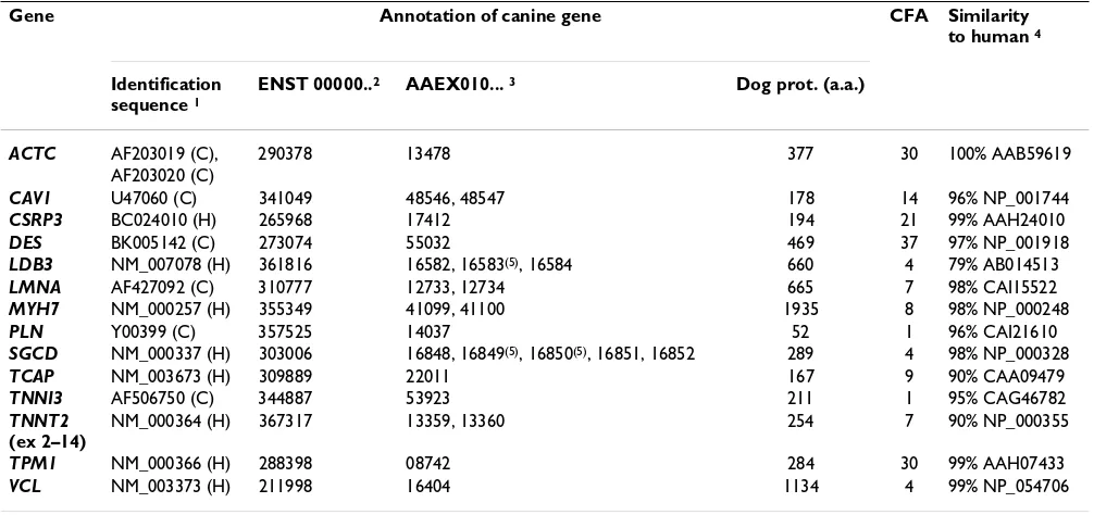
The conservation of the coding region of each gene was assessed by BLAST comparison of the cDNA and derived amino acid sequences with those of human (at the web-site of NCBI [36], BLASTN and TBLASTX analysis, respec-tively). The percentages of identity at the nucleotide level varied between 88 and 95% (Table 1). At the amino acid level, the percentages of identity varied in general between 90–100%, except for the canine LDB3 protein, that was 79% identical to the human protein. The canine ACTC protein appeared to be identical to the human protein. In
LDB3, a relatively low percentage of identity was found between the canine and human gene, both at the cDNA and the protein level. This was caused by the large (inframe) loss of part of exons (i.e. 4, 7, 8 and 9) com-pared to the human gene: the canine gene had 660 codons, the human gene had 734 codons.
When analysing the location of the genes in the dog genome (Table 1), using the canine-human comparative map of Guyon et al. [37], each was found to be syntenic to the human location.
Polymorphisms
Single Nucleotide Polymorphism detection
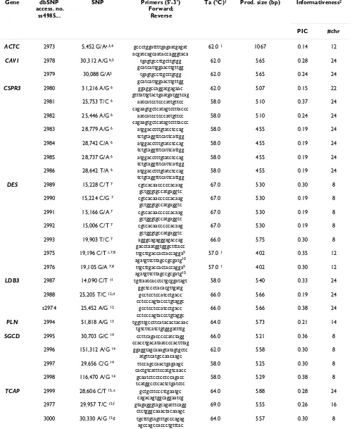
We used denaturing high-performance liquid chromatog-raphy (DHPLC) analysis for the detection of SNPs in amplified genomic canine DNA fragments. Polymor-phisms were assessed in DNA from Newfoundland dogs. For each gene, several DNA fragments of approximately 500 bp were selected based on melting profile (analyzed with WAVEMAKER™ software from Transgenomic) with a maximum of 2 melting temperatures covering each prod-uct. The melting behaviour of a fragment depends on the fragment's DNA sequence. Primers were designed using Primer3 [38] and annealing temperatures of the PCRs were optimized (Table 2). Touchdown PCR amplification of these fragments was performed with DNA of New-foundland dogs (n = 16; 8 unrelated founders of a pedi-gree of Newfoundland dogs and 8 family members), using HotStartTaq DNA Polymerase (Qiagen). The Touch-down (TD) PCR program consisted of a denaturing step of 5 min at 95°C, followed by 14 cycles of 95°C 30 sec, Ta +7°C 30 sec, 72°C 20 sec, with a Ta decrease of 0.5°C/ cycle, followed by 25 cycles of 30 sec at 94°C, 30 sec at Ta°C, 30 sec at 72°C, followed by a final extension at
72°C for 2 min (Ta in Table 2). Subsequently, a heterodu-plex formation step was carried out to allow formation of hetero- and homo-duplex products; the PCR products were heated 5 min at 95°C, after which the temperature was decreased gradually (38 cycles of 1 min, temperature decreasing 1.5°C/cycle), followed by a final step of 5 min at 10°C. Mutation analysis of the PCR products, based on the presence of heteroduplexes, followed on a WAVE instrument (WAVE Nucleic Acid Fragment Analysis Sys-tem, Transgenomic). Multiple WAVE patterns of a single PCR fragment in different dogs pointed at existence of both homoduplexes and heteroduplexes and, therefore, indicated potential presence of SNPs in the fragment. In that case, the PCR fragment (of at least of 2 dogs per WAVE pattern) was cleaned (Shrimp Alkaline Phos-phatase/ExoI) and the DNA sequence was obtained to determine the identity of the SNPs, by a commercial com-pany (Lark Technologies™, UK).
Twenty-eight SNPs were discovered by WAVE analysis (Table 2). No indication of the presence of a SNP was found in WAVE fragments of LMNA, MYH7 and TNNI3
(3, 5 and 3 fragments analyzed, respectively). One new SNP, TCAP SNP 29,957 T/C in genomic contig [Genbank: AAEX01022011], was found when we resequenced a
[image:3.612.54.557.110.347.2]TCAP fragment in a group of Newfoundland dogs. WAVE analysis of this fragment had not indicated presence of a potential SNP – although the obtained DNA sequences
Table 1: Assignment, genomic location and the degree of sequence conservation compared to human of the canine DCM candidate genes.
Gene Annotation of canine gene CFA Similarity
to human 4
Identification sequence 1
ENST 00000..2 AAEX010... 3 Dog prot. (a.a.)
ACTC AF203019 (C), 290378 13478 377 30 100% AAB59619
AF203020 (C)
CAV1 U47060 (C) 341049 48546, 48547 178 14 96% NP_001744
CSRP3 BC024010 (H) 265968 17412 194 21 99% AAH24010
DES BK005142 (C) 273074 55032 469 37 97% NP_001918
LDB3 NM_007078 (H) 361816 16582, 16583(5), 16584 660 4 79% AB014513
LMNA AF427092 (C) 310777 12733, 12734 665 7 98% CAI15522
MYH7 NM_000257 (H) 355349 41099, 41100 1935 8 98% NP_000248
PLN Y00399 (C) 357525 14037 52 1 96% CAI21610
SGCD NM_000337 (H) 303006 16848, 16849(5), 16850(5), 16851, 16852 289 4 98% NP_000328
TCAP NM_003673 (H) 309889 22011 167 9 90% CAA09479
TNNI3 AF506750 (C) 344887 53923 211 1 95% CAG46782
TNNT2 (ex 2–14)
NM_000364 (H) 367317 13359, 13360 254 7 90% NP_000355
TPM1 NM_000366 (H) 288398 08742 284 30 99% AAH07433
VCL NM_003373 (H) 211998 16404 1134 4 99% NP_054706
1 Sequence used to identify the canine gene in the dog genome, Genbank accession numbers; C = canine sequence, H = human sequence; 2
Transcript ID numbers of human annotation [33] used to annotate the canine gene; 3 canine genomic contig in which the gene's coding exons were
identified; 4 the percentage identity of each canine protein compared to the human protein (Genbank accession number is listed); 5 canine genomic
Table 2: Single Nucleotide Polymorphisms in the DCM candidate genes. For each SNP its origin, its primers and the PCR conditions, and its informativity are listed.
Gene dbSNP
access. no. ss4985...
SNP Primers (5'-3') Forward;
Reverse
Ta (°C)1 Prod. size (bp) Informativeness2
PIC #chr
ACTC 2973 5,452 G/Aa,3,4 gccctggattttgagaatgagat
acgatcagcaataccagggtaca
62.0 1 1067 0.14 12
CAV1 2978 30,312 A/G b,5 tgagtgccttgcttgtgg
gcatcattggaacttgttgg
62.0 565 0.28 24
2979 30,088 G/A5 tgagtgccttgcttgtgg
gcatcattggaacttgttgg
62.0 565 0.24 24
CSPR3 2980 31,216 A/G 6 ggaggccaggatgagaac
gtttattgtactgaatgatggtcag
62.0 507 0.15 22
2981 25,753 T/C 6 aatcatcctcccattgttcc
cagaagtgctcatagtctttaccc
58.0 510 0.37 24
2982 25,446 A/G 6 aatcatcctcccattgttcc
cagaagtgctcatagtctttaccc
58.0 510 0.24 24
2983 28,779 A/G 6 atggacctttgtatctccag
tctgtaggtttcattcattgg
58.0 455 0.19 24
2984 28,742 C/A 6 atggacctttgtatctccag
tctgtaggtttcattcattgg
58.0 455 0.19 24
2985 28,737 G/A 6 atggacctttgtatctccag
tctgtaggtttcattcattgg
58.0 455 0.19 24
2986 28,642 T/A 6 atggacctttgtatctccag
tctgtaggtttcattcattgg
58.0 455 0.19 24
DES 2989 15,228 C/T 7 cgtcacaacccccacaag
gctgggtgccatgaggtc
67.0 530 0.30 8
2990 15,224 C/G 7 cgtcacaacccccacaag
gctgggtgccatgaggtc
67.0 530 0.19 8
2991 15,166 G/A 7 cgtcacaacccccacaag
gctgggtgccatgaggtc
67.0 530 0.19 8
2992 15,006 C/T 7 cgtcacaacccccacaag
gctgggtgccatgaggtc
67.0 530 0.19 8
2993 19,903 T/C 7 agggcagagggagaccag
gacctaatggtgggctttacc
66.0 575 0.30 8
2975 19,196 C/T c,7,8 ttgcttgaccactaccagga9
agatgttcttagccgcgatg10
57.0 1 402 0.35 12
2976 19,105 G/A 7,8 ttgcttgaccactaccagga9
agatgttcttagccgcgatg10
57.0 1 402 0.30 12
LDB3 2987 14,090 C/T 11 tgttaatcacctctgcggatagt
ggctccctacacgttgatg
58.0 540 0.33 24
2988 25,205 T/C 12,d gcctcctccatcctgacc
cctcccagtaccctgtaggc
66.0 566 0.19 24
s2974 25,452 A/G 12 gcctcctccatcctgacc
cctcccagtaccctgtaggc
66.0 566 0.38 24
PLN 2994 51,818 A/G 13 tggtttgccttcatacactacaac
tgtcttcatctgtgggattttg
64.0 573 0.21 14
SGCD 2995 30,703 G/C 14 ccttcagacccccatctagg
ccacctgacataatcccactttag
66.0 521 0.36 8
2996 151,312 A/G 14 ggaggtagcaaagtatagtgctc
atgttcatgccaacaagc
62.0 558 0.30 8
2997 29,656 C/G 14 ttccagccaactgagaagc
cactgtcatttccatgtcaacc
58.0 525 0.30 8
2998 116,470 A/G 14 gcaatctcctcctccagacc
tcatggcctcactctgatctc
58.0 529 0.38 8
TCAP 2999 28,606 C/T 15,e gctgcttcccttgaatgc
cagacagtggcaggaatcg
64.0 588 0.28 24
2977 29,957 T/C 15,f gtagagggtagcagatttcagg
ctctgggcaaactacaaagc
69.0 555 0.26 16
3000 30,330 A/G 15,g tgctttgtagtttgcccagag
agccagccaccctgtttac
3001 30,687 C/T 15,h tgctttgtagtttgcccagag
agccagccaccctgtttac
64.0 557 0.30 8
TNNT2 3002 10,466 C/T 16 tgaccctcacttggggaac
cgcagggctcttccagac
58.0 519 0.38 24
3003 10,577 T/C 16 tgaccctcacttggggaac
cgcagggctcttccagac
58.0 519 0.38 24
3004 10,671 T/C 16 tgaccctcacttggggaac
cgcagggctcttccagac
58.0 519 0.38 24
VCL 3005 177,743 G/A17 tgcaggccacagagatgc
ggaatgagggcggagcag
62.0 491 0.30 8
1 All PCR program were Touchdown (TD) at the listed Ta, accept for the ACTC SNP: 94°C 5 min, 35× (94°C 30 sec, 62°C 1 min, 72°C 1 min), 72°C
10 min, 20°C ∞; and for the DES SNPs 19,196 C/T and 19,105 G/A: 94°C 10 min, 35× (94°C 30 sec, 57°C 30 sec, 72°30 sec), 72°C 10 min, 20°∞; 2
The informativeness of each SNP was described by its polymorphism information content (PIC), based on the number of genotyped chromosomes (#chr) listed; 3 SNP detected while sequencing available ACTC SNPs (166C/T and 38C/T of [Genbank: AF203019] and 289T/A of [Genbank:
AF203020], these were in the dog genome, respectively, 4,871G/A, 5,000 G/A and 5,454 A/T in [Genbank: AAEX01013478)] [26]; 4 in genomic
contig AAEX01013478; 5 AAEX01048546; 6 AAEX01017412; 7 AAEX01055032; 8 SNP detected while sequencing available DES SNPs (1,808C/T and
1,851G/C of [Genbank: BK005142], these were in the dog genome, respectively, 19,262 G/A and 19,218C/G in [Genbank: AAEX01055032]) [28]; 9
M13-tailed F-primer: 5'- GTTTTCCCAGTCACGAC---- 3'; 10 M13-tailed R-primer: 5'- CAGGAAACAGCTATGAC----3'; 11 in genomic contig
AAEX01016584; 12 AAEX01016582; 13 AAEX01014037; 14 AAEX01016848; 15 AAEX01022011; 16 AAEX01013360; 17 AAEX01016404; a is identical
to SNP BICF237J37997 (Broad, at [39]); b identical to BICFPJ1220038; c identical to BICFPJ152241; d identical to BICF231J18538; e identical to
[image:5.612.57.555.101.206.2]BICFG630J165218; f identical to BIFG630J165217; g identical to BICFG630J165215; h identical to BICFG630J165213.
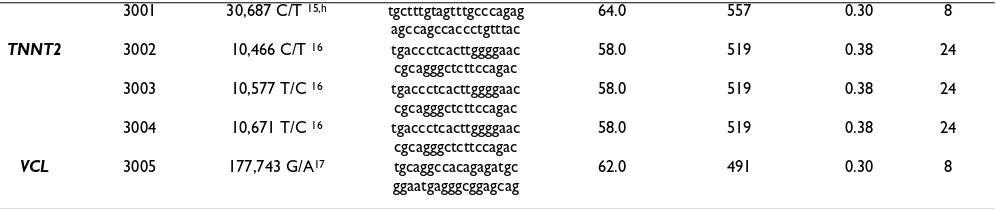
Table 2: Single Nucleotide Polymorphisms in the DCM candidate genes. For each SNP its origin, its primers and the PCR conditions,
and its informativity are listed. (Continued)
showed that both homozygous and heterozygous animals were among the dogs used for WAVE analysis. Conversely, sometimes WAVE analysis indicated potential presence of SNPs, yet sequencing of dogs with different WAVE pat-terns did not confirm these. This could be due to the sequencing procedure used.
In search of additional SNPs for canine ACTC and DES, genomic DNA fragments containing SNPs annotated by others (Table 2) were resequenced. After PCR amplifica-tion of these fragments, 1 µl of 1:15 diluted PCR product was used in a Tercycle big dye reaction with the PCR-primer for the ACTC SNP and a HPLC-purified M13 F-primer (5'-GTTTTCCCAGTCACGAC-3') for the DES SNPs. The Tercycle consisted of 25 cycles of 30 sec at 96°C, 15 sec at 55°C and 2 min at 60°C. After purifica-tion (Sephadex TM G50 Superfine, Amersham Bio-sciences), each product was processed with an ABI PRISM®
3100 Genetic Analyzer (Applied Biosystems). Five SNPs (ACTC 5,452G/A; DES 19,196C/T and 19,105G/A; LDB3
25,452A/G and TCAP 29.957 T/C) were identified by rese-quencing areas of earlier described SNPs (Table 2).
Of the total of 33 identified SNPs, 4 were in coding regions (DES 15,006C/T, LDB3 14,090C/T, TCAP
29,957T/C and TNNT2 10,466C/T). These exonic SNPs, however, did not cause polymorphisms at the amino acid level. Comparing the 33 newly discovered SNPs to the dog SNP database of the Broad Institute [39] showed 25 of our SNPs to be new, the remaining 8 SNPs matched SNPs present in the Broad database (see Table 2). This indicates that, in addition to the many SNPs that have become available by random sequencing of the dog genome,
many more canine SNPs exist. Our limited search for SNPs in 14 DCM candidate genes took place in a single breed, the Newfoundland dog. However, a high percent-age of SNPs found in one breed can be expected to be pol-ymorphic in other breeds too [40]. All identified SNPs were submitted to dbSNP and the respective accession numbers are listed in Table 2.
Detection of microsatellite polymorphisms
Simple DNA sequences composed of CA, GAAA or GA repeats were identified in the genomic contigs that con-tain the candidate genes or in neighbouring contigs. For
VCL, a polymorphic microsatellite became available through personal communication with P.Stabej (Table 3; a repeat was obtained from BAC RP81-251B5, isolated using methods as described in [28] with an overgo probe based on murine VCL exon 17, F-overgo CCAAGGTCA-GAGAAGCCTTCCAAC, R-overgo AAGTCAGGCTCCT-GAGGTTGGAAG). Primers were designed from the DNA sequence flanking the repeats and the forward primer was fluorescently labelled with 6-FAM or HEX. For some mic-rosatellites, a 3-primer protocol was used for the PCR amplification (Table 3), using an M13-tailed (GTTTTC-CCAGTCACGAC--- (5'-3')) F-primer, a 6-FAM-labelled M13 primer (GTTTTCCCAGTCACGAC (5'-3')) and a R-primer. Genotyping PCR reactions were incubated 12 min at 94°C, followed by 35 cycles of 10 sec at 94°C, 15 sec at Ta°C and 30 sec at 72°C, and a final step of 20 min at 72°C (Ta in Table 3). An ABI PRISM ® 3100 Genetic
BM
C Veterinary Research
20
07,
3
:2
8
http
://www.bio
m
e
dcent
ral.com/174
6-614 Page
(page nu
mber not
for
cit
a
tion pur
Gene Repeat Primers (5'-3') Forward; Reverse
Ta (°C) Detected alleles Informativeness2 Origin (bp ... of contig AAEX 010...) Distance to gene3
bp # PIC #chr
ACTC 15CA actccgaagaaggaagtcaac4
gttcccatctatgagggctat
57.0 234–238 2 0.17 10 bp 37,720/..13479 69.2 kb downstr. Stop
20CA ggaacaaggtgctgttagacc5
cacattccaccgagtaggc7
59.0 338–356 5 0.42 24 bp 5,945/..13478 intragenic (intron 4)
CAV1 13CA ccacagagctagaaagctacg4
tgttgcaaacaccctatgat
54.5 240–242 2 0.28 24 bp 39,315/...48546 8.2 kb downstr. Stop
CSRP3 15CA catgtcctgcaagttaatggt4
ggatttctattctgggtttcc
53.0 237–245 3 0.41 24 bp 38,367/..17412 2.8 kb upstr. Start
LMNA 16CA gggtggtagatgagcatttc6
gaagagaacaagtgggcaag
54.5 204–212 4 0.36 12 bp 5,128/..12734 9.3 kb downstr. Stop
18GAAA ggaagatgagactgttagaatgc5
caggccatgattacttttcc7
57.0 321–344 6 0.67 24 bp 26,754/..12735 22.6 kb downstr. Stop
MYH7 21CA gatatcctgggattaaagactgg5
ctattttgccctcttcatgg7
58.0 351–363 4 0.37 24 bp 1,418/..41098 36.6 kb downstr. Stop
TNNI3 20CAa tcaaacagggaaacctgaac6
gattattcagctcccagaacc7
57.0 297–301 3 0.38 24 bp 597/..53929 119.3 kb upstr. Start
20CAb ttccagttgattgtttctctgc5 gcggtttagcactgcattc7 59.0 302–306 2 0.08 24 bp 13,248/..53916 110.2 kb downstr. Stop
17GA tccaacctcagggtactgg5 catgccatggagctatgc7 59.0 304–312 3 0.37 24 bp 48,910/..53930 179.8 kb upstr. Start
TPM1 19CA8 actgtgtccagagtgcagcta4
gattgctagactggc
60.0 467–483 4 0.67 12 bp 88,113/..08742 6.5 kb downstr. Stop
VCL 15GAAA9 caatttcttttccaatcacattag 10
gccattttgcattctcttcaa
54.0 150–170 6 0.69 24 bp 12,680/..16406 88.6 kb downstr. Stop
1 Microsatellites for DES and SGCD were demonstrated in, respectively, [28] and [30]; 2 The informativeness of each SNP was described by its polymorphism information content (PIC), based on the
number of genotyped chromosomes (#chr) listed; 3 Based on genomic build 1.1; 4 F-primer fluorescently labelled with 6FAM; 5 Three-primer protocol used; 6 F-primer fluorescently labelled with
HEX; 7 extra tail on R-primer: GTGTCTT---- (5'-3') to promote addition of an Adenosine residue at the 3'-end of the complementary DNA strand; 8 Microsatellite demonstrated in [31]; 9 Personal
[image:6.612.77.722.76.482.2]markers), CAV1 (1), CSRP3 (1), LMNA (2), MYH7 (1),
TNNI3 (3) and VCL (1) (Table 3). The markers, mostly CA-repeats, showed multiple allele sizes (2–6 alleles/ marker) in a group of 16 Newfoundland dogs (Table 3). To describe the informativeness of our microsatellite markers, the polymorphism information content (PIC) was obtained based on the genotypes of unrelated found-ers of a family of Newfoundland dogs (Table 3). Accord-ing to [41], 2 of the 11 newly designed microsatellites were considered highly informative (PIC>0.50), 7 reason-able informative (0.25<PIC<0.50) and 2 slightly informa-tive (PIC<0.25) in the Newfoundland founder dogs. Besides the 11 polymorphic microsatellites, 2 other mark-ers were found to be monomorphic in the group of New-foundland dogs, but might be polymorphic in other breeds. This was a MYH7 CA-repeat (position 11,730 of [Genbank: AAEX010141100]) and a TNNI3 CA-repeat (position 17,739 of [Genbank: AAEX01053915]. An already available microsatellite for TPM1 [31] was shown to be highly informative in our group (Table 3).
The distance between the microsatellite and the corre-sponding gene was derived from the dog genome build 1.1 [42] and can be found in Table 3. This distance varied from zero for an intragenic microsatellite to 179.8 kb. The genomic locations of polymorphic microsatellites, already available for DES, SGCD, TPM1 and VCL, were determined. For DES a CA-repeat [28] was located at posi-tion 5,688 of [Genbank: AAEX01055032], 9.0 kb down-stream of the stop codon. For SGCD both a GAAA-repeat and a CA-repeat were available [30]. The first was located at position 76,364 of [Genbank: AAEX4801016848], the second at position 42,047 of the same genomic contig and both markers are in intron 7 of SGCD. For TPM1 a GA-repeat [31] was located at position 88,113 of [Gen-bank: AAEX01008742], 6.5 kb downstream of the stop codon. A polymorphic GAAA-repeat for VCL showed to be located at position 12,680 of [Genbank: AAEX01016406] in the dog genome, 88.6 kb downstream of the stop codon.
Conclusion
With the annotation of these 14 candidate genes for DCM and the identification of polymorphic markers, the genes can be evaluated for the involvement in breed specific DCM. The SNPs and microsatellites presented in this paper are a powerful tool to analyse linkage between the fourteen candidate genes encoding cytoskeletal proteins and DCM in the dog. The annotation of each gene facili-tates screening of these genes for mutations in naturally occurring canine DCM in specific breeds, potential mod-els for forms of human DCM.
Authors' contributions
ACW carried out the molecular genetic studies and drafted the manuscript. PAJL participated in the design of the study, helped to draft the manuscript. BAvO participated in the design of the study and was co-applicant for fund-ing. WEO participated in the coordination of the study. JDMcE conceived of the study and was main applicant for funding. She phenotyped the dogs, collected the samples and extracted the genomic DNA.
All authors had read and approved the final manuscript.
Additional material
Acknowledgements
This study and A.C. Wiersma were supported by a grant from the Kennel Club Charitable Trust Canine Health Foundation Fund, United Kingdom, and by the Faculty of Veterinary Science, University of Liverpool, United Kingdom. Special thanks to Francine Jury of the Centre for Integrated Genomic Medical Research (CIGMR, University of Manchester, United Kingdom) for her help with the WAVE analyses. Thanks to Polona Stabej (Faculty of Veterinary Medicine, University of Utrecht, The Netherlands) for the VCL microsatellite data.
References
1. Burkett EL, Hershberger RE: Clinical and Genetic Issues in
Familial Dilated Cardiomyopathy. Journal of the American College
of Cardiology 2005, 45:969-981.
2. Zhao YY, Liu Y, Stan RV, Fan L, Gu Y, Dalton N, Chu PH, Peterson K, Ross JJ, Chien KR: Defects in caveolin-1 cause dilated cardi-omyopathy and pulmonary hypertension in knockout mice. Proceedings of the National Academy of Sciences of the United States of America 2002, 99:11375-11380.
3. Nigro V, Okazaki Y, Belsito A, Piluso G, Matsuda Y, Politano L, Nigro G, Ventura C, Abbondanza C, Molinari AM, Acampora D, Nishimura M, Hayashizaki Y, Puca GA: Identification of the Syrian hamster
cardiomyopathy gene. Human Molecular Genetics 1997, 6:601-607.
4. Olson TM, Michels VV, Thibodeau SN, Tai YS, Keating MT: Actin mutations in dilated cardiomyopathy, a heritable form of
heart failure. Science 1998, 280:750-752.
5. Knoll R, Hoshijima ML, Bang ML, Hayashi H, Shiga N, Yasukawa H, Schaper W, McKenna W, Yokoyama M, Schork NJ, Omens JH, McCulloch AD, Kimura A, Gregorio CC, Poller W, Schaper J, Schultheiss HP, Chien KR: The cardiac mechanical stretch
sen-Additional file 1
Overview of the genomic organization of the canine ACTC (A), CAV1 (B), CSRP3 (C), DES (D), LDB3 (E), LMNA (F), MYH7 (G), PLN (H), SGCD (J), TCAP (K), TNN-I3 (L), TNN-T2 (M), TPM1 (N) and VCL (P) gene, in build 1.1 of the canine genome. The size of each coding exon, its actual location in bp in the respective genomic contig, 10 bp of DNA sequence at the 5'end and 3'end of the exon, 10 bp of the flanking intron and the intron sizes are listed. In case the coding sequence of a gene was covered by multiple Canis familiaris genomic contigs, the size of the intron covered by more than one contig was based on information of the respective chromosome. For the exons containing the start and the stop codon, the number of coding bp is listed as ORF (open reading frame); the location of the respective codon is listed between brackets.
Click here for file
sor machinery involves a Z disc complex that is defective in
a subset of human dilated cardiomyopathy. Cell 2002,
111:943-955.
6. Arimura T, Hayashi T, Terada H, Lee SY, Zhou Q, Takahashi M, Ueda K, Nouchi T, Hohda S, Shibutani M, Hirose M, Chen J, Park JE, Yasu-nami M, Hayashi H, Kimura A: A Cypher/ZASP mutation associ-ated with dilassoci-ated cardiomyopathy alters the binding affinity
to protein kinase C. The Journal of Biological Chemistry 2004,
279:6746-6752.
7. Kamisago M, Sharma SD, DePalma SR, Solomon S, Sharma P, McDonough R, Smoot L, Mullen MP, Woolf PK, Wigle ED, Seidman JG, Seidman CE: Mutations in sarcomere protein genes as a
cause of dilated cardiomyopathy. The New England journal of
medicine 2000, 343:1688-1696.
8. Hayashi T, Arimura T, Itoh-Satoh M, Ueda K, Hohda S, Inagaki N, Takahashi M, Hori H, Yasunami M, Nishi H, Koga Y, Nakamura H, Matsuzaki M, Choi BY, Bae SW, You CW, Han KH, Park JE, Knoll R, Hoshijima M, Chien KR, Kimura A: Tcap gene mutations in hypertrophic cardiomyopathy and dilated cardiomyopathy. Journal of the American College of Cardiology 2004, 44:2192-2201. 9. Murphy RT, Mogensen J, Shaw A, Kubo T, Hughes SS, McKenna WJ:
Novel mutation in cardiac troponin I in recessive idiopathic
dilated cardiomyopathy. Lancet 2004, 363(9406):371-372.
10. Itoh-Satoh M, Hayashi H, Nishi H, Koga Y, Arimura T, Koyanagi T, Takahashi M, Hohda S, Ueda K, Nouchi T, Hiroe M, Marumo F, Imai-zumi T, Yasunami M, Kimura A: Titin mutations as the molecular
basis for dilated cardiomyopathy. Biochem Biophys Res Commun
2002, 291(2):385-393.
11. Olson TM, Illenberger S, Kishimoto NY, Huttelmaier S, Keating MT, Jockusch BM: Metavinculin mutations alter actin interaction in
dilated cardiomyopathy. Circulation 2002, 105:431-437.
12. Li D, Tapscoft T, Gonzalez O, Burch PE, Quinones MA, Zoghbi WA, Hill R, Bachinski LL, Mann DL, Roberts R: Desmin mutation
responsible for idiopathic dilated cardiomyopathy. Circulation
1999, 100(5):461-464.
13. Burke B, Stewart CL: Life at the edge: the nuclear envelope and
human disease. Nature reviews Molecular Cell Biology 2002,
3:575-585.
14. MacLennan DH, Kranias EG: Phospholamban: a crucial
regula-tor of cardiac contractility. Nature Reviews Molecular cell biology
2003, 4:566-577.
15. Cohen N, Muntoni F: Multiple pathogenetic mechanisms in X
linked dilated cardiomyopathy. Heart 2004, 90:835-841.
16. D'Adamo P, Fassone L, Gedeon A, Janssen EA, Bione S, Bolhuis PA, Barth PG, Wilson M, Haan E, Orstavik KH, Patton MA, Green AJ, Zammarchi E, Donati MA, Toniolo D: The X-linked gene G4.5 is responsible for different infantile dilated cardiomyopathies. American Journal of Human Genetics 1997, 61:862-886.
17. Suomalainen A, Paetau A, Leinonen A, Majander A, Peltonen L, Somer
H: Inherited idiopathic dilated cardiomyopathy with multiple
deletions of mitochondrial DNA. Lancet 1992, 340:1319-1320.
18. McNair WP, Ku L, Taylor MR, Fain PR, Dao D, Wolfel E, Mestroni L, Group FCRR: SCN5A mutation associated with dilated
cardi-omyopathy, conduction disorder, and arrhythmia. Circulation
2004, 110:2163-2167.
19. Bienengraeber M, Olson TM, Selivanov VA, Kathmann EC, O'Coch-lain F, Gao F, Karger AB, Ballew JD, Hodgson DM, Zingman LV, Pang YP, Alekseev AE, Terzic A: ABCC9 mutations identified in human dilated cardiomyopathy disrupt catalytic KATP
channel gating. Nature Genetics 2004, 36:382-387.
20. Domanjko-Petric A, Stabej P, Zemva A: Dilated cardiomyopathy in Dobermanns, survival, causes of death and pedigree
review in a related line. Journal of Veterinary Cardiology 2002,
4:17-24.
21. Meurs KM, Miller MW, Wright NA: Clinical features of dilated cardiomyopathy in Great Danes and results of a pedigree
analysis. Journal of the American Veterinary Medical Association 2001,
218:729-732.
22. Tidholm A, Jonsson L: Dilated cardiomyopathy in the
New-foundland: a study of 37 cases (1983-1994). Journal of the
Amer-ican Animal Hospital Association 1996, 32:465-471.
23. Brownlie SE, Cobb MA: Observations on the development of congestive heart failure in Irish wolfhounds with dilated
car-diomyopathy. Journal of Small Animal Practice 1999, 40:371-377.
24. Tidholm A, Haggstrom J, Jonsson L: Detection of attenuated wavy fibers in the myocardium of Newfoundlands without
clinical or echocardiographic evidence of heart disease. American journal of veterinary research 2000, 61:238-241.
25. Dukes-McEwan J, Borgarelli M, Tidholm A, Vollmar AC, Häggström J: Proposed Guidelines for the Diagnosis of Canine Idiopathic
Dilated Cardiomyopathy. Journal of Veterinary Cardiology 2003,
5:7-19.
26. Brouillette JA, Andrew JR, Venta PJ: Estimate of nucleotide
diver-sity in dogs with a pool-and-sequence method. Mammalian
Genome 2000, 11:1079-1086.
27. Meurs KM, Magnon AL, Spier AW, Miller MW, Lehmkuhl LB, Towbin
JA: Evaluation of the cardiac actin gene in Doberman
Pin-schers with dilated cardiomyopathy. American journal of
veteri-nary research 2001, 62:33-36.
28. Stabej P, Imholz S, Versteeg SA, Zijlstra C, Stokhof AA, Domanjko-Petric A, Leegwater PA, Van Oost BA: Characterization of the canine desmin (DES) gene and evaluation as a candidate
gene for dilated cardiomyopathy in the Dobermann. Gene
2004, 340:241-249.
29. Stabej P, Leegwater PA, Stokhof AA, Domanjko-Petric A, Van Oost
BA: Evaluation of the phospholamban gene in purebred
large-breed dogs with dilated cardiomyopathy. American
jour-nal of veterinary research 2005, 66:432-436.
30. Stabej P, Leegwater PA, Imholz S, Versteeg SA, Zijlstra C, Stokhof AA, Domanjko-Petric A, Van Oost BA: The canine sarcoglycan delta gene: BAC clone contig assembly, chromosome assignment and interrogation as a candidate gene for dilated
cardiomy-opathy in Dobermann dogs. Cytogenet Genome Res 2005,
111(2):140-146.
31. Stabej P: Molecular Genetics of Dilated Cardiomyopathy in
the Dobermann Dog. In PhD thesis University of Utrecht, Faculty
of Veterinary Medicine, Dept of Clinical Sciences of Companion Ani-mals; 2005:183.
32. BLAST Dog Sequences section of NCBI BLAST
[http:www.ncbi.nlm.nih.gov/genome/seq/BlastGen/Blast Gen.cgi?taxid=9615]
33. Ensembl [http://www.ensembl.org]
34. NCBI Trace Archive database Mega BLAST search [http://
www.ncbi.nlm.nih.gov/blast/mmtrace.shtml]
35. Schott EJ, Robledo JA, Wright AC, Silva AM, Vasta GR: Gene organ-ization and homology modeling of two iron superoxide
dis-mutases of early branching protist Perkinsus marinus. Gene
2003, 309:1-9.
36. NCBI BLAST Assembled Genomes [http://
www.ncbi.nlm.nih.gov/BLAST/]
37. Guyon R, Lorentzen TD, Hitte C, Kim L, Cadieu E, Parker HG, Qui-gnon P, Lowe JK, Renier C, Gelfenbeyn B, Vignaux F, DeFrance HB, Gloux S, Mahairas GG, Andre C, Galibert F, Ostrander EA: A 1-Mb
resolution radiation hybrid map of the canine genome.
Pro-ceedings of the National Academy of Sciences of the United States of Amer-ica 2003, 100:5296-5301.
38. Rozen S, Skaletsky HJ: Primer3 on the WWW for general users
and for biologist programmers. In Bioinformatics Methods and
Pro-tocols: Methods in Molecular Biology Edited by: Krawetz S and Misener S. Totowa, NJ, Humana Press; 2000:365-386.
39. Dog SNPs - CanFam 1.0 database, BROAD Institute [http://
www.broad.mit.edu/mammals/dog/snp/]
40. Parker HG, Kim LV, Sutter NB, Carlson S, Lorentzen TD, Malek TB, Johnson GS, DeFrance HB, Ostrander EA, Kruglyak L: Genetic
structure of the purebred domestic dog. Science 2004,
304:1160-1164.
41. Botstein D, White RL, Skolnick M, Davis RW: Construction of a genetic linkage map in man using restriction fragment
length polymorphisms. American Journal of Human Genetics 1980,
32:314-331.