0022-538X/87/030667-06$02.00/0
CopyrightC)1987, American Society forMicrobiology
Unusual Transcripts in La Crosse Virus-Infected Cells and the Site
for Nucleocapsid
Assembly
RAMASWAMYRAJU ANDDANIEL KOLAKOFSKY*
Department of Microbiology, University of Geneva Medical School, 1211
Geneva,
SwitzerlandReceived25August 1986/Accepted17November 1986
The LaCrosse virus S genome segment is known to code for two plus-strand transcripts, free S-mRNA (nucleotide
[nt]
-15 to 886) and encapsidated antigenome RNA (nt 1 to 983). Early in infection only these plus-strand transcripts could be detected, but at later times plus-strand RNAs representing nt 1 to 886 in an encapsidated form and nt -15 to 983 as a free RNA could also be seen, as well as S-mRNA in an encapsidated form.TheencapsidatedS-mRNA became relatively important at later times because the free S-mRNA turned overrapidly after 5 h postinfection. The existence of these unusual RNAs and their presence as either free orencapsidatedspecies suggests that the sitefor nucleocapsid assemblyislocatedat the 5' ends of the genome and antigenome chains.
La Crosse virus (LAC) is a member of the California
encephalitis serogroup of the insect-transmitted bunyavirus family (13). The genome of these viruses consists of three segments of single-strandedRNAofnegative polarity, each
contained within a separate nucleocapsid (NC), which are
labeled small(S), medium (M), and large (L). The viral NCs have helical symmetry and often appear in the electron
microscope ascircularandsupercoiled structures (9).
The mechanism by which bunyavirusesinitiate transcrip-tion of their mRNAs is remarkably similarto that of influ-enza viruses (8). Bunyavirus S-mRNAs in vivo contain 5'
nontemplated extensions ofca. 15 nucleotides (nt) in length
whichareheterogeneous in sequence(2, 12),presumably the
result ofahost cell primer usedto initiate transcription. In
vitro, purified LAC virions were found to contain a
tran-scriptase which is stimulated by natural mRNAs such as
alfalfa mosaic virus (AlMV)RNA4,whichwasshown to be
actingas aprimer. LAC virionswerealso foundtocontaina
methylated cap-dependent endonuclease which cleaves AIMV RNA 4 at thepositions expected from the length of the 5'extensions foundon thetranscriptsmade invitro(11).
Bunyaviruses, like influenza viruses,thusapparentlysnatch
capped
oligonucleotides
from host cell mRNAs to prime their mRNA synthesis and to provide it with a cap grouprequired for efficient translation. However,unlike influenza viruses, LAC transcription takes place in the host cell cytoplasm rather than in the nucleus (16), in which ituses a
stable pool ofmRNAs as substratesforprimers, consistent withtheknownimmunityofbunyavirusreplication todrugs
which disrupthostcell mRNAsynthesis (10). The 3' endof
theLACS-mRNA, which isapparentlynot
polyadenylated,
hasbeenmappedtoposition 886,ca.100 ntbeforethe endof the negativegenome template (12).
Since the S-mRNA is only some 80 nt shorter than the
antigenome RNA and does not selectively bind to olig(dT) cellulose, we have taken advantage of the fact that
antigenomes, like genomes, are almost
exclusively
found intracellularly in nucleocapsid structures as the basis fortheir separation from mRNAs. Intracellular mRNAs were routinelyseparatedfromgenomes and
antigenomes
by
cen-*Correspondingauthor.
trifuging cytoplasmicextractstoequilibriumonCsCl density gradients under conditions in which the unencapsidated
mRNAs pellet, whereas the nucleocapsids are found as a
sharp bandattheirbuoyant density of1.31g/ml.Whenthese separated fractionswereexaminedbyNorthernblotanalysis byusingstrand-specific riboprobes,thebandedmaterialwas
found to contain all the intracellulargenomes and the vast
majority ofthe antigenomes, whereas the pelleted material contained all the mRNA (14). More recently, however, we havefoundthatunder someconditions thebanded material unexpectedly contained asizableproportion ofthe
intracel-lularS-mRNA(Fig. 1). Thisunusualfinding formsthebasis ofthe presentcommunication.
MATERIALS AND METHODS
Isolation of cytoplasmic RNAs. Confluent cultures of
BHK-21 cells in 10-cm-diameterpetrie disheswereinfected with 20 to 50 PFU of LAC virions per cell. At the times indicated, the cells were harvested by being scraped into
phosphate-buffered
saline
and were recovered by centrifu-gation.Cytoplasmicextractswereprepared byvortexing107
cells in0.2 ml of 0.5% Nonidet P-40-0.15 MNaCl-10 mM Tris hydrochloride (pH
7.4)-i
mM EDTA (except as indi-cated fortheexperiment
inFig.
2) followedby
centrifugation
for4 minat4,000 x g. When
denaturing
agents were used (see Fig. 4), thecytoplasmic supernatant
wasimmediately
mixed with 6 M
guanidinium-thiocyanate-1%
sarcosyl-0.2
M
P-mercaptoethanol
(5).Alternatively,
either 1 mg of heparinper ml or 2 mMvanadyl ribonucleosidecomplex
was added to limitdegradation
whendenaturing
agents were omitted. The extracts were thencentrifuged
on20 to 40% preformed CsCl densitygradients
for 16 h at45,000
rpm in the SW60 rotor. In the absence ofdenaturing
agents, theNCs formedasharp visible band in thecenterof the
gradient
(1.31 g/ml) and were removed with a
hypodermic
syringe
fromthesideofthe tube.This CsClsolutionwasthenmixed with 2volumesoftheabove
denaturing
agentsplus
40 ,ug of carrieruninfected BHK cellRNA,
and the NC RNAswere recovered bycentrifugation
through
aCsClgradient.
Northernblotanalysis.The RNA
samples
wereheated for 2minat900C
in80% formamideand0.1%xylene
cyanol
FF andelectrophoresed
on either a 4.0or2.8%,
1.5-mm-thick 667on November 10, 2019 by guest
http://jvi.asm.org/
Pst 764 Pst
196
4...e
[image:2.612.138.502.69.240.2] [image:2.612.331.529.529.620.2]4..
P
F. ......
Alu 896
4....0
...
4.. 5'
-l5 + oligomer 51
1
m
5'S::
-15
3'
lWw Yo3
mRNA
886
u)
983antigenome .Q
983~ ~
1-886
886~~ c
8-)15-983 _
983 Jo
983 gernome
-FIG. 1. Schematic diagram of LAC S-RNAs. The LACS-genomesegmentis shown inthemiddle,withopen bars above thelineindicating theoverlapping N and NS coding regionsandthe numbers belowindicating nucleotide positions.Aboveareshown the variousriboprobes used, with arrowheads showing the direction of transcription from their SP6plasmids andkey restriction sites used in theirgeneration.Below areshown theS-genome segmenttranscripts found in infected cells. Thick andnarrowlines indicate abundantandrarespecies,respectively, with the numbers at the left showing the positions of their5'and 3'ends. The numbers below the lines show the nucleotidepositionsof the ends of the chains. The closed circle followedbyanopen bar represents thenontemplatedcapped primer.Thethickarrowlabeledoligomer shows theposition of theoligodeoxynucleotide primer usedtolocate the positions ofthe 5'ends(Fig.5B).
polyacrylamide gel containing 8 M urea (17). The gel was
then soaked for 10 min in TAE (10 mMTris, 5 mM sodium acetate, 0.5 mM EDTA, pH 7.8) and electroblotted onto zeta-probe membranes in the same buffer. The membrane was dried, prehybridized with 5x SSC (lx SSC is0.15 M
NaCl plus 0.015 M sodium citrate)-5x Denhardt solution-50% formamide-200 ,ug of yeast RNA per mlfor4 h,andthenhybridized inthe samesolutioncontainingca. 5 x 10⁶ cpm of
[a-32P]UTP
(400Ci/mmol)-labeled riboprobesfor8 to 12h. Thefinalstringent washwasfor1h at70°Cin 0.1 x SSC.
Primer extension analysis. The oligonucleotide
represent-ingnt38 to50 asminus-strandDNAwas 5' end labeled with
[_y-32P]ATP
and T4 polynucleotide kinase, separated on an18% sequencing gel, eluted, and ethanol precipitated with the template RNAs. The pellets were suspended in 6 ,ul of
water, heated at 90°C for 2 min, and quick chilled. The
reaction conditionswereadjustedto 50 mMTris
hydrochlo-ride (pH 8.3)-8 mM
MgCl2-80
mM KCl-20 U of murineleukemiavirus reverse transcriptase in atotal volume of 20
,ul and incubated at 38°C for 45 min. The reaction mixture was then phenol extracted, ethanol precipitated, and
ana-lyzed on a9%sequencing gel.
RESULTS
As described in the Introduction, when intracellular
nu-cleocapsid RNA isolated from CsCl density gradients is
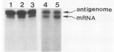
examinedbyNorthernblotanalysis with S genome negative-strandriboprobes,only the S antigenome RNA can normally bedetected. However, in some cases,S-mRNA-sized mate-rial can also bedetected (Fig. 2). Since there is little or no
precedent among minus-strand RNA viruses for the
encapsidation ofmRNAs, we first suspectedthat the pres-ence ofmRNA in the nucleocapsid fraction was due to a
nonspecific contamination, e.g., free mRNA which was
sticking
tothe NCs or sticking to other cytoplasmic materialwhoseaggregate buoyant density was similar to that of viral NCs. In anattempt to limit suchcontamination,cytoplasmic
extracts were prepared from replicate samples of infected cells in the presence of increasing concentrations of salt which mightpreventformation of such complexes. Varying
the NaCl concentrationfrom 0to0.75 M in the presence of 1 mMEDTA, however, had no effect on the relative amount of mRNA in the banded material(Fig. 2, lanes 1 to 3). The presenceof EDTA in the extraction buffers should also have eliminated complexformation dueto Mgsaltbridges.
Simi-larly, the use oflysolecithin rather than Nonidet P-40 to
solubilize membranes during sample preparation had no
effect(data not shown).
We also argued that if the mRNA wassimply stuck to the NC, the buoyant density of the complexes containing addi-tionalcomplements of RNA should besufficiently higher to allow thesecomplexestobe separated from free NCsduring
centrifugation. For example, assuming buoyant densities in CsCl of 2.1 and 1.28 g/ml for RNA and the NC protein,
respectively (3),andthat, like Sendai virus NCs or tobacco
1
2
3
4
5
'I 'W
-0--MRNA
FIG. 2. Effect ofpreparation conditionson amountofS-mRNA in CsCl-banded NCs. Cytoplasmic extractsof LAC-infected BHK cellsat8 hp.i. wereprepared with Nonidet P-40extraction buffer containing either 0, 0.15,or0.75 MNaCl (lanes1to 3,respectively) andtheir NCRNAswereisolatedandanalyzed onNorthern blots of a4%polyacrylamide gelwitha negativeriboprobe representing nt 1 to196(seeFig. 1).In aseparateexperiment, half of the NCs isolated from cells at 20 hp.i. werecentrifuged to equilibrium on a second CsCl density gradient. Lanes 4 and 5 show the Northern blot analysis as above for the once- and twice-banded NC RNAs, respectively.
I. Sam 4
ani
on November 10, 2019 by guest
http://jvi.asm.org/
pellet
(p)
band(NC)
5 8 12 16 20 3 5 8 12 16 20 P
_qlom
0
a)
(U)
4)
l._
81
2-51
mRNA( P)
I
151
mRNA(NC)
4 12
** a1-wMRNA
-"Kl-886
20 4 12
[image:3.612.136.474.67.366.2]harvest
time(hrs)
FIG. 3. Kinetics of encapsidated and unencapsidatedplus-strand S-RNAaccumulation inLAC-infectedcells. Fourpetrie dishes eachof LAC-infected BHKcellswereharvestedatthe timesindicated, and 5% of their CsCl pelleted (P) and15%of their banded (NC)RNAswere
analyzedonNorthernblots ofa2.8%polyacrylamide gel by usinganegativeriboprobe representingnt1%to764 (seeFig. 1). Lanepshows aduplicate of the 3-h-pelletedRNAas amarker. Below is shown thedensitometricanalysisof theautoradiogram normalized forthedifferent
amounts of pelleted andbanded material used. Note that the ordinate ofthe right-handgraph has been amplified 16-fold relativeto the left-handgraph. The closed circles andopentrianglesatthe baseline of thelower right-hand graph markedPrefertotheantigenome and1
to886RNAs, respectively, found in the CsClpellet.
mosaic virus, LAC NCs are composed of 96% protein and
4% RNAby weight, free LAC NCs would have a buoyant
density of 1.31, whereas an NC containing only one addi-tional complement of RNA would bandat 1.35 g/ml. Since these large structures form extremely sharp bands even in relatively shallowCsCl density gradients,aseparatebandat
1.35 g/ml should have been visible, but no satellite band below the free NC band could be detected from cells harvested at any time postinfection (p.i.) (see below). In addition, when the hypersharp band from the preparative gradient was carefully removed and subjected to a second
cycle ofCsCl density gradient centrifugation andthe RNAs weresimilarly examined, the second cycle ofcentrifugation
was found not toeliminate any of the mRNA found in the banded material (Fig. 2,lanes 4 and 5). Since onlythe viral
N protein can be detected by dye staining of the banded material run on protein gels (H. Lindsey-Regnary, Ph.D. thesis, Emory University, Atlanta, Ga., 1983; M. Hewlitt,
personal communication; results not shown), it would
ap-pearthat under someconditions, significant amountsof the
intracellular S-mRNA canbefound in bona fide NC struc-tures.Unfortunately, since, unlike Sendai virusorvesicular
stomatitis virus NCs, the RNA within LAC NCs is highly
sensitive to RNase A (9), this criterion cannot be used to further characterize the S-mRNA-containing structures
which cobandwith viralNCs inCsCldensity gradients.
As mentionedabove, mRNAs of other minus-strand RNA viruses are not known to be encapsidated. In the case of
vesicular stomatitis virus, interestingly, two groups have
neverthelessdetected mRNAs associated with the Nprotein late in infection (7, 15). However, unlike the encapsidated LAC S-mRNA described above, these vesicular stomatitis virus complexes could clearly be separated from genome NCsonCsCl density gradients, since they containedalesser
amountofprotein relativetoRNA.
Kinetics of S-RNA accumulation in LAC-infected celis.
Replicate samples ofcellswere harvestedatdifferent times after infection, and their cytoplasmic extracts were again
separated into CsCl-pelleted and -banded material. This
material was then examined by Northern blot analysis as above, exceptthat 2.8% polyacrylamide gels were used to
increasetheresolutionbetween theS-antigenomeRNA and
the mRNA. When the pelleted material was examined,
S-mRNA was clearlydetectableby 3 hp.i. and reached its
maximumat5 hp.i. However,after 5 hp.i.thesteady-state
concentration of the S-mRNAunexpectedlydecreased such
that itwas atthe limit of detection by12 h p.i. Fig. 3also
showsthat the S-mRNA wasthe onlyband detectedinthe
pelleted material by the probe. When the banded material
from the same infection was examined, at 3 h p.i. only antigenomeRNA could be seen.At 5 hp.i., someS-mRNA
could nowalso be detected. Curiously, by 8h p.i., aband
hrs(peij):
3
-.wantigenome
on November 10, 2019 by guest
http://jvi.asm.org/
196-764 (+)
' 3 5 8 12 16 20 24'
to4amp
genome
* I ~antigenomet
. .
/mRNA
+A 1-886
4 12 20
harvest time(hrs)
FIG. 4. Kinetics of total S-RNA accumulation in LAC-infected
cells. One-fourth of the
cytoplasmic
extractsfrom the experimentshown in
Fig.
3 were denatured directly withguanidinium-thiocyanate,
and the total RNAs were pelleted through a CsCldensity gradient. Duplicate samples
of eachpelletedRNA(amount-ingto0.2ofa
petrie
dish) werethenanalyzed
byNorthern blotting ofa2.8%polyacrylamide
gel.
Thereplicate
blotsthusobtainedwereprobed
with eitherpositive
(+) ornegative
(-) riboprobesrepre-senting
nt196to764,asindicated. The densitometric analysis of theautoradiogramis shown below.
just
belowthe S-mRNAwasalso nowdetectable, andthesethree bands increased
slowly throughout
the remainder of the infection.Inthe above
experiment,
the RNA in the bandedmaterialfrom the CsCl
gradient
was recovered by mixing the CsClsolution with
guanidinium-thiocyanate
todissociate the NCand
repelleting
the RNA in the presence of carrier RNAthrough
asecondCsClgradient.
This secondultracentrifuga-tion
step
may have led tounequal
recoveries ofthe RNAsbetween
samples,
since itappeared
unlikelythatantigenomeRNA,
forexample,
would continue to accumulate after8 hp.i.
when the S-mRNA level was barely detectable (theS-mRNA codes for the NC
protein).
To control for thispossibility,
total RNA was also recovered from the same infectionby
mixing
thecytoplasmic
extracts directly withdenaturing
agents beforecentrifugation,
such that all theviral RNAs are now recovered in the
pellet
in a single step(14).
In thisexperiment,
the Northern blots were alsoexamined with
riboprobes
for bothplus
and minus strand S-RNAs. As shown inFig.
4,
thesteady-state
concentrationof both genomes and
antigenomes
reached amaximum at 5to 8 h
p.i.
and then increasedonly
very slowly throughoutthe remainder of the infection. The continuous increase in
the accumulation of
antigenomes
found in the Fig. 3 wouldthusappeartobe duetoincreasedrecoveriesoftheRNAsat
later times
p.i.
for reasons whichare notclear. The patternof
S-mRNA,
on the otherhand,
did not change from theprevious experiment.
The S-mRNA reachedits maximumat5h
p.i.
and thenagain
decreased dramatically from thenon. The bandjust
below the S-mRNAis alsodetectablehere,but its ratio relative to the S-mRNA is lower since in thisexperiment
both thepelleted
and banded material arecom-bined,
and the bandjust
below the mRNA cannot bedetected in the
pelleted
material at any time p.i.The above
experiments
demonstrate that the S-mRNA-sizedmaterial whichbands in CsCldensity
gradients
can beresolved into two RNA
species,
one which migratespre-cisely
with S-mRNA(Fig.
3,
lanep)
and onewhichmigrates
slightly
faster,
called 1 to 886(see
below).
Neither ofthesespecies
can be detectedearly
ininfection. At 5 h p.i. whenfree
(i.e.,
unencapsidated)
S-mRNAaccumulation and viralprotein synthesis
aremaximal,
theencapsidated
S-mRNA and the 1 to 886 RNA areextremely
minor species,repre-senting
less than 2% of theunencapsidated
S-mRNA. Atlater times
p.i.,
however,
sincetheunencapsidated
S-mRNA has turned over almostcompletely,
the encapsidatedS-mRNA and 1 to 886 RNA become the predominant
S-mRNA-sized material intracellularly.
Mapping
the 5' and 3' ends of the encapsidatedRNAs. Tofurther characterize the
plus-strand
S-RNAs which wereencapsidated,
NC RNAsisolated from cells at 20 hp.i. wererecovered and
electrophoresed
on a 2.8% acrylamide gelalong
with radiolabeled S-mRNA made(1)
asamarker. Theantigenome
RNA was isolated from thegel
separately, but noattempt
was made toseparate
the S-mRNA from the bandjust
below it because of theircloseness,
and both the bandswere isolated
together.
Unencapsidated
(i.e.,
CsCl pellet) S-mRNA wassimilarly
isolated fromthe same gel to serve asa marker. To determine the
position
of the 5' ends of theseplus-strand
RNAs,
a radiolabeledoligodeoxynucleotide
rep-resenting
nt38 to 50 as minus-strand DNAwas extended on thesetemplates
with reversetranscriptase,
as well as on tRNA as a control. As shown inFig.
5B,
the primerextended on the
antigenome
RNAto asingle
band (position +1)
and on thepelleted
mRNA to a ladder of bands atpositions
-12 to-18,
aspreviously
described (11). Whenthe
primer
was extended on theencapsidated
S-mRNA andthe RNA
just
below
it,
both
a band atposition
+ 1 and theladder of bands
atpositions
-12to -18
were found. Themost
straightforward
interpretation
of these
results is thatthe RNA in the banded material which comigrates with
authentic
(i.e.,
unencapsidated)
mRNA contains anontem-plated primer
12 to 18 nt inlength
at its 5' end like thepelleted
S-mRNA,
whereas the bandjust
belowthe mRNAstarts at
position
+1 like theantigenome
RNA. Thediffer-ence in
migration
of these two bands is alsoconsistent withadifference of 12 to 18 nt.
The 3' ends of the
encapsidated
RNAs wereexamined byNorthern blot
analysis
with two
riboprobes
representing nt764
to 983 and 896 to 983 asminus-strand
RNA(the 3' end ofthe S-mRNA
maps
at
position
886,
see
Fig.
1).
As shown inFig.
5 theriboprobe representing
nt 764 to
983 annealed tothe
antigenome
RNA,
theS-mRNA,
and the bandjust belowit,
whereas the
riboprobe representing
nt
896
to
983annealedonly
to theantigenome
RNA.These results indicate
that theencapsidated
S-mRNA and
the
band
just
below
it terminatebefore
position
896. Taken
together,
the above
evidencesuggests
that
(i)
a small
fraction of
the bona
fide S-mRNAcan
be
encapsidated
and
(ii)
the
encapsidated
RNA whichmigrates just
ahead of
the S-mRNA
represents
a positivetranscript starting
at
position
+1
and
most
probably
endingat
position
886.Unusual
LAC S
positive
transcripts.
The
RNA whichbegins
atposition
1and
ends at 886 is
presumably
neither anprobe: 196-764 (-)
hrsp.i.i: 3 5 8 12
1-983-
-mnRNAR
1
-888= , r 4m 4
-16 20 24
6
4
C')
c
(L 2
on November 10, 2019 by guest
http://jvi.asm.org/
[image:4.612.57.300.62.327.2]mRNA (it would not be expected to have a cap group nor
could it be translated as an encapsidated species) nor an
antigenome (it is incomplete) or a defective interfering genome (its complement as minus-strand RNA cannot be
detected [Fig.
SA]).
This RNApresumably resultsfrom the polymerase starting the chain as an antigenome but ending it as an mRNA. We therefore examined whether a moleculerepresenting theinverse parallel, namely a transcript which starts like an mRNA (position ca. -15) but ends like an
antigenome (position983) could also be detected in infected cells. Indeed, acandidate forthis molecule could be found
butonly in the pelleted material. As shown in Fig. 6, when
theNorthern blotofthe pelleted material from two separate
infectionsisoverexposed, adoubletband above the mRNA canclearly beseen,the lower band of which comigrates with
antigenomeRNA, and the upper band migrating at a position
consistentwithit containing ca. 15 extra nucleotides. Both ofthese bands reachtheir maximumswith themRNA or just
after it and like the mRNA in the pelleted material are
unstablewith time p.i. Noattempt was made to characterize
themfurtherdue totheir extremely lowabundance.
DISCUSSION
The work reported here has described two unusual S genome positive-strand transcripts in LAC-infected cells (see Fig. 1). One transcript starts like an antigenome at
position +1but ends likethe S-mRNA atposition 886 (called 1 to886).Theothertranscriptstarts on a primer of ca. 15 nt in length likethe mRNA but like theantigenome continues on to the end ofthe genome template (called -15 to 983).
The -15to983transcript is foundonly asan unencapsidated
species and never represents more than a smallfraction of
1% of theS-mRNA at anytimeduringtheinfection. Like the
unencapsidated S-mRNA, the -15 to 983 transcript is
un-stable withtime and is drasticallyreduced later ininfection.
The 1 to 886 species, on the other hand, is foundonly as an
encapsidated form and like the minor form of encapsidated S-mRNA appears to be stableatlater times ofinfection.
The most plausible explanation forthese findings is that
both the 1 to 886 and -15 to 983 RNAs are made at
extremely lowfrequencies relativetothe S-mRNA and that the relative steady-state concentrations of the various S
positive transcripts laterininfectionsimply reflectsthe fact that the encapsidated species are stable, whereas the
unencapsidated RNAs turn over rapidly with time. In the case of the
encapsidated
form ofthe mRNA, ifan exceed-inglysmallfraction of the freepopulation becomesencapsid-ated at a constant rate whereas the remaining
unencapsid-ated molecules turned over rapidly with time, this would
explainwhy late ininfectionthere isactuallymore
encapsid-ated thanunencapsidatedmRNAinthe cell. In this
view,
the -15 to 983 and the 1 to 886 transcripts are the result of polymerase errors, whereas the encapsidated mRNA is aresult ofan encapsidation error, all of which occur
infre-quently. Theynevertheless indicatethatatleastthis part of theviralreplication machinery does notfunctionwith abso-lute specificity.
The existence ofthe 1 to 886
transcript
and its relativepredominance late in infection may also
explain
the unex-pected results ofBouloy
et al. (4)working
withGermiston virus, another member of the bunyamwera virus genus. These workers reportedtheexistence ofan S-mRNA-sizedtranscript which couldbe
capped
in vitroand thuscontained a di- ortriphosphate
at its 5' end andsuggested
that thistranscriptmight functionas amRNA. Inthese
experiments,
PROBE: (-) (-) (+)
764-983 896-983 764-983
SAMPLE: P NC P NC P NC
1-983---& s
H-15-886
t-86
2
z
I
0E
CL
4 4)
z
.o
E c
z
-z
E
4
z M
-18[
-121
f
+1-FIG. 5. (A) Mapping the 3' ends of the encapsidated S-RNAs. Triplicate samples of CsCl pelleted (P) and banded(NC)RNAfrom cells harvested at 5 and 20 h p.i., respectively were analyzed on
Northern blots ofa2.8%polyacrylamide gel by using the riboprobes indicated. (B) Mapping the5' ends of the encapsidated S-RNAs. CsClpelleted and bandedRNAsfromcells harvestedat5and 20h p.i., respectively, were separated on a 2.8% polyacrylamide gel along with radiolabeled S-mRNA made in vitro (1)as amarker.The positions correspondingtothe SantigenomeRNAand theS-mRNA
(including the band just below it)wereexcised from thegel, andthe RNA waselectroeluted and recovered byethanol precipitation in
the presence of carrier tRNA. A 5'-end-labeled oligonucleotide
representingnt38to50asminus-strandDNA(seeFig. 1)wasthen primer extendedontheseRNAsalong withtRNAas acontrol.The products were separated on a 9% sequencing gel. The genome
positionsof the 5' endsof thetemplateRNAsareindicatedonthe
left.
S-mRNA which contained 5'
nontemplated primers
would not have been scored, and so the relative amounts of the mRNA-sizedtranscripts
with different 5' ends could notbe determined. Morerecently, work from the samelaboratory (6) has shown that this material can also beseparated
intotwobands, theuppermost of whichappearedtocontain ca. 15 nontemplated nucleotides at its 5'
end,
whereas the bottomband didnot. Itthus seems reasonableto conclude that the Germistontranscript
which contains a 5' triphos-phateis theequivalent
of the LAC 1to886transcript.
In thison November 10, 2019 by guest
http://jvi.asm.org/
[image:5.612.354.527.69.443.2]hrs
(pDi):
(-)15-983 o-1-.983
(-)15-886-_
a
2
5
8
12
b
2
4
6
8
12
- .U ~P-
-.:
FIG. 6. Detection of a -15 to 983 positive S transcript in LAC-infected cells. Twopetriedishes each of LAC-infected BHK cellswere harvested at thetimes indicated, and 10% of the
CsCl-pelletedRNAsfrom twoseparateinfections(aandb)wereanalyzed onNorthernblotson2.8%polyacrylamide gels by usinganegative
riboproberepresentingnt 1 to195.
case, it would not be functional as a mRNA whether it is
capped or uncapped, if indeed this transcript were also
encapsidated.
Regardless oftheprecise mechanism oftheirgeneration,
theexistenceofthe -15to983 and1 to 886transcriptsand their presence intracellularly as unencapsidated and
encapsidated species, respectively, offers some insight on
thelocationof thosesequenceswhichdetermine
encapsida-tion. Since the vast majorityofthe mRNA is not
encapsid-ated when mRNA accumulation is maximal, whereas
antigenomesare encapsidated,the encapsidationsite of the
antigenome could have beenlocated betweenpositions 886 and 983. However since the 1 to 886 transcript is also
encapsidated, this would appear to eliminate these
se-quences nearthe 3' end of theantigenome. The next most
likely site is therefore the 5' end, since both genomes and
antigenomes are encapsidated and only at the two ends of thechainsaretheirsequenceshighlyconserved. Inthiscase,
one explanationforwhy the vastmajority of the mRNA is notencapsidated when mRNA accumulation is maximal is that this recognition sequence at the 5' end has now been
placed out of context by the addition of the ca. 15 nt
transcription primer, and the Nproteincannot now initiate
encapsidation herefor this reason. Alternatively, the pres-enceofacap groupincloseproximitytothissequencecould
bind a cytoplasmic cap-binding factor, for example, and
therebymasktheinitiation site.The presenceof theprimer, however, does not appearto confer complete immunity to
encapsidation, but it is relatively effective; when mRNA accumulationis atits maximum,less than2%of the mRNA has beenencapsidated (Fig. 3).Moredirectexperimentswill
be required, however, to determine whetherthe conserved
sequences at the 5' ends of the antigenome and genome
chains do indeed contain the site for the initiation of NC
assembly.
Finally, sincethe Northernblots ofFig. 4 wereanalyzed
with riboprobes made with [32P]UTP of identical specific activity, the relative specific activities of the positive and negative probes is then dependent only on their uridine
composition. From the data in Fig. 4, we can therefore
calculate the ratioofgenomesto antigenome intracellularly
to be 5.8. Curiously, this number does notagree wellwith
the 60% self-annealing ofS-sized NC RNA previously
de-termined by solution hybridization (16), which suggests a
ratio of only 2.3. Further, the ratio of 5.8 may be an
underestimate, since thegenomebands mayhave exceeded
thelinearrangeofthe filmontheexposureshown in Fig. 4.
Onepossible explanationforthisdiscrepancy is that the NC
RNA used in the solution hybridization experiment came
from cellsharvestedat8 hp.i. andwasseparated into
S, M,
and L RNAs by velocity sedimentation which would not
have separated S-mRNA, 1 to 886, or antigenome RNA. Inspectionofthe datain
Fig.
3 showsthatat8hp.i.,
the NCbandmaterial containsasmuch S-mRNA and 1to886RNA as antigenome RNA, all of which would anneal to the negative genomeRNAandtherebydouble the
self-annealing
value. Our more recent data would then indicate that
genomesare atleast 5.8timesasabundantas
antigenomes
inLAC-infected BHK cells. Further, since bothgenomes and
antigenomes appear stable throughout the infection, their accumulation would then reflecttheirrate of
synthesis.
LITERATURE CITED
1. Bellocq, C., R. Raju, J. Patterson, and D. Kolakofsky. 1987. TranslationalrequirementofLaCrosse virus S-mRNA
synthe-sis: Invitrostudies. J. Virol. 61:87-95.
2. Bishop, D. H.L.,M. E.Gay,and Y.Matsuoko. 1983. Nonviral heterogeneous sequences are present at the 5' ends ofone
species ofsnowshoe harebunyavirus S complementary RNA. Nucleic Acids Res. 11:6409-6418.
3. Blumberg,B.M., C. Giorgi, K. Rose, and D. Kolakofsky.1984. Preparation andanalysis of the nucleocapsidproteins of vesic-ular stomatitisvirus and Sendaivirus, and analysisof theSendai virusleader-NPgene region. J. Gen Virol. 65:769-779. 4. Bouloy, M., P. Vialat, M. Girard, and N. Pardigon. 1984. A
transcriptfrom the S segmentof the Germiston bunyavirus is uncappedand codes for the nucleoprotein and anonstructural protein.J. Virol. 49:717-723.
5. Chirgwin, J.M., A. E. Przybyla,R.J. MacDonald, and W. J. Rutter. 1979. Isolation of biologically active ribonucleic acid from sourcesenriches in ribonuclease. Biochemistry 18:5294-5300.
6. Gerbaud, S.,N. Pardigon, P.Vialat, andM. Bouloy. 1986. InD. Kolakofsky and B. W. J. Mahy (ed.), Biology ofnegativestrand viruses. Elsevier BiomedicalPress, Amsterdam.
7. Grubman, M. J., and D. A. Shafritz. 1977. Identification and characterization of messenger ribonucleoprotein complexes from vesicular stomatitis virus-infected HeLa cells. Virology 81:1-16.
8. Krug, R. M.1981. Priming ofinfluenzaviral RNAtranscription bycapped heterologousRNAs.Curr. Top. Microbiol. Immunol. 93:125-149.
9. Obijeski, J. F., D. H. L. Bishop, E. L. Palmer, and F. A. Murphy. 1976. Segmented genome and nucleocapsid of La Crosse virus. J.Virol. 20:664-675.
10. ObiJeski,J. F., and F. A. Murphy. 1977. Bunyaviridae: recent biochemical developments. J. Gen. Virol.37:1-14.
11. Patterson, J. L., B. Holloway, and D. Kolakofsky. 1984. La Crosse virions contain a primer-stimulated RNA polymerase and a methylated cap-dependent endonuclease. J. Virol. 52: 215-222.
12. Patterson, J. L., and D. Kolakofsky. 1984. Characterization of La Crossevirus small-genome transcripts. J. Virol. 49:680-685. 13. Porterfield, J. S., J. Casals, M. P. Chumakov, S. Y. Gaidamovich, C. Hannoun, I. Holmes, M. C. Horzinek, M. Mussgay, N. Oker-Blom, and P. K. Russel. 1975-1976. Bunyaviruses and bunyaviridae. Intervirology6:13-14. 14. Raju,R.,and D.Kolakofsky.1986. Inhibitors of protein
synthe-sis inhibit both La Crosse virus S-mRNA and S genome syntheses in vivo. VirusRes5:1-9.
15. Rosen, C. A., H. L.Ennis,and P.S.Cohen. 1982. Translational control ofvesicular stomatitis virus protein synthesis: isolation ofanmRNA-sequestering particle. J. Virol.44:932-938. 16. Rossier, C., J. Patterson, and D. Kolakofsky. 1986. La Crosse
virus small genome mRNA is made in thecytoplasm. J. Virol. 58:647-650.
17. Sanger, F.,and A. R. Coulson. 1978. The use of thin acrylamide gels forDNA sequencing. FEBS Lett. 87:107-110.