Open Access
Research article
Surface proteins that promote adherence of
Staphylococcus aureus
to human desquamated nasal epithelial cells
Rebecca M Corrigan, Helen Miajlovic and Timothy J Foster*
Address: Microbiology Department, Moyne Institute of Preventive Medicine, Trinity College, Dublin 2, Ireland
Email: Rebecca M Corrigan - [email protected]; Helen Miajlovic - [email protected]; Timothy J Foster* - [email protected] * Corresponding author
Abstract
Background: The natural habitat of Staphylococcus aureus is the moist squamous epithelium in the anterior nares. About 20% of the human population carry S. aureus permanently in their noses and another 60% of individuals are intermittent carriers. The ability of S. aureus to colonize the nasal epithelium is in part due to expression of surface proteins clumping factor B (ClfB) and the iron-regulated surface determinant A (IsdA), which promote adhesion to desquamated epithelial cells present in the anterior part of the nasal vestibule. S. aureus strain Newman defective in IsdA and ClfB exhibited reduced but not completely defective adherence to squamous cells in indicating that other cell surface components might also contribute.
Results: Surface proteins IsdA, ClfB, and the serine-aspartic acid repeat proteins SdrC, SdrD and SdrE were investigated to determine their contribution to the adherence of S. aureus to desquamated nasal epithelial cells. This was achieved by expression of ClfB, IsdA, SdrC, SdrD and SdrE on the surface of the surrogate Gram-positive host Lactococcus lactis and by isolating mutants
of S. aureus Newman defective in one or more factor. The level of adherence of strains to
squamous cells isolated from the nares of volunteers was measured. Results consistently showed that ClfB, IsdA, SdrC and SdrD each contributed to the ability of S. aureus to adhere to squamous cells. A mutant lacking all four proteins was completely defective in adherence.
Conclusion: The ability of S. aureus Newman to adhere to desquamated nasal epithelial cells is multifactorial and involves SdrD and SdrC as well as ClfB and IsdA.
Background
Staphylococcus aureus causes community-acquired and
nosocomial infections. Although multiple body sites such as the axilla and the perineum can be colonized, the most frequent site of carriage is the moist squamous epithelium of the anterior nares. About 20% of the human
popula-tion carry S. aureus permanently in their noses and
another 60% of individuals are intermittent carriers [1].
The reasons for the variable tropism of S. aureus for the
human nares are unclear. Higher carriage rates occur in
white people [2], in men [2], in certain age groups [3] and in dialysis [4], diabetic [5] and AIDS patients [6]. Infec-tion rates are higher in carriers than in non-carriers and invasive disease is often caused by a patients' carried strain [7]. However when infected, carriers suffer significantly fewer fatalities, suggesting that carriage stimulates a degree of protective immunity [8].
It has been suggested that the ability of S. aureus to adhere
to human desquamated nasal epithelial cells is an impor-Published: 30 January 2009
BMC Microbiology 2009, 9:22 doi:10.1186/1471-2180-9-22
Received: 22 September 2008 Accepted: 30 January 2009
This article is available from: http://www.biomedcentral.com/1471-2180/9/22
© 2009 Corrigan et al; licensee BioMed Central Ltd.
tant factor in determining nasal colonization [9]. Both clumping factor B (ClfB) and iron regulated surface deter-minant protein A (IsdA) are expressed on the bacterial cell surface and promote adhesion to desquamated epithelial
cells in vitro and colonization of the nares of rodents in in
vivo models [10,11], and in the case of ClfB [12], humans.
Protection against colonization was elicited by active immunization of rodents with recombinant ClfB or IsdA, and in the case of ClfB, with a function-blocking mono-clonal antibody.
The surface protein SasG can also promote adhesion to
desquamated nasal epithelial cells in vitro [13,14].
How-ever SasG is not expressed by many strains including
New-man [14]. A mutant of S. aureus strain Newman defective
in IsdA and ClfB had reduced adherence to squamous cells but still bound at about 40% of the level of the wild-type [10]. Since SasG is not expressed by strain Newman [14], other cell surface components are likely to be involved. It had been noted that the serine-aspartic acid repeat proteins SdrC and SdrD can also promote adhesion to squamous cells [11], although this has never been examined in detail. In this paper the role of surface pro-teins IsdA, ClfB, SdrC and SdrD in adhesion to desqua-mated cell has been systematically analyzed in order to determine the contribution of each under the same condi-tions. This was achieved by expression of ClfB, IsdA, SdrC and SdrD on the surface of the Gram-positive surrogate
host Lactococcus lactis and by testing single and combined
mutants of S. aureus Newman.
Results
Expression of IsdA, ClfB, SdrC and SdrD by the surrogate host L. lactis
To determine the ability of the IsdA, ClfB, SdrC, SdrD and SdrE proteins to promote adhesion to human
desqua-mated nasal epithelial cells, L. lactis cells expressing each
protein [9] were incubated with squamous cells from the
anterior nares of healthy volunteers. L. lactis containing
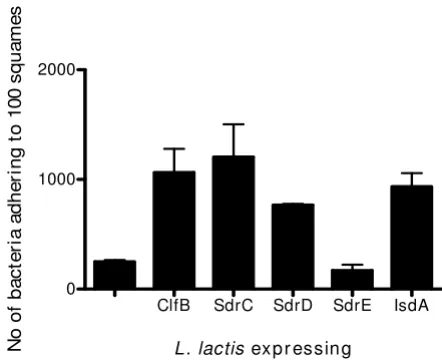
the empty vector pKS80 adhered poorly (Figure 1). L.
lac-tis expressing SdrE was not significantly different to L.
lac-tis carrying pKS80 (P = 0.2055; Figure 1) indicating that
this protein cannot promote adhesion to squamous cells. In contrast, a significant increase in adherence to
squa-mous cells was observed when L. lactis cells expressed
SdrC, SdrD, ClfB or IsdA (P values of 0.0339, SdrC; P =
0.0003, SdrD; P = 0.0396, ClfB and P = 0.0178, IsdA;
Fig-ure 1) showing that each of these proteins can promote adhesion when expressed on the surface of a Gram posi-tive coccus. It was shown previously that ClfA expressed
by L. lactis did not promote adhesion [15].
Adherence of S. aureus mutants to desquamated nasal epithelial cells
In order to investigate the role of surface proteins in
pro-moting adherence of S. aureus to desquamated nasal
epi-thelial cells a set of isogenic mutants was constructed and
compared. Strain Newman defective in clfA was used as
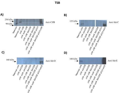
the starting point in the strain construction but this muta-tion had no bearing on adhesion since ClfA is known not to promote adhesion to squamous cells [9]. Each strain was examined by Western immunoblotting in order to show that the relevant proteins were missing in the mutants and that the remaining proteins were expressed
at the same level as in the wild type. Newman clfA grown
to exponential phase in TSB expressed ClfB, SdrC and SdrE but not SdrD (Figure 2). Since bacteria were grown in TSB they did not express Isd proteins. Introduction of
the multicopy shuttle plasmid pCU1 bearing the clfB, sdrC
or sdrE genes resulted in expression of proteins at levels
equivalent to or higher than the wild-type. In the case of SdrD expression was not seen in the wild-type strain and
was only detected when the pCU1sdrD+ plasmid was
present (Figure 2C). This may be due to amplification of low level expression under these growth conditions due to a gene dosage affect by a multicopy plasmid.
[image:2.612.314.535.87.269.2]Bacteria were also grown to stationary phase in RPMI. The wild-type strain expressed ClfB, IsdA, SdrD and SdrE, but not SdrC at levels that were detectable by Western immu-Adherence of L. lactis expressing different surface proteins to desquamated nasal epithelial cells
Figure 1
Adherence of L. lactis expressing different surface proteins to desquamated nasal epithelial cells. L. lactis (pKS80), L. lactis (pKS80clfB+), L. lactis (pKS80sdrC+), L. lactis
(pKS80sdrD+), L. lactis (pKS80sdrE+) and L. lactis (pKS80isdA+)
grown to stationary phase were tested for their ability to bind to human desquamated epithelial cells. Counts repre-sent the number of bacterial cells adhering to 100 squamous cells. Results are expressed as the mean of triplicate experi-ments +/- standard deviations.
ClfB SdrC SdrD SdrE IsdA
0 1000 2000
L . lactis exp r essin g
No
o
f b
ac
ter
ia
ad
h
er
in
g
t
o
100 s
q
u
am
noblotting (Figure 3). The Sdr proteins were detected with antibodies that recognized the conserved B domains (Fig-ure 3C) and specific anti-A domain antibodies (not shown). Complementation of the mutant strain lacking
these surface proteins with pCU1clfB+, pCU isdAB+,
pCU1sdrD+ or pCU1sdrE+ resulted in restoration of
expres-sion of the appropriate protein at levels similar to (IsdA) or higher than wild-type (ClfB, SdrD, SdrE). In the case of
pCU1sdrC+ low level expression was achieved.
With Newman clfA grown in TSB approximately 800
bac-teria adhered per 100 squamous cells (Figure 4A). The level of adhesion was reduced to ca 500 bacteria per 100 squamous cells when either ClfB or a combination of
SdrC, SdrD and SdrE proteins were missing (Figure 4A, P
= 0.0392, ClfB; P = 0.0441, SdrCDE). Adherence was even
lower when the clfB and sdrCDE mutations were
com-bined (Figure 4A, P = 0.0233 for Newman clfA clfB sdrCDE
compared to Newman clfA). As only SdrC and SdrE were
expressed under these conditions (Figure 2) and as
exper-iments with L. lactis (pKS80sdrE+) indicated that SdrE did
[image:3.612.54.556.83.475.2]Western immunoblot to detect expression of ClfB, SdrC, SdrD and SdrE Figure 2
Western immunoblot to detect expression of ClfB, SdrC, SdrD and SdrE. A-D. Cell wall associated proteins from strains Newman, Newman clfA, Newman clfA clfB, Newman clfA isdA clfB, Newman clfA clfB sdrCDE, Newman clfA isdA clfB
sdrCDE, Newman clfA sdrCDE, Newman clfA isdA sdrCDE and Newman clfA isdA clfB sdrCDE (pCU1) and (A) Newman clfA isdA
clfB sdrCDE (pCU1clfB+), (B) Newman clfA isdA clfB sdrCDE (pCU1sdrC+), (C) Newman clfA isdA clfB sdrCDE (pCU1sdrD+), (D)
Newman clfA isdA clfB sdrCDE (pCU1sdrE+) were grown to the exponential phase in TSB, cell wall associated proteins were
sol-ubilised by lysostaphin and run on 7.5% SDS-PAGE gels. Western immunoblotting was performed with (A) rabbit anti-ClfB antibodies, (B) rabbit anti-SdrC antibodies, (C) rabbit anti-SdrD antibodies and (D) rabbit anti-SdrE antibodies and subse-quently with HRP-conjugated protein A-peroxidase.
250 kDa
98 kDa Anti-ClfB
Ne wm an Ne wm an clf A -clf A -clf B -clf A -isd A -clf B -clf A
-clf
B -Sd rCD E -clf A -c lfB
-is
dA -S drC DE -clf A -clf B
-isd A -Sd rC DE -p CU1 clf A -clf B
-is
dA -S drC DE -p CU1 clf B+ 155 kDa Anti-Sdr C Ne wm an clf A -clf A -sd rC DE -clf A -isd A -sd rC DE -clf A -clf B -Sd rC DE -clf A
-clf
B -isd
A -Sd rCD E -clf A -clf B
-is
dA -S drC DE -p CU1 clf A -clf B
-isd A -Sd rC DE -p CU1 sd rC + 180 kDa Anti-Sdr E Ne wm an clf A -clf A -sd rCDE -clf A -is dA -s drC DE -clf A -clf B -Sd rC DE -clf A -clf B
-isd A -S drC DE -clf A -clf B
-is
dA -S drC DE -p CU1 clf A -clf B -isd
A -Sd rC DE -p CU1 sd rE +
160 kDa Anti-Sdr D
clf A
-clf B
-isd A -Sd rC DE -p CU1 sd rD + Ne wm an clf A -clf A -sd rCDE -clf A -isd A -sd rC DE -clf A -clf B -Sd rC DE -clf A
-clf
B -isd
A -Sd rCD E -clf A -clf B
not promote adhesion to squamous cells (Figure 1), it is
likely that the decrease observed by disrupting the sdrCDE
genes is due to the loss of SdrC.
In order to determine the role of IsdA in adherence, mutants were grown to stationary phase in the iron lim-ited medium RPMI and tested for adhesion to squamous
cells. Newman wild-type and Newman clfA adhered at
similar levels of ca 1300 bacteria per 100 squamous cells
(Figure 4B). This confirms that ClfA does not promote adhesion to squamous cells. Disruption of ClfB, IsdA or
SdrCDE in the clfA mutant host each caused a drop in
adherence to ca 800 bacteria per 100 squamous cells (Fig-ure 4B). The decrease was statistically significant for IsdA
(P = 0.0389, compared to Newman clfA) but not for ClfB
or SdrCDE (P = 0.0662 and 0.1852, respectively
com-pared to Newman clfA). Combining the isdA and sdrCDE
mutations, the clfB and sdrCDE mutations or the isdA and
[image:4.612.61.555.79.475.2]Western immunoblot to detect expression of surface protein under iron-limiting conditions Figure 3
Western immunoblot to detect expression of surface protein under iron-limiting conditions. Bacteria were grown to stationary phase in RPMI. Cell wall associated proteins were solubilized with lysostaphin and separated on a 7.5% SDS-PAGE gel and detected with rabbit antibodies followed by HRP-conjugated protein A-peroxidase. (A). Newman wild-type, Newman clfA, Newman clfA clfB, Newman clfA isdA clfB, Newman clfA clfB sdrCDE, Newman clfA isdA clfB sdrCDE, Newman clfA
isdA clfB sdrCDE (pCU1) and Newman clfA isdA clfB sdrCDE (pCU1clfB+). (B). Newman wild type, Newman clfA, Newman clfA
isdA, Newman clfA isdA clfB, Newman clfA isdA sdrCDE, Newman clfA isdA clfB sdrCDE, Newman clfA isdA clfB sdrCDE (pCU1) and Newman clfA isdA clfB sdrCDE (pCU1isdAB+). (C). Newman clfA, Newman clfA sdrCDE, Newman clfA isdA sdrCDE, Newman clfA
clfB sdrCDE, Newman clfA isdA clfB sdrCDE, Newman clfA isdA clfB sdrCDE (pCU1), Newman clfA isdA clfB sdrCDE (pCU1sdrC+),
Newman clfA isdA clfB sdrCDE (pCU1sdrD+) and Newman clfA isdA clfB sdrCDE (pCU1sdrE+). The primary antibodies used were
(A) rabbit anti-ClfB (B) rabbit anti-IsdA and (C) rabbit anti-SdrD B repeats. Anti-ClfB 250 kDa 98 kDa Ne wm an Ne wm an clf A -clf A
-clf
B -clf A -isd A -c lfB -clf A -clf
B-S
drC
DE
-clf
A-c
lfB
-is
dA -S drC DE -clf
A-c
lfB
-is
dA -S drC DE -p CU1 clf
A-c
lfB
-is
dA -S drC DE -p CU1 clf B + Anti-IsdA 50 kDa 36 kDa Ne wm an Ne wm an clf A -clf A -is dA -clf
A-is
dA
-clf B
-clfA
-is dA -S drC DE -clf A -clf B
-isd A -Sd rC DE -clf
A-c
lfB
-is
dA -S drC DE -p CU1 clf A
-clfB
-is
dA -S drC DE -p CU1 isd AB +
Sdr D and Sdr E 155 kDa 180 kDa Ne wm an clf A -clf
A-s
drC DE -clf A -is dA -s drC DE -clf A
-clf
B
-S
drC
DE
-clfA
-clf
B -isd
A -S drC DE -clf A -clf B
-isd A
-S
drC
DE
-pCU1
clf A
-clf
B-i
sd
A
-S
drC
DE
-pCU1
sdrC
+
clf A
-clf B
-isd
A-S
drC DE -p CU1 sd rD + clf A -clf
B-i
sd
A
-Sd
rCD
E
-pCU1
sd
rE
+
clfB mutations decreased adherence further (Figure 4B, P = 0.0352, 0.0135 and 0.0183, respectively compared to
Newman clfA). Finally when a mutant lacking ClfA, ClfB,
IsdA and SdrCDE was tested, only 200 bacteria adhered per 100 squamous cells. Of the Sdr proteins only SdrD and SdrE were expressed by Newman growing in RPMI (Figure 3) (IsdA and ClfB are also expressed under these conditions [12,15]) and as SdrE does not promote adhe-sion it can be concluded that the decrease associated with
the deletion of sdrCDE was due to the loss of the SdrD
protein. In conclusion, these results are consistent with
the data obtained with L. lactis and demonstrate a similar
role for ClfB, IsdA, SdrC and SdrD in adhesion to squa-mous cells.
Complementation
To confirm the roles of surface proteins in S. aureus
deduced from the analysis of mutants, a strain of Newman that was defective in all four adherent surface proteins (in addition to ClfA and SdrE) was complemented by intro-ducing multicopy shuttle plasmids expressing ClfB, SdrC, SdrD, SdrE, IsdAIsdB and IsdB. When bacteria were grown
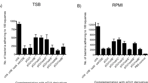
to exponential phase in TSB, ClfB, SdrC and SdrD each promoted increased adhesion to squamous cells above
the background level (Figure 5A; P = 0.0554, ClfB; P =
0.0282, SdrC; P = 0.0449, SdrD; P = 0.8803, SdrE; P =
0.533, IsdA). The differences were statistically significant for SdrC and SdrD, but not for ClfB. Expression of SdrE did not promote adhesion which is consistent with results described above. The Isd proteins were not expressed in TSB-grown bacteria.
When the strains were grown under iron restricted condi-tions in RPMI, complementation with ClfB, IsdA, SdrC or
SdrD each promoted adhesion (Figure 5B, P = 0.029, ClfB;
P = 0.0536, SdrC; P = 0.0908, SdrD; P = 0.0384, IsdA). The
conclusion about IsdA was drawn by comparing the level of adhesion promoted by the plasmid expressing both IsdA and IsdB with that expressing IsdB alone. Attempts to express IsdA alone in pCU1 were unsuccessful. These results were statistically significant except for those involv-ing SdrC and SdrD. Expression of SdrE did not promote adhesion (Figure 5B). These results confirm that ClfB,
IsdA, SdrC and SdrD are all important in adherence of S.
[image:5.612.73.559.80.359.2]Adherence of Newman mutants to desquamated nasal epithelial cells Figure 4
Adherence of Newman mutants to desquamated nasal epithelial cells. The ability of (A) Newman clfA, Newman clfA clfB, Newman clfA sdrCDE and Newman clfA clfB sdrCDE grown to exponential phase in TSB and (B) Newman, Newman clfA, Newman clfA clfB, Newman clfA sdrCDE, Newman clfA isdA, Newman clfA isdA sdrCDE, Newman clfA clfB sdrCDE, Newman clfA
isdA clfB, and Newman clfA isdA clfB sdrCDE grown to stationary phase in RPMI to adhere to desquamated human nasal epithelial
cells was measured. The tenth track is a control without S. aureus showing background due to adherent bacteria from the donor. Counts represent the number of bacterial cells adhering to 100 squamous cells. Results are expressed as the mean of triplicate experiments +/- standard deviations.
-clfA
clfB
-clfA
sd rCDE
-clfA
isdA
-clfA
sdr CDE
isdA
-clfA
sd rCDE
clfB
-clfA
clfB
isd A
-clfA
sd rCDE
clfB
isdA
-clfA
PB S c
ontro l
0 1000 2000
Mutants of Newman
N
o
. of
ba
ct
e
ri
a
a
dhe
ri
ng t
o
1
0
0
squa
m
e
s
RPMI
B)
-clfA
clfB
-clfA
sd rCDE
-clfA
-sdrC DE
clfB
-clfA 0
250 500 750 1000
Mu t an t s o f New man
No
. o
f b
ac
ter
ia ad
h
er
in
g
t
o
100 s
q
u
am
es
aureus to desquamated nasal epithelial cells under growth
conditions that pertain in vivo.
Discussion
S. aureus is a commensal of the moist squamous
epithe-lium of the anterior nares of a significant proportion of the population. Colonization is a known risk factor for the development of staphylococcal infections in the com-munity and hospital. The causes of intermittent and per-sistent carriage are believed to be different. Perper-sistent
carriers are often colonised by a single strain of S. aureus
over a long period of time, while intermittent carriers tend to carry different strains for briefer time periods [16,17]. Persistent carriers also carry a higher load of bacteria in the nares than intermittent carriers [18,19]. When volun-teers were decolonized and were then inoculated with a
mixture of S. aureus strains non-carriers eliminated the
bacteria, whereas persistent carriers selected their original
S. aureus colonizing strain from the mixture [20]. These
results indicate that certain host characteristics determine carriage status in particular individuals. This view is
sup-ported by the fact that certain age or ethnic groups seem to be predisposed to carriage [2,3]. One determinant of varying patterns of nasal carriage may be differing
expres-sion levels of ligands for S. aureus on the surface of
desq-uamated nasal epithelial cells.
In this study we used three donors to provide the desqua-mated nasal epithelial cells for adhesion experiments. They were selected because their cells supported a consist-ent level of adhesion. It has been noted that cells from dif-ferent donors can provide widely variable levels of adhesion [21]. The reason for this is not known. One pos-sibility is different levels of expression of the ligands responsible for adherence promoted by one or more of
the S. aureus surface proteins. It is imperative to perform a
detailed comparative study of the ability of the surface proteins described here to support adhesion of bacteria to squamous cells from donors who are persistent carriers and those who are non-carriers. This could contribute to the knowledge of the contribution of host factors to car-riage.
[image:6.612.61.548.82.354.2]Adherence of S. aureus Newman complemented mutants grown in TSB and iron restricted conditions to desquamated nasal epithelial cells
Figure 5
Adherence of S. aureus Newman complemented mutants grown in TSB and iron restricted conditions to desq-uamated nasal epithelial cells. The ability of mutants of strain Newman carrying complementing pCU1 plasmids carrying surface protein genes to adhere to desquamated nasal epithelial cells was tested. Strains Newman clfA, Newman clfA isdA clfB
sdrCDE, Newman clfA isdA clfB sdrCDE (pCU1), Newman clfA isdA clfB sdrCDE (pCU1clfB+), Newman clfA isdA clfB sdrCDE
(pCU1sdrC+), Newman clfA isdA clfB sdrCDE (pCU1sdrD+), Newman clfA isdA clfB sdrCDE (pCU1sdrE+),) Newman clfA isdA clfB
sdrCDE (pCU1isdAB+) and Newman clfA isdA clfB sdrCDE (pCU1isdB+) grown to the exponential phase in (A) TSB and to the
sta-tionary phase in (B) RPMI were tested for adherence. Counts represent the number of bacterial cells adhering to 100 squa-mous cells. Results are expressed as the mean of triplicate experiments +/- standard deviations.
-clfA
sdrC DE
isdA
clfB
-clfA
pC U1 + pCU1c
lfB
+
pCU1 sdrC
+
pCU1s drD
+
pCU1s drE
+
pCU1i sdA
B + pCU
1isdB
0 250 500 750 1000
Complementation with pCU1 derivatives
N
o
. o
f b
a
ct
e
ri
a
a
d
he
ri
ng
t
o
1
0
0
sq
ua
m
e
s
A)
TSB
-clfA
sdrC DE
isdA
clf B
-clfA
pCU 1 +
pCU 1cl
fB
+
pCU 1sdrC
+
pCU1s drD
+
pCU 1sdrE
+
pC U1isdAB
+
pCU 1isd
B
PB S c
ontro l
0 200 400 600 800 1000 1200
Co mp lemen t at io n w it h p CU1 d er ivat ives
No
. o
f b
ac
ter
ia ad
h
er
in
g
t
o
100 s
q
u
am
es
Surface proteins ClfB and IsdA have previously been shown to promote adhesion to squamous epithelial cells [9,15] and are required for colonization of the nares of rodents [11,15]. Both ClfB and IsdA have been shown to bind to proteins present in the envelope of cornified squa-mous epithelial cells. IsdA and ClfB both bind to cytoker-atin 10 and loricrin [22] (Clarke, S. Walsh, E. J. Andre, G. Dufrene, Y. Foster, T. J. Foster, S. J. manuscript submitted). Loricrin accounts for 70 – 85% of the cornified envelope [23-25]. It is possible that differences in the level of expression of these proteins could contribute to the
varia-tion in carriage of S. aureus in the nares.
To investigate the contribution of each of five surface pro-teins (IsdA, ClfB, SdrC, SdrD and SdrE) to squamous cell adhesion, the proteins were expressed from the surrogate
host L. lactis. Expression of IsdA, ClfB, SdrC and SdrD each
resulted in increased adherence. Gene disruption and
complementation experiments in S. aureus also showed a
role for IsdA, ClfB, SdrC and SdrD in adhesion. SdrE did
not promote adhesion by either L. lactis or S. aureus.
Schaffer et al 2006 investigated whether SdrC or SdrD had
a role in colonization of the nares in a mouse model. Mutants defective in SdrC or SdrD colonized mice to the same extent as the wild-type indicating that these proteins do not play a role colonization of the nares of mice [11]. However, this does not necessarily mean that SdrC and SdrD have no role to play in colonization of the human nares.
Adherence to desquamated epithelial cells from the ante-rior nares is clearly multifactorial. When expression of IsdA, ClfB, SdrC and SdrD was disrupted in strain New-man the level of adherence was reduced to background. This does not support a role for wall teichoic acid (WTA) in promoting adhesion to squamous cells [21,26]. SasG did not play a role in adherence of Newman to squamous cells because this protein was not expressed detectably by
this strain despite the intact sasG gene being present [14].
SasG might play a role in clinical isolates where expres-sion occurs at higher levels.
It has been reported that WTA plays a prominent part in nasal colonization of the cotton rat model [26]. The authors also demonstrated that teichoic acid promoted bacterial adhesion to normal epithelial cells. However the WTA apparently does not contribute to bacterial adhesion to desquamated nasal cells epithelial cells [21]. This is consistent with the data reported here which indicates that only surface proteins are required for adhesion to squames. A multiple mutant defective in ClfB, SdrC, SdrD and IsdA did not adhere. If WTA contributed to adherence the multiple mutant would still have bound above back-ground levels. Thus colonization of the cotton rat requires both WTA [26] and surface proteins [15] albeit at different
stages in the process [21] and in different parts of the nares.
Conclusion
Eradication of carriage of S. aureus has been shown to
reduce infection rates in dialysis, diabetic and AIDS patients [4-6]. Vaccination with IsdA and ClfB was
effec-tive in reducing S. aureus carriage in animal models
[11,15]. It has been suggested that immune responses in
part determine the ability of humans to carry S. aureus in
the nares. This study has confirmed the role of ClfB and IsdA in adhesion to desquamated nasal epithelial cells and has revealed important roles for SdrC and SdrD. Vac-cination against two or more of these surface proteins could provide significant reduction in carriage and could potentially reduce the rate of infection and dissemination.
Methods
Growth conditionsEscherichia coli strains were grown on Luria (L) agar or in
L-broth (Difco). S. aureus strains were grown on tryptic
soy agar (TSA; Oxoid), tryptic soy broth (TSB) or RPMI 1640 (Sigma). Cultures were grown in an orbital shaker at 200 rpm at 37°C. RPMI cultures were subcultured into fresh broth and grown for a further 15 h before harvesting.
L. lactis strains were cultured in M17 medium (Difco)
con-taining 0.5% (v/v) glucose without shaking at 30°C. Anti-biotics (Sigma) were added when needed as follows:
ampicillin (100 μg ml-1), erythromycin (10 μg ml-1),
chlo-ramphenicol (10 μg ml-1) or tetracycline (2 μg ml-1).
Bacterial strains
The wild-type strain S. aureus strain Newman (10) and
Newman isdA (RC107 ΔisdA [27]) were subjected to allele
replacement mutagenesis with the temperature sensitive
plasmid pJH1 [28] forming strains DU5999 clfA5 [28]
and DU6020 clfA5 isdA. The clfB::Emr mutation [29] and
the ΔsdrCDE::Tcr mutation [22] were introduced by
trans-duction using phage 85 [30] in order to construct the
fol-lowing mutants of Newman: DU6000 clfA5 clfB::Emr [28],
DU6021 clfA5 ΔsdrCDE::Tcr, DU6001 clfA5 clfB::Emr
ΔsdrCDE::Tcr [28], DU6022 clfA5 isdA ΔsdrCDE::Tcr,
DU6023 clfA5 isdA clfB::Emr ΔsdrCDE::Tcr. For each
trans-ductant the mutations were verified by PCR and Southern blotting.
Complementation analysis was performed by transferring
into DU6023 clfA5 isdA clfB::Emr ΔsdrCDE::Tcr plasmid
pCU1 containing the full length structural genes for S.
aureus surface proteins as follows: pCU1sdrC+,
pCU1sdrD+, pCU1sdrE+, pCU1clfB+ [31], pCU1isdAisdB+
and pCU1isdB+. The plasmids were maintained by
select-ing resistance to chloramphenicol (10 μg ml-1). In each
down-stream transcription terminator. In the case of isd proteins
both the closely linked isdA and isdB genes and their
cog-nate promoters were cloned together. The primers are described below.
Expression of surface proteins in L. lactis MG1363 [32]
was achieved by cloning open reading frames from New-man genomic DNA in-frame into the expression vector
pKS80 [33] forming pKS80sdrC+ (25), pKS80sdrD+ (25),
pKS80sdrE+ (25), pKS80clfB+ (25) and pKS80isdA+ (this
study). Plasmid transformants were selected and main-tained in M17 medium containing erythromycin.
Molecular techniques
Standard procedures were used [34]. Restriction enzymes and ligase (New England Biolabs or Roche) were used according to manufacturer's protocol, as was Pfu DNA polymerase (Roche). Oligonucleotides were purchased from Sigma-Genosys.
Plasmid and strain construction
Primers pCU1sdrCF
(5'-CGGGGATCCAAGCTTAGAT-TAAAAGTGAG-'3) and pCU1sdrCR
(5'-GCTCTAGACT-GGGAATTTCTAAACAG-'3), pCU1sdrDF
(5'-CGGGGATCCTTCTGTTTAGAAATTCCCAG-'3) and
pCU1sdrDR (5'-GCTCTAGACCAGGCCTCACGGAC-'3)
and pCU1sdrEF
(5'-CCGGATCCGTAGAAACGAATAA-GAAAAAGC-'3) and pCU1sdrER
(5'-GCTCTAGAGTAAT-TCATATTATCGCCTC-'3) which all incorporate a 5'
BamHI and '3 XbaI site, respectively, were used to amplify
the sdrC, sdrD and sdrE genes, respectively, from strain
Newman genomic DNA. The DNA containing the sdrC,
sdrD and sdrE genes were digested with BamHI and XbaI
and cloned between the BamHI and XbaI sites of plasmid
pCU1.
Primers pCU1isdBF
(5'-CAGCTGCAGCCTATGTCATAGA-TATTTCATAATC-'3) and pCU1isdBR
(5'-CAGGAGCTCA-GAGATTCTAAACGTATTCGTAAG-'3) which incorporate
a 5' PstI and 3' XbaI site, respectively, were used to amplify
the isdB coding sequence including the upstream
pro-moter and Fur consensus sequence from strain Newman
genomic DNA. The isdB coding sequence is located 203 bp
downstream of the isdA coding sequence on the S. aureus
chromosome. Primers pCU1isdAF
(5'-CAGCTGCAGA-CATAATCCTCCTTTTTATGATTG-'3) and pCU1isdBR
(5'-CAGGAGCTCAGAGATTCTAAACGTATTCGTAAG-'3)
were used to amplify the isdA and isdB coding sequence
including the upstream promoter and Fur consensus
sequence of both genes. The 2.3 kb isdB and 3.6 kb isdAB
coding sequences were digested with PstI and XbaI and
cloned between the PstI and XbaI sites of plasmid pCU1.
Plasmids pCU1isdB+ and pCU1isdAB+ were sequenced and
screened by restriction mapping.
Plasmids pCU1, pCU1clfB+, pCU1sdrC+, pCU1sdrD+,
pCU1sdrE+, pCU1isdB+ and pCU1isdAB+ were transformed
into S. aureus RN4220 and transduced into strain
New-man clfA clfB isdA sdrCDE selecting for chloramphenicol
resistance.
Primers FpKisdA (5'-CGCTGATCAAACATTATT-TAAACAGTAAGTATC-'3) and RpKisdA (5'-CGCTGAT-CATTATTTAGATTCTTTTCTTTTGA-'3) which incorporate
a 5' and a 3' BclI site, respectively, were used to amplify the
isdA coding sequence from genomic DNA. The PCR
prod-uct was digested with BclI and cloned into BclI digested
pKS80. This resulted in the open reading frame of isdA
being fused to the ATG codon of the expression cassette to
optimize translation and created the plasmid pKS80isdA+.
The plasmid was sequenced, screened by restriction
map-ping and electroporated into competent L. lactis strain
MG1363.
Western immunoblot analysis
Cell wall-associated proteins of S. aureus and L. lactis were
prepared as previously described [35,22]. For S. aureus
exponential phase cultures were grown to an OD600 of 0.6.
Stationary phase cultures were grown for 16 – 24 h. Cells were harvested, washed in PBS and resuspended to an
OD600 of 1 in lysis buffer (50 mM Tris/HCl, 20 mM
MgCl2, pH 7.5) supplemented with 30% (w/v) raffinose
and 40 μl ml-1 protease inhibitors (Roche). Cell wall
pro-teins were solubilized by incubation with lysostaphin
(200 μgml-1) for 10 minutes at 37°C. Cell wall fractions
were separated on 7.5% (w/v) polyacrylamide gels, elec-trophoretically transferred onto PVDF membranes (Roche), blocked in 10% (w/v) skimmed milk (Marvel) and probed with ClfB antibodies (1:5,000; [31], anti-IsdA antibodies (1:2,000; a gift from Prof. P. Speziale, Department of Biochemistry, University of Pavia, Pavia, Italy) and anti-SdrC, anti-SdrD, anti-SdrE or anti-Sdr region B antibodies (1:2,000) [22]. The specificity of each antibody is indicated by the fact that no immnocrossreac-tive bands appeared in mutant strains lacking the relevant antigen. Membranes were washed three times with gentle agitation for 15 min in TS-Tween (10 mM Tris-HCl pH 7.4, 150 mM NaCl, 0.05% (v/v) Tween 20 (Sigma)). Bound antibodies were detected using horseradish perox-idase-conjugated protein A-peroxidase (1:500; Sigma). Proteins were visualised using the LumiGLO™ Reagent and peroxide detection system (Cell Signalling
Technol-ogy®). Membranes were detected using Kodak X-ray film.
The exposed films were fixed and developed using a Kodak X-OMAT 1000 Processor developing machine.
swabbing of the anterior nares. One donor was a carrier of
S. aureus while the other two were not. After washing in
PBS, cells were adjusted to 1 × 10⁵cell ml-1. Bacterial cells
were washed with PBS and adjusted to 1 × 10⁹cells ml-1.
100 μl volumes of bacterial and epithelial cells were
mixed and incubated at 37°C for 1 h with occasional
shaking before being captured on 12 μm isopore
polycar-bonate filters, washed with PBS, fixed and stained with 5% (w/v) crystal violet. The filters were mounted onto glass slides. The number of bacteria per 100 squames was counted using light microscopy.
Statisical Analysis
Statistical analyses were determined by the Student t-test, using the online GraphPad software. Differences were
considered significant if p values were less than 0.05.
Authors' contributions
RMC carried out strain construction, performed Western immunoblotting, all squamous cell adhesion assays and drafted the manuscript. HM constructed five plasmids/ strains for this study and helped to draft the manuscript and TJF conceived and coordinated the study, and helped to draft the manuscript. All authors read and approved the final manuscript.
Acknowledgements
Grants from Science Foundation Ireland and the Health Research Board are acknowledged. We thank Professor Simon Foster (University of Sheffield) for sending the isdA mutant of S. aureus Newman
References
1. Kluytmans J, van Belkum A, Verbrugh H: Nasal carriage of Staphy-lococcus aureus : epidemiology, underlying mechanisms, and associated risks. Clin Microbiol Rev 1997, 10(3):505-520. 2. Cole AM, Tahk S, Oren A, Yoshioka D, Kim YH, Park A, Ganz T:
Determinants of Staphylococcus aureus nasal carriage. Clin Diagn Lab Immunol 2001, 8(6):1064-1069.
3. Armstrong-Esther CA: Carriage patterns of Staphylococcus aureus in a healthy non-hospital population of adults and chil-dren. Ann Hum Biol 1976, 3(3):221-227.
4. Yu VL, Goetz A, Wagener M, Smith PB, Rihs JD, Hanchett J, Zuravleff JJ: Staphylococcus aureus nasal carriage and infection in patients on hemodialysis. Efficacy of antibiotic prophylaxis.
N Engl J Med 1986, 315(2):91-96.
5. Lipsky BA, Pecoraro RE, Chen MS, Koepsell TD: Factors affecting staphylococcal colonization among NIDDM outpatients. Dia-betes Care 1987, 10(4):483-486.
6. Nguyen MH, Kauffman CA, Goodman RP, Squier C, Arbeit RD, Singh N, Wagener MM, Yu VL: Nasal carriage of and infection with
Staphylococcus aureus in HIV-infected patients. Ann Intern Med
1999, 130(3):221-225.
7. von Eiff C, Becker K, Machka K, Stammer H, Peters G: Nasal car-riage as a source of Staphylococcus aureus bacteremia. Study Group. N Engl J Med 2001, 344(1):11-16.
8. Wertheim HF, Vos MC, Ott A, van Belkum A, Voss A, Kluytmans JA, van Keulen PH, Vandenbroucke-Grauls CM, Meester MH, Verbrugh HA: Risk and outcome of nosocomial Staphylococcus aureus
bacteraemia in nasal carriers versus non-carriers. Lancet
2004, 364(9435):703-705.
9. O'Brien LM, Walsh EJ, Massey RC, Peacock SJ, Foster TJ: Staphylo-coccus aureus clumping factor B (ClfB) promotes adherence to human type I cytokeratin 10: implications for nasal colo-nization. Cell Microbiol 2002, 4(11):759-770.
10. Clarke SR, Wiltshire MD, Foster SJ: IsdA of Staphylococcus aureus
is a broad spectrum, iron-regulated adhesin. Mol Microbiol
2004, 51(5):1509-1519.
11. Schaffer AC, Solinga RM, Cocchiaro J, Portoles M, Kiser KB, Risley A, Randall SM, Valtulina V, Speziale P, Walsh E, et al.: Immunization with Staphylococcus aureus clumping factor B, a major deter-minant in nasal carriage, reduces nasal colonization in a murine model. Infect Immun 2006, 74(4):2145-2153.
12. Wertheim HF, Walsh E, Choudhurry R, Melles DC, Boelens HA, Mia-jlovic H, Verbrugh HA, Foster T, van Belkum A: Key role for clump-ing factor B in Staphylococcus aureus nasal colonization of humans. PLoS Med 2008, 5(1):e17.
13. Roche FM, Meehan M, Foster TJ: The Staphylococcus aureus sur-face protein SasG and its homologues promote bacterial adherence to human desquamated nasal epithelial cells.
Microbiology 2003, 149(Pt 10):2759-2767.
14. Corrigan RM, Rigby D, Handley P, Foster TJ: The role of Staphylo-coccus aureus surface protein SasG in adherence and biofilm formation. Microbiology 2007, 153(Pt 8):2435-2446.
15. Clarke SR, Brummell KJ, Horsburgh MJ, McDowell PW, Mohamad SA, Stapleton MR, Acevedo J, Read RC, Day NP, Peacock SJ, et al.: Iden-tification of in vivo-expressed antigens of Staphylococcus aureus and their use in vaccinations for protection against nasal carriage. J Infect Dis 2006, 193(8):1098-1108.
16. Eriksen NH, Espersen F, Rosdahl VT, Jensen K: Carriage of Staphy-lococcus aureus among 104 healthy persons during a 19-month period. Epidemiol Infect 1995, 115(1):51-60.
17. VandenBergh MF, Yzerman EP, van Belkum A, Boelens HA, Sijmons M, Verbrugh HA: Follow-up of Staphylococcus aureus nasal car-riage after 8 years: redefining the persistent carrier state. J Clin Microbiol 1999, 37(10):3133-3140.
18. Nouwen JL, Fieren MW, Snijders S, Verbrugh HA, van Belkum A: Per-sistent (not intermittent) nasal carriage of Staphylococcus aureus is the determinant of CPD-related infections. Kidney Int 2005, 67(3):1084-1092.
19. Nouwen JL, Ott A, Kluytmans-Vandenbergh MF, Boelens HA, Hof-man A, van Belkum A, Verbrugh HA: Predicting the Staphylococ-cus aureus nasal carrier state: derivation and validation of a "culture rule". Clin Infect Dis 2004, 39(6):806-811.
20. Nouwen J, Boelens H, van Belkum A, Verbrugh H: Human factor in
Staphylococcus aureus nasal carriage. Infect Immun 2004, 72(11):6685-6688.
21. Weidenmaier C, Kokai-Kun JF, Kulauzovic E, Kohler T, Thumm G, Stoll H, Gotz F, Peschel A: Differential roles of sortase-anchored surface proteins and wall teichoic acid in Staphylococcus aureus nasal colonization. Int J Med Microbiol 2008, 298(5– 6):505-513.
22. O'Brien L, Kerrigan SW, Kaw G, Hogan M, Penades J, Litt D, Fitzger-ald DJ, Foster TJ, Cox D: Multiple mechanisms for the activa-tion of human platelet aggregaactiva-tion by Staphylococcus aureus
: roles for the clumping factors ClfA and ClfB, the serine-aspartate repeat protein SdrE and protein A. Mol Microbiol
2002, 44(4):1033-1044.
23. Candi E, Schmidt R, Melino G: The cornified envelope: a model of cell death in the skin. Nat Rev Mol Cell Biol 2005, 6(4):328-340. 24. Steinert PM, Marekov LN: The proteins elafin, filaggrin, keratin intermediate filaments, loricrin, and small proline-rich pro-teins 1 and 2 are isodipeptide cross-linked components of the human epidermal cornified cell envelope. J Biol Chem 1995, 270(30):17702-17711.
25. Kalinin A, Marekov LN, Steinert PM: Assembly of the epidermal cornified cell envelope. J Cell Sci 2001, 114(Pt 17):3069-3070. 26. Weidenmaier C, Kokai-Kun JF, Kristian SA, Chanturiya T, Kalbacher
H, Gross M, Nicholson G, Neumeister B, Mond JJ, Peschel A: Role of teichoic acids in Staphylococcus aureus nasal colonization, a major risk factor in nosocomial infections. Nat Med 2004, 10(3):243-245.
27. Clarke SR, Foster SJ: IsdA protects Staphylococcus aureus
against the bactericidal protease activity of apolactoferrin.
Infect Immun 2008, 76(4):1518-1526.
Publish with BioMed Central and every scientist can read your work free of charge
"BioMed Central will be the most significant development for disseminating the results of biomedical researc h in our lifetime."
Sir Paul Nurse, Cancer Research UK
Your research papers will be:
available free of charge to the entire biomedical community
peer reviewed and published immediately upon acceptance
cited in PubMed and archived on PubMed Central
yours — you keep the copyright
Submit your manuscript here:
http://www.biomedcentral.com/info/publishing_adv.asp
BioMedcentral 29. McAleese FM, Walsh EJ, Sieprawska M, Potempa J, Foster TJ: Loss of
clumping factor B fibrinogen binding activity by Staphyloco-ccus aureus involves cessation of transcription, shedding and cleavage by metalloprotease. J Biol Chem 2001, 276(32):29969-29978.
30. Foster TJ: Molecular genetic analysis of staphylococcal viru-lence. Methods in Microbiology 1998, 27:433-454.
31. Ni Eidhin D, Perkins S, Francois P, Vaudaux P, Hook M, Foster TJ: Clumping factor B (ClfB), a new surface-located fibrinogen-binding adhesin of Staphylococcus aureus. Mol Microbiol 1998, 30(2):245-257.
32. Gasson MJ: Genetic transfer systems in lactic acid bacteria.
Antonie Van Leeuwenhoek 1983, 49(3):275-282.
33. Hartford O, O'Brien L, Schofield K, Wells J, Foster TJ: The Fbe (SdrG) protein of Staphylococcus epidermidis HB promotes bacterial adherence to fibrinogen. Microbiology 2001, 147:2545-2552.
34. Sambrook J, Russell DW: Molecular cloning, a labratory man-ual. 3rd edition. Cold Spring Harbor, New York: Cold Spring Har-bour Laboratory Press; 2001.