"MORPHOMETERIC STUDY OF THE ORBIT IN HUMAN DRY SKULLS AND HIGH RESOLUTION COMPUTED TOMOGRAPHIC
SCANS"
Dissertation submitted in
Partial fulfillment of the regulations required for the award of
M.D. DEGREE
In
ANATOMY - BRANCH V
THE TAMIL NADU DR.M.G.R MEDICAL UNIVERSITY
CHENNAI
CERTIFICATE
This is to certify that the dissertation "MORPHOMETERIC STUDY OF THE ORBIT IN HUMAN DRY SKULLS AND HIGH RESOLUTION COMPUTED TOMOGRAPHIC SCANS" is an original work done by Dr.Subbulakshmi A, Post Graduate student, PSG Institute of Medical Sciences and Research, Coimbatore, under my supervision and guidance.
Dr.M.Jamuna, M.S Dr.S.Ramalingam, M.D
Professor and HOD Dean
Department of Anatomy PSG IMS & R, Coimbatore
DECLARATION
I solemnly declare that this dissertation "Morphometeric Study Of The Orbit In Human Dry Skulls And High Resolution Computed Tomographic Scans" was done by me in the Department of Anatomy, PSG Institute of Medical Sciences & Research, Coimbatore, under the guidance of Dr.M.Jamuna, M.S, Professor and Head of the Department, Anatomy, PSG Institute of Medical Sciences & Research Coimbatore.
This dissertation is submitted to the Tamil Nadu Dr.M.G.R. Medical University, Chennai in partial fulfillment of the university regulations for the award of degree of M.D. Anatomy – Branch V examinations to be held in April 2016.
Place: Coimbatore Dr. Subbulakshmi. A
ACKNOWLEDGMENT I THANK GOD FOR HIS GRACE
I render my gratefulness and sincere thanks to Dr. M. Jamuna, professor and HOD of anatomy for her valuable ideas in shaping the dissertation and kind support.
I express my gratitude to Dr. Ramalingam, Dean PSG IMS and R Coimbatore for facilitating me to undertake this project in this esteemed institution.
I extend my sincere thanks to Dr. Elango, Professor of Radiology, for his skillful ideas in the radiological aspect of dissertation.
I thank Dr. G.Amudha, Professor of anatomy for her timely support.
I am thankful to Dr. P.A.Kumar professor of anatomy for his enthusiastic support.
I thank Dr. M.Nirmala Devi, Associate professor of anatomy, who encouraged me with my work.
I thank Dr. Beula, Dr. Manikavasuki, Dr. D.Mathivanan, Mrs. Radhika and Mrs. Joy Deborah, assistant professors of anatomy for their support.
I thank my colleagues, senior and junior postgraduate friends in helping me for handling the computer techniques.
I am also thankful to all the technicians and non-teaching staff of our department for their dedicated work in making arrangements of the lab facilities and cleanliness.
I extend my heartful gratitude to my mother and father for supporting me throughout my life morally.
TABLE OF CONTENTS
S.NO
CONTENTS
PAGE
1.
INTRODUCTION
1
2.
AIMS AND OBJECTIVES
17
3.
REVIEW OF LITERATURE
18
4.
MATERIALS AND METHODS
53
5.
RESULTS
60
6.
DISCUSSION
76
7.
CONCLUSION
92
8.
BIBLIOGRAPHY
INTRODUCTION
The eyes speak without words which enhance the beauty of face. The malposition of eyeballs leads to unacceptable cosmetic problem for the people of both sexes and all age groups. The word eye constitutes eye ball and orbital cavity. The orbital cavities are intended as a socket for eyeballs, muscles and fascia which keeps the eyeballs in position, nerves and vessels associated with vision, orbital pad of fat and lacrimal apparatus.
The eyeballs are situated in anterior one third of orbital cavities. The anterior aspect of eye ball project outside the orbital cavity so that when a needle is passed from lateral orbital margin to bridge of nose, the needle will pass behind the lens.
Development of orbit:
The development of orbital cavity is simultaneous process with the development of eye ball which begins during third week of embryonic life, when the embryo is of 2.6mm length. There are four sources contributing to the development of eye ball and orbital cavity.
Neuroectoderm of forebrain Surface ectoderm
Paraxial mesoderm
The optic pit appears in the anterior neural folds around 22nd day of fertilization, which invaginate to form optic groove or sulcus. When the embryo is about 3.2mm stage, optic groove further invaginate to form primary optic vesicle. The proximal part of optic vesicle is constricted to form optic stalk. The surface ectoderm which comes in contact with optic vesicle is thickened to form lens placode. The lens placode deepens to form lens pit. As the lens pit get deepened, simultaneous approachment of their edges form spherical lens vesicle.
At the same time optic vesicle invaginates to form optic cup to accommodate the spherical lens vesicle. The process of transformation of optic vesicle to optic cup takes place during 4th week (4 – 5 mm stage) to 6th week (15 – 18 mm stage).
The mesoderm around the growing eyeball differentiates to form orbital cavity in order to accommodate them. The bony orbital cavities are developed from visceral mesoderm (mesenchyme) of maxillary process and paraxial mesoderm. The mesenchyme of maxillary process forms the floor and lateral wall of orbital cavity. The roof of orbital cavity is formed by paraxial mesoderm which is the part of mesodermal capsule enveloping the brain. The medial wall of orbital cavity is formed from the portion of paraxial mesoderm of lateral nasal process (Cooper WC 1985)
The ossification centers for bony orbital cavity appear during 6th and 7th week and their fusion takes place at 6th and 7th month of intra uterine life. Initially the orbital cavity seems to be rounded in appearance to accommodate circular optic cup, later maturation of skull and developing orbital contents make it more ovoid.
The growth of orbital cavity is influenced by following factors: Growth of surrounding structures
Growth of intraorbital structures
Pneumatization of paranasal air sinuses Suture growth
Appositional growth
Growth of surrounding structures and sutural growth:
The growth of bony orbit is highly influenced by developing brain rather than the developing eyeballs. The deformities of brain and skull lead to major orbital abnormalities. In anencephaly the orbital cavities are extremely small and shallow and the roof of orbital cavity is severely deranged. With the growth of brain, there is expansion of neurocranium which induces the sutural growth in following sutures.
Frontal suture – fuses by one year of age.
Frontoethmoid suture – fuses by two years of age.
Internasal suture – growth continues until adolescence period.
Appositional growth and growth of intraorbital structures:
Appositional growth is defined as growth process where new bone is formed on one surface and resorption takes place on the opposite surface of bone. This growth increases the capacity of orbital cavity. The growth of orbital contents enhances the appositional growth. This explains why anophthalmic and microphthalmic orbits are likely to be smaller than the normal orbital cavities.
Gross anatomy of orbital cavity:
The orbital cavities are four walled, quadrilateral pyramidal cavities with their apices directed posteromedially at the optic canal and their bases directed anterolaterally at the orbital opening in the face bounded by orbital margins. The medial walls of the orbital cavities are parallel to each other with the distance of 25mm apart. The ethmoidal air sinuses are present between the medial walls of both orbital cavities. The lateral walls of the orbital cavities are inclined at an angle of 900. The axes of the orbital cavities seem to diverging at an angle of 450 but the optical axes of both eye balls are parallel to one another.
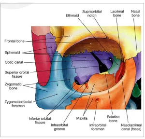
The superior wall (roof) of orbital cavity is markedly concave on its orbital aspect. It is formed by orbital plate of frontal bone in its major anterior aspect and lesser wing of sphenoid bone in its minor posterior aspect. The superior wall in its anterolateral aspect has fossa tolodge lacrimal gland and in its anteromedial aspect has fovea which serves as a pulley for superior oblique muscle.
of palatine bone in its small posterior aspect. The major part of floor of the orbit is contributed by maxilla which is traversed by infraorbital groove and canal from inferior orbital fissure to infra orbital foramen. The infraorbital canal transmits the nerves and vessels of the same name. The medial aspect of floor is weakened by this canal and this part of floor is often involved in blow out fractures.
Fig 1a: Bones forming the orbital walls
transmits anterior and posterior ethmoidal vessels and nerves respectively. The thinnest part of medial wall (orbital plate of ethmoid) is called lamina papyracea.
The lateral wall is the strongest wall of orbital cavity formed by zygomatic bone in its anterior aspect and orbital surface of greater wing of sphenoid bone in its posterior aspect. The lateral wall presents foramina to transmit zygomaticofacial and zygomaticotemporal nerves. The lateral wall presents a palpable elevation called Whitnall’s tubercle, situated posterior to lateral margin and slightly inferior to frontozygomatic suture. The Whitnall’s tubercle gives attachment to suspensory ligament of eyeball, lateral palpebral ligament, check ligament of lateral rectus muscle and levator palpebral superioris muscle.
Optic foramen is situated at the posterior most aspect of roof of orbital cavity, between the body of sphenoid bone and two roots of lesser wing of sphenoid bone. The optic foramen transmits the optic nerve which is invested by three layers of meninges and ophthalmic artery.
Orbital margin is continuous curved margin and forms the rim of orbital cavity.
Supraorbital margin is formed by frontal bone which presents a supraorbital notch or foramen at its junction of medial one third and lateral two third which transmits the corresponding nerves and vessels.
Infraorbital margin is formed by zygomatic and maxillary bone.
Lateral orbital margin is formed by frontal process of zygomatic bone and zygomatic process of frontal bone.
The three bones, frontal, zygomatic and maxillary bone contributes nearly equal third in the formation of orbital margin.
Apart from optic canal the orbital cavity has superior and inferior orbital fissures. The superior orbital fissure is retort shaped gap between posterior aspect of lateral wall and superior wall of orbit through which the orbital cavity communicates with middle cranial fossa. The superior orbital fissure transmits third, fourth and sixth cranial nerves which innervate the extra ocular muscles, recurrent meningeal branch of lacrimal artery, superior and inferior ophthalmic vein. The inferior orbital fissure lies at the junction of posterior aspect of lateral wall and inferior wall of the orbit. The orbital cavity communicates with pterygopalatine fossa and infratemporal fossa respectively through medial and lateral aspect of inferior orbital fissure. The fissure transmits inferior orbital vessels and nerves, zygomatic nerve and orbital branch of pterygopalatine ganglion.
Nowadays because of increasing road traffic accidents, facial skeleton fractures have become common. The orbital fractures are seen commonly in midfacial trauma. The severity of fracture ranges from small fracture of an isolated wall without any displacement to disruption of entire orbital cavity. There are two major types of fracture of orbit, blow out fractures and blow in fractures.
Though the orbital contents are protected by strong orbital margins, the heavy blow over the central orbit will produce fracture of inferior wall (most common) or medial wall of orbital cavity with intact orbital margins. Direct blow to the central orbit compress the orbital contents and there is sudden increase in intraorbital pressure which produces fracture of one of the orbital walls most commonly inferior wall.
In fracture of floor of orbital cavity, the orbital contents get herniated into maxillary sinus, which leads to following complications.
Enophthalmos due to increased orbital volume. Diplopia (double vision) due to displaced eye ball
Loss of sensation of skin over cheek and gum of the affected side due to entrapment of infraorbital nerve in the fractured segment.
Difficulty in upward gaze due to entrapped inferior rectus muscle within fractured segment.
Medial wall fractures may be isolated or associated with fractures of floor of orbit. The fracture of medial wall of orbital cavity ends in following complications.
Enophthalmos due to herniation of orbital contents into ethmoidal sinuses. Difficulty in lateral gaze due to entrapment of medial rectus muscle within
fractured segment. Severe epistaxis.
Lacrimal drainage problems.
The fracture complexes of both medial wall and floor of orbit will definitely produce volume expansion in which enophthalmos is the predicted complication. Increase in orbital volume of 1ml will produce enophthalmos of 0.8mm. Surgical correction is mandatory in patients with obvious enophthalmos.
Blow in fractures
This type of fractures decreases the orbital volume and produces proptosis. This is an inwardly displaced fracture of either orbital rim or walls of orbital cavity which decreases the orbital volume. Isolated blow in fracture of any one of the walls of orbital cavity is described as pure fractures. When there is inward disruption of orbital rim along with the walls of orbital cavity is described as impure fracture. The complications of these types of fractures are
Proptosis.
Restricted ocular motility and diplopia.
Rupture of eye globe due to displacement of fractured fragments. Superior orbital fissure syndrome.
Optic nerve injury is reported in 1% of cases. Lateral wall fractures
The mode of management of orbital fractures
The mode of management is based on two criteria Disturbances in the movement of eye ball.
Change in the orbital volume which produces prompt disfigurement of face by enophthalmos and proptosis.
Medical management alone is not sufficient if above said complications are present. Surgical reconstruction of orbital cavity is required
To reconstitute the orbital volume to its prepathological state. To reposition the eye globe to its normal position.
To align the fractured wall or walls of orbital cavity properly so that they are restored to their normal length.
To release the entrapped muscle or orbital soft tissue so that the eye ball motility is restored.
Enophthalmos which is the major complication of orbital wall fractures may not be apparent in the early weeks of trauma because of associated periorbital and intraorbital edema or hemorrhage. It will develop after several weeks to months after trauma and so the complication is described as “Late Enophthalmos”. To correct enophthalmos of 2mm, orbital volume is reduced up to 2.86cm3.
The other conditions producing profound alteration in the orbital volume:
Apart from fractures, orbital volume can be altered by several congenital, neoplastic, vascular, inflammatory and endocrine disorders.
1. Congenital anomalies of brain 2. Congenital defects of skull 3. Congenital anomalies of eye
4. Over expansion of paranasal sinuses
1. Congenital anomalies of brain affecting the growth of the orbit: a) Anencephaly
b) Holoprosencephaly
2. Congenital defects of skull affecting the orbital cavity:
a) Craniostenosis: The major aspect of brain growth takes place in uterus itself and for first two years of life. Expansion of skull takes place simultaneously with growing brain and total closure of sutures occurring around 25 years of age. Premature closure of one or more cranial sutures is defined as Craniostenosis.
i) Oxycephaly: This is major form of Craniostenosis in which shape of skull is like a tower. The orbital cavities are very shallow and lead to severe proptosis.
ii) Plagiocephaly: This condition presents with asymmetric skull with shallow, elevated and elliptical shaped orbital cavities.
3. Congenital anomalies of eye affecting growth of orbital cavity: a. Anophthalmos:
Primary (true) anophthalmos is the condition in which optic pit fails to develop. Secondary (apparent) anophthalmos is the condition in which there is arrest in
the growth of optic vesicle. b. Microphthalmos:
In extreme microphthalmos, there is a degeneration of optic vesicle during the organogenesis period.
Both the conditions affect the growth of orbital cavity. 4. Over expansion of paranasal sinuses
With relation to orbit, the paranasal sinuses are divided into 1. Anterior group comprised of
Frontal sinus related to orbital roof
Anterior ethmoidal sinus related to medial wall Maxillary sinus related to floor
2. Posterior group comprised of Posterior ethmoidal air sinus Sphenoid sinus
Both the posterior group sinuses are related to medial aspect of orbital apex.
A. Neoplastic disorders:
Neoplastic tumors like dermoid cyst, optic nerve glioma or meningioma, neurofibroma, neurolemmoma, mixed tumor of lacrimal gland, rhabdomyosarcoma, lymphoma, lymphangioma, malignant melanoma, acute myeloid leukemia and secondary metastasis are space occupying lesions of orbital cavity and results in protrusion of eye ball.
B. Endocrine disorder:
Exophthalmos is the term used to specify the bulging of eye balls due to endocrine thyroid disorders. There is an autoimmune-mediated inflammatory process of all orbital contents, especially the orbital fat and the extraocular muscles. The cellular constituents of this inflammatory process are lymphocytes, mast cells and plasma cells. There is deposition of glycosaminoglycans and the water influx in to the orbital contents. All these increase the volume of contents of orbital cavity which eventually pushes the eyeballs forward.
C. Vascular lesions:
Vascular lesions like capillary heamangioma, cavernous heamangioma and orbital varices are the space occupying lesions of orbit which leads to proptosis.
D. Inflammatory process:
There are four self contained spaces in the orbital cavity. Subperiosteal space- between bone and periorbita.
Peripheral orbital space- between periorbita and extraocular muscles. Central space- within muscle cone.
Chronic inflammatory process in these spaces produce pseudotumor which pushes the eyeball forward.
All these malformations or diseases require their own specific line of management. But the correction of malposition of eye globe is mandatory in all above said conditions for which the idea of orbital morphometry and bony orbital volume are very essential.
Numerous genetic and environmental factors determine the body physique. The relative contributions of these factors are not known. This gives the identity of individual in same species (Tanner JM 1964). The size, shape, capacity and alignment of orbital cavity vary with different races, regions and ethnic groups. The morphometry of orbit also shows variation between both sexes. Female orbital cavity seems to be smaller than male.
Many studies have been conducted in adult dry skulls which enumerate orbital height, orbital breadth, and orbital index of the corresponding population. These studies categorize the orbital cavity as microseme, mesoseme and megaseme based on the value of orbital index.
parameters of orbital cavity for south Indian population which are very essential for assessing the deformed orbit as well as for planning reconstructive surgery.
Compared to other parameters, bony orbital volume stands as an important orbital measurement because accurate reconstruction of orbital volume to its prepathological state is very necessary to prevent the disfigurement of face due to enophthalmos or proptosis. Realising this fact there are many studies since 1870’s to till date, estimate the orbital volume.
In 18th century, orbital volume was measured in dry skulls by using lead pullets, fine sand and water. In 1970’s imprint method was used to determine the volume of orbital cavity in skulls.
The term ‘volume’ denotes the quantitative measurement of three dimensional space enclosed by closed boundary, measured in cubic units. Volume of regular shaped space can be easily calculated with arithmetic formula. Since the orbital cavities are irregular cavities, their volume calculation in living people is challenging procedure. In late nineteenth and twentieth century there were many studies trying to set the standard method to calculate the bony orbital volume by using radiological images of living people. In this study, we are going to measure the bony orbital volume of South Indian people by using their computed tomographic images.
With the invention of x ray, studies are conducted in anterior and lateral view of x ray skull. The measurement of length of orbital walls and orbital volume is not possible in x rays. With the evolution of computerized tomography, CT it is considered as best mode for measurement because of the following reasons.
It is possible to appreciate the orbital cavity in all the views. The main three views are axial, coronal and saggital.
In the bone window of computerized tomographic images, it is possible to take measurements along the bony walls of orbital cavity.
With the invention of three dimensional reconstruction of two dimensional computerized tomographic images, it is possible to make out the bony landmarks of skull in three dimensional images which are super imposed in two dimensional images. Then the measurements are taken in two dimensional images between the superimposed points which are very accurate to that of direct measurement in dry skulls.
Based on accuracy, two modes of measurements are selected. Direct measurement in dry skulls
AIM
To assess and document the quantitative morphometry of orbital cavity for south Indian population.
OBJECTIVES For dry bone study
To analyse and compare the morphometric measurements of right and left orbital cavity in adult dry skulls and to see the statistical significance in it. To assess the influence of other parameters over bony orbital volume and to
evaluate its significance. For radiological study
To analyse and compare the quantitative morphometry of right and left orbital cavity in high resolution computerized tomographic scans and to observe the statistical significance in it.
To study and compare the quantitative orbital morphometry of male and female in high resolution computerized tomographic scans and to observe the statistical significant parameters.
To assess the influence of other parameters over bony orbital volume and to evaluate its significance.
REVIEW OF LITERATURE- DRY BONE STUDY
Patnaik et al (2001) reported that the orbital width was usually greater than the orbital height. The relation between these two anatomical parameters was the orbital index.
The orbital index =
Orbital height
X 100
Orbital breadth
With the orbital index as the standard, he categorized the orbital cavity into three classes.
Megaseme (large): when the orbital index ≥89.Megaseme type of orbital cavity was
characteristically seen in yellow races, except the equimaux. The orbital opening was round.
Mesoseme (intermediate): When the orbital index ranges between 89 and 83. Mesoseme type orbital cavity was seen in white races. (European 87, English 88.4)
Microseme (small): When the orbital index ≤83. Microseme type of orbital cavity
was characteristics of the black races. In this type the orbital opening is rectangular. Karakas P et al (2002) conducted this study in 31 adult skulls of male Caucasians. He studied the morphometry of various reference points in all the four walls of orbit to avoid complications during orbital surgery. He reported that the
Distance between infraorbital foramen and inferior aspect of optic foramen (length of inferior wall) was 50.3±3.2mm.
Distance between supraorbital foramen and superior aspect of optic foramen (length of superior wall) was 45.3±3.2mm.
Distance between frontozygomatic suture and lateral aspect of optic foramen (length of lateral wall) was 44.9±2.5mm.
Briggs CA (2005) stated that the accuracy of sex determination in adults by using skull alone was 90%. He tabulated the “sex differences” of the skull. In that tabulation he quoted that the orbits of males were square with blunted margins and orbits of females were round with sharp margins.
Marks MK and Synstelien JA observed the craniofacial difference in people of different races in USA. They stated that
Orbit of American black was rectangular. Orbit of American Indian was round. Orbit of American White was angled.
Sangvichien S et al (2007) assessed the sexual dimorphism of 101 adult dry skulls of known sex (66 males and 35 females) belonged to Thai population. He took 30 measurements and calculated 14 indices in skulls of both sex, out of which 26 measurements and 5 indices showed significant difference between both sexes. From the results of the study,
Mean orbital breadth for male and female was 40.10 mm and 38.09 mm respectively. The difference was statistically significant.
Mean orbital height for male and female was 33.44 mm and 32.89 mm respectively. The difference was not statistically significant.
Mean orbital index for male and female was 83.50 and 86.61 respectively. The orbital index of males was significantly lower than females.
Orbital dimensions of the adult male Nigerians were studied by using seventy dry skulls of adult males by Ukoha et al (2011). He concluded that the mean height for the right sided orbital cavity was 31.90 ± 0.70 mm and for the left sided orbital cavity was 31.45 ± 0.71mm. Similarly the mean breadth for the right sided orbital cavity was 36.03 ± 0.37 and for the left sided orbital cavity was 34.98 ± 0.38mm. The mean orbital index was 89.21. He categorized the study population under megaseme (large) orbital cavity which coincides with the study of Patnaik et al (2001).
Kaur et al (2012) studied the orbital dimensions of north Indian population using 30 adult dry skulls. He reported that the mean orbital height was 31.9 ± 2.2 mm and 32.2 ± 1.8 mm for right and left orbital cavity respectively. The mean orbital breath for right and left orbital cavity was 39.7 ± 2.2 mm and 38.8 ± 3.1 mm respectively. The mean orbital index was 81.65. With these results the orbital cavity of north Indian population was classified under microseme category.
were male skulls and 33 were female skulls. Using sliding vernier caliper, measurements were taken. The superior orbital depth was defined as the distance between supra orbital foramen to superior margin of optic canal. Similarly inferior orbital depth was defined as the distance between infra orbital foramen to inferior margin of optic canal. The biorbital distance was distance between ectochion of right and left orbit. The point ectochion was the anterior most point in the lateral rim of each orbit intersected by bisecting line of orbit along its long axis. The interorbital distance was calculated as a distance between two dacryon. The point dacryon is the point on the medial orbital rim where there is intersection of frontal, maxillary and lacrimal bone.
The mean right superior orbital depth was 52.9 ± 2.86 mm, ranged between 43mm and 62mm.
The mean left superior orbital depth was 53.1 ± 2.60 mm, ranged from 48 mm and 61mm.
The mean right inferior orbital depth was 54.7 ± 2.88 mm, ranged between 48mm and 64mm.
The mean left inferior orbital depth was 54.8 ± 2.74 mm, ranged between 51mm and 63mm.
There was no statistical significant difference between the measurements of right and left orbit (p<0.05).
In females, the mean biorbital distance was 96.43 ± 4.86mm ranged from 80.70mm to 104.00mm.
The biorbital distance showed statistical significant difference between male and female sex (p=0.001).
The mean inter orbital distance of male and female was 18.91±3.18mm and 18.26±3.32mm respectively, which showed no statistical significance difference between both sex (p=0.331). He concluded that during orbital reconstruction surgical procedures, in order to avoid injury to optic nerve, the safe superior orbital depth was 53mm and the safe inferior orbital depth was 55mm.
Xing S et al (2012) studied the morphometry of orbital cavity with the application of a novel standardized technique, the geometric morphometrics. He conducted the study in different populations and discussed about the variations in the shape of orbital cavity in each individual population. He stated that the variations are mainly seen in the inferior aspect of orbital cavity. The bones forming inferior aspect of orbital cavity were maxilla and zygoma which showed larger variations than the frontal bone which forms superior aspect of orbital cavity. He reported that the Asian orbital cavity tended to be round and tall, African orbital cavity seemed to be shorter and European orbital cavity was likely to be square and more inclined. This study confirmed the regional and ethnic variations in the shape of orbital cavity.
sliding caliper. He quoted that the mean orbital height was 3.11cm and the mean orbital breadth was 3.62 cm. The total orbital index was 86.4.
Rajangam s et al (2012) studied the morphometry of orbit in 51 male and 21 female skulls of Indian population. He measured directly in the skull with the help of thread and later applied the thread to the scale. He measured the orbital height as the maximum distance between supraorbital margin and infraorbital margin. The orbital width was measured as the maximum distance between medial margin and lateral margin of orbital cavity. He derived the results as
The height of right orbital cavity of male and female was 3.5±0.27cm and 3.2±0.28cm respectively.
The height of left orbital cavity of male and female was 3.37±0.26cm and 3.08±0.21cm respectively.
The width of right orbital cavity of male and female was 4.17±0.21cm and 3.72±0.16cm respectively.
The width of left orbital cavity of male and female was 4.08±0.19cm and 3.69±0.16cm respectively.
The orbital index of right orbital cavity of male and female was 73.55±12.9 and 66.79±7.46 respectively.
The orbital index of left orbital cavity of male and female was 75.27±11.13 and 65.03±15.77 respectively.
Stephan CN (2013) estimated the facial soft tissue depth at different skeletal landmarks using three dimensional reconstruction image of skull. He stated that corneal apex lies 16.0 mm anterior to most posterior point of lateral orbital margin. Jeremiah M et al (2013) studied the cranial index and the orbital index of black Kenyan population using adult dry skulls of known sex (age ranged between 22 and 67 years) from national museum of Kenya. The cranial index and orbital index were employed together to determine the sex of the skull. By using sliding vernier caliper, orbital breadth (OB) was measured as the distance between ectochion and dacryon and orbital height (OH) was measured as the distance between upper and lower orbital margins which was perpendicular to orbital breadth. The orbital index was (Orbital height/Orbital breadth) x100. The results were reported as
Mean Orbital index of right orbit of male and female was 82.75±5.01 mm and 83.50±5.84 mm respectively.
Mean Orbital index of left orbit of male and female was 82.42±3.50 and 83.46±3.50 respectively.
It was observed that there was no significant difference in the orbital index of both sexes. He concluded that with the orbital index as an independent factor, the determination of sex of the skull was not possible.
opposite to examiner chair. The orbital height was measured as the distance between supraorbital margin and infraorbital margin. The orbital width was measured as the distance between medial and lateral orbital margin. The adjustments were made so that level of examiner head was in line with subject head. He reported that the mean orbital height for male and female was 33.01mm±3.22mm and 31.92±3.07mm respectively. The mean orbital width for male and female was 42.24±2.64mm and 40.82±3.29mm respectively. The average orbital index for male and female was 78.15±0.82 and 78.57±0.6 which classified the orbital cavity of Urhobos men and women under microseme category. He concluded that all the three parameters showed statistical significant difference between male and female (p<0.05). Igbigbi PS et al (2010), classified orbit of Malawian population of Nigeria as megaseme category. He reasoned out this difference was because of difference in the mode of measurement. Igbigbi measured orbital parameters in x rays whereas this study was conducted directly in living population.
Patil GV et al (2014) studied in 200 skulls of known sex of south Indian origin. In 200 skulls 130 were male skulls and 70 were female skulls. With digital vernier caliper the measurement were taken twice to avoid intra observer error. He reported that
Orbital height of male and female was 34.04±3.12mm and 32.12±2.89mm respectively.
Orbital width of male and female was 41.89±2.34mm and 39.02±3.08mm respectively.
According to this study the orbital cavity of south Indian population was categorized as microseme. He concluded that orbital height and orbital width of males were higher than that of females which was statistically significant.
Fetouh FA et al (2014) studied the morphometry of orbital cavity in adult dry skulls of Egyptian population. He collected 52 adult dry skulls from Zagazig University of Egypt. He identified the sex of the adult dry skulls by using standard forensic medicine criteria such as prominence of superciliary arch, mastoid region and occipital region. He divided 52 skulls into 30 male skulls and 22 female skulls. He utilized 4 fixed points over the rim of orbital cavity which was also utilized by Ji Y et al (2010).
Maxillofrontale point (MF): intersection of frontal bone, maxillary bone and medial orbital rim.
Ectochion point (EC): intersection of anterior most point on the lateral orbital rim and horizontal bisecting line of the orbit.
Supraorbital point (SO): intersection of superior orbital rim and the perpendicular bisector of the line joining MF and EC.
Infraorbiat point (IO): intersection of inferior orbital rim and the perpendicular bisector of the line joining MF and EC.
With this four fixed points, the following parameters were studied,
a) Orbital height (OH): Distance between the points (SO) on the superior orbital rim and (IO) on the inferior orbital rim.
c) Orbital index (OI) :
Orbital height
X 100
Orbital breadth
d) Orbital rim perimeter: Silk thread was coursed along the entire orbital rim and ended at the beginning point itself without any overlapping. The beginning point and the ending point of the thread were noted and the distance between them was measured using vernier caliper.
e) Orbital opening area : 22/7 x (half of Orbital height) (half of Orbital breadth)
f) Bony orbital volume : By water displacement method
g) Superior wall : Distance between supraorbital foramen to optic foramen h) Inferior wall : Distance between infraorbital foramen to optic foramen i) Medial wall : Distance between anterior lacrimal crest to optic foremen j) Lateral wall: Distance between frontozygomatic suture to optic foremen. The reports of the study were
a. The average orbital height in male and female was 35.57±1.37 mm and 35.12±1.10 mm respectively.
b. The average orbital breadth in male and female was 43.25±1.25 mm and 42.37±1.39 mm respectively.
c. The average orbital index in male and female was 82.27±3.18 mm and 83.50±3.53 mm respectively.
d. The average orbital rim perimeter in male and female was 12.60±0.202 cm and 12.28±0.35 cm respectively.
f. The average bony orbital volume in male and female was 28.75±1.57ml and 25.68±1.21 ml respectively.
g. The average superior wall length in male and female was 49.64 mm and 48.16 mm respectively.
h. The average inferior wall length in male and female was 51.76 mm and 50.53 mm respectively.
i. The average medial wall length in male and female was 47.25 mm and 46.21 mm respectively.
j. The average lateral wall length in male and female was 44.25 mm and 43.58 mm respectively.
There was statistical significant difference found between male and female orbital cavity and between right and left orbit.
Gosavi SN et al (2014) studied the morphometry of orbital cavity of Indian population. The sample size of the study was 64 intact dry skulls of Indian population. The measurements were taken by using digital vernier caliper. He defined
Orbital height (OH) as distance between superior orbital margin and inferior orbital margin,
Orbital width (OW) as distance between medial orbital wall and lateral orbital wall.
Orbital index as (Orbital height/Orbital width)x100
Interorbital distance (IOD) as distance between right and left anterior lacrimal crest.
According to this study,
a. The orbital height of right orbit was 31.97±2.39mm and left orbit was 32.66±2.71mm.
b. The mean orbital height was 32.31±2.52mm.
c. The orbital width was 39.71±2.65mm and 39.22±2.5mm for right and left orbit respectively.
d. The mean orbital width was 39.46±2.57mm. e. The orbital index was 81.88mm.
f. The biorbital distance ranged between 88.5mm and 102.5mm. g. The interorbital distance ranged between 11.8mm and 27.6mm.
Kumar A et al (2014) studied the morphometry of orbit from 68 Indian dry skulls using vernier caliper with accuracy of 0.1mm. The orbital index of Indian population was compared with other population which showed significant variation.
He defined orbital height as the maximum distance between superior orbital margin and inferior orbital margin which was perpendicular to horizontal axis of orbital cavity.
He defined orbital width as the distance from maxillofrontale suture to ectoconchion.
The results of the study were
The orbital width of left and right orbital cavity was 41.88±1.73mm and 42.06±1.69 mm respectively.
The orbital index was ranged from 79.65 to 80.49. This classified the orbital cavity of Indian population under microseme category. There was no statistical significant difference found between right and left orbital cavity.
Maharana SS et al (2015) studied the orbital morphometry in 100 dry skulls of known sex (60 male and 40 female skulls) from Maharishi Markandeshwar medical college, Himachal Pradesh. The measurements were taken by using vernier caliper calibrated in millimeters. He described the orbital length as the maximum distance between supraorbital margin and infraorbital margin and orbital breadth as the distance between midpoints of medial and lateral orbital margin. He reported that
The mean orbital length of male and female was 32.91±2.47mm and 31.83±2.85mm respectively.
The mean orbital breadth of male and female was 40.55±3.37mm and 38.73±3.93mm respectively.
The average orbital index of male and female was 81.15 and 82.18 respectively.
According to this study the orbital cavity of north Indian population was classified as microseme category.
Orbital height of male and female was 3.62±0.23cm and 3.45±0.2cm respectively.
Orbital breadth of male and female was 4.29±0.27cm and 4.05±0.24cm respectively.
Orbital index of male and female was 84.62±8.21cm and 85.46±5.93cm respectively.
The orbital height and orbital breadth showed significant difference between male and female sexes. There was no significant difference in orbital index of both sexes. This study classified the orbital cavity of south Indian population under mesoseme category (OI ranged between 83 and 89).
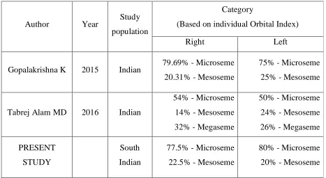
Gopalakrishna K et al (2015) studied the orbital parameters of 64 dry skulls which belonged to Indian population. The measurements were taken using vernier caliper. The author stated the results as
Vertical diameter of right and left orbit was 32.75±2.21mm and 33.05±1.99mm respectively.
Horizontal diameter of right and left orbit was 40.62±3.06mm and 40.75±2.69 mm respectively.
Orbital index of right and left orbit was 80.69±2.19 and 81.16±2.02 respectively.
He classified 79.69% of right orbit and 75.0% of left orbit under microseme category. The rest of the orbits were classified under mesoseme category.
for the horizontal diameter and orbital index. The Pearson correlation coefficient and regression analysis was done to evaluate the strength and relationship of studied parameters. The strength of relationship between the pairs of orbit for vertical diameter was upto 92.16%, horizontal diameter upto 90.25% and for orbital index was 60.22%.
Rao NB et al (2015) studied the orbital index for North coastal Andhra Pradesh people. He utilized 70 adult dry skulls of that population. He reported that the mean orbital height of right sided orbital cavity was 32.62±2.03mm and left sided orbital cavity was 32.89±2.2mm. The mean orbital breadth of right sided orbital cavity was 36.5±1.92mm and left sided orbital cavity was 36.41±1.78mm. The mean orbital index was 86.13 for right orbit and 90.69 for left orbit. He concluded by classifying the orbital cavity of study population between mesoseme and megaseme category.
Orish CN et al (2016) studied the craniometric indices in 100 adult dry skulls of Nigerian population. The craniometric indices he studied were gnathic, cranial, palatal, orbital and nasal indices. These craniometric indices were useful for identification of sex and race of skulls of unknown identity. The results of the study were
Mean orbital breadth of male and female was 40.76 mm and 37.73 mm respectively.
Mean orbital height of male and female was 36.52 mm and 32.85 mm respectively.
The average orbital index was 89.59 for males which classified the orbital cavity of Nigerian males under megaseme category.
The average orbital index was 87.04 for females which classified the orbital cavity of Nigerian females under mesoseme category.
Tabrej Alam MD et al (2016) studied the dimensions of orbital cavity in 50 adult dry skulls of Indian population. The measurements were taken directly in the skull with the help of Vernier caliper. He measured
Orbital length as the maximum distance between superior and inferior margins of orbital rim.
Orbital breadth as the distance between midpoints of medial and lateral margins of orbital rim.
Orbital index as (orbital length/orbital breadth)×100
According to this study,
a. Orbital length of right and left orbit was 34.3±2.09mm and 34.4±1.68mm respectively.
b. Orbital breadth of right and left orbit was 41.6±3.26mm and 41.8±2.48mm respectively.
c. Orbital index of right and left orbit was 82.60±4.43mm and 82.76±6.96mm respectively.
REVIEW OF LITERATURE – RADIOLOGICAL STUDY
Forbes G et al (1985) studied the volume of total bony orbit, volume of orbital soft tissue, volume of extra ocular muscle and volume of orbital fat, in vivo in 29 patients (58 orbits). He found the volume of these structures by summation of pixel count in continuous thin axial slices of 1.5mm. He stated that the mean total bony orbital volume for right orbit was 22.06 cm3 and left orbit was 22.70 cm3. The percentage of difference for right orbit was 3.0 cm3 and left orbit was 7.5 cm3.
Waitzman AA et al (1992) studied the normal values and growth trends using the measurement of the upper craniofacial region which were taken from 542 CT scan series of normal subjects. The age range of the subjects was 1 to 17 years. In his study he quoted that at the age of 17 years
a. Anterior interorbital distance i.e. the distance between the points on each lacrimal bone was 23.8mm ±1.7mm
b. The biorbital distance i.e. the distance between the most anterior point of each lateral orbital wall 95.3mm ± 5.9mm,
c. The medial orbital wall length was 44.2 ± 3.1mm, d. The lateral orbital wall length was 47.1 ± 2.7mm.
orbital cavity was calculated. They calculated the orbital volume by multiplying the area of orbital cavity with section thickness. He also demonstrated the position of the globe which was the distance between zygomaticofrontal process baseline and posterior surface of the lens in each orbit. The distance of enophthalmos was calculated by subtracting fractured eye globe distance from normal eye globe distance. By using Pearson correlation coefficient he found significant correlation between the increase in volume of the total bony orbit (because of blowout fracture) and enophthalmos. He reported that the increase in total bony orbital volume by 1 cm3 would produce enophthalmos of 0.8mm.
between the two methods was 0.93 ± 1.08 ml for each orbit. The correlation between these two techniques was found to be high (r=0.887, P<0.01).
Furuta M (2000) studied the orbital volume in computed tomographic scans of 109 patients. He measured the orbital volume in serial coronal sections of 2 mm thickness. These coronal sections were taken between the lateral orbital rim and the optic canal. In his study, he reported that the mean orbital volume in adult Japanese was 23.6 ± 2.0cm3 in men and 20.9 ± 1.3 cm3 in women.
Ramieri G et al (2000) observed the dimensions and volume of orbital cavity in two dimensional and three dimensional computerized tomographic images of post traumatic patients. He conducted the study in twenty five adult patients in which thirteen were males and twelve were females who underwent surgical intervention for severe orbital fractures either unilateral or bilateral. Out of fifty orbits, thirty four were fractured orbits, fourteen were normal orbits and two were anophthalmic orbits. He included both fractured orbits and normal orbits in the study. The normal orbits were considered as control group. The anophthalmic orbits were excluded from the study. After getting informed consent from each patient, post operative computerized tomographic image was taken using contiguous axial slices of thickness 1mm. Three dimensional reconstruction of 2D image was done. With the help of Mimics software version 4.0 (materialized Leuven, Belgium) linear dimensions and volumetric measurements were made.
anterior lacrimal crest and the lateral wall length was measured from optic foramen to lateral rim.
In three dimensional images, with the saggital cut which was 3mm lateral to medial wall, the orbital height was measured as the vertical distance between the supraorbital rim and the infraorbital rim and the bony orbital volume was measured with the help of application, voxel-based volume reconstruction.
He reported that for control group
The mean medial wall length was 41.69±4.7mm. The mean lateral wall length was 44.82±3.2mm. The mean orbital height at rim was 36.61±3.3mm. The mean bony orbital volume was 24975±2486mm3. He stated that
The mean bony orbital volume of all fractured orbits was 28516±2876mm3 which was significantly higher than control group.
The mean bony orbital volume of fractured orbits with no enophthalmos was 27742±2654mm3 and with enophthalmos was 29567±2922mm3 which showed high correlation between increased bony orbital volume and enophthalmos.
method and 3D-CT method. He compared these with direct anatomical measurement and evaluated with Lin’s concordance coefficient (Þc). The concordance coefficient between direct anatomical measurement and 3D-CT based method for area of fracture was Þc=0.962 and for volume of silicone was Þc=0.872. The concordance coefficient between direct anatomical measurement and 2D-CT based method for area of fracture was Þc=0.981 and for volume of silicone was Þc=0.952. This showed the accuracy of both 2D-CT based method and 3D-CT based method. The mean processing time allocated for 2D-CT method was 6.5 ± 1.3 min and 3D-CT method was 20.8 ± 4.6 min. In this study he proved that 2D-CT based calculation method utilized lesser processing time than 3D-CT based method which was statistically significant.
post-operative bony orbital volume (after surgical expansion of orbital cavity) was 31.6 ml/cm3.
Ye J et al (2006) studied the orbital volume in two dimensional computed tomographic scans. He measured the orbital volume in coronal section of 3 mm thickness. He stated that mean orbital volume was 23.94 ± 3.47 cm3.
Kahn DM et al (2008) studied the age changes in the orbital cavity of both sexes in three dimensional computed tomographic scans. He had chosen three age groups in both sexes. The mean age of young age group for male was 33.8 years and for female was 30.4 years. The mean age of middle age group for male was 57.7 years and for female was 54.5 years. The mean age of old age group for male was 75.4 years and for female was 75 years. He stated that the mean orbital aperture width of young age group for male was 39.0 mm and for female was 35.7 mm. The mean orbital aperture width of middle age group for male was 40.2mm and for female was 37.7mm. The mean orbital aperture width of old age group for male was 40.6 mm and for female was 38.7 mm. He stated that the mean orbital aperture area of young age group was 1082.7 mm2 and 945.3 mm2 for male and female respectively and middle age group was 1104.6 mm2 and 1047.9 mm2 for male and female respectively and old age group was 1191.2 mm2 and 1100.1 mm2 for male and female respectively.
Seiji F et al (2009) studied the orbital asymmetry in one hundred and twenty seven dry human skulls with known age and sex. He grouped the skulls according to its age.
Group I – Intrauterine life (n=20)
Group III – Three to 20 years old (n=27); Group IV – 21 to 76 years old (n=37);
He photographed these skulls in Norma frontalis and measurements were obtained by using software program. The parameters he observed were greater horizontal diameter, greater vertical diameter, orbital perimeter and orbital base area. In this study he stated that the orbital cavity of all skulls were asymmetrical except the orbit of four skulls which were symmetrical in their vertical diameter and perimeter.
Acer N et al (2009) studied the orbital volume in 9 dry skulls by two methods. Method I was water filling method which is the standard method for volume determination. Method II was point counting method.
Method I water filling method
In this method he introduced the ordinary balloon into the orbital cavity and filled the balloon with water under pressure from tap. When the orbital cavity was filled, he poured the water into the measuring cylinder which was taken as the total volume of the orbit. Water filling method is the gold standard criterion method for the measurement of volume in vitro.
Method II Point counting method
Using a conventional scanner, computed tomographic images of 9 skulls were obtained. All skulls were scanned in axial plane; the thickness of slice was 1.5 mm without interval. Point counting was done in all series of axial section and orbital volume was calculated.
were analysed statistically and it was found that there was no statistical significant difference between the two methods. He stated that the point counting method was very accurate to that of standard water filling method and it was used for orbital volume calculation in vivo.
Weaver AA (2010) studied the orbit and eye anthropometry in three dimensional reconstructions of computerized tomographic images of 39 subjects of different age ranges from 17 to 76 years. He stated that orbit widens with age. In this study he quoted that the mean orbital width in male was 37.42 ± 2.4 mm and in female was 36.60 ± 1.71 mm. The mean orbital height in male was 32.44 mm ± 1.89mm, and in female was 31.75 ± 2.51 mm. The total rim perimeter in male and female was 114.74 ± 5.49 mm and 112.15 ± 5.44 mm respectively.
the mean intra orbital distance was 27.18 mm and 25.11 mm, and the mean extra orbital distance was 98.77 mm and 93.69 mm. With all the above mentioned parameters, he concluded that the orbital size of women was significantly smaller than the orbital size of men but there was no significant difference in orbital height between both the sexes.
Igbigbi PS et al (2010) studied the morphometry of orbit from x rays of adult Malawian population. He studied the orbital index of both the sex of Malawian population in 136 x rays in which 70 were x rays of men and 66 were x rays of women. He selected anteroposterior radiographs with occipito – mental views (waters’ view) and placed it on the x ray viewer. He measured orbital width and height and calculated the orbital index. He stated that the mean orbital height of Malawian male and female was 42.07±2.96mm and 40.26±1.92mm respectively. The mean orbital width of Malawian male and female was 44.57±2.24 mm and 42.15±2.20mm respectively. The total orbital index of Malawian male and female was 94.35±5.56mm and 96.03±3.34mm respectively. He classified the orbital cavity of Malawian population under megaseme category (square type). The orbital index of Malawian female was greater than the Malawian male. He reported that the orbital index of some Malawian females were nearer to 100 (i.e. orbital height = orbital breadth).
the bony orbital margin were counted. The volume of orbital cavity was calculated by multiplying slice thickness, total number of points and the area represented by each point. In this study he stated that the mean bony orbital volume of right orbital cavity was 22.35 ± 3.05 cm3 and 18.47 ± 2.52 cm3 for male and female respectively. Similarly the mean bony orbital volume of left orbital cavity was 22.71 ± 2.80 cm3 and 18.16 ± 2.52 cm3 for male and female respectively.
In the orbital floor fractures, orbital contents were herniated into maxillary sinus and the orbital volume was decreased. This leads to the complication enophthalmos which would require surgical management. The volume of orbital contents herniated into maxillary sinus seems to determine the mode of management. Alinasab B et al (2011) did a study in 23 patients with untreated unilateral isolated fracture of the orbital floor to evaluate whether the change in the orbital volume would serve as an indicator for surgical management. He measured the orbital volume in two dimensional computed tomographic scans of above said patients and also for 18 normal subjects (as control group) for reference value. He concluded that there is no clear relation found between the herniated orbital contents and development of enophthalmos. He reported that the volume of right orbit of control group ranges from 17.7 ml to 24.7 ml and the volume of left orbit of control group ranges from 18.6 ml to 25.6 ml.
radiological volumetric measurement was done in both affected and unaffected eye preoperatively and as well as post operatively. The orbital volume was measured with the help of volume viewer (an installed software application program) in two dimensional computed tomographic scans using 0.625mm slice thickness which was measured in cubic centimeter (cm3). He reported that the bony orbital volume of unaffected side ranges from 20.015 cm3 to 29.395 cm3.
Lukats O et al (2012) conducted a study in 20 patients who underwent enucleation and orbital implantation for various pathological reasons. He observed the differences in the orbital volume between the orbit containing artificial implant and the healthy opposite side orbit. He also selected 20 patients with various dental problems and unaffected orbital cavities and measured the orbital volume in these patients to observe the differences in the orbital volume of right and left orbit. And he concluded that there was no significant difference between the volume of right and left orbital cavity.
Bankole JL et al (2012) conducted the study in Ikwerre and Kalabari tribes of Rivers state, Nigeria. He studied their orbital morphometry from 300 normal radiographs of skull. In which 150 radiographs belonged to females of both tribes and 150 belonged to males of both tribes. He measured the orbital height and orbital width from frontal view of x ray and orbital depth from lateral view of x ray. He reported that
Mean orbital width of Ikwerre and Kalabari males was 42.87±3.88mm and 41.14±3.09 mm respectively. This difference was statistically significant. Mean orbital index of Ikwerre and Kalabari males was 103.98±8.22 and
102.92±9.49 respectively. This difference was not statistically significant. Similarly the mean orbital length of females of both tribes showed statistical
significant difference but not the orbital width and index.
Erkoc MF et al (2014) conducted a retrospective study using magnetic resonance imaging scans of 1,453 subjects of different age groups. He divided these 1,453 subjects into five age groups with interval of 10 year. He measured the volume of orbit and volume of total orbital fat and observed their correlation with age, gender, height and weight of the subject. The orbital volume was measured in T1-weighted magnetic resonance imaging scans in axial view with slice thickness of 3mm. In each axial slice, orbital cavity was selected by taking optic foramen as posterior boundary and the line connecting the anterior most points of medial and lateral wall as anterior boundary. Then they calculated the area of selected orbital cavity by using software program. The volume was calculated by multiplying the area of orbital cavity with slice thickness. The summation of volume of all axial slices gave the total orbital volume. He reported that mean total orbital volume of men was 32.21 ± 1.55 cm3 and for women was 31.11 ± 1.87 cm3. He stated that there was significant difference between total orbital volume of men and women. Total orbital volume of men was significantly greater than that of women.
ethnic groups. He reported that the mean orbital volume for male in right orbit was 24.7 ± 1.17 ml and in left orbit was 24.3 ± 1.51 ml. The mean orbital volume for female in right orbit was 16.1 ± 0.92 ml and left orbit was 16.0 ± 1.01 ml. He observed the mean orbital volume of males was higher than the mean orbital of females and the difference was statistically significant (p<0.001).
Methodology of this study
He chosen 20 patients randomly (10 males & 10 females) who already underwent facial CT scan for any other craniofacial abnormality excluding orbital cavity. The two dimensional scans were reconstructed to three dimensional image using 3D multiplanar reconstruction (3D MPR). Then the 3D images were oriented to Frankfurt plane.
He selected the following points in 3D image. Laterally - Zygomatico frontal process
Inferomedially - Anterior lacrimal crest
Superomedially – Nasal process of frontal bone Superiorly – supraorbital margin
Inferiorly – infraorbital margin
Anterior limit – a line connecting lateral and medial orbital rim landmarks Posterior limit – optic foramen
called compute ROI volume tool (OsiriX software). He finally confirmed that 3D assisted method as accurate method which corresponds to gold standard water displacement method for volume calculation.
Wu D et al (2014) observed absolute hertel exophthalmometric values (EVs) for Chinese Han population. The device called Hertel exophthalmometer was used to measure exophthalmometric value (EV) and distance between the two lateral orbital rims. The absolute exophthalmometric value was used for diagnosis of proptosis and to monitor the progress of disease. In this study he reported that the distance between the two lateral orbital rims was 111.0±4.0mm (mean ± SD) in adults. The range was 90mm to 122mm.
Jeong HC et al (2015) did a retrospective study using 44 computed tomographic scans, in which 22 belonged to Korean population and 22 belonged to Caucasian population. He studied the morphometry of orbit and compared the orbital anatomy in both populations. He measured
a. Medial wall of orbital cavity in the axial view as the distance between posterior lacrimal crest to optic canal
b. Lateral wall of orbital cavity in the axial view as the distance between anterior most extent of zygomatic bone and optic canal.
c. Horizontal orbital length in the axial view as the distance between the anterior most points on the medial and lateral rim of orbit.
e. Interorbital length in the axial view as the distance between the right and left posterior lacrimal crests.
f. Lateral wall interorbital length in the axial view as the distance between the right and left zygomatic orbital rim.
He reported that
a) The length of medial wall of orbital cavity for Korean and Caucasian population was 35.54±1.40mm and 35.51±1.81mm respectively.
b) The length of lateral wall of orbital cavity for Korean and Caucasian population was 44.60±2.08 mm and 44.82±2.44 mm respectively.
c) The horizontal orbital length for Korean and Caucasian population was 34.22±1.78mm and 34.66±1.39mm respectively.
d) The vertical orbital length for Korean and Caucasian population was 34.19±1.67 mm and 35.03 ±1.18 mm respectively. The vertical orbital length showed statistical significant difference between the two populations. (p<0.05) e) The inter orbital length for Korean and Caucasian population was 24.05±2.00
mm and 21.96±1.96 mm respectively which showed statistical significant difference between the two population (p<0.05).
f) The lateral wall inter orbital length for Korean and Caucasian population was 97.73 ±2.46 mm and 97.69±2.66 mm respectively.
thickness was 0.625mm. The morphometric measurements were taken with the help of GE Advantage Windows 4.3 workstation which had three dimensional options. He stated that the morphometric measurements which were taken in three dimensional images were not accurate. To avoid this inaccuracy the measuring line was first placed in axial view of two dimensional images over the appropriate bony prominences. Then he placed the same measuring line over the same bony points in three dimensional images. The total length of measuring line would give the readings.
The results were
In the lateral wall, the distance between frontozygomatic suture and optic canal was 46.15mm and 43.58mm in males and females respectively.
In the medial wall, the distance between anterior lacrimal crest and optic canal was 40.40mm and 38.39mm in males and females respectively.
In the superior wall, the distance between supra optic foramen and optic canal was 46.49mm and 43.29mm in males and females respectively.
In the inferior wall, the distance between zygomaticomaxillary suture and optic canal was 45.24mm and 42.80mm in males and females respectively.
MATERIALS AND METHOD
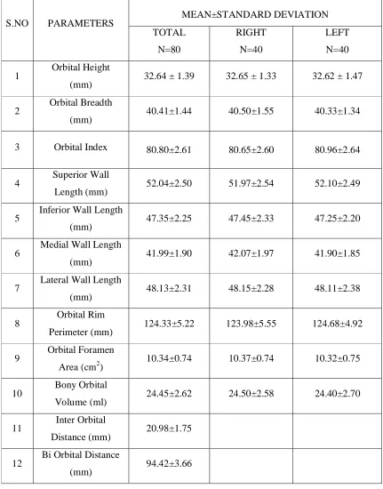
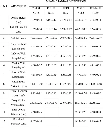
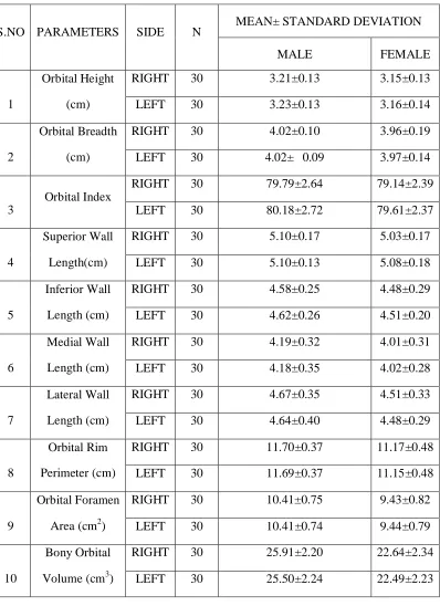
The quantitative morphometry of orbital cavity was studied in 40 adult dry skulls and computed tomographic images of brain belonging to 60 patients (30 males and 30 females). Adult dry skulls with intact orbital cavities and the computed tomographic images of brain reported as ‘normal study’ were only included in this study. The patients with any disease affecting eye and orbital cavity such as thyroid disease, Intra orbital tumor and congenital abnormalities like microphthalmia, anophthalmia and orbitofacial cleft were excluded from the study. The study was conducted after obtaining approval from ethics committee of institution.
METHODOLOGY FOR DRY BONE STUDY
In adult dry skulls, measurements were taken by using digital vernier caliper. The following points were marked over the orbital margins of skulls, between which the measurements were taken (Figure 2).
1.A point MF was marked on the medial orbital margin over maxillofrontal suture. 2.A point EC was marked on the lateral orbital margin over ectoconchion. The point ectoconchion was defined as the anterior most point on the lateral orbital margin intersected by the horizontal bisecting line of orbital cavity.
3.A point SO was marked on the supraorbital margin, over the point of intersection of supraorbital margin and perpendicular bisector of the line joining MF and EC.
4.A point IO was marked on the infra orbital margin, over the point of intersection of infra orbital margin and perpendicular bisector of the line joining MF and EC.
]
Orbital index was calculated as
Orbital height
X 100
Orbital breadth
The length of superior wall was measured as the distance from the marked point on the supraorbital margin to the superior most point on the superior border of optic foramen (Figure 5a, 5b). The length of inferior wall was measured as the distance from the marked point on the infraorbital margin to the inferior most point on the inferior border of optic foramen (Figure 6a, 6b). The length of medial wall was measured as the distance from the marked point on the medial orbital margin to the medial most point on the medial border of optic foramen (Figure 7a, 7b) The length of lateral wall was measured as the distance from the marked point on the lateral orbital margin to the lateral most point on