Transcriptase in
cis
and in
trans
during Protein-Primed Initiation of
DNA Synthesis
In Vitro
Rajeev K. Boregowda, Christina Adams, and Jianming Hu
Department of Microbiology and Immunology, The Penn State University College of Medicine, Hershey, Pennsylvania, USA
The hepadnavirus reverse transcriptase (RT) has the unique ability to initiate viral DNA synthesis using RT itself as a protein primer. Protein priming requires complex interactions between the N-terminal TP (terminal protein) domain, where the primer (a specific Y residue) resides, and the central RT domain, which harbors the polymerase active site. While it normally utilizes the
cis-linked TP to prime DNA synthesis (cis-priming), we found that the duck hepatitis B virus (DHBV) RT domain, in the context of the full-length RT protein or a mini-RT construct containing only truncated TP and RT domains, could additionally use a sep-arate TP or RT domain intransas a primer (trans-priming).transinteraction could also be demonstrated by the inhibitory effect (trans-inhibition) oncis-priming by TP and RT domain sequences provided intrans. Protein priming was further shown to in-duce RT conformational changes that resulted in TP-RT domain dissociation, altered priming site selection, and a gain of sensi-tivity to a pyrophosphate analog inhibitor.trans-priming,trans-inhibition, andtrans-complementation, which requires sepa-rate TP and RT domains to reconstitute a functional RT protein, were employed to define the sequences in the TP and RT domains that could mediate physical or functional inter- and intradomain interactions. These results provide new insights into TP-RT domain interactions and conformational dynamics during protein priming and suggest novel means to inhibit protein priming by targeting these interactions and the associated conformational transitions.
T
he hepatitis B virus (HBV) is a major human pathogen thatchronically infects over 350 million people worldwide (15,32).
Chronic HBV infection is a major cause of end-stage liver diseases, including cirrhosis and hepatocellular carcinoma, resulting in a
million fatalities annually. HBV is a member of the
Hepadnaviri-daefamily, which also includes related viruses that infect other
mammalian and avian species (39). In particular, the duck hepa-titis B virus (DHBV) has been a widely used model to study many different aspects of HBV replication and pathogenesis. All hepad-naviruses, as pararetroviruses, replicate a short (ca. 3-kb), partially double-stranded (DS), relaxed circular DNA genome via packag-ing and reverse transcription of a pregenomic RNA (pgRNA) by a
virally encoded reverse transcriptase (RT) (37,39,41).
The hepadnavirus RT is a multifunctional protein with unique
structural and functional properties (20, 21). Like its retroviral
counterparts, RT catalyzes the synthesis of the DS viral DNA, first the minus strand from the pgRNA template and then the plus
strand from the minus-strand DNA template (10,12,35,47). Also
in common with retroviral RTs, the hepadnavirus RT has an RNase H activity that degrades the pgRNA template during the
synthesis of the viral minus-strand DNA (10,11,35). Thus, the
central and C-terminal regions of RT harbor, respectively, the RT and RNase H domains that are homologous to retroviral RTs. Uniquely, however, the hepadnavirus RT has a so-called N-termi-nal, terminal protein (TP) domain, which is conserved among all hepadnaviruses but absent from all other RTs. TP is linked to the RT domain via a flexible spacer region. Furthermore, the hepad-navirus RT is able to initiate minus-strand DNA synthesis using itself as a protein primer, via a complex protein priming mecha-nism whereby a specific tyrosine residue in the TP domain is used
as a primer, resulting in a covalent linkage between the 5=end of
the viral minus-strand DNA and the RT protein via a
phosphoty-rosyl bond (4,28,43,49,53,56). Furthermore, protein priming
requires RT recognition of a specific viral RNA, an RNA
stem-loop structure located on the 5=end of pgRNA calledε(14,20,23,
33, 34,36,44,46,48,50). In particular, the sequence from an
internal bulge ofεserves as the specific template for protein
prim-ing to direct the synthesis of a short (3- to 4-nucleotide [nt]) minus-strand DNA oligomer that is covalently linked to RT. Based on distinct sequence and structural requirements, protein
primingin vitroby the DHBV RT has been subdivided into two
sequential stages, i.e., the first stage of priming initiation resulting in the formation of the phosphotyrosyl bond between the primer Y residue (Y96 in DHBV) and the first nucleotide (dGMP) of the minus-strand DNA and the second stage of DNA polymerization involving the addition of the next 2 to 3 nt to the initiating dGMP
via conventional DNA phosphodiester linkages (31,51).
Extensive genetic and biochemical studies have shown that both the TP and RT domains of the hepadnavirus RT protein are
required to interact with ε and to carry out protein priming
whereas the spacer and the C-terminal RNase H are dispensable
(17,27,34,50,51). For protein priming to occur, the TP and RT
domains must interact precisely so that the primer Y residing in TP is properly positioned into the DNA polymerase active site in the RT domain. Furthermore, these two domains must
simulta-neously engage theεRNA so that its internal bulge template
se-quence is properly positioned next to the TP primer as well as the
Received11 January 2012 Accepted9 April 2012
Published ahead of print18 April 2012
Address correspondence to Jianming Hu, [email protected].
Supplemental material for this article may be found athttp://jvi.asm.org/. Copyright © 2012, American Society for Microbiology. All Rights Reserved.
doi:10.1128/JVI.00086-12
on November 7, 2019 by guest
http://jvi.asm.org/
RT active site for phosphotyrosyl bond formation (i.e., initiation of protein priming or TP deoxynucleotidylation). In order for RT
to adopt a conformation competent for interaction withε, specific
host factors, including a cellular chaperone complex consisting of the heat shock protein 90 (Hsp90), Hsp70, and other cochaper-ones, are recruited to associate with RT and facilitate the
establish-ment of theε-binding competent state (18,19,22,25).
Further-more, upon specific RT andεinteraction, conformational changes
are triggered in both the RT protein and theεRNA of the resulting
ribonucleoprotein (RNP) complex through an induced-fit mech-anism and are thought to activate the RT enzymatic activity and
theεtemplate function (6,40,42,45).
We and others have reconstituted DHBV RT-εinteraction and
protein priming using purified, bacterially expressed RT, its
cog-nateεRNA, and the eukaryotic chaperone proteins (7,17,24,40).
Detailed analyses of the DHBV RT requirements for protein prim-ing also led us to the construction of a severely truncated RT protein, MiniRT2, which lacks part of the TP, the spacer, part of the RT, and the entire RNase H domain and retains the ability to
carry out authentic,ε-dependent protein primingin vitrobut is no
longer dependent on the host chaperone proteins forεbinding
and protein priming (31,52). In addition, separately expressed TP
and RT domains, containing the minimal TP and RT domain
sequences as defined in MiniRT2, can interact intrans, i.e.,
inter-molecularly, to reconstitute a functional RT protein and carry out
protein priming (7,27,29,31).
Recent work using the simplifiedin vitroDHBV priming
sys-tems, including MiniRT2, has demonstrated that hepadnavirus RT is remarkably flexible and dynamic in structure and function.
For example, we have shown that the divalent metal ions, Mn2⫹
versus Mg2⫹, can induce significantly different RT conformations
that dramatically affect the RT protein priming functions, includ-ing catalytic efficiency, template and nucleotide selectivity, and the transition from priming initiation to DNA polymerization (31). In addition, sensitivity to inhibition by a pyrophosphate an-alog, phosphonoformic acid (PFA), can be induced during the polymerization but not the initiation stage of protein priming in
the presence of Mn2⫹, whereas PFA shows no effect on protein
priming (either initiation or polymerization) in the presence of
Mg2⫹and inhibits viral DNA synthesis only following protein
priming (31,49). Furthermore, we and others have recently
dis-covered that DHBV RT protein can utilize so-called cryptic prim-ing sites, i.e., Y as well as S/T residues, other than Y96, in both the
TP and RT domains to initiate DNA synthesis (5, 8). As with
authentic priming at Y96, priming at the cryptic sites requires both the TP and RT (in particular, its polymerase active site)
do-mains and theεRNA and is stimulated by Mn2⫹relative to Mg2⫹.
In the present study, we found that MiniRT2, and the full-length DHBV RT, was able to initiate protein priming on a
sepa-rate TP or RT domain provided in trans (trans-priming), in
addition to carrying out protein priming from thecis-linked TP
(cis-priming). Further studies showed that TP and RT domain
sequences provided intranscould also exert an inhibitory effect
on cis-priming (trans-inhibition). Taking advantage of these
trans-priming andtrans-inhibition effects, as well as the trans -complementation priming assay, we have defined the TP and RT domain sequences that were necessary for primer function, prim-ing inhibition, or primprim-ing reconstitution through inter- and
in-tradomain interactions incis(i.e., intramolecular) or intrans(i.e.,
intermolecular).
MATERIALS AND METHODS
Plasmids. pGEX-MiniRT2 and pQE-MiniRT2 express the truncated DHBV MiniRT2 protein that is fused to the glutathioneS-transferase (GST) and the six-histidine (6⫻His) tag, respectively (17). pSUMO-MiniRT2 expresses the same DHBV pSUMO-MiniRT2 protein with an N-terminal SUMO tag as well as the 6⫻His tag. It was created by subcloning the MiniRT2 coding sequence downstream of the SUMO coding sequence in pSUMO-T7-Amp (Lifesensors) with an additional triple-Flag tag inserted between SUMO and the MiniRT2 sequences. pGEX-MiniRT2-YMHA was derived from pGEX-MiniRT2 and contains two amino acid substitu-tions in the RT active site (changing the conserved YMDD motif into YMHA) (10,51) that abolish the polymerase activity of RT. pQE-TP and pQE-RT express the 6⫻His-tagged, truncated TP (residues 75 to 220 [75-220]) and RT (residues 349 to 575 [349-575]) domains, respectively, and were derived from pQE-MiniRT2 by removing the RT and TP coding sequences, respectively (31). Similarly, pGEX-TP and pGEX-RT were de-rived from pGEX-MiniRT2 and express the GST-tagged, truncated DHBV TP and DHBV RT domains, respectively (30). The pQE-TP-Y96F was derived from pQE-TP by changing the tyrosine residue at position 96 to phenylalanine (8,56). Additional truncated pGEX-TP and -RT frag-ments were generated by PCR. The PCR-amplified TP or RT fragfrag-ments were then cloned into the pGEX vector fused in frame downstream of the GST coding sequence. pHP expresses the full-length DHBV RT under the phage SP6 promoterin vitro(56). All mutations were verified by DNA sequencing.
Protein expression and purification.The DHBV full-length RT pro-tein and SUMO-MiniRT2 were expressedin vitrousing a coupledin vitro transcription and translation reaction kit, the TnT rabbit reticulocyte lysate (RRL) system (Promega), according to the manufacturer’s instruc-tions. The amount of translated SUMO-MiniRT2 (3⫻Flag tagged) was estimated to be 5 to 10 ng/l translation reaction mixture, based on West-ern blotting using the anti-Flag antibody (Sigma) and SUMO-MiniRT2 standards purified from bacteria. The translation yield of the full-length RT protein was similar to that of SUMO-MiniRT2. GST-MiniRT2, -RT, and -TP were expressed in the BL21(DE3)-CodonPlus-RIL cells and pu-rified using the glutathione resin, and SUMO-MiniRT2 was expressed using the same cells and purified using Ni⫹affinity resins (17,19). His-MiniRT2, -RT, and -TP were expressed in M15(pREP4) cells and purified using Ni⫹affinity resins under native conditions (Qiagen) (17). His-RT, His-TP, and SUMO-MiniRT2 proteins were also purified under denatur-ing conditions and refolded as described previously (8,52).
In vitroprotein priming.The protein priming reaction was carried out as previously described (8). Briefly, 1 pmol of purified RT or 2.5l of thein vitrotranslation reaction mixture was mixed with the DHBV mini-ε RNA (6 pmol) (17), 1⫻EDTA-free protease inhibitor cocktail (Roche), 0.5l [␣-32P]dGTP (3,000 Ci/mmol and 10 mCi/ml) or another labeled deoxynucleoside triphosphate (dNTP) as indicated per 10-l reaction mixture, and TMnNK (10 mM Tris-HCl, pH 8.0, 1 mM MnCl2, 15 mM NaCl, 20 mM KCl) or TMgNK (same as TMnNK, except containing 2 mM MgCl2instead of 1 mM MnCl2) buffer. NP-40 (0.2%, vol/vol) was added to stimulate protein priming (52). For thetrans-complementation assay, equimolar amounts (1 pmol each) of TP and RT domains were used. For thetrans-priming assay, 1 pmol of MiniRT2 was used. In addition, 1 pmol (unless indicated otherwise) of the RT or TP domain fragments was also used. To test the inhibitory effect of the different TP and RT fragments on protein priming, TP and RT fragments (32 pmol unless otherwise indi-cated) were mixed with 1 pmol of purified MiniRT2 or 2.5l of the full-length RT or MiniRT2 translated in RRL, for 15 min at room temper-ature. TheεRNA was then added, and the reaction mixtures were incu-bated for an additional 15 min at room temperature before adding the TMnNK or TMgNK buffer and [␣-32P]dGTP. Alternatively, 1 pmol of purified MiniRT2 was mixed first with theεRNA and incubated for 15 min at room temperature. For the full-length RT and MiniRT2 translated in RRL, theεRNA was added during translation to allow the RNA binding to RT. TP and RT fragments (32 pmol unless indicated otherwise) were
on November 7, 2019 by guest
http://jvi.asm.org/
then added to the RNP complex, and the samples were further incubated for 15 min at room temperature before adding the TMnNK or TMgNK buffer and [␣-32P]dGTP. The protein priming reaction was carried out at 30°C for 2 h. The reaction products were resolved by sodium dodecyl sulfate-polyacrylamide gel electrophoresis (SDS-PAGE) and quantified by phosphorimaging.
Phosphoamino acid analysis.Phosphoamino acid analysis was car-ried out as described previously (8). Briefly, following SDS-PAGE, the gel was cut into two pieces. Both pieces were fixed with a 10% acetic acid and isopropanol mixture for 1 h with two changes. The gel pieces were rinsed with distilled water twice. One piece was then treated with 3 M KOH at 55°C for 14 h, and the other was mock treated with water. After KOH or water treatment, the gel pieces were treated with a 10% acetic acid and isopropanol mixture and then with water. Subsequently, the gel pieces were dried and protein priming signals were quantified by phosphorim-aging.
RESULTS
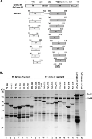
Expression and purification of DHBV RT proteins and do-mains.To facilitate expression and purification, we produced the DHBV MiniRT2 protein and the individual TP (75–220) and RT (349 –575) domains derived from MiniRT2 (Fig. 1A) with the
GST or 6⫻His tag and purified them by glutathione or Ni⫹
affin-ity methods as described before (17, 19, 30). As an additional
attempt to enhance solubility, MiniRT2 was also fused to the SUMO tag. Under native purification conditions, the TP and RT domains and SUMO-MiniRT2 were copurified with GroEL and DnaK, two bacterial chaperone proteins known to bind the DHBV
and HBV RT proteins (Fig. 1B, lanes 1, 8, and 18) (17,19). The
SUMO-MiniRT2 that was purified under denaturing conditions
did not contain the bacterial chaperone proteins and was⬎95%
pure (Fig. 1B, lane 19), with only some degradation products as minor contaminants. Additional TP and RT domain constructs (other than 75–220 and 349 –575) derived from further trunca-tions were also made as GST fusions and similarly purified using glutathione resins (Fig. 1) and will be described below.
TP and RT domains served as primers intransas well as in
cis.It is known that the separate TP and RT domains cantrans
-complement each other to carry out protein priming using the authentic Y96 site in TP and, to a much lesser degree, the cryptic
priming sites in both the TP and RT domains (7,8,29,31) (Fig. 2,
lanes 1 and 2). When the priming reaction was carried out using
MiniRT2 in the presence of an extra RT or TP domain, intrans, we
found, surprisingly, that the separate RT and TP domains were also apparently used as protein primers for initiating DNA syn-thesis (Fig. 2) by MiniRT2. Furthermore, the ability of the TP and RT domains to serve as primers used by MiniRT2 was indepen-dent of the nature of the tag on the RT or TP domain. Thus,
6⫻His-tagged (Fig. 2, lanes 1 to 4, 6, 9, 13, and 17) or GST-tagged
(lanes 7 and 10) TP and 6⫻His-tagged RT (lanes 12 and 16) all
served as primers. As MiniRT2 was able to utilize the separate TP
(and apparently separate RT, but see below) intransas well as its
owncis-linked TP as a primer, we called priming from the separate
TP by MiniRT2trans-priming to differentiate it fromcis-priming,
whereby MiniRT2 uses its owncis-linked TP as the primer. As with
cis-priming, trans-priming was also detected from TP-Y96F,
which lacks the authentic priming site, indicating thattrans
-prim-ing was occurr-prim-ing at the cryptic site(s) (Fig. 2, lanes 14 and 18).
Both refolded and natively purified TP domain served as a
trans-primer (Fig. 2, lanes 3, 4, 6, 9, 13, and 17). The ability of MiniRT2 to initiate protein priming on the separate RT and TP domains in
transwas also independent of the nature of its own tag. Thus,
SUMO-tagged (Fig. 2, lanes 3, 4, and 8 to 10), 6⫻His-tagged
(lanes 12 to 14), and GST-tagged (lanes 5 to 7) MiniRT2s were all
able to carry out priming using the TP and RT domains intrans.
Both refolded and natively purified SUMO-MiniRT2 was able to
carry outtrans-priming (Fig. 2, lane 4 versus lane 3). Equimolar
amounts of the MiniRT2 proteins and the individual TP or RT domains (1 pmol each) were used in all the priming reactions, except that 4 pmol of His-TP or His-RT was used in the reactions
shown in lanes 11 to 18 inFig. 2, due to the lower priming activity
of His-MiniRT2 than of SUMO-MiniRT2 or GST-MiniRT2. When the RT or TP domain was mixed with MiniRT2-Y96F,
the mutant MiniRT2 was able to initiate protein priming incis
(i.e., using cryptic sites) (Fig. 2, lanes 15 to 18), as shown
previ-ously (8), but also intranson the RT (lane 16) and the wild-type
(WT)- and Y96F-TP (lanes 17 and 18) domain, indicating that
Y96 was not essential fortrans-priming, as was true forcis
-prim-ing and prim-prim-ing viatrans-complementation (i.e., priming in the
presence of separate TP and RT domains but no MiniRT2 or full-length RT; see below) (8). On the other hand, a mutant (YMHA) RT domain that lacks a functional polymerase active site was not able to serve as a primer when mixed with MiniRT2 (see Fig. S1A, lane 3, in the supplemental material), indicating that the apparent
trans-priming observed on the WT RT domain (Fig. 2, lanes 12
and 16; see also Fig. S1A, lane 2) was in fact priming incis(i.e., the
RT domain acting on itself) and that the RT domain was unable to
serve as a primer intransto be used by another RT domain (also
see more results below using additional RT domain constructs), in agreement with a recent report (5). As the RT domain by itself, in the absence of a functional TP, is unable to carry out protein prim-ing on either the TP or the RT domain (8), this result indicated
that the RT domain intranswas able to access TP in MiniRT2.
The above reactions were all carried out in the presence of
Mn2⫹due to the higher priming activity with this metal cofactor
(31). Since RT presumably utilizes Mg2⫹rather than Mn2⫹for
protein priming underin vivoconditions, we also tested the ability
of MiniRT2 to carry out protein priming on the separate TP
do-main intrans, in the presence of Mg2⫹. The result showed that the
GST- or SUMO-tagged MiniRT2 protein was able to carry out
protein priming on TP intransin the presence of Mg2⫹(see Fig.
S1B in the supplemental material, lanes 2 and 4), though much
less efficiently than with Mn2⫹. Furthermore, the full-length
DHBV RT protein was also able to initiate priming on TP intrans,
although excess TP (40 pmol of GST-TP) was needed to obtained
a cleartrans-priming signal (see Fig. S1C, lane 2). In summary, the
RT domain clearly could use a TP domain intrans, as well as a
cis-linked TP, as a protein primer to initiate DNA synthesis.
The nucleotide selectivity intrans-priming was similar to that duringcis-priming.The initiating nucleotide (dGMP) in DHBV protein priming is strictly dependent on (i.e., templated
by) the last nucleotide of theεRNA bulge (CMP) when Mg2⫹is
used as the metal cofactor (48,49,51). We have found previously
that when Mn2⫹, instead of Mg2⫹, is used, the preferred initiating
nucleotide for protein priming is still dGMP, but at a lower level, dAMP and TMP (and, to a still much smaller extent, dCMP) can also be used as the initiating nucleotide, at both the authentic Y96 TP site and the cryptic priming sites in the TP and RT domains (8,
31). This indicates that under the Mn2⫹condition, the RT protein
still uses the correctεtemplate site for priming. To test iftrans
-priming discovered here also used the sameεtemplate site, we
on November 7, 2019 by guest
http://jvi.asm.org/
determined the nucleotide selectivity oftrans-priming in
compar-ison tocis-priming. As shown in Fig. S2 in the supplemental
ma-terial, the dNTP selectivity intrans-priming (labeling of His-TP by
SUMO-MiniRT2) was the same as that incis-priming (labeling of
SUMO-MiniRT2 itself), with the following nucleotide selectivity in
both cases: dGTP⬎⬎dATP⫽TTP⬎⬎dCTP. Thus,trans-priming
appeared to use the sameεRNA template site as didcis-priming.
Protein priming incisled to a change in priming site selec-tion intrans.Since MiniRT2 was able to initiate protein priming from a separate TP domain, we reasoned that protein priming in
FIG 1Construction of DHBV RT proteins and individual TP and RT domains. (A) Schematic diagrams of the RT protein and individual TP and RT domain constructs. The top diagram depicts the full-length DHBV RT protein, with the primer Y residue (Y96) in the TP domain and the511YMDD514active site in the
RT domain denoted. The boundaries (in amino acid positions) of the truncated MiniRT2 (second diagram) and the TP (left) and RT (right) domain constructs (below the MiniRT2 diagram) are indicated. (B) Purification of RT proteins and domains. The DHBV SUMO-MiniRT2 protein and the GST-tagged TP and RT domain constructs were expressed inEscherichia coliand purified by affinity methods as described in Materials and Methods. The GST-tagged TP and RT domains were purified under native conditions (lanes 1 to 17). SUMO-MiniRT2 (His tagged) was purified under either native (N, lane 18) or denaturing (and refolding [R], lane 19) conditions. The purified MiniRT2 protein and domains were analyzed by SDS-PAGE and Coomassie blue staining. The intact protein or domain species are denoted by the stars to the left of the corresponding bands. The copurifying bacterial chaperone proteins, DnaK and GroEL, are also indicated. The absence of GroEL in TP/110 –220 (lane 7) is denoted by the dashed box. The protein molecular mass markers are indicated on the left in kDa. The bracket denotes the degradation products from the GST-tagged TP and RT domain constructs or SUMO-MiniRT2, consisting mostly of the GST or SUMO tag plus variable amounts of the TP or RT sequences remaining attached to the tag.
on November 7, 2019 by guest
http://jvi.asm.org/
[image:4.585.135.447.68.537.2]cismight have triggered a conformational change in the TP and/or
RT domain that dissociated the two domains linked incisand
allowed the RT domain in MiniRT2 to initiate DNA synthesis
from the separate TP domain intrans. As one potential indication
for the putative conformational change occurring in the RT
do-main followingcis-priming, we determined the priming site
se-lected on the separate TP domain by MiniRT2. Recently, we showed that KOH treatment of the primed RT or TP domain can be used to distinguish the protenucleotide (DNA) linkage in-volving either serine/threonine (sensitive to KOH cleavage) or tyrosine (resistant to KOH) from the TP or RT domain. Using this
method, we showed previously that duringtrans
-complementa-tion with the RT and TP domains, the RT domain was able to initiate protein priming using S/T on TP as an alternative priming site(s), in addition to the authentic Y96 priming site (8).
There-fore, we treated thetrans-primed TP (WT or Y96F) domain with 3
M KOH following resolution of the labeled proteins by SDS-PAGE to determine the nature of the phosphoester linkage be-tween the labeled nucleotide (dGMP) and TP (Fig. 3). The
prim-ing signal from the WT TP with Mg2⫹served as an internal control
since priming in the presence of Mg2⫹ is almost exclusively
(⬎95%) from the authentic Y96 site and was not affected by KOH
(Fig. 3, lane 5), whereas the priming signal from TP-Y96F
pro-duced by the RT domain with Mn2⫹served as a control for
prim-ing exclusively from the cryptic S/T site but not the Y site, which was completely eliminated by the KOH treatment (lane 8) (8). Withtrans-complementation, the priming signal on the RT do-main was decreased only slightly, suggesting that priming on the RT domain predominantly occurred at Y (Fig. 3, lanes 6 and 8, top
versus bottom panels), as reported recently by us and others (5,8).
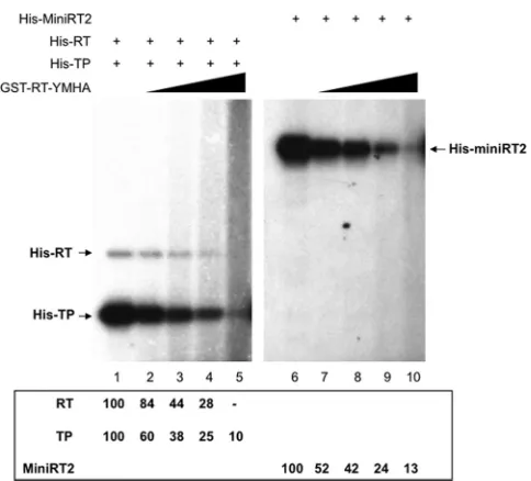
FIG 2trans-priming from the TP and RT domains by purified MiniRT2. Purified MiniRT2, TP, and RT domains were mixed together as indicated for protein priming in the presence of [␣-32P]dGTP and the DHBVεRNA. The RT proteins used were SUMO-MiniRT2 refolded (lane 4) or natively purified (lanes 3 and
8 to 10), natively purified GST-MiniRT2 (lanes 5 to 7), His-MiniRT2 (lanes 11 to 14), and His-MiniRT2-Y96F (lanes 15 to 18). The individual domains used were His-tagged and natively purified (lanes 1, 3, 13, and 17) or refolded (lanes 2, 4, 6, and 9) TP or TP-Y96F (lanes 14 and 18), natively purified GST-TP (lanes 7 and 10), and His-tagged RT domain natively purified (lanes 1, 12, and 16) or refolded (lane 2). All priming reactions were conducted in TMnNK. Priming reaction products were resolved by SDS-PAGE and detected by autoradiography. The labeled RT proteins and domains, as a result ofcis-ortrans-priming, are indicated. All reactions used 1 pmol of proteins/domains, except those shown in lanes 11 to 18, which used 4 pmol of His-TP or His-RT.
FIG 3Determination of priming site usage incis- andtrans-priming by alka-line treatment of the priming products. Priming reactions were carried out in TMnNK (lanes 1 to 4, 6, and 8) or TMgNK (lanes 5 and 7), using SUMO-MiniRT2 (lanes 1 to 4), GST-RT (lanes 5 to 8), His-RT (lane 2), His-TP (lanes 3, 5, and 6), or His-TP-Y96F (lanes 4 and 8). The reaction products were resolved by SDS-PAGE. The gel was then cut into two parts; one part, contain-ing one set of the primcontain-ing reaction products, was mock treated (top panel), and the other part, containing a second set of the same priming reaction products, was treated with 3 M KOH (bottom panels). The priming products were then detected by autoradiography. Lanes 9 to 16 represent a longer exposure of lanes 1 to 8 of the bottom panel.
on November 7, 2019 by guest
http://jvi.asm.org/
[image:5.585.138.449.66.309.2] [image:5.585.300.542.389.622.2]Similarly, the cryptic priming site(s) on the RT domain used dur-ingtrans-priming was also predominantly Y (Fig. 3, lane 2). When
the WT TP domain wastrans-complemented with the RT domain,
the priming signal on the TP domain was decreased by 3- to 4-fold by the KOH treatment, indicating that priming initiation in the
presence of Mn2⫹occurred (mostly) at the cryptic S/T site(s) as
well as at the authentic Y96 site (Fig. 3, lane 6, top versus bottom panels), as we reported recently (8). In contrast, the priming signal
from WT or mutant (Y96F) TP domains trans-primed with
MiniRT2 was not reduced upon KOH treatment (Fig. 3, lanes 3 and 4, top versus bottom panels; also lanes 11 and 12), indicating that the RT domain from MiniRT2 initiated protein priming on TP using mostly, if not exclusively, Y residues (i.e., Y96 plus at least one other Y on TP). Thus, differently from the usage of S/T
cryptic priming sites on TP duringtrans-complementation and
cis-priming (8),trans-priming on TP carried out by MiniRT2
uti-lized predominantly or exclusively a Y residue(s).
PFA sensitivity was induced following initiation of protein priming at Y96.The pyrophosphate analog PFA is reported to block hepadnavirus DNA synthesis but only after protein prim-ing, as it fails to inhibit protein priming either at the initiation or
at the polymerization stage (31,49). However, we recently showed
that PFA could inhibit the polymerization stage of protein
prim-ing when Mn2⫹, instead of Mg2⫹, was used as the polymerase
cofactor, though it still failed to inhibit the initiation stage of
priming even with Mn2⫹(31). These results suggest that RT
un-dergoes a conformational change immediately after initiation of
protein priming in the presence of Mn2⫹that renders it sensitive
to PFA inhibition, whereas with Mg2⫹, this PFA-sensitive RT
con-formation is not adopted until after the polymerization stage of priming. Thus, PFA can be a useful tool to probe the RT confor-mational change during protein priming (and subsequent viral
DNA synthesis). Since priming incisapparently induced a
con-formational change in the RT (and possibly TP) domain of MiniRT2 as evidenced by the altered priming site selection on the independent TP domain, we were next interested in determining
the effect of PFA on priming initiation on TP intransby MiniRT2.
Therefore, we carried out the trans-priming reaction using
MiniRT2 and different concentrations of WT or mutant (Y96F)
TP, in the presence of Mn2⫹, with or without PFA. As reported
before (31), PFA did not inhibit priming initiation incis(i.e., the
labeling of MiniRT2 itself) (Fig. 4A, lane 2 versus lane 1). On the other hand, PFA inhibited (by ca. 2-fold) priming initiation on
the TP domain intransby MiniRT2 (lanes 6 to 8 versus lanes 3 to
FIG 4Sensitivity ofcis- andtrans-priming to inhibition by PFA. (A) Priming reactions were performed in TMnNK using SUMO-MiniRT2 (1 pmol) alone (lanes 1 and 2) or together with increasing amounts of His-TP (1 pmol, lanes 3 and 6; 2 pmol, lanes 4 and 7; 4 pmol, lanes 5 and 8). The pyrophosphate analog PFA (at a 1 mM final concentration) was added to the priming reaction mixtures shown in lanes 2 and 6 to 8. The arrowhead denotes a degradation product from SUMO-MiniRT2 that apparently was able to serve as a primer intransand most likely consisted of the SUMO tag plus the TP domain remaining attached (i.e., SUMO-TP). (B)trans-complementation priming reactions were performed in TMnNK using GST-RT (lanes 1 to 4) and His-TP (lanes 1 and 2) or His-TP-Y96F (lanes 3 and 4). PFA (at a 1 mM final concentration) was added to the reaction mixtures shown in lanes 3 and 4. Priming products were resolved by SDS-PAGE and detected by autoradiography. Priming signals were quantified using phosphorimaging and are represented at the graphs shown at the bottom, with those in the absence of PFA set at 100. The means and standard errors are shown. Statistical significance was calculated using the one-tailed, unpaired Studentttest. *,P⬍ 0.05; **,P⬍0.01; ***,P⬍0.001; ns, not statistically significant.
on November 7, 2019 by guest
http://jvi.asm.org/
[image:6.585.138.446.67.369.2]5). Interestingly, a degradation product from SUMO-MiniRT2
(marked by the arrowhead inFig. 4A) that migrated above His-TP
and presumably corresponded to SUMO fused to the TP domain (with the RT domain being degraded) also was labeled, probably
through trans-priming by the intact SUMO-MiniRT2. trans
-priming from this endogenous degradation product (i.e., SUMO-TP) was sensitive to PFA inhibition, too (lanes 2, 6, 7, and 8). It was also apparent that the addition of increasing amounts of
His-TP diminishedtrans-priming initiation from this putative
degradation product (lanes 3 to 5), probably through competition for access to the RT domain in SUMO-MiniRT2. Also, the slightly
decreasedcis-priming signal of MiniRT2 in the presence of
in-creasing amounts of TP (lanes 3 to 5 versus lane 1) might be attributed to the inhibitory effect exerted by the excess TP domain intrans(see below). Thus, the PFA inhibition of priming
initia-tion on the TP domain intransby MiniRT2 was consistent with
the notion thatcis-priming, which likely occurred beforetrans
-priming (Fig. 4Bbelow; see also the Discussion), induced the
PFA-sensitive conformation in MiniRT2, which subsequently acted on
the TP intransto initiate another round of priming.
We next tested the effect of PFA on protein priming during
trans-complementation where separate TP and RT domains
re-constitute a priming active polymerase. As shown inFig. 4B, PFA
did not inhibit priming initiation from the TP domain, whether WT or Y96F. However, PFA was able to inhibit priming initiation on the RT domain strongly (by 6-fold) when the WT TP and RT
domains were used intrans-complementation (Fig. 4B, lane 3
ver-sus lane 1). In contrast, when the mutant Y96F TP was used, PFA had only a modest effect (2-fold inhibition) on priming initiation from the RT domain (lane 2 versus lane 4). As reported earlier (8), the mutation at TP (Y96F) also decreased priming from the RT domain (lane 2 versus lane 1), suggesting that priming at Y96 could stimulate RT catalytic activity. Together, these results sug-gested that it was priming initiation specifically at Y96 that
in-duced the putative RT conformational state that was more cata-lytically active but also more sensitive to PFA inhibition, whereas priming at the cryptic sites was not able or less able to do so. This
result also suggested that duringtrans-complementation, priming
at the RT domain probably occurred only after that at TP.
An inactive RT domain blocked the interaction between its
cis-linked TP and another independent RT domain intrans.The results above indicated that the RT domain in MiniRT2 could
initiate protein priming on a separate TP domain intransthrough
intermolecular interaction. As intramolecular TP-RT interaction
(i.e., in ciswithin MiniRT2) is expected to dominate over the
intermolecular (intrans) TP-RT interaction, we hypothesized that
in order fortrans-priming to occur, protein priming incismight
induce a conformational change, as already suggested above based on the alteration in priming site selection and the induction of
PFA sensitivity followingcis-priming, which would also weaken
the intramolecular TP-RT interaction in MiniRT2 (i.e., at least partially dissociate the RT from the TP domain) so that the RT
domain in MiniRT2 could access, intrans, a separate TP domain
to initiate priming there. In support of this hypothesis, we found that the priming sites on TP were apparently inaccessible to an RT
domain intranswhen the TP was linked incisto an RT domain
that is catalytically inactive. Thus, when MiniRT2-YMHA, which contains a mutant RT domain with no catalytic activity and is thus unable to carry out protein priming from either the TP or the RT
domain (Fig. 5A, lane 1), was complemented intranswith a
func-tional RT domain (either 6⫻His or GST tagged), little to no
prim-ing from the mutant MiniRT2 was observed (lanes 2 to 5), even though the TP domain, as contained in the mutant MiniRT2,
tagged with either 6⫻His (lane 5) or GST (lane 4), was able to
serve readily as a functional protein primer when provided as an isolated domain by itself (without any RT domain sequences) to
the same RT domain intransin the same reaction (alsoFig. 2to4),
as reported before (31). Furthermore, the small amounts of
ap-FIG 5Failure of an active RT domain intransto rescue the catalytically inactive MiniRT2-YMHA mutant. (A) The priming reactions were performed in the presence of TMnNK. Proteins or domains used were GST-MiniRT2-YMHA (lanes 1 to 5), GST-MiniRT2 (lane 6), GST-RT (lanes 2, 4, and 7), His-RT (lanes 3 and 5), GST-TP (lanes 4 and 7), and His-TP (lane 5). The bracket denotes degradation products from GST-MiniRT2-YMHA that were able to serve as primers intrans. (B) SDS-PAGE and Coomassie blue staining of natively purified GST-MiniRT2 (lane 1) and GST-MiniRT2-YMHA (lane 2). The intact MiniRT2 fusion protein and two copurifying bacterial chaperone proteins, DnaK and GroEL, are indicated. The bracket denotes degradation products from GST-MiniRT2-YMHA (representing mostly just the GST tag itself).
on November 7, 2019 by guest
http://jvi.asm.org/
[image:7.585.139.448.66.270.2]parent degradation products (indicated by the bracket) from GST-MiniRT2-YMHA, invisible on the stained gel (Fig. 5B) but comigrating or migrating just above GST-TP upon labeling by protein priming (Fig. 5A, lanes 2 to 5), were nevertheless labeled at even higher levels than was the intact MiniRT2-YMHA protein, suggesting that the degradation products, containing the TP do-main but with little RT dodo-main sequence redo-maining attached, served as a much more efficient primer than did MiniRT2-YMHA
for the active RT domain intrans. These results thus further
sup-ported the interpretation that the TP domain was sequestered by the inactive RT domain in MiniRT2-YMHA and was inaccessible
to the active RT domain intrans, as a result of the strongcisTP-RT
interaction dominant over TP-RT interactions intrans.
Further-more, these results combined suggested that protein priming incis
indeed dissociated or weakened intramolecular TP-RT domain interactions.
The inactive RT domain intransinhibited protein priming in a dose-dependent manner.Since the inactive RT domain that
wascis-linked to a functional TP domain in the MiniRT2-YMHA
mutant blocked the function of the TP to serve as a primer by an
active RT domain intrans, it was possible that an inactive RT
domain (with the same YMHA catalytic mutations as those in
MiniRT2-YMHA), when added intrans, could also be employed
to inhibit protein priming via nonproductive interaction with TP and thus block the interactions between TP and an active RT do-main. We tested this notion under two different conditions of
protein priming, either throughtrans-complementation
reconsti-tuted with functional TP and RT domains or throughcis-priming
with MiniRT2. As the above results suggested that TP-RT
interac-tion inciswould dominate over the same interaction intrans(Fig.
5), increasing amounts of the mutant RT-YMHA domain were added to the priming reactions. The mutant (YMHA) RT domain was indeed able to inhibit, dose dependently, protein priming on
both the TP and RT domains in thetrans-complementation
reac-tion (Fig. 6, lanes 1 to 5). Furthermore, the mutant RT domain
also inhibitedcis-priming by MiniRT2 in a dose-dependent
man-ner (lanes 6 to 10), suggesting that the mutant RT domain, acting intrans, could indeed block the intramolecular (i.e., incis) TP-RT domain interactions, especially when provided in excess.
Inhibition of protein priming by sequences derived from the RT domain.The strong inhibitory effect of the RT-YMHA mutant on protein priming prompted us to determine in more detail the sequences in the RT domain that could inhibit protein priming
when provided intrans. To this end, a series of RT domain
frag-ments derived from C- and N-terminal truncations were con-structed as GST fusions (Fig. 1A). All the fragments could be ex-pressed in bacteria and purified at levels similar to or higher than that of the initial RT (349 –575) domain construct derived from MiniRT2, with RT/366 –555 producing the largest amount of pu-rified proteins among all the RT fragments. As with the initial RT domain construct (17), all the newly derived RT fragments puri-fied were associated with the bacterial chaperones DnaK and GroEL (Fig. 1B).
To test the inhibitory effects of the RT fragments on protein
priming, we added an excess amount (32 pmol per 10-l reaction
mixture, or 3.2M) of these fragments to SUMO-MiniRT2 (1
pmol), astrans-inhibition by the mutant RT (YMHA) domain was
more effective when it was present in excess over the target of
inhibition (either MiniRT2 or the TP plus RT domain intrans
-complementation) (Fig. 6). Priming reactions were conducted
with Mg2⫹, which is presumably the relevant ion for RT function
in vivo. The mutant RT (YMHA) domain again strongly inhibited protein priming by SUMO-MiniRT2 (Fig. 7A, lane 2 versus lane 1). The other RT domain fragments also showed a strong (though not as strong as the RT-YMHA mutant) inhibitory effect (lanes 3 to 11). Surprisingly, even 349 –575, the initial RT domain con-struct derived from MiniRT2 that is active in reconstituting a
functional RT protein with TP intrans-complementation (Fig. 2
to6) (8,30,31), also blocked MiniRT2 priming, though less
effec-tively than did its mutant (RT-YMHA) counterpart (Fig. 7A, lane 3 versus lane 2). Also, RT/349 –555 and RT/366 –555, which were as active as or more active than the starting RT fragment (349 –
575) intrans-complementation (seeFig. 9below), still inhibited
priming (lanes 4 and 8). The RT fragments also inhibited priming when used at smaller amounts (16 pmol), albeit the inhibition was less efficient, as anticipated (Fig. 7B). These results suggested that multiple inhibitory sequences might be distributed across the RT domain.
Given the inhibitory effect of the RT fragments on priming by MiniRT2, we were next interested in determining their potential effect on protein priming by the full-length RT protein. Therefore,
priming active full-length DHBV RT protein was expressed byin
vitrotranslation in RRL as described previously (16,49), and the RT fragments (32 pmol each) were mixed with the translated full-length RT (ca. 0.1 pmol). Also, to test the potential influence of the
εRNA on the inhibitory effect of the TP and RT fragments, we
addedεeither during translation (i.e., to allow RT-εRNP
forma-tion before the addiforma-tion of the RT fragments) or after preincubat-ing the translated full-length RT with the RT fragments. Primpreincubat-ing
reactions were then performed in the presence of Mg2⫹. When the
RT fragments were added beforeε, none of the RT fragments,
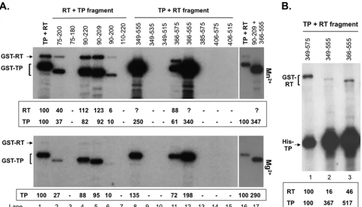
FIG 6Inhibition of protein priming by the RT-YMHA mutant intrans. trans-complementation priming reactions were performed in TMnNK using His-RT and His-TP (lanes 1 to 5), andcis-priming was performed using His-MiniRT2 (lanes 6 to 10). RT-YMHA at a 1-, 2-, 4-, or 8-fold molar excess (i.e., 1, 2, 4, or 8 pmol) was added to thetrans-complementation (lanes 2 to 5) or cis-priming (lanes 7 to 10) reactions. The priming signals of the RT and TP domains or MiniRT2 in the presence of RT-YMHA are indicated at the bottom as percentages of those in the absence of RT-YMHA (lanes 1 and 6).
on November 7, 2019 by guest
http://jvi.asm.org/
[image:8.585.301.543.62.281.2]including the RT-YMHA mutant, inhibited protein priming by the full-length RT (see Fig. S3A in the supplemental material). In contrast, when the RT fragments were added to the preformed
full-length RT-ε RNP complex, some inhibitory effect (2- to
3-fold reduction) on priming was observed in most cases (see Fig. S3B), except for 366-555 (see Fig. S3B, lane 8) and 406-515 (lane 11), which showed only a minor effect (a less-than-2-fold reduc-tion). These results suggested that some of the interacting sites in the full-length RT protein may become more accessible upon RNP complex formation and thus could be more readily targeted by the
inhibitory sequences added intrans.
In general, the inhibitory effect of the RT domain fragments on the full-length RT was less than that on SUMO-MiniRT2, suggest-ing that factors in RRL may be affectsuggest-ing the inhibitory effect of the fragments on priming or that the full-length RT was less sensitive to inhibition than was MiniRT2. To differentiate between these possibilities, we translated SUMO-MiniRT2 as well as the full-length RT in RRL and tested the effects of the RT fragments on
priming by thein vitro-translated MiniRT2. SUMO-MiniRT2,
even when translated in RRL, was also more sensitive to the inhib-itory effect of the RT domain fragments than was the full-length RT expressed in the same system (data not shown), thus indicating that the truncated MiniRT2 was indeed more amenable than the
full-length RT protein totrans-inhibition. This could be explained
by the more-extensive intramolecular (i.e.,cis) interactions in the
full-length RT than in MiniRT2, which would be contributed by the C-terminal RT and RNase H domain sequences absent from MiniRT2 and might make it more difficult for the separate TP or
RT domain fragments to insert into the full-length RT intrans. We
also attempted to determine if TP domain sequences could exert thetrans-inhibitory effects as well. Although some inhibitory ef-fects were observed with some of the TP domain fragments (Fig. 1
and4A; data not shown), the effects were much weaker than those
with the RT domain fragments and were more variable, preclud-ing a definitive analysis uspreclud-ing the current system.
Although it is well established that the TP and RT domains
together are required for specific interaction with theεRNA, it
was possible that the excess RT fragments added intransmight
bindεnonspecifically and thus make it unavailable to support
protein priming by MiniRT2 or the full-length RT in the priming reaction. However, this was made unlikely by the fact that
addi-tion of a large excess of tRNA (100-fold excess over theεRNA and
20-fold excess over the TP or RT fragments) to the priming reac-tion mixtures did not alleviate the inhibitory effect of these frag-ments on protein priming (data not shown). The observation that adding the inhibitory RT fragments to the preformed full-length
RT-εor MiniRT2-εcomplex was as effective or even more
effec-tive in inhibiting protein priming than was adding these fragments
before theε RNA (Fig. 7; see also Fig. S3 in the supplemental
material; also data not shown) also helped exclude this nonspecific RNA binding effect. If the inhibitory RT fragments had simply
inhibited priming by sequestering theεRNA away from MiniRT2
or the full-length RT, little inhibition of the preformed RT-εRNP
complex would have been expected.
There was also a concern that some of the RT domain frag-ments might not remain soluble during the priming reaction and could have aggregated and caused precipitation of the MiniRT2 or full-length RT protein, accounting for their inhibitory effects on priming. This was unlikely because all the RT domain fragments were purified under native conditions routinely at 0.3 to 1.2 mg/ ml, levels which were 2- to 10-fold above those used in the priming reactions. To formally exclude this possibility, SUMO-MiniRT2 as well as all the RT domain fragments remaining in solution after the priming reactions was visualized by SDS-PAGE and silver staining. The amount of SUMO-MiniRT2 remaining in solution was constant whether it was incubated with the GST-RT domain fragments or GST alone, and the GST-RT fragments remained in solution during the priming reactions (see Fig. S4 in the supple-mental material).
Mapping of protein primer sequences in the TP and RT do-mains duringtrans-priming.Given the ability of the TP fragment
75 to 220 to serve as primer in thetrans-priming reaction (Fig. 2to
5; see also Fig. S1 and S2 in the supplemental material), we were interested in determining the potential of additional TP trunca-tion fragments, as well as the series of RT domain fragments
de-scribed above, to serve as a protein primer intrans. As with the RT
domain series described above, a series of TP domain fragments derived from C- and N-terminal truncations were constructed as
FIG 7Inhibition of MiniRT2 priming by RT domain fragments. Protein priming reactions were conducted in TMgNK using natively purified SUMO-MiniRT2 (1 pmol), in the presence of the indicated (GST-tagged) RT domain fragments (lanes 3 to 11) (32 pmol in panel A and 16 pmol in panel B). GST (lane 1) and the RT-YMHA mutant (lane 2) (32 pmol in panel A and 16 pmol in panel B) were used as negative and positive controls fortrans-inhibition, respectively. TheεRNA was preincubated with MiniRT2 before the RT frag-ments were added. The protein priming signals are indicated in panel C as percentages of those without inhibition (i.e., in the presence of GST; lane 1). The means and standard errors are shown in the bar graph. Statistical signifi-cance was calculated using the one-tailed, unpaired Studentttest. *,P⬍0.05; **,P⬍0.01; ***,P⬍0.001.
on November 7, 2019 by guest
http://jvi.asm.org/
[image:9.585.42.283.67.395.2]GST fusions (Fig. 1A). All the fragments could be expressed in bacteria and purified at levels similar to or higher than that of the initial TP (75–220) fragment derived from MiniRT2, with TP/90 – 200 producing the largest amount of purified proteins among all the TP fragments. As with the initial TP (17) and all the RT do-main constructs (Fig. 1B), all the newly derived TP fragments purified were associated with the bacterial chaperone DnaK and, with the exception of TP/110 –220, also with another bacterial chaperone, GroEL (Fig. 1B).
While thetrans-inhibitory effect of the RT domain fragments
was dominant when added in excess to MiniRT2 or the full-length
RT (Fig. 6and7; see also Fig. S3 in the supplemental material),
trans-priming was readily detectable when an equimolar amount (1 pmol) or a slightly larger amount (4 pmol) of the TP or RT
domain (relative to MiniRT2) was used (Fig. 2to4; see also Fig. S1
and S2). Therefore, a slight excess of the newly constructed TP or RT domain fragments (4 pmol each) was mixed with SUMO-MiniRT2 (1 pmol) and priming reactions were carried out. Simi-lar to the starting TP construct (75–220), all new TP constructs (Fig. 8, lanes 2 to 7) except one (110 –220) (lane 8) were able to
serve as atransprimer used by MiniRT2. With the RT domain, in
addition to the starting construct (349 –575) (lane 9), three other constructs, 349 –555, 366 –575, and 366 –555 (lanes 10, 13, and 14,
respectively), also apparently served as primers fortrans-priming.
The priming signals from 349 –575 (lane 9) and 366 –575 (lane 13)
were more difficult to visualize inFig. 8due to their weak labeling
and their comigration with MiniRT2 degradation products, but
these two RT fragments clearly showedtrans-priming signals in
other experiments (e.g., see Fig. S1A, lane 2; alsoFig. 3, lane 2, and
data not shown). As described below (seeFig. 9), these three RT
domain constructs, like the starting RT construct (349 –575), also retained the ability to reconstitute priming (i.e., catalytic activity)
in thetrans-complementation assay and thus, like 349 –575,
prob-ably carried out priming really incis: i.e., the same RT domain,
upon interaction with the TP domain in MiniRT2, initiated prim-ing from itself (see Fig. S1A). In contrast, the shortest RT domain construct, 406 –515, could still serve as a primer even though it
completely lacked any RT activity intrans-complementation (see
Fig. 9, below) and thus was incapable of self-priming incis,
indi-cating that it truly served as a primer intrans.
Mapping of minimal TP and RT domain sequences required to reconstitute protein priming throughtrans -complementa-tion.The availability of the various TP and RT domain fragments also provided the opportunity to map further the minimal TP and RT domain sequences required to reconstitute a priming active
RT protein throughtrans-complementation and to compare the
TP and RT domain sequence requirements fortrans-inhibition
(Fig. 7; see Fig. S3 in the supplemental material) andtrans-
prim-ing (Fig. 8) with those fortrans-complementation. Therefore, we
performed a trans-complementation priming assay using the
newly constructed (further truncated) TP and RT fragments,
un-der either Mn2⫹(Fig. 9A, top, and 9B) or Mg2⫹(Fig. 9A, bottom)
conditions. Except for the stronger priming signals overall and the
clearer RT domain (cryptic sites) priming signals with Mn2⫹, as
reported recently (8,31), the results obtained with Mn2⫹and
those obtained with Mg2⫹were identical in terms of functional
mapping.
The priming activity of the new TP and RT fragments was normalized to that obtained with the starting TP and RT domain constructs (75–220 and 349 –575, respectively) (Fig. 9A, lanes 1 and 16). TP/75–200 retained low priming activity (ca. 1/3) (lane 2), whereas the further C-terminal deletion (75–180) lost all prim-ing activity (lane 3), which placed the TP C-terminal boundary essential for protein priming between position 180 and 200. On the other hand, TP/90 –220 (lane 4) was almost as active as the starting TP while 110 –220 lost all priming activity (lane 7), plac-ing the N-terminal boundary of TP between position 90 and 110. Further truncations showed that 90 –209 retained full priming activity (lane 5) while 90 –200 retained 10% activity (lane 6). Thus, residues from position 90 to 200 of TP were determined to be the minimal TP domain sequences essential for priming and the TP sequences from position 200 to 209 contributed significantly to TP priming function, although they were not absolutely required. With the RT domain, the sequence from position 366 to 555 (lane 12) was shown to be the minimal region that retained efficient priming function (even better than that of the starting construct 349 –575). Also, extending this minimal sequence either N or C terminally (349 –555, lane 8, or 366 –575, lane 11) or both (349 – 575, lane 1) decreased the RT priming function, suggesting that these additional sequences (349 –366 and 555–575) might
inter-fere with 366 –555 function under thesein vitroconditions (see
Discussion). None of the other RT fragments showed any priming activity (lanes 9, 10, and 13 to 15), suggesting that sequences after
position 366 and before position 555 were essential for primingin
vitro. When TP/90 –209 and RT/366 –555 were combined, they
also reconstituted high priming activityin vitro(lane 17).
One cryptic priming site in the RT domain was mapped to Y561 (5), but our previous results also indicated the existence of additional cryptic sites in the RT domain (8). The strong priming function of 349 –555 and 366 –555 provided the opportunity to
FIG 8trans-priming from TP and RT domain fragments by MiniRT2. The indicated TP (lanes 2 to 8) or RT (lanes 9 to 17) domain fragments (at 4 pmol each) or GST (4 pmol, as a negative control; lane 1) was added to SUMO-MiniRT2, natively purified (N, top) or refolded (R, bottom), and priming reactions were conducted in TMnNK. Thetrans-complementation priming reaction conducted with GST-TP (75 to 220) and GST-RT (349 to 575) served as a positive control (lane 18). The asterisks to the left of the labeled bands denote thetrans-priming signals from the corresponding TP or RT domain fragments. Note that the RT domain priming signal shown in lanes 9, 10, 13, and 14 probably represented RT domain self-priming (i.e., incis) instead of truetrans-priming, as in Fig. S1A in the supplemental material (see the text for details).
on November 7, 2019 by guest
http://jvi.asm.org/
[image:10.585.44.284.63.253.2]determine the presence of a cryptic RT domain priming site(s) N terminal to Y561. Indeed, both 349 –555 and 366 –555 showed a clear priming signal, albeit less than that of 349 –575, suggesting that an additional priming site(s) was indeed present N terminal to position 555 in the RT domain. The (GST-tagged) RT domain priming signals were not clearly separated from the strong (GST-tagged) TP domain signal (Fig. 9A, lanes 8 and 12), but they were
detected clearly when trans-complemented with the shorter,
6⫻His-tagged TP domain (Fig. 9B). As withtrans
-complementa-tion using GST-TP (Fig. 9A), trans-complementation using
His-TP also showed that 355–566 was the most active RT frag-ment, followed by 349 –555 and 349 –575 (Fig. 9B).
DISCUSSION
Protein-primed initiation of reverse transcription in hepadnavi-ruses is a highly dynamic process that requires precise interactions between the TP and RT domains of the RT protein and between
these domains and the specific viral RNA signalε. In the present
study, we have discovered that the DHBV RT domain, with its
cis-linked TP in the context of a truncated RT protein called
MiniRT2 and, to a lesser extent, the full-length RT protein, was
able to use a separate TP domain provided intransto prime DNA
synthesis. We call thistrans-priming, to differentiate it fromcis
-priming, in which the RT domain uses itscis-linked TP domain as
a primer, as well as from trans-complementation priming,
whereby the separate TP and RT domains come together to
recon-stitute a functional protein. We have exploited thetrans-priming
system to show that the RT (and possibly TP) domain underwent
a conformational change upon protein priming incis, which
dis-sociated, or at least weakened, the intramolecular (i.e.,cis) TP-RT
domain interactions to allow the RT domain to initiate protein
priming on a separate TP intrans(Fig. 10A). The altered RT
con-formation followingcis-priming could also be demonstrated as a
change in priming site selection on TP intransand a gain of
sus-ceptibility to inhibition by the pyrophosphate analog PFA. In
ad-dition totrans-priming, intermolecular (i.e.,trans) interactions
between the TP and RT domains were also indicated bytrans
-inhibition, whereby sequences derived from the RT domain were
able to inhibitcis-priming. Comparison of the sequence
require-ments fortrans-priming,trans-inhibition, andtrans
-complemen-tation priming allowed further mapping of the minimal TP and RT sequences essential not only for productive priming (requiring
functional TP-RT interactions as well as TP/RT-εinteractions)
but also for the capacity to serve as a protein primerper se
(mini-mally requiring productive TP-RT domain interactions) and of
those fortrans-inhibition (requiring physical TP-RT interactions
that are not necessarily productive for priming) (Fig. 10B).
The fact that RT proteins withcis-linked TP and RT domains
could utilize a separate TP domain intransto prime DNA
synthe-sis indicates that the RT domain in these proteins is able to
inter-act, intermolecularly, with TP intrans, as well as intramolecularly
with theircis-linked TP domain. The difference between primer
site selection on TP duringcis-priming and that during trans
-priming (8) suggests that the RT domain adopts a conformation
duringtrans-priming that is different from that duringcis
-prim-ing (Fig. 10A). Two additional lines of evidence argue thattrans
-priming occurred aftercis-priming, which further supports the
putative RT conformational change occurring aftercis-priming
(Fig. 10A). First, the intramolecular interactions between thecis
-linked TP and RT domains have to be at least partially disrupted or
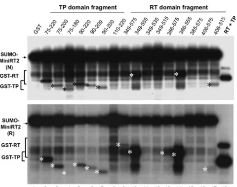
FIG 9trans-complementation priming reaction using TP and RT domain fragments. (A)trans-complementation priming reactions were performed using the starting GST-RT (RT) plus the starting GST-TP domain (TP, lanes 1 and 16) or the newly constructed TP fragments (lanes 2 to 7), using the starting GST-TP plus the newly constructed RT domain fragments (lanes 8 to 15), or using TP/90 –209 plus RT/366 –555 (lane 17) in TMnNK (top panel) or TMgNK (bottom panel). The protein priming signals on the TP (top and bottom panels) and RT (top panel) domain fragments are indicated at the bottom of the images, as percentages of the priming signals using the starting TP and RT domain constructs (lanes 1 and 16). The question marks denote that the RT domain signals from these constructs were difficult to quantify due to their comigration with the TP signal (see also panel B). (B)trans-complementation priming reactions were performed using GST-RT/349 –575 (lane 1), GST-RT/349 –555 (lane 2), or GST-RT/366 –555 (lane 3) plus His-TP (lanes 1 to 3) in TMnNK, to show more clearly the priming signals on GST-RT/349 –555 and GST-RT/366 –555, which were not well resolved from the strong and closely migrating GST-TP signals in panel A (lanes 8 and 12). The protein priming signals on the RT and TP domain fragments are indicated at the bottom of the image, as percentages of the priming signals using the starting (His-) TP and (GST-) RT domain constructs (lane 1).
on November 7, 2019 by guest
http://jvi.asm.org/
[image:11.585.113.473.66.272.2]weakened to allow the intermolecular TP-RT interactions
re-quired fortrans-priming, as indicated by the inaccessibility of a TP
domain that is linked incisto a catalytically inactive RT domain
(i.e., as in MiniRT2-YMHA) to a functional RT domain intrans.
The result is consistent with the previous finding that two full-length RT mutants containing either the Y96F mutation (in TP) or the YMHA mutation (in the RT domain) could not complement
each other for protein priming (55,56) and supports the notion
that the RT protein functions as a monomer (55). This apparent
sequestration of thecis-linked TP, together with the ability of an
active RT domain with acis-linked TP to use a separate TP intrans
as a primer, supports the notion that some conformational change
in the RT and/or TP domain occurs followingcis-priming, which
dissociates, at least partially, the intermolecular TP-RT interac-tions and allows the RT domain to interact with a separate TP for
trans-priming. Second, in contrast tocis-priming initiation,trans -priming initiation was found to be sensitive to inhibition by PFA.
As we have previously shown that PFA, under the Mn2⫹priming
conditions as used here, also inhibits the second stage ofcis
-prim-ing (polymerization) follow-prim-ing initiation (31), these results
sug-gest that the RT conformation duringtrans-priming initiation is
similar to that during polymerization ofcis-priming and that both
trans-priming and polymerization occur following cis-priming initiation, which induces the PFA-sensitive RT conformational state. Furthermore, the efficient induction of the PFA-sensitive RT
conformation followingcis-priming required the authentic Y96
site, which on the one hand stimulates the enzymatic activity of
the RT (8) and on the other enhances PFA sensitivity. A recent crystal structure of PFA in complex with a DNA polymerase in-deed shows that PFA sensitivity is determined by a specific poly-merase conformation rather than specific side chains (54), affirm-ing the utility of PFA as a sensitive probe for polymerase conformational changes.
The TP-RT domain interactions incis, while strong, were
evi-dently dynamic and could be disrupted by excess RT domain
frag-ments provided intrans, which were shown to inhibitcis-priming
in a dose-dependent manner. Efforts to localize the inhibitory sequences within the RT domain showed that multiple sequences
within it could function, in trans, to inhibit cis-priming by
MiniRT2 and, to a lesser extent, by the full-length RT protein,
through intermolecular (trans) RT-TP and RT-RT domain
inter-actions (Fig. 10B). Even catalytically active RT domain fragments, including the starting construct 349 –575 and the newly con-structed 349 –555, 366 –555, and 366 –575 (see also below), could
inhibitcis-priming. In these cases, simple competition by the RT
domain intrans to snatch (deprive) TP from itscis-linked RT
domain in MiniRT2 or the full-length RT may not account en-tirely for the inhibitory effect, as the catalytically active RT domain
fragments intrans, having interacted with TP, would be able to
carry out priming viatrans-complementation (see below). Even
considering that priming viatrans-complementation may be less
efficient thancis-priming, the rather strong inhibitory effects of
the RT domain fragments on protein priming were likely
medi-ated in part viatransRT-RT domain interactions, as well as RT-TP
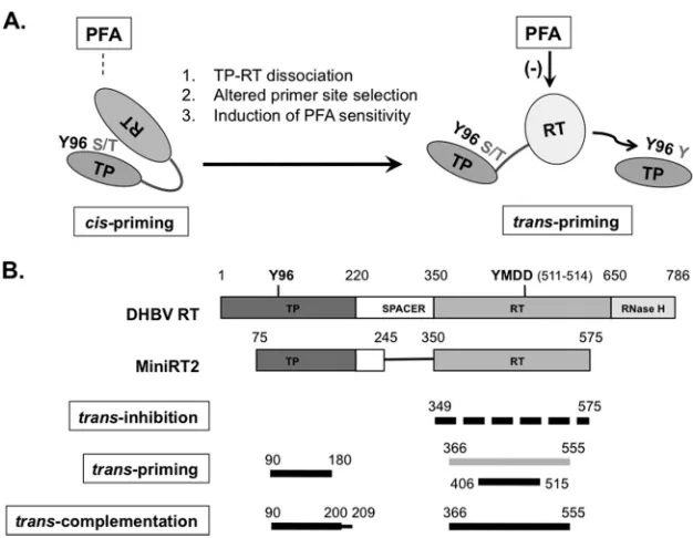
FIG 10Proposed RT conformational dynamics and TP and RT domain interactions in protein priming. (A) RT conformational changes following initiation of protein priming. The authentic primer site (Y96 in TP) and cryptic priming sites (S/T or Y in TP) are indicated. For clarity, the cryptic priming sites in the RT domain are omitted. The resistance (left) ofcis-priming and sensitivity (right) oftrans-priming to PFA inhibition are also indicated. The dissociation ofcis-linked TP and RT domains uponcis-priming is depicted as an opening of the protein structure. The proposed conformational change in the RT domain is depicted as a change in the shape and shading of the RT domain. (B) Definition of TP and RT domain sequences required fortrans-inhibition,trans-priming, and trans-complementation. The top two diagrams depict the domain structures of the full-length DHBV RT and MiniRT2, as explained inFig. 1A. The dashed lines on the third diagram indicate that multiple sequences in the RT domain could function to inhibit priming intrans. The light shading in the fourth diagram signifies the fact that the longer RT domain construct (containing position 366 to 555) failed to serve as a primer, intrans, to be used by another RT domain but could prime from itself incis(when provided with a functional TP domain). The thinner line in the fifth diagram (denoting residues 200 to 209) signifies that these TP sequences, while not essential fortrans-complementation, nevertheless contribute substantially to the reaction. See the text for details.
![FIG 2 trans-priming from the TP and RT domains by purified MiniRT2. Purified MiniRT2, TP, and RT domains were mixed together as indicated for proteinpriming in the presence of [�-32P]dGTP and the DHBV ε RNA](https://thumb-us.123doks.com/thumbv2/123dok_us/153706.30850/5.585.300.542.389.622/priming-domains-puried-minirt-puried-indicated-proteinpriming-presence.webp)