Copyright © 2003, American Society for Microbiology. All Rights Reserved.
Analysis of Early Human Immunodeficiency Virus Type 1 DNA
Synthesis by Use of a New Sensitive Assay for Quantifying
Integrated Provirus
Audrey Brussel and Pierre Sonigo*
De´partement des Maladies Infectieuses, Institut Cochin, INSERM U567, CNRS UMR 8104, Universite´ Rene´ Descartes, 75014 Paris, France
Received 7 February 2003/Accepted 19 June 2003
A novel Alu-long terminal repeat (LTR)-based real-time nested-PCR assay was developed to quantify integrated human immunodeficiency virus type 1 (HIV-1) DNA in infected cells with both accuracy and high sensitivity (six proviruses within 50,000 cell equivalents). Parallel assays for total HIV-1 DNA and two-LTR HIV-1 DNA circles allowed the synthesis and fate of the different HIV-1 DNA species to be monitored upon a single round of viral replication.
The RNA genome of human immunodeficiency virus type 1 (HIV-1) is, like those of other retroviruses, reverse transcribed in the cytoplasm into a double-stranded linear DNA contain-ing a copy of the long terminal repeats (LTR) at each terminus. The resulting linear DNA molecule moves into the nucleus as a component of the preintegration complex, where it may integrate into the host cell genome (17). This process is de-pendent on viral integrase activity, which is essential for a productive infection (7, 8, 13, 16, 19, 21). In addition to linear DNA species, HIV-1 DNA circles with one or two LTR are detected in the nucleus (1, 11).
While the unintegrated forms of HIV-1 DNA can be readily detected by using standard PCR or Southern hybridization techniques, quantitative analysis of integrated HIV-1 DNA has been hampered by the lack of a reliable assay. Early studies have attempted to quantify integrated viral DNA by PCR using one primer annealing in the HIV-1 LTR and a second in the
highly repeated chromosomalAluelement (Alu-LTR PCR) (6,
18). Alu repeated sequences are interspersed in the human
genome and number at least 900,000 copies per haploid
ge-nome, giving an average distance of 4 kb betweenAluelements
(2). However, previousAlu-LTR PCR methods have limited
sensitivity and do not take into account that the distance
be-tween an integrated viral DNA and its nearest Alurepeat is
variable.
To detect and accurately quantify integrated HIV-1 DNA with high sensitivity, we have developed a new method, based
on Alu-LTR real-time nested PCR. In conjunction with the
study of HIV-1 DNA integration, real-time quantitative PCR was used to analyze the synthesis and fate of total HIV-1 DNA and two-LTR HIV-1 circles during a single-round of viral rep-lication.
Generation of an integrated HIV-1 DNA standard.To ac-curately determine the integrated HIV-1 DNA copy number
within a sample, the standard should be representative of a natural viral infection and contain numerous integration sites with a wide distribution of distances between the provirus LTR
and the nearestAlusequence. To generate a proper integrated
HIV-1 DNA standard, HeLa cells were infected with a⌬env
HIV-1 R7 Neo virus pseudotyped with the G glycoprotein from vesicular stomatitis virus (VSV-G). VSV-G-pseudotyped HIV-1 R7 Neo virus stocks were prepared by cotransfection
into 293T cells of the pR7 Neo ⌬env vector (gift from U.
Hazan) with an expression vector encoding VSV-G. The pR7
Neo ⌬env vector was constructed by deleting the envelope
coding sequence of the HIV-1 R7 Neo genome (9), in which a
neomycin resistance gene replaces the HIV-1 nef coding
se-quence. Infected cells were then cultured for several weeks in
the presence of G418 (500g/ml) to select cells that contained
integrated viral DNA and to allow the loss of all unintegrated forms. Ten days after initiation of the G418 selection, neomy-cin-resistant cell clones were counted; they numbered approx-imately 5,000. For our standard cell line, designated HeLa R7 Neo, the copy number of integrated viral DNA matched the total HIV-1 DNA copy number. Thus, based on total HIV-1
DNA and-globin quantifications (see below), we estimated
that the standard cell line contained 1.24⫾0.03 proviruses per
cell (data not shown).
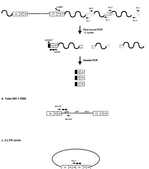
Alu-LTR-based real-time nested-PCR procedure. Amplifica-tion reacAmplifica-tions were performed with the Light Cycler instru-ment (Roche Diagnostics, Meylan, France). In a first round of PCR, integrated HIV-1 sequences were amplified with two
outward-facingAlu primers that anneal within conserved
re-gions of theAlurepeat element together with an HIV-1
LTR-specific primer (Fig. 1a). As previously proposed (18), the use
of two outward-facingAluprimers optimizes the probability of
amplifying an LTR sequence, asAluelements are present in
either orientation relative to the integrated provirus (Fig. 1a). In addition, we used an LTR primer extended with a lambda
phage-specific heel sequence at the 5⬘end of the
oligonucleo-tide (L-M667) in this first amplification step (Fig. 1a).
Alu-LTR sequences were amplified in duplicate from 1/50 of total
* Corresponding author. Mailing address: De´partement des Mala-dies Infectieuses, Institut Cochin, INSERM U567, CNRS UMR 8104, Universite´ Rene´ Descartes, 22 rue Me´chain, 75014 Paris, France. Phone: 33 1 40 51 64 13. Fax: 33 1 40 51 64 30. E-mail: sonigo@cochin .inserm.fr.
10119
on November 8, 2019 by guest
http://jvi.asm.org/
cell DNA in a 20-l reaction mixture comprising 1⫻ Light Cycler Fast Start DNA master hybridization probes (Roche), 4
mM MgCl2, 100 nM L-M667 primer, and 300 nM (each)
prim-ersAlu1 andAlu2 (18) (Table 1). Given the high number of
Aluelements within the human genome, abundant
amplifica-tions of inter-Alusequences occurred simultaneously with the
amplification of Alu-LTR sequences. To remain in the
[image:2.603.56.541.92.642.2]expo-nential phase, only 12 cycles of amplification were performed.
FIG. 1. Real-time PCR strategies and location of primers and probes for the quantification of integrated HIV-1 DNA (a), total HIV-1 DNA (b), and two-LTR circles (c). Thin arrows, primers; thick arrows, probes.
10120 NOTES J. VIROL.
on November 8, 2019 by guest
http://jvi.asm.org/
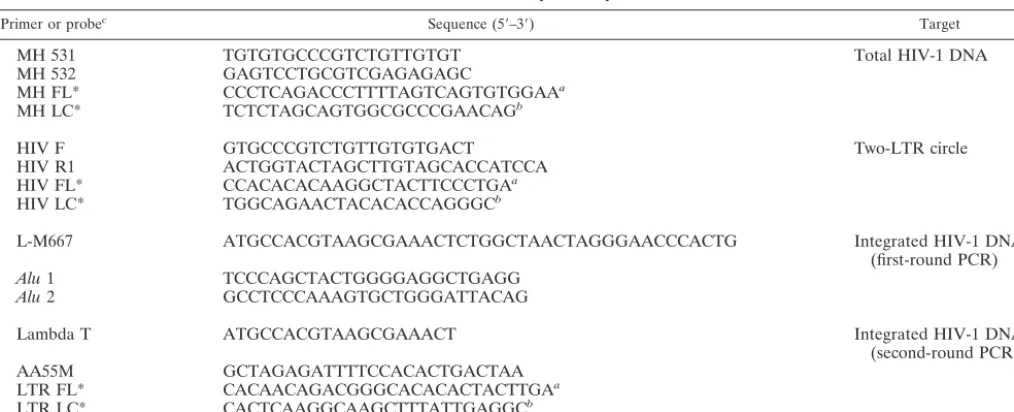
FIG. 2. Characteristics ofAlu-LTR nested PCR. (a) Fluorescence curves generated by two-step amplification of serial dilutions of HeLa R7 Neo cell DNA. The copy numbers for each standard dilution are shown over the corresponding fluorescence curve. (b) Linear regression for quantifying integrated HIV-1 DNA. (c) Specificity of theAlu-LTR nested PCR. Fluorescence curves generated by amplification of viral DNA from CEM cells infected with the VSV-G-pseudotyped HIV-1 R7 Neo virus and collected in the presence ofAluprimers (pink, 8 h p.i.; green, 48 h p.i.), in the absence ofAluprimers (blue, 8 h p.i.; red, 48 h p.i.), or without a preamplification step (purple, 8 h p.i.; orange, 48 h p.i.) are shown.
10121
on November 8, 2019 by guest
Thus, the first-round PCR cycle conditions were as follows: a denaturation step of 8 min at 95°C and then 12 cycles of amplification (95°C for 10 s, 60°C for 10 s, and 72°C for 170 s). In a second round of PCR using the lambda-specific primer (Lambda T) and an LTR primer (AA55M), only products from the first-round PCR could be amplified (Fig. 1a). In contrast to the first-round PCR, which generated fragments of various lengths, the nested amplification resulted in discrete DNA fragments. Nested PCR was performed on 1/10 of the
first-round PCR product in a mixture comprising 1⫻Light Cycler
Fast Start DNA master hybridization probes, 4 mM MgCl2,
300 nM Lambda T primer, 300 nM AA55M primer, and 200 nM (each) hybridization probes LTR FL and LTR LC (Table 1). The nested-PCR cycling profile began with a denaturation step (95°C for 8 min), followed by 50 cycles of amplification (95°C for 10 s, 60°C for 10 s, and 72°C for 9 s).
The copy number of integrated HIV-1 DNA was determined in reference to a standard curve generated by concomitant two-stage PCR amplification of a serial dilution of the standard HeLa R7 Neo cell DNA (Fig. 2a) mixed with uninfected-cell DNA to yield 50,000 cell equivalents. Quantification was achieved by the second-derivative maximum method provided by the Light Cycler quantification software, version 3.5 (Roche Diagnostics). In our system, the linear regression obtained from amplification of serial dilutions of the integrated HIV-1
DNA standard was linear over a 5-log10-unit range, and our
Alu-LTR nested-PCR procedure allowed the detection of ap-proximately six proviruses within 50,000 cell equivalents (Fig. 2b).
Specificity of the Alu-LTR nested-PCR assay.During the first-round PCR, the L-M667 oligonucleotide can prime the formation of a single-stranded DNA from all LTR containing HIV-1 DNA, leading to an overestimation of the actual inte-grated HIV-1 DNA copy number. To control these linear am-plifications, we performed a nested-PCR procedure omitting
or notAluprimers in the first-round PCR. Figure 2c displays
fluorescence curves obtained during amplification of cell DNA from CEM lymphoid cells infected with the VSV-G-pseudotyped HIV-1 R7 Neo virus and collected 8 or 48 h
postinfection (p.i.). In the presence of Aluprimers, the
fluo-rescence curves shifted, moderately but reproductively, to the left, compared to an amplification performed in the absence of
Alu primers (Fig. 2c). The shifts were about 1 cycle and 7
cycles, corresponding to 2-fold and 50-fold increases in copy numbers, for the 8- and 48-h time points, respectively. Fur-thermore, to determine the extent of second-round amplifica-tion of nonpreamplified viral DNA, we performed an addi-tional control where the first-round PCR was replaced by a 4°C incubation of the PCR mixture. As expected, the amplification curves shifted dramatically to the left when a preamplification step was performed. The shift corresponded to approximately
2.3- (9 cycles) and 3.5-log10-unit (14 cycles) increases in copy
numbers for the 8- and 48-h time points, respectively. Taken together, these findings indicate that the PCR signal obtained
in the absence ofAluprimers must be subtracted from the total
signal, especially at early times following infection, and that the second-round amplification of nonpreamplified viral DNA is efficiently prevented.
Synthesis and fate of HIV-1 DNA species in a single-round infection assay.To validate our integration assay, we quanti-fied integrated HIV-1 DNA, together with total HIV-1 DNA and two-LTR circles in a single round of viral replication. Eighteen million CEM cells were infected by
VSV-G-pseudotyped HIV-1 R7 Neo virus by using 35 ng of p24gag
antigen (measured by enzyme-linked immunosorbent assay;
Perkin-Elmer Life Sciences, Paris, France) per 106cells. One
hour p.i., cells were washed in phosphate-buffered saline
(PBS), exposed to trypsin (25 g/ml) for 1 min at 37°C, and
washed once with medium and twice with PBS. The cells were then cultured in RPMI 1640 supplemented with 10% fetal calf
serum. At each time point, 1⫻106to 3⫻106infected cells
[image:4.603.45.552.79.285.2]were collected. To eliminate residual pR7 Neo ⌬env vector
TABLE 1. Primer and probe sequences
Primer or probec Sequence (5⬘–3⬘) Target
MH 531 TGTGTGCCCGTCTGTTGTGT Total HIV-1 DNA
MH 532 GAGTCCTGCGTCGAGAGAGC
MH FL* CCCTCAGACCCTTTTAGTCAGTGTGGAAa
MH LC* TCTCTAGCAGTGGCGCCCGAACAGb
HIV F GTGCCCGTCTGTTGTGTGACT Two-LTR circle
HIV R1 ACTGGTACTAGCTTGTAGCACCATCCA
HIV FL* CCACACACAAGGCTACTTCCCTGAa
HIV LC* TGGCAGAACTACACACCAGGGCb
L-M667 ATGCCACGTAAGCGAAACTCTGGCTAACTAGGGAACCCACTG Integrated HIV-1 DNA
(first-round PCR)
Alu1 TCCCAGCTACTGGGGAGGCTGAGG
Alu2 GCCTCCCAAAGTGCTGGGATTACAG
Lambda T ATGCCACGTAAGCGAAACT Integrated HIV-1 DNA
(second-round PCR)
AA55M GCTAGAGATTTTCCACACTGACTAA
LTR FL* CACAACAGACGGGCACACACTACTTGAa
LTR LC* CACTCAAGGCAAGCTTTATTGAGGCb
aModified with fluorescein at the 3⬘end.
bPhosphorylated at the 3⬘end and modified with LC red 640 dye at the 5⬘end.
cPrimers and probes were purchased from TIB MOLBIOL (Berlin, Germany).ⴱ, probe sequence.
10122 NOTES J. VIROL.
on November 8, 2019 by guest
http://jvi.asm.org/
DNA, the samples were washed in PBS and incubated with 750 to 1,500 U of DNase I (Invitrogen, Cergy Pontoise, France) in a buffer comprising 20 mM Tris-Cl, pH 8.3, 50 mM KCl, and 2
mM MgCl2for 1 h at room temperature. Cells were washed in
PBS, and dry cell pellets were frozen at⫺80°C until use. Total
cell DNA was extracted with a QIAamp blood DNA minikit (Qiagen, Courtaboeuf, France).
The total HIV-1 DNA copy number was determined with previously described primers (4) that annealed in the U5
re-gion of the LTR (MH 531) and in the 5⬘end of thegaggene
(MH 532) (Fig. 1b). The two-LTR circles were amplified with primers spanning the LTR-LTR junction (HIV F and HIV
R1), as described elsewhere (3) (Fig. 1c). U5-gag sequences
and two-LTR junctions were amplified in duplicate from 1/50
of total cell DNA. Reaction mixtures contained 1⫻Light
Cy-cler Fast Start DNA master hybridization probes (Roche
Di-agnostics), 4 mM MgCl2, 300 nM (each) forward and reverse
primers, and 200 nM (each) fluorogenic hybridization probes
in a final volume of 20 l. After an initial denaturation step
(95°C for 8 min), the cycling profile for total HIV-1 DNA was 50 cycles consisting of 95°C for 10 s, 60°C for 10 s, and 72°C for 6 s and that for two-LTR circles was 15 cycles consisting of 95°C for 10 s, 66°C for 10 s, and 72°C for 10 s, followed by 35 cycles at the beginning of which the annealing temperature was decreased by 0.5°C per cycle to the secondary target temper-ature (59°C). The copy numbers of total HIV-1 DNA and two-LTR circles were determined in reference to a standard curve prepared by amplification of quantities ranging from 10
to 105 copies of cloned DNA with matching sequences. The
cell equivalents in sample DNA were calculated based on the
amplification of the-globin gene (two copies per diploid cell)
with the Light Cycler instrument and commercially available materials (Control kit DNA; Roche Diagnostics). The quanti-fication results for two-LTR circle, total HIV-1 DNA, and integrated HIV-1 DNA were expressed as copy numbers per
106cells.
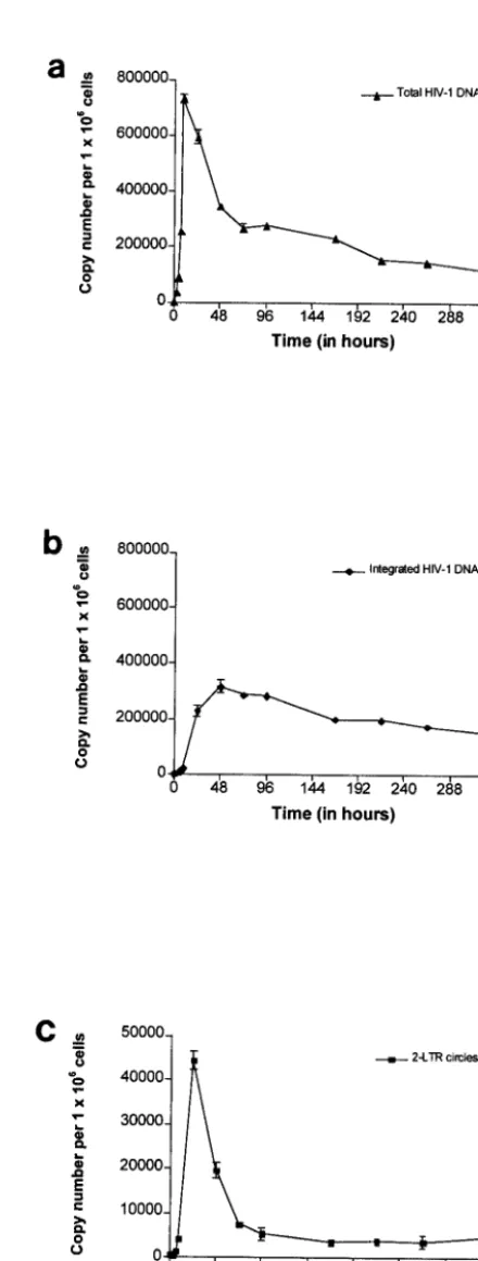
Viral DNA was detected by 3 h p.i. The level of total HIV-1 DNA increased rapidly until 9 h p.i., reaching a maximum of
734,996 copies per 106cells (Fig. 3a). Then the level of total
HIV-1 DNA underwent a steep decrease until 72 h p.i., fol-lowed by a slow decay phase. In previous single-round infec-tion assays, the initial steep decrease in the level of total HIV-1 DNA was reported to represent the proteasome-mediated deg-radation of unintegrated linear HIV-1 DNA (5). We detected integrated HIV-1 DNA by 3 h p.i., attesting to the rapid nu-clear import of reverse transcription products and emphasizing the sensitivity of our integration assay. The level of integrated HIV-1 DNA increased until 48 h p.i to reach a maximum of
316,578 copies per 106cells (Fig. 3b). By 72 h p.i., the level of
integrated HIV-1 DNA matched that of total HIV-1 DNA, as expected in a single-round of viral replication. This confirmed the accuracy of our quantification method. The slow decay in the level of integrated HIV-1 DNA may reflect the death of infected cells and/or the slow division rate of infected cells carrying an integrated provirus compared to that of uninfected ones. Two-LTR circles, as detected by the presence of LTR-LTR junctions, were observed as early as 3 h p.i. The level of
two-LTR circles reached 44,253 copies per 106cells by 24 h p.i.
[image:5.603.307.527.71.650.2]and declined thereafter, reaching a quasisteady level from 96 h p.i. until the end of the culture (Fig. 3c). Two-LTR circles were
FIG. 3. Quantification of total HIV-1 DNA (a), integrated HIV-1 DNA (b), and two-LTR circles (c) during a single round of replication of the VSV-G-pseudotyped HIV-1 R7 Neo virus in CEM cells. Values are means⫾standard errors of the means. The depicted results were obtained from a representative experiment.
on November 8, 2019 by guest
http://jvi.asm.org/
shown to be stable DNA forms incapable of self-replication, and, consequently, they are diluted as a function of cell division (5, 15). Accordingly, a dilution effect resulting from cell divi-sion may account for the decrease in the level of two-LTR circles observed before 96 h p.i. In contrast, steady LTR-LTR junction levels from 96 h p.i. until the end of the culture were unexpected. Nevertheless, the nonspecific integration of some two-LTR circles (less than 10% in this experiment) into the host cell genome might explain the stability of LTR-LTR junc-tion levels. Integrated LTR-LTR juncjunc-tions may have been transmitted from mother to daughter cells at each division, leading to steady LTR-LTR junction levels in spite of cell proliferation. Likewise, it was previously shown that some lin-ear HIV-1 DNA could be integrated into the cell genome by an integrase-independent process (10). This finding indicates that detection of LTR-LTR junctions does not necessarily indicate the presence of unintegrated forms of HIV-1 DNA.
Using a new sensitive assay to quantify integrated viral DNA, we analyzed the integration kinetics of viral DNA fol-lowing a single cycle of replication. Our integration kinetics were similar to those obtained by Butler et al., who employed
a one-step real-time Alu-LTR PCR-based amplification
method (4). However, we were able to detect integrated HIV-1 DNA as early as 3 h p.i., while the one-step quantification method allowed the detection of proviral DNA no earlier than 12 (5) or 24 h p.i. (4). The limited sensitivity of the one-step Alu-LTR PCR method (1,000 copies in 100,000 cell equiva-lents) should account for the delayed detection of integrated HIV-1 DNA. Recently, a second method for the quantification of integrated HIV-1 DNA was devised. This method is based on a linker-primer PCR (LP-PCR) amplification (12, 20). The LP-PCR method was shown to detect four copies of proviruses in 200,000 cell equivalents (12), allowing the detection of in-tegrated HIV-1 DNA as early as 4 h p.i. Unfortunately, the LP-PCR protocol involves many experimental steps. More re-cently, a real-time nested-PCR method (14) which used one
primer annealing in the HIV-1 gag sequence and a second
annealing in theAluelement was described. This assay has a
detection limit of one provirus per 10,000 cells, i.e.,
compara-ble to ours. However, this Alu-LTR PCR method did not
prevent the second-round amplification of nonpreamplified HIV-1 DNA, and its range of linearity was less extensive than
ours (2.5 versus 5 log10units).
The rapidity and sensitivity of ourAlu-LTR-based real-time
nested PCR should allow the routine analysis of HIV-1 DNA integration and may be a valuable tool to test future integrase inhibitors or to evaluate the integration efficiency of retroviral vectors designed for gene therapy.
A. Brussel was supported by a fellowship from the Agence Nationale de Recherche sur le SIDA.
We thank U. Hazan for constant advice and help in this work. We acknowledge Olfert Landt (TIB MOLBIOL) for excellent technical assistance with the design of primers and hybridization probes. We also thank C. Petit, O. Delelis, S. Pierre, M. Alizon, and especially B.
Canque for testing and optimizing the assay and C. Sonnenschein for reviewing the manuscript.
REFERENCES
1. Barbosa, P., P. Charneau, N. Dumey, and F. Clavel.1994. Kinetic analysis of HIV-1 early replicative steps in a coculture system. AIDS Res. Hum. Ret-rovir.10:53–59.
2. Britten, R. J., W. F. Baron, D. B. Stout, and E. H. Davidson.1988. Sources and evolution of human Alu repeated sequences. Proc. Natl. Acad. Sci. USA
85:4770–4774.
3. Brussel, A., D. Mathez, S. Broche-Pierre, R. Lancar, T. Calvez, P. Sonigo, and J. Leibowitch.2003. Longitudinal monitoring of 2-long terminal repeat circles in peripheral blood mononuclear cells from patients with chronic HIV-1 infection. AIDS17:645–652.
4. Butler, S. L., M. S. Hansen, and F. D. Bushman.2001. A quantitative assay for HIV DNA integration in vivo. Nat. Med.7:631–634.
5. Butler, S. L., E. P. Johnson, and F. D. Bushman.2002. Human immunode-ficiency virus cDNA metabolism: notable stability of two-long terminal re-peat circles. J. Virol.76:3739–3747.
6. Chun, T. W., L. Stuyver, S. B. Mizell, L. A. Ehler, J. A. Mican, M. Baseler, A. L. Lloyd, M. A. Nowak, and A. S. Fauci.1997. Presence of an inducible HIV-1 latent reservoir during highly active antiretroviral therapy. Proc. Natl. Acad. Sci. USA94:13193–13197.
7. Engelman, A., G. Englund, J. M. Orenstein, M. A. Martin, and R. Craigie.
1995. Multiple effects of mutations in human immunodeficiency virus type 1 integrase on viral replication. J. Virol.69:2729–2736.
8. Englund, G., T. S. Theodore, E. O. Freed, A. Engleman, and M. A. Martin.
1995. Integration is required for productive infection of monocyte-derived macrophages by human immunodeficiency virus type 1. J. Virol.69:3216– 3219.
9. Feinberg, M. B., D. Baltimore, and A. D. Frankel.1991. The role of Tat in the human immunodeficiency virus life cycle indicates a primary effect on transcriptional elongation. Proc. Natl. Acad. Sci. USA88:4045–4049. 10. Gaur, M., and A. D. Leavitt.1998. Mutations in the human
immunodefi-ciency virus type 1 integrase D, D(35)E motif do not eliminate provirus formation. J. Virol.72:4678–4685.
11. Kim, S. Y., R. Byrn, J. Groopman, and D. Baltimore.1989. Temporal aspects of DNA and RNA synthesis during human immunodeficiency virus infection: evidence for differential gene expression. J. Virol.63:3708–3713. 12. Kumar, R., N. Vandegraaff, L. Mundy, C. J. Burrell, and P. Li. 2002.
Evaluation of PCR-based methods for the quantitation of integrated HIV-1 DNA. J. Virol. Methods105:233–246.
13. LaFemina, R. L., C. L. Schneider, H. L. Robbins, P. L. Callahan, K. LeGrow, E. Roth, W. A. Schleif, and E. A. Emini.1992. Requirement of active human immunodeficiency virus type 1 integrase enzyme for productive infection of human T-lymphoid cells. J. Virol.66:7414–7419.
14. O’Doherty, U., W. J. Swiggard, D. Jeyakumar, D. McGain, and M. H. Malim.
2002. A sensitive, quantitative assay for human immunodeficiency virus type 1 integration. J. Virol.76:10942–10950.
15. Pierson, T. C., T. L. Kieffer, C. T. Ruff, C. Buck, S. J. Gange, and R. F. Siliciano.2002. Intrinsic stability of episomal circles formed during human immunodeficiency virus type 1 replication. J. Virol.76:4138–4144. 16. Sakai, H., M. Kawamura, J. Sakuragi, S. Sakuragi, R. Shibata, A. Ishimoto,
N. Ono, S. Ueda, and A. Adachi.1993. Integration is essential for efficient gene expression of human immunodeficiency virus type 1. J. Virol.67:1169– 1174.
17. Sherman, M. P., and W. C. Greene.2002. Slipping through the door: HIV entry into the nucleus. Microbes Infect.4:67–73.
18. Sonza, S., A. Maerz, N. Deacon, J. Meanger, J. Mills, and S. Crowe.1996. Human immunodeficiency virus type 1 replication is blocked prior to reverse transcription and integration in freshly isolated peripheral blood monocytes. J. Virol.70:3863–3869.
19. Stevenson, M., T. L. Stanwick, M. P. Dempsey, and C. A. Lamonica.1990. HIV-1 replication is controlled at the level of T cell activation and proviral integration. EMBO J.9:1551–1560.
20. Vandegraaff, N., R. Kumar, C. J. Burrell, and P. Li.2001. Kinetics of human immunodeficiency virus type 1 (HIV) DNA integration in acutely infected cells as determined using a novel assay for detection of integrated HIV DNA. J. Virol.75:11253–11260.
21. Wiskerchen, M., and M. A. Muesing.1995. Human immunodeficiency virus type 1 integrase: effects of mutations on viral ability to integrate, direct viral gene expression from unintegrated viral DNA templates, and sustain viral propagation in primary cells. J. Virol.69:376–386.
10124 NOTES J. VIROL.