White Rose Research Online URL for this paper: http://eprints.whiterose.ac.uk/98869/
Version: Accepted Version
Article:
Barrie, J, Jayne, DG, Neville, A et al. (3 more authors) (2016) Real-time measurement of the tool-tissue interaction in minimally invasive abdominal surgery; the first step to
developing the next generation of smart laparoscopic instruments. Surgical Innovation, 23 (5). pp. 463-468. ISSN 1553-3506
https://doi.org/10.1177/1553350616646475
© 2016, The Author(s). This is an author produced version of a paper accepted for publication in Surgical Innovation. Uploaded in accordance with the publisher's self-archiving policy.
[email protected] https://eprints.whiterose.ac.uk/ Reuse
Unless indicated otherwise, fulltext items are protected by copyright with all rights reserved. The copyright exception in section 29 of the Copyright, Designs and Patents Act 1988 allows the making of a single copy solely for the purpose of non-commercial research or private study within the limits of fair dealing. The publisher or other rights-holder may allow further reproduction and re-use of this version - refer to the White Rose Research Online record for this item. Where records identify the publisher as the copyright holder, users can verify any specific terms of use on the publisher’s website.
Takedown
If you consider content in White Rose Research Online to be in breach of UK law, please notify us by
Title Page
Title: Real-time measurement of the tool-tissue interaction in minimally invasive abdominal
surgery; the first step to developing the next generation of smart laparoscopic instruments
Jenifer Barrie (MRCS), Peter R. Culmer (PhD), Louise Hunter (PhD), Adrian J. Hood
(MRCS), David G. Jayne (MD), Anne Neville (PhD).
Authors:
Miss Jenifer Barrie (MRCS)
School of Mechanical Engineering, University of Leeds, Woodhouse Lane, Leeds. LS2 9JT.
Dr Peter R. Culmer (PhD)
School of Mechanical Engineering, University of Leeds, Woodhouse Lane, Leeds. LS2 9JT.
Miss Louise Hunter
School of Mechanical Engineering, University of Leeds, Woodhouse Lane, Leeds. LS2 9JT.
Mr Adrian J. Hood (MRCS)
School of Mechanical Engineering, University of Leeds, Woodhouse Lane, Leeds. LS2 9JT.
Professor David G. Jayne (MD)
Professor of Surgery & Honorary Consultant Surgeon
Division of Clinical Sciences, St. James’s University Hospital, Beckett Street, Leeds, LS9 7TF.
School of Mechanical Engineering, University of Leeds, Woodhouse Lane, Leeds. LS2 9JT.
Corresponding Author:
Professor David G. Jayne (MD)
Level 7 Clinical Sciences building,
St. James’s University Hospital,
Beckett Street,
Leeds,
LS9 7TF.
Tel: +44 113 2065281
Fax: +44113 244 9168
E-mail: [email protected]
Source of funding:
This research received no specific grant from any funding agency in the public, commercial, or
not-for-profit sectors.
Co-author email addresses
Miss Jenifer Barrie: [email protected]
Miss Louise Hunter: [email protected]
Mr Adrian J. Hood:[email protected]
Prof A Neville: [email protected]
This paper is based on an oral presentation at the Association of Surgeons of Great Britain
and Ireland Annual Meeting; 01/05/2013. Glasgow, UK.
Abstract
Introduction
Analysis of force application in laparoscopic surgery is critical to understanding the nature of
the tool-tissue interaction. The aim of this study is to provide real time data about
manipulations to abdominal organs.
Methods
An instrumented short fenestrated grasper was used in an in vivo porcine model, measuring
force at the grasper handle. Grasping force and duration over five small bowel manipulation
tasks were analysed. Forces required to retract gallbladder, bladder, small bowel, large bowel
and rectum were measured over 30 seconds. Four parameters were calculated; T (hold) ; the
grasp time, T(close); time taken for the jaws to close, F(max); maximum force reached,
F(rms); root mean square force (representing the average force across the grasp time).
Results
Mean F (max) to manipulate the small bowel was 20.5N (+-7.2N) and F (rms) was 13.7N (+-
5.4). Mean T (close) was 0.52s (+-0.26) and T (hold) was 3.87s (+-1.5). In individual organs
mean F (max) was 49N (+-15) to manipulate the rectum and 59N (+-13.4) for the colon. The
mean F (max) for bladder and gallbladder retraction was 28.8N (+-7.4) and 50.7 (+-3.8)
respectively. All organs exhibited force relaxation, the F (rms) reduced to below 25N for all
organs except the small bowel, with a mean F (rms) of under 10N.
This study has commenced the process of quantifying tool-tissue interaction. The static
measurements discussed here should evolve to include dynamic measurements such as shear,
Introduction
Basic laparoscopic instrumentation has changed very little in the three decades since the first
laparoscopic cholecystectomy, whilst the spectrum of both elective and emergency
procedures performed laparoscopically has widened (1-8). The true nature of the tool-tissue
interaction in laparoscopic surgery is not fully understood and the contribution of
laparoscopic instruments to bowel perforations, serosal tears and the development of an ileus
or adhesion formation have not been quantified. In laparoscopic colorectal cancer operations,
iatrogenic bowel injury is reported as a complication in 2% of colonic and 1% of rectal
resections (9). A bowel injury rate of 1.8% has been found in a recent study of laparoscopic
resections for gynaecology malignancies (10). Although the majority of grasper injuries are
probably of minor clinical significance the occurrence of a bowel perforation is a disastrous
yet wholly avoidable event. The mortality rate associated with laparoscopy induced bowel
injury is high at 3.6% (9). Reports of other visceral injuries in laparoscopic surgery can be
found in the literature and include bladder injuries (11) and splenic injuries (12).
The ideal laparoscopic grasper will grip tissue without slippage and allow the surgeon to
perform the required movement without causing damage to the grasped tissue or adjacent
structures. This not only depends on the properties of the grasper jaws but the force applied
by the surgeon and the mechanical properties of the manipulated tissue. Analysis of force in
minimally invasive surgery is imperative in understanding the nature of the tool-tissue
interaction and the degree of macroscopic and microscopic tissue trauma incurred to
abdominal organs. Such data contributes to the understanding of instrument design, surgical
simulators and surgical training. Attempts have been made over the past decade to measure
the forces in minimally invasive surgery (MIS) manipulations (13-18) but no real-time
durations. Several studies have advanced upon this and attempted to relate grasping force to
measureable histological damage (19-24).
The aim of this study is to use an instrumented grasper to provide real time data about the
duration and force applied by the surgeon during laparoscopic manipulations on abdominal
Materials and methods
Instrumented Grasper System
The instrumented grasper was developed by adapting a commercially-available reusable
Johan grasper (Surgical Innovations Ltd UK- as shown in figure 1) to integrate a bespoke
sensor module at the instrument handle. This configuration positions the electronic sensing
elements outside the abdominal cavity, thus removing the risk of sensor contamination and
ensuring that the tool-tissue interface is identical to that in a conventional grasper system.
The sensor module comprises a force sensor and a potentiometer position sensor which are
connected to the shaft that controls the grasper jaws (see figure 2). A custom computer
measurement system (LabVIEW, National Instruments Inc.) was used to measure and log
data at 100Hz. This enabled real-time measurement of the surgeon’s interaction with the
instrument. Real- time force measurements and grasping durations are displayed on a monitor
as manipulations occur. Data was produced in the form of a force-time graph for analysis.
Experimental protocol
The instrumented grasper was tested in in vivo porcine experiments. Manipulations in a
specific surgical task were measured in five single abdominal organs in an anaesthetised 40kg
Large White pig. All experiments were performed under Home Office licence (number PPL
40/3662). 12mmHg pneumoperitoneum was instituted using an open Hassan technique and
the laparoscope was inserted through a 12mm suprapubic port. Working -ports were placed
on the left and right lateral positions in line with the umbilicus and in the left and right iliac
fossae in order to access the abdominal organs manipulated. Tasks were performed by a
able to perform basic laparoscopic procedures, such as appendicectomy and cholecystectomy,
under minimal supervision.
Manipulations were divided into grasps on specific abdominal organs and then one simple
surgical task. The specific surgical task chosen was running the small bowel which is
performed to examine the length of the small bowel for pathology. The task was conducted
by alternately passing the small bowel between the right and left handed grasper. Ten
manipulations were performed to achieve this, five of these with a standard grasper in the
surgeon’s left hand and five with the instrumented grasper in the surgeon’s right hand. The
force and duration of grasping was measured for each manipulation with the right hand. The
bowel running task was then performed five times over as described, resulting in five
data-sets containing five individual grasps in each one, giving a total of 25 measurements. Each
grasp manipulation was analysed to calculate four summary parameters; T (hold); the time
taken to grasp the small bowel over one manipulation, T (close); the time taken for the
grasper jaws to close when manipulating tissue, F (max); the maximum force reached in the
hold time, F (rms); the root mean square force over the hold time, illustrating an average
force. The force-time graph of a typical bowel running task containing 5 grasps is shown in
figure 3a, with 3b illustrating a single grasp with the summary parameters that have been
measured.
There is little existing data on the forces required to manipulate individual abdominal organs.
In this study a spectrum of abdominal organs were manipulated to investigate how these
forces may vary across the gallbladder, bladder, rectum, large bowel and small bowel. Each
organ was grasped with the instrumented short fenestrated grasper for 30 seconds and a single
grasp was defined by the ability to lift and retract the organ successfully without slip. This
Results
Small bowel running task: manipulation force
Each of the five small bowel running tasks consisted of five measurements. Mean F (max)
over all 25 measurements was 20.5N (+- 7.2N) and mean F (rms) was 13.7N (+- 5.4N).
Figure 4shows a boxplot of the forces measured at the grasper handle for each individual
bowel running task. In task number four and task number five the forces of both F (max) and
F (rms) has reduced compared to task numbers one, two and three. The reduced forces in
tasks four and five could be explained by the surgeon becoming accustomed to the
manipulations performed, indicating that there may be an experience effect involved.
Small bowel running task: time measurements
The mean T (close) was 0.52s (+-0.26). The time to close the grasper jaws appears to reduce
after the first task, at 0.5 seconds or less. The longest T (close) occurred in the first bowel
running task at 2.03 seconds. This would again indicate that there is some experience effect.
The manipulation time, mean T (hold) over all 25 manipulations was 0.52s (+-1.5). The hold
time did not appear to show the same experience effect over the five manipulations. Figure 5
shows the results for the hold time across all 5 manipulation tasks.
Individual organ grasping
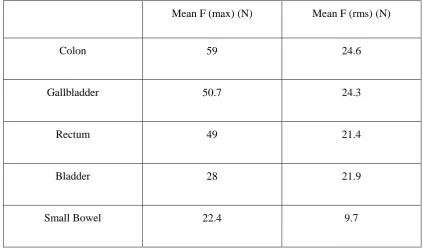
The F (max) and F (rms) for each individual organ grasped is shown in Table 1. The largest
the range of forces, up to 75N were used to grasp the colon and rectum. Much lower
maximum forces were required for bladder and small bowel retraction.
All organs exhibited force relaxation so that the F (rms) reduced to between 21 and 25N for
all organs except the small bowel, with a mean F (rms) of under 10N. When comparing the
maximum force reached with the root mean squared force, the RMS has more than halved for
the colon, gallbladder, rectum and small bowel. The bladder does not exhibit such large force
Discussion
The focus of this study was to present an instrumented grasper which can be used to measure
grasp durations and forces applied by surgeons during laparoscopic abdominal manipulations.
These results demonstrate that we have an instrument and methodology for analyzing forces
used by surgeons, with the potential for further studies identifying critical forces that result in
tissue damage. The results demonstrate the range of forces that are applied to a spectrum of
abdominal organs, each with varied mechanical properties. Each force-time output graph in
our series indicated an initial maximum force that was applied to lift the organ, followed by a
period of force relaxation that we believe is a combination of tissue response and the pressure
applied to the grasper handle.
The work presented has commenced the important process of quantifying tool-tissue
interaction in MIS, and in particular providing an experimental methodology for these
investigationsThe limitations of this preliminary study are the use of a single porcine model
and constraining experimental variables to a single laparoscopic grasper type operated by a
single surgeon. The single porcine model reflects the scope of this preliminary work in which
our emphasis is to demonstrate a methodology of assessing the tool-tissue interaction.
Additionally, ethical considerations negate a human model prior to this animal model. Time
constraints in conducting these in vivo experiments limited sample size in the selected
grasping procedures. In vivo testing was performed in an anaesthetised 40 kg large white
Yorkshire pig because the intestinal size at this weight resembles the adult human. The Johan
grasper was selected because it is commonly used in a wide variety of laparoscopic
procedures, however, the eventual aim of this research is to broaden the scope of testing to
Brown and Colleagues used a system known as the Blue Dragon (13), which consisted of an
actuated Babcock grasper, to measure the force at the grasper handle. This group measured
the force required to run the bowel and to pass the stomach behind the oesophogus (stomach
wrap). Published data combined these tasks, therefore comparisons cannot be made between
the forces applied to the small bowel in the current experiments and those from the Brown
study group, as significantly greater forces were applied during the stomach wrap task
compared to bowel running (13). Their results showed that the mean force applied to the tool
handles during tissue grasps was 8.52 N ± 2.77 N and the maximum force was 68.17 N (13).
In our data-set the force application to the small bowel tended to be larger than 10N, except
in the final two out of the five tasks. These lower forces later on in the task may be indicative
of an experience effect. It is noteworthy that the grasper jaws were of different dimension and
design in the two studies, preventing an exact comparison. A Johan grasper contains surface
fenestrations and a Babcock grasper has a smooth, complete grasping surface. Analysis of
force applied to the small bowel is of increasing clinical relevance as the laparoscopic
approach in treating acute adhesive small bowel obstruction becomes more popular, with
evidence of low postoperative complication rate, a quicker recovery of bowel function and a
shorter hospital stay (25). One concern in this approach is in the handling of the bowel, which
is often thin walled and dilated or friable and inflamed. When comparing laparoscopic versus
open surgery for mechanical small bowel obstruction, Wullstein (25) reported a 26.9% rate of
intra-operative bowel perforation in the laparoscopy group compared 13.5% in the
laparotomy group. Data on safe thresholds for small bowel manipulation, with particular
emphasis on diseased tissue, would result in the application of active constraints on
The forces applied to the colon were the largest in this series, reaching up to 75N maximum
force. There is concern when grasping the colon that excessive force may result in a serosal
tear or perforation. A mean perforation force of 13.5N for the large bowel was identified by
Heijnsdijk et al in a study investigating safety margins for laparoscopic forces (20). The
forces in the Heijnsdijk study do not correspond with those used in our study for safe
grasping, however there is wide variation in the methodology used to measure force between
the two studies. Heijnsdijk’s group pinched bowel tissue between hemispheres at the end of a
lever and a perforation was identified when the electrical resistance decreased to zero (20).
Their results on small bowel also showed a low mean perforation force of 11.0 ± 2.5 N,
which differs from both our study results and that of Brown et al (13). Analysis of tool-tip
force may be a beneficial method of truly understanding the force applied at the
instrument-tissue interface. This can be calculated by converting handle forces using a mathematical
model and initial measures have been carried out in work performed by our study group (26).
The area of interest to surgeons will be that they are able to perform a successful grasp,
without slip, avoiding excessive and unnecessary force application for the manipulation being
performed. Handle force analysis, rather than tool tip analysis, may be more intuitive for this
aim. Analysis of tool-tip force may be useful in correlating force application with evidence of
microscopic or macroscopic tissue trauma in further experiments.
Increased morbidity due to intra-operative gallbladder perforation in laparoscopic
cholecystectomy has been reported (27). In the case of a perforation, spilled gallstones should
be collected to prevent further complications (28). Although intra-operative gallbladder
perforations are largely caused by dissection of the gallbladder off the gallbladder fossa,
grasper related perforation can occur. Marucci et al (23) studied the area of the gallbladder
(control sample). They devised a grading system of histological change to represent mild,
moderate and severe damage. The histological features measured included focal thinning of
the gallbladder wall, epithelial loss, interstitial blood loss and serosal change. The presence of
these changes versus the control samples was statistically significant (23). The mean F (max)
for gallbladder grasping in our study was 51N (+- 8N) with an F (rms) of 24N (+-3.8) and
this did not result in macroscopic evidence of gallbladder perforation in the in vivo porcine
experiment. This is the first published literature of the forces used to manipulate the
gallbladder and may be another area of surgery where active constraints on laparoscopic
To compare data and compile a database of the forces that result in tissue damage,
confounding variables should be minimised so that force measurements are taken uniformly,
either at the grasper jaws or grasper handle. Variation in tissue properties due to age, disease
or bowel contents are difficult to account for, emphasising the need to identify a range of
forces and large safety margins. The static measurements discussed here should evolve to
include dynamic measurements such as shear, torque and retraction forces and be correlated
with evidence of histological damage to tissue. Furthermore, it is critical that these methods,
results and understanding are translated to consider human tissue. Using an instrumented
laparoscopic grasper to quantify tool-tissue interactions during surgery in humans has the
potential to bring improvements to laparoscopic instrumentation design and ultimately deliver
a new generation of ‘smart’, truly atraumatic laparoscopic graspers, which reduce
complications in laparoscopic abdominal surgery.
Conflicts of interest
References
1. Turley RS, Barbas AS, Lidsky ME, Mantyh CR, Migaly J, Scarborough JE.
Laparoscopic versus open Hartmann procedure for the emergency treatment of diverticulitis:
a propensity-matched analysis. Diseases of the Colon & Rectum. 2013;56(1):72-82.
2. Ballian N, Weisensel N, Rajamanickam V, Foley EF, Heise CP, Harms BA, et al.
Comparable postoperative morbidity and mortality after laparoscopic and open emergent
restorative colectomy: outcomes from the ACS NSQIP. World J Surg. 2012;36(10):2488-96.
3. Tierris I, Mavrantonis C, Stratoulias C, Panousis G, Mpetsou A, Kalochristianakis N.
Laparoscopy for acute small bowel obstruction: indication or contraindication? Surg Endosc.
2011;25(2):531-5.
4. Bertleff MJ, Lange JF. Laparoscopic correction of perforated peptic ulcer: first choice?
A review of literature. Surg Endosc. 2010;24(6):1231-9.
5. Gash K, Chambers W, Ghosh A, Dixon A. The role of laparoscopic surgery for the
management of acute large bowel obstruction. Colorectal Disease. 2011;13(3):263-6.
6. Royds J, O’Riordan J, Eguare E, O’Riordan D, Neary P. Laparoscopic surgery for
complicated diverticular disease: a single centre experience. Colorectal disease.
2012;14(10):1248-54.
7. Vettoretto N, Gazzola L, Giovanetti M. Emergency Laparoscopic Ileocecal Resection
for Crohn's Acute Obstruction. JSLS: Journal of the Society of Laparoendoscopic Surgeons.
2013;17(3):499-502.
8. O’Connor DB, Winter DC. The role of laparoscopy in the management of acute
small-bowel obstruction: a review of over 2,000 cases. Surg Endosc. 2012;26(1):12-7.
9. Van der Voort M, Heijnsdijk E, Gouma D. Bowel injury as a complication of
10. Puntambekar SP, Agrawal GA, Joshi SN, Rayate NV, Saravana D, Deshmukh AV.
Laparoscopic Gynae-oncological Procedures: Lessons Learnt After a Single Institution Audit
of Complications and Their Management in 567 Consecutive Patients. The Journal of
Obstetrics and Gynecology of India. 2014;64(1):36-40.
11. Levy B, De Guara J, Willson P, Soon Y, Kent A, Rockall T. Bladder injuries in
emergency/expedited laparoscopic surgery in the absence of previous surgery: a case series.
Annals of the Royal College of Surgeons of England. 2012;94(3):e118-20.
12. Chung BI, Desai MM, Gill IS. Management of intraoperative splenic injury during
laparoscopic urological surgery. BJU international. 2011;108(4):572-6.
13. Brown JD, Rosen J, Chang L, Sinanan MN, Hannaford B. Quantifying surgeon
grasping mechanics in laparoscopy using the blue dragon system. Studies in health
technology and informatics. 2004:34-6.
14. Hanna G, Drew T, Arnold G, Fakhry M, Cuschieri A. Development of force
measurement system for clinical use in minimal access surgery. Surg Endosc.
2008;22(2):467-71.
15. Moradi Dalvand M, Shirinzadeh B, Shamdani AH, Smith J, Zhong Y. An actuated
force feedback-enabled laparoscopic instrument for robotic-assisted surgery. The
International Journal of Medical Robotics and Computer Assisted Surgery. 2013;10(1):11-21.
16. Horeman T, Rodrigues S, Jansen F-W, Dankelman J, Dobbelsteen J. Force
measurement platform for training and assessment of laparoscopic skills. Surg Endosc.
2010;24(12):3102-8.
17. Mirbagheri A, Farahmand F. A triple jaw actuated and sensorized instrument for
grasping large organs during minimally invasive robotic surgery. The International Journal of
18. Richards C, Rosen J, Hannaford B, Pellegrini C, Sinanan M. Skills evaluation in
minimally invasive surgery using force/torque signatures. Surg Endosc. 2000;14(9):791-8.
19. De S. The Grasper-tissue interface in minimally invasive surgery:stress and acute
indication of injury. Washington: The University of Washington, USA; 2008.
20. Heijnsdijk EAM, van der Voort M, de Visser H, Dankelman J, Gouma DJ. Inter- and
intraindividual variabilities of perforation forces of human and pig bowel tissue. Surg Endosc.
2003;17(12):1923-6.
21. Famaey N, Verbeken E, Vinckier S, Willaert B, Herijgers P, Sloten JV. In vivo soft
tissue damage assessment for applications in surgery. Medical Engineering & Physics.
2010;32(5):437-43.
22. Brown J, Rosen J, Sinanan M, Hannaford B. In Vivo and Postmortem Compressive
Properties of Porcine Abdominal Organs. In: Peters REEaTM, editor. Medical Image
Computing and Computer-Assisted Intervention - MICCAI 2003. Lecture Notes in Computer
Science. 2878: Springer Berlin / Heidelberg; 2003. p. 238-45.
23. Marucci D, Shakeshaft A, Cartmill A, Cox M, Adams S, Martin C. Grasper Trauma
During Laparoscopic Cholecystectomy. Aust NZ J Surg. 2000(70):578-81.
24. Vonck D, Goossens RHM, Eijk DJ, Hingh IHJT, Jakimowicz JJ. Vacuum grasping as
a manipulation technique for minimally invasive surgery. Surg Endosc. 2010;24(10):2418-23.
25. Wullstein C, Gross E. Laparoscopic compared with conventional treatment of acute
adhesive small bowel obstruction. British journal of surgery. 2003;90(9):1147-51.
26. Hunter L. The Design and Development of an Intelligent Atraumatic Laparoscopic
27. Suh SW, Park JM, Lee SE, Choi YS. Accidental gallbladder perforation during
laparoscopic cholecystectomy: does it have an effect on the clinical outcomes? Journal of
Laparoendoscopic & Advanced Surgical Techniques. 2012;22(1):40-5.
28. Demirbas BT, Gulluoglu BM, Aktan AO. Retained Abdominal Gallstones After
Laparoscopic Cholecystectomy: A Systematic Review. Surgical laparoscopy, endoscopy &
Table 1: F (max) and F (rms) shown for each abdominal organ grasped for 30 seconds
Mean F (max) (N) Mean F (rms) (N)
Colon 59 24.6
Gallbladder 50.7 24.3
Rectum 49 21.4
Bladder 28 21.9
Small Bowel 22.4 9.7
Author contributions:
Study concept and design: Jenifer Barrie, Louise Hunter, Peter Culmer, Anne Neville and David Jayne.
Acquisition of data: Jenifer Barrie, Louise Hunter and Adrian Hood.
Analysis and interpretation: Jenifer Barrie, Louise Hunter and Peter Culmer.
[image:21.595.85.509.143.394.2]