0022-538X/05/$08.00⫹0 doi:10.1128/JVI.79.22.14079–14087.2005
Laser-Capture Microdissection: Refining Estimates of the Quantity and
Distribution of Latent Herpes Simplex Virus 1 and Varicella-Zoster
Virus DNA in Human Trigeminal Ganglia at the Single-Cell Level
Kening Wang,* Tsz Y. Lau, Melissa Morales, Erik K. Mont,† and Stephen E. Straus
Medical Virology Section, Laboratory of Clinical Infectious Diseases, National Institute of Allergy and Infectious Diseases, Bethesda, Maryland
Received 9 June 2005/Accepted 15 August 2005
There remains uncertainty and some controversy about the percentages and types of cells in human sensory nerve ganglia that harbor latent herpes simplex virus 1 (HSV-1) and varicella-zoster virus (VZV) DNA. We developed and validated laser-capture microdissection and real-time PCR (LCM/PCR) assays for the presence and copy numbers of HSV-1 gG and VZV gene 62 sequences in single cells recovered from sections of human trigeminal ganglia (TG) obtained at autopsy. Among 970 individual sensory neurons from five subjects, 2.0 to 10.5% were positive for HSV-1 DNA, with a median of 11.3 copies/positive cell, compared with 0.2 to 1.5% of neurons found to be positive by in situ hybridization (ISH) for HSV-1 latency-associated transcripts (LAT), the classical surrogate marker for HSV latency. This indicates a more pervasive latent HSV-1 infection of human TG neurons than originally thought. Combined ISH/LCM/PCR assays revealed that the majority of the latently infected neurons do not accumulate LAT to detectable levels. We detected VZV DNA in 1.0 to 6.9% of individual neurons from 10 subjects. Of the total 1,722 neurons tested, 4.1% were VZV DNA positive, with a median of 6.9 viral genomes/positive cell. After removal by LCM of all visible neurons on a slide, all surrounding nonneu-ronal cells were harvested and assayed: 21 copies of HSV-1 DNA were detected in⬃5,200 nonneuronal cells, while nine VZV genomes were detected in⬃14,200 nonneuronal cells. These data indicate that both HSV-1 and VZV DNAs persist in human TG primarily, if not exclusively, in a moderate percentage of neuronal cells.
Herpes simplex virus type 1 (HSV-1) and varicella-zoster virus (VZV) establish lifelong latent infections in human sen-sory ganglia, processes that have been investigated extensively but are still not fully elucidated. It is well established that during latency, infectious HSV-1 and VZV particles are not produced (40, 41, 56), but small subsets of their genes are expressed (11, 12, 13, 21, 25, 27, 31, 35, 44, 52). These latent viruses, however, are subject to single or multiple rounds of reactivation and can result in recrudescent disease (7, 59). For HSV, gene expression in latency is extremely restricted in that only the latency-associated transcripts (LAT) accumulate to levels high enough to be detected by in situ hybridization (ISH) (4, 11, 13, 44, 49, 50, 52). Although there is no confirmation that LAT encodes a protein that regulates HSV latency and reactivation, it has been documented that the 5⬘portion of the LAT gene facilitates the efficient establishment and reactiva-tion from latency in experimentally infected animals (3, 10, 16, 23, 38, 48, 54, 55).
The discovery and detection of LAT by ISH enabled an indirect estimate of the percentages of animal and human sensory ganglion neurons that are latently infected. ISH stud-ies in our laboratory, for example, showed that LAT can be detected in 0.2 to 4.3% of the neurons in human trigeminal ganglia (TG) (11), a range similar to that reported for
exper-imentally infected mice, rabbits, and guinea pigs (4, 22, 44). Because the in situ detection of LAT is merely a surrogate marker for HSV latency, it is possible that many more neurons are latently infected but their LAT expression or accumulation is too low to be detected by this technique. In accord with this possibility, a variety of tools have been used to better estimate the numbers of HSV-1 DNA-containing neurons in experi-mental animals, including in situ PCR (27, 33, 34, 42, 43), contextual analysis (45, 47), and laser-capture microdissection (LCM) (6). These studies revealed that, in mice and rats, neurons that are LAT positive by ISH represent but a fraction of those harboring HSV-1 DNA (6, 33, 42, 45).
In the past several years, using real-time DNA PCR assays, the latent HSV-1 DNA load in human TG was estimated to be hundreds to thousands of copies/g of total ganglion DNA (9, 39), suggesting that a higher proportion of cells might be latently infected than are identified by ISH. The actual per-centages of neurons that harbor latent HSV-1 DNA and the ranges of viral genome copy numbers in individual neurons, however, were addressed more directly only recently by PCR of dissociated human TG cells (5).
As complex as the analysis of HSV latency in human ganglia has been, studies of the cellular distribution of latent VZV DNA in human ganglia have been even more so, despite nu-merous attempts using ISH (12, 21, 25, 27, 31), in situ PCR (17, 27), immunohistochemical staining (8, 26, 32, 36), and PCR of dissociated ganglion cells (28, 30). There had long remained controversy, for example, as to which cells primarily harbor latent VZV. Our laboratory reported many years ago, for example, that by ISH, VZV transcripts were most evident in nonneuronal, satellite cells (12). Yet, aggregate data from * Corresponding author. Mailing address: Medical Virology Section,
Laboratory of Clinical Infectious Diseases, NIAID/NIH, Building 10, Room 11N-228, 10 Center Drive, Bethesda, MD 20892. Phone: (301) 496-7895. Fax: (301) 496-7383. E-mail: [email protected].
† Present address: Miami-Dade County Medical Examiner Depart-ment, Miami, FL 33136.
14079
on November 8, 2019 by guest
http://jvi.asm.org/
many other laboratories have since built a compelling case that the primary cellular locus of VZV latency is the sensory ron (17, 21, 25, 27, 28, 30), although the percentages of neu-rons reported to be VZV positive in these prior studies have ranged very widely, from 0 to 1.5% of cells in some studies to 20 to 30% of cells in others, depending on the methodologies employed (25, 30, 31).
To further refine estimates of the cellular distribution of latent HSV-1 infection and to clarify the cellular locus and distribution of latent VZV infection in human TG, we have been exploring the use of LCM, a method for visualizing and procuring single cells from conventional histological sections for analysis. LCM utilizes an infrared laser integrated into a microscope to activate a small area of a thermoplastic mem-brane attached to a transparent cap that is placed directly on the surface of the section mounted on a glass slide. When heated by the laser pulse, the membrane is activated, causing it to bind the targeted cells below it (Fig. 1) so that their DNA, RNA, or protein contents can be extracted for analyses (19). To date, LCM has been used primarily for analyses of gene expression profiles in cancer cells (14, 18, 20). By using LCM followed by real-time PCR assays (LCM/PCR) for HSV-1 and
VZV DNA in human TG and comparing the results with those of ISH for LAT, we have documented the persistence of HSV-1 in a percentage of human neurons, of which only a subset accumulates detectable LAT. Moreover, we detect VZV DNA in a moderate percentage of neurons, with little or no viral DNA in nonneuronal cells.
MATERIALS AND METHODS
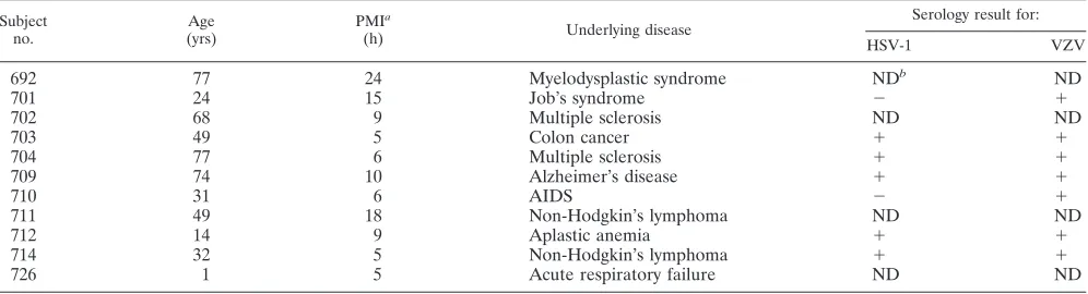
TG and serum samples.TG were collected at autopsy at a postmortem interval of less than 24 h from 11 subjects, whose salient historical and clinical features are summarized in Table 1, and were immediately fixed in 10% formalin in phosphate-buffered saline at room temperature overnight and then embedded in paraffin. One TG of each subject from whom serum was not available for antibody assays was snap-frozen on dry ice and stored at⫺80°C for later extrac-tion of total DNA and determinaextrac-tion of the status of HSV-1 and VZV infecextrac-tion. From 7 of the 11 subjects postmortem serum samples were collected and frozen at⫺20°C for later serological testing.
Serological analyses.Titers of type-specific human serum antibodies against HSV-1 were determined by means of Western blot analysis in the laboratory of Rhoda Ashley at the University of Washington in Seattle, as described previously (1). Titers of serum antibodies to VZV were determined in the Department of Clinical Pathology, Clinical Center, National Institutes of Health, using the Captia VZV immunoglobulin G kit (Trinity Biotech USA, New York), according to the manufacturer’s instructions. With these assays we determined that all subjects tested were seropositive for both VZV and HSV-1, with the exception of subjects 701 and 710, who lacked detectable circulating antibodies to HSV-1.
HSV-2 LAT transgenic mice.TG from the HSV-2 LAT transgenic mice we engineered and have described previously (57) were used to help define the efficiency of LCM in recovering individual neurons and the overall sensitivity of the assay. For these experiments, TG harvested from transgenic line 5238, which bears⬃40 copies of the transgene/cell, were processed as described above for human TG.
In situ hybridization.RNA sequences antisense to LAT, corresponding to nucleotides 119,505 to 120,669 of the HSV-1 genome, were transcribed in vitro from plasmid pG.1LAT using a SP6/T7 transcription kit (Roche Molecular Biochemicals), according to the manufacturer’s instructions. This RNA product was then fragmented by partial alkaline hydrolysis. Briefly, a 100-l reaction mixture containing 48l of RNA, 10 mM dithiothreitol, 60 mM Na2CO3, and 40 mM NaHCO3was incubated at 64°C for 50 min. The reaction mixture was then chilled on ice, 5l of 10% glacial acetic acid and 10l of 4 M sodium acetate, pH 5.2, were added to it and mixed, 300l of cold (⫺20°C) ethanol was then added, and the mixture was incubated at⫺20°C overnight. After centrifu-gation at 18,000⫻g, the pellet was washed with 70% ethanol and resuspended in 40l of RNase-free water.
[image:2.585.47.283.65.196.2]To prepare the biotin-labeled RNA probe, 5g of RNA in 20l Tris-EDTA buffer was mixed with 20l of 1g/l PHOTOPROBE biotin reagent (Vector Laboratories, Burlingame, Calif.), according to the manufacturer’s instructions for the thermal coupling method. Briefly, the reaction mixture was covered with 30l of mineral oil and incubated at 95°C for 30 min. The aqueous phase was FIG. 1. Schematic representation of LCM. An infrared laser is
integrated into a PixCell II microscope. A cap containing a transpar-ent, thermoplastic membrane is placed directly on the surface of a tissue section on a glass slide. A laser pulse heats and activates a defined area of the membrane, causing it to adhere to (A) and bind the targeted portion of the tissue (B). DNA is then extracted from the captured cells for PCR analyses.
TABLE 1. Subjects from whom trigeminal ganglia were retrieved for the study
Subject no.
Age (yrs)
PMIa
(h) Underlying disease
Serology result for:
HSV-1 VZV
692 77 24 Myelodysplastic syndrome NDb ND
701 24 15 Job’s syndrome ⫺ ⫹
702 68 9 Multiple sclerosis ND ND
703 49 5 Colon cancer ⫹ ⫹
704 77 6 Multiple sclerosis ⫹ ⫹
709 74 10 Alzheimer’s disease ⫹ ⫹
710 31 6 AIDS ⫺ ⫹
711 49 18 Non-Hodgkin’s lymphoma ND ND
712 14 9 Aplastic anemia ⫹ ⫹
714 32 5 Non-Hodgkin’s lymphoma ⫹ ⫹
726 1 5 Acute respiratory failure ND ND
aPostmortem interval in hours. bND, not done.
on November 8, 2019 by guest
http://jvi.asm.org/
[image:2.585.44.542.573.708.2]taken and mixed with 40l RNase-free water, 80l of 100 mM Tris-HCl pH 9.5, and extracted twice with 160l of 2-butanol to remove the unincorporated biotin. The aqueous phase recovered from this extraction procedure was sup-plemented with 10l of 10 M ammonium acetate, 2l of 1 M MgCl2, and 125l of cold (⫺20°C) ethanol and then incubated at⫺20°C for 15 min. After centrif-ugation at 18,000⫻gfor 15 min, the pellet was washed with 70% ethanol and resuspended in 40l Tris-EDTA buffer. This probe was stored at⫺80°C in aliquots sufficient for individual reactions.
ISH was then performed, as described previously (11), with some modifica-tions required to adapt the procedure for the use of the biotinylated probe and so that the sections could be later subjected to LCM. Briefly, TG sections were deparaffinized twice in serial xylene baths for 5 min each; incubations were carried out at room temperature unless otherwise specified, followed by rehy-dration sequentially in 100%, 100%, 95%, 70%, and 50% ethanol for 2 min each and 1 min twice in water. Slides were then transferred to 0.2 N HCl for 20 min, rinsed in water, treated with prewarmed 25g/ml proteinase K, 10 mM Tris-HCl, pH 7.4, 2 mM CaCl2at 37°C for 15 min, and finally rinsed in water. Acetylation was performed by incubating the slides in freshly prepared 0.25% (vol/vol) acetic anhydride/1.33% (vol/vol) triethanolamine at pH 8.0 for 10 min and finally rinsing the slides in water. Excess water was wiped from the slides, taking care to not touch the tissue sections themselves. Then, 30l of prehybridization solution (50% formamide, 2⫻SSC [1⫻SSC is 0.15 M NaCl plus 0.015 M sodium citrate], 50 mM Tris, 1 mM EDTA, 0.02% Ficoll, 0.02% polyvinylpyrrolidone, 500g/ml tRNA, 500g/ml bovine serum albumin) was added onto each section. One minute later the liquid was drained, another 20 l of the prehybridization solution was added onto each section, and then the section was covered with a glass coverslip. After incubation at 42°C for 2 h, the coverslip was removed and 30l of hybridization solution (50% formamide, 20% dextran sulfate, 1 mM EDTA, 10 mM dithiothreitol, 300 mM NaCl, 50 mM Tris-HCl, pH 7.4, 0.02% Ficoll, 0.02% polyvinylpyrrolidone, 500g/ml tRNA, 500g/ml bovine serum albumin) with 500 ng/ml of heat-denatured (65°C for 20 min) probe was added. New coverslips were applied, the edges were sealed with rubber cement, and slides were incubated at 55°C overnight. The next day, after the sealant and coverslips were carefully removed, slides were washed twice with 2⫻SSC for 10 min each at room temperature, twice at 55°C for 20 min each with buffer containing 2⫻SSC, 1 mM EDTA, and 0.1% Triton X-100, and once more for 30 min at 55°C with buffer containing 0.1⫻SSC, 1 mM EDTA, and 0.1% Triton X-100. To remove any unhybridized RNA probe, sections were treated with an RNase mixture containing 40g/ml RNase A, 10 U/ml RNase T1, 10 mM Tris-HCl, pH 7.4, and 300 mM NaCl, incubated at 37°C for 40 min, and finally washed twice for 20 min each with 2⫻SSC at 55°C. To detect the hybridized probe, slides were incubated in a blocking solution of 1⫻casein in 100 mM Tris-HCl (pH 7.5), 150 mM NaCl, and 0.1% Tween 20 (TBST) for 40 min. Horseradish peroxidase-streptavidin (Vector Laboratories) was added to the blocking solution to yield a final concentration of 4g/ml, and slides were incubated for another 30 min. After three washes in TBST, the slides were developed using a TMB substrate kit for horseradish peroxidase (Vector Labo-ratories) for 5 min and then rinsed in water. Slides were dehydrated in graded 70%, 95%, 100%, and 100% ethanol for 1 min each and 1 min twice in xylene, and then air dried.
LCM.Serial 5-m sections from different anatomical regions of each TG were cut from each tissue block and mounted on plain glass slides. In order to better represent the neurons in whole ganglia with limited numbers of cells that can be processed by LCM/PCR, cells from the first, middle, and last 10 slides of a ganglion were selected for LCM. The tissue sections were deparaffinized twice in
xylene at room temperature for 5 min each and then air dried. LCM was performed using a PixCell II (Arcturus Engineering, Inc., Mountain View, Calif.). The laser beam was set to be 7.5m in diameter, with a power of 25 to 35 mW and for 30 to 40 ms in duration. These optimal settings were selected for capturing single neurons from human TG without apparent contamination with adjacent cells. After placing CapSure LCM caps (Arcturus Engineering, Inc.) on top of each tissue section, single neurons with visible nuclei were chosen under the microscope and a laser pulse was fired to capture that neuron onto the cap. Satellite cells, hundreds on each cap, were harvested only after all of the neurons in the desired area were removed.
To procure LAT-positive and LAT-negative neurons separately, sections were first subjected to ISH for HSV-1 LAT, as described in the previous section. The in situ-stained cells were then visualized and photomicrographed while the sec-tions were still covered with xylene. After each section was air dried, LAT-positive and -negative cells were identified with the guidance of the photographs and captured using LCM.
DNA extraction of LCM samples.Caps with captured cells were inserted into 0.5-ml GeneAmp tubes (Applied Biosystems) containing 14 or 50l of DNA extraction buffer (10 mM Tris-HCl, pH 8.0, 1 mM EDTA, 1% Tween 20, and freshly added proteinase K to yield 0.04%) for single neurons or multiple satellite cells, respectively. At 37°C overnight, the tubes were incubated upside down so that all captured cells were covered with the extraction buffer. After 1 min of centrifugation at 18,000⫻g, the caps were removed and the tubes were sealed and heated at 95°C for 8 min to inactivate any remaining proteinase K. From each sample, 12l/reaction mixture was subjected to real-time PCR specific for HSV-1 glycoprotein G (gG) or VZV immediate-early gene 62, as described previously (39). The sequences of the primers and probes used are shown in Table 2. Each 25l of the PCR mixture contained 12.5l of 2⫻Universal TaqMan PCR master mix (Applied Biosystems), forward and reverse primers at a final concentration of 1.0M each, 0.2M of probe, and 12l of DNA extract from the LCM sample. In every PCR assay performed, four no-template control reactions and a standard panel of DNA copy-number reference samples were run. Each reference panel consisted of six dilutions of the pcDNA3.gG1 plasmid DNA for quantifying HSV-1 DNA, or pCMV62 DNA for VZV DNA, at 1.5, 5, 50, 500, 5,000, or 50,000 copies/reaction mixture. All standards were run in duplicate. All reactions were carried out in an ABI PRISM 7700 sequence detector (Applied Biosystems, Foster City, CA) with optimized thermal cycle conditions: 50°C for 2 min, 95°C for 10 min, and 40 cycles at 95°C for 20 seconds and 60°C for 1 min. By comparing the threshold cycle (Ct) of each sample to the Cts from the standard reference panel, the copy number of each experimental reaction was then determined. To estimate the cellular genome content of LCM samples of satellite cells, 24l out of 50l DNA extract from each cap with satellite cells was subjected to PCR in duplicate with primers and probes specific for human albumin, with human genomic DNA (Applied Biosystems) as copy number reference.
Statistical analyses.Analysis of variance and linear regression analyses were performed with the computer software package JMP version 3.0 (SAS Institute, Cary, NC).
RESULTS
LCM/PCR detection of viral DNA in single cells is sensitive, specific, and efficient.Extensive developmental work was un-dertaken to define the operational characteristics and limits of TABLE 2. Primers and probes used for real-time PCR
Primer or probe Sequence
Primers
gG1 forward...5⬘CTGTTCTCGTTCCTCACTGCCT 3⬘ gG1 reverse...5⬘CAAAAACGATAAGGTGTGGATGAC 3⬘ ORF62 forward ...5⬘TCTTGTCGAGGAGGCTTCTG 3⬘ ORF62 reverse ...5⬘TGTGTGTCCACCGGATGAT 3⬘ hAlbumin forward...5⬘TGCATGAGAAAACGCCAGTAA 3⬘ hAlbumin reverse...5⬘ATGGTCGCCTGTTCACCAA 3⬘ Probes
gG1 P...5⬘FAM-CCCTGGACACCCTCTTCGTCGTCAG-TAMRA 3⬘ ORF62 P ...5⬘FAM-TCTCGACTGGCTGGGACTTGCG-TAMRA 3⬘ hAlbumin P...5⬘FAM-TGACAGAGTCACCAAATGCTGCACAGAA-QSY 3⬘
on November 8, 2019 by guest
http://jvi.asm.org/
the LCM/PCR assays for detecting viral DNA sequences in single human neurons. We began by determining the optimal LCM pulse characteristics. In this regard, we determined mi-croscopically that the diameter of human TG neurons in fixed tissue sections ranged from 25 to 60m. When the energy and diameter of the laser beam were set for each desired TG neuron, as specified in Materials and Methods, it activated an area of the thermal membrane slightly smaller than the dia-meter of that cell, such that the content of the targeted single neuron was captured while all surrounding satellite and other nonneuronal cells were left attached to the slide (Fig. 2).
Even though we performed LCM only on cells whose nuclei were visible in the section, because of both the depth of the tissue sections and potential inefficiencies in extraction and detection, we expected that the DNA recovered from each cell might represent only a fraction of its entire content. Thus, we set out to estimate the efficiency of cellular DNA recovery and detection by LCM/PCR. To this end, we first tested the LCM/ PCR efficiency of single neurons recovered from TG sections of heterozygous HSV-2 LAT transgenic mice that we had en-gineered and estimated previously by Southern hybridization to harbor about 40 copies of the LAT transgene/cell (57) in-stead of directly testing single neurons from human TG, be-cause the copy number of the cellular gene is too low to be detected. By LCM/PCR an average of 37.4 ⫾ 3.6 (standard error of the mean) copies of the HSV-2 LAT genomic se-quence were detected from DNA extracted from single TG neurons of the transgenic mice. The result was not affected by adding 10 ng of human genomic DNA or total RNA isolated from the transgenic mouse liver in an amount equivalent to about 5,000 copies of LAT RNA. Thus, by this method, it
appeared that the combined efficiency of LCM/PCR in recov-ering DNA from a single neuron was roughly 93.4⫾9%.
[image:4.585.81.512.69.285.2]The combined efficiency of LCM/PCR of sensory neurons from human TG was also examined by subjecting DNA ex-tracted from LCM caps containing known numbers (50 to 250) of neurons to PCR with primers and a probe for the human albumin gene (there are two copies of the gene per cell). Based on the numbers of albumin gene copies recovered, we esti-mated an efficiency of 64⫾ 15% for LCM/PCR of neurons from human TG sections. Considering that the diameter of a human neuronal nucleus is greater than the thickness of a 5-m tissue section, it is likely that portions of human nuclei were excluded from the sections. This is evident by observing a neuronal nucleus on both of two adjacent sections under mi-croscopy. By comparing the diameters of human neuronal nu-clei with that of mouse neuronal nunu-clei microscopically, we found that the diameter of the mouse neuronal nucleus is FIG. 2. LCM of neurons and satellite cells from human trigeminal ganglion sections. (A and B) Photomicrographs (100⫻) of a ganglion section before and after dissection of single neurons, respectively. The arrows indicate the neurons in which nuclei were clearly visible (A) and those which were microdissected completely (B). It is evident in panel B that nonneuronal, satellite cells surrounding the targeted neurons were retained in the tissue section. (C) Several captured neurons on a cap. (D) Photomicrograph of a tissue section in which neurons had been removed from the circled clusters but the satellite cells surrounding those neurons were retained in the section. (E) The same section as in panel D after the satellite cells in the circled clusters had been removed by LCM. (F) Some captured satellite cells on a cap.
TABLE 3. Detection of HSV-1 DNA in individual neurons
Subject no. No. of neurons tested
No. (%) of positive neurons
Median copies/ positive cell
701 119 1 (0.8) 12.1
702 153 3 (2.0) 12.7
703 206 15 (7.3) 30.7
704 162 17 (10.5) 9.0
711 214 21 (9.8) 10.1
712 235 5 (2.1) 7.1
Totala 970 61 (6.3) 11.3
aTotal data do not include results for subject 701, who was HSV-1 sero-negative.
on November 8, 2019 by guest
http://jvi.asm.org/
[image:4.585.301.542.608.707.2]about three-fourths that of its human equivalent. This could explain the difference in efficiency of neuronal DNA recovery, being 64% for human sections and 93% for mouse sections, because the 5-m section contained a greater percentage of the mouse nucleus. Nonetheless, by two independent tech-niques, we determined that LCM/PCR detects the majority of the DNA content of a single neuron.
The sensitivity and specificity of the PCR assay for HSV-1 gG and VZV ORF62 sequences were first determined by anal-yses of serial dilutions of plasmid standards. We routinely detected ⱖ5 copies of target sequences in a 25-l reaction mixture with or without carrier DNA when all HSV-1-negative DNA samples were read as negative. Assay specificity was then further determined by testing DNA from single neurons re-covered from HSV-1-seronegative subject 701 in parallel with every assay of samples from HSV-1-positive subjects. When the cutoff threshold was set at⬍5 copies/reaction mixture, 1 out of a total of 119 independent reactions with DNA from
subject 701 was read as positive (equivalent to 12 copies of gG1), being the only occasion, or false positive, detected from HSV-1-seronegative samples. These data suggest that, as per-formed, the LCM/PCR assay yields a real but very low rate of false-positive reactions.
[image:5.585.142.446.70.208.2]HSV-1 genome copy numbers vary widely in individual PCR-positive neurons.From 119 to 235 single neurons were microdissected from sections of each of six study subjects and analyzed for HSV-1 DNA by means of real-time PCR with primers and a probe specific for gG sequences. Among 970 single neurons from five HSV-1-positive subjects (three of whom were seropositive, while HSV-1 DNA was detected from the total TG DNA of subjects 702 and 711, from whom post-mortem sera were not available), 61 cells were PCR positive for gG sequences, with copy numbers/cell ranging from 5 (the limit of assay sensitivity) to 3,955 and a median of 11.3 genome copies/positive cell (Table 3). The majority of positive neurons harbored less than 20 copies of HSV-1 genomes: 43% had FIG. 3. HSV-1 and VZV genome copy numbers in individual PCR-positive neurons. A total of 970 single neurons captured from sections from five study subjects were tested for HSV-1 gG, of which 61 were PCR positive, with genome copy numbers/cell ranging from 5 (the lower limit of detection of the PCR assay) to 3,955 and with a median of 11.3 copies/positive cell. A total of 71% of all positive neurons contained 20 or fewer copies of HSV-1 DNA (A). The VZV genome copy number detected in single neurons ranged from 2.6 copies (the lower limit of detection of the assay being 5 copies of the diploid gene ORF62, or 2.5 genomes) to 5,773 copies, with a median of 6.9 copies per positive cell (B). The VZV genome copies per positive cell wereⱕ20 in 70% of all positive neurons.
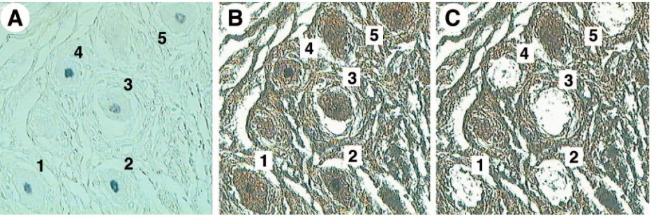
FIG. 4. LCM of LAT-positive and LAT-negative neurons. Human TG sections were subjected to in situ hybridization for HSV-1 LAT. The hybridization signals were visualized microscopically and photographed while sections were covered with xylene. (A) ISH section covered with xylene, in which five LAT-positive neurons could be identified (200⫻). (B and C) The section was then air dried (B), and each LAT-positive neuron was harvested by LCM (C). The LAT-negative neurons were then harvested from these sections after all clearly LAT-positive neurons were removed.
on November 8, 2019 by guest
http://jvi.asm.org/
[image:5.585.60.535.522.677.2]ⱕ10 copies and 28% hadⱖ10 butⱕ20 copies per neuron. In 14% of the DNA-positive cells, however, the viral DNA copy numbers wereⱖ100, and a few of them wereⱖ1,000 copies/ cell (Fig. 3).
More neurons contain HSV-1 DNA than are found to con-tain LAT.As indicated in Table 3, the percentages of neurons positive for HSV-1 DNA ranged from 2.0 to 10.5% in the five HSV-1-positive subjects. This range of percentages is higher than the 0.2 to 4% of LAT-positive neurons we reported pre-viously in human TG using ISH (11), and similar percentages have been found by others in their ISH studies of infected animals (4, 22, 44).
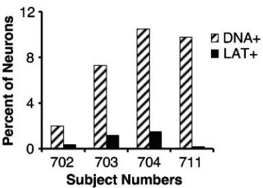
To determine whether the percentages of LAT-positive neu-rons in the TG studied now differ from those prior observa-tions, sections adjacent to ones from which single neurons were captured for LCM/PCR were subjected to ISH using a biotin-ylated cRNA probe antisense to HSV-1 LAT and visualized with streptavidin-conjugated peroxidase and its substrate, TMB (Fig. 4). Even though some of our preparatory work (data not shown) suggested that the nonradioactive probe used here is less sensitive than the radioactive probe we had used in our previous ISH studies, we found that the percentages of LAT-positive neurons were similar to those previously re-ported (11), being 0.4, 1.2, 1.5, and 0.2% in TG of subjects 702, 703, 704, and 711, respectively. In these four subjects the per-centages of their neurons positive for HSV-1 DNA were 2.0, 7.3, 10.5, and 9.8%, respectively. The percentages of HSV-1 DNA-positive neurons were 5- to 41-fold higher than those positive for LAT by ISH (Fig. 5). This finding strongly suggests that there are neurons with HSV-1 DNA detectable by PCR that are not ISH positive for LAT.
HSV-1 DNA is detected in LAT-negative neurons.To deter-mine that HSV-1 DNA is detectable in LAT-negative neurons in human TG, as reported in mice (6), single neurons were microdissected from sections of human TG from subjects 702, 703, 709, and 711 after undergoing ISH for LAT. As shown in Fig. 6, HSV-1 DNA was detected from 4, 6, 11, and 7% of LAT-negative neurons from these subjects, respectively, while
among LAT-positive neurons the percentages of cells positive for HSV-1 DNA were 27, 21, 16, and 40%, respectively.
A comparison of the HSV-1 DNA content in single LAT-positive neurons with that in LAT-negative ones is shown in Table 4. Since the percentage of LAT-positive neurons in these sections proved very low (e.g., 4/1,000, or 0.4%), most of the HSV-1 DNA-positive cells (e.g., 63/1,000, or 6.3%) did not accumulate LAT to levels detectable by ISH. On the other hand, the HSV-1 genome copy numbers in the positive neu-rons of these two groups were comparable, with medians of 18 and 12 copies in LAT-positive and LAT-negative neurons, respectively.
Detection of VZV DNA in human TG neurons. From 10 subjects, 146 to 209 single neurons were captured from their TG sections by LCM and analyzed for VZV DNA content using PCR primers and a probe specific for the diploid VZV gene 62 (Table 2). The percentages of VZV DNA-positive neurons ranged from 1.0 to 6.9%, with the median VZV ge-nome copy numbers among positive neurons ranging from 4.5 to 29.8 (Table 5). Overall, a total of 4.1% (71 out of 1722) of the neurons were found to harbor 2.6 to 5,773 copies of the VZV genome, with a median of 6.9 copies per positive cell. Among the VZV DNA-positive neurons, 70% harboredⱕ20 viral genomes while 9% harboredⱖ100 copies (Fig. 3).
HSV-1 and VZV DNAs are rarely detected in satellite cells.
To determine if HSV-1 and VZV DNAs can be found in nonneuronal cells, hundreds of satellite cells were microdis-FIG. 5. More neurons contain HSV-1 DNA than are found to
[image:6.585.68.260.66.203.2]contain LAT. DNA extracted from LCM caps bearing single neurons was subjected to real-time PCR for HSV-1 gG sequences. Adjacent sections were subjected to ISH for HSV-1 LAT. For sections of all four subjects studied, the percentages (2.0, 7.3, 10.5, and 9.8) of neurons that were PCR positive for HSV-1 DNA (DNA⫹) were higher than the percentages (0.4, 1.2, 1.5, and 0.2) of cells that were LAT positive (LAT⫹).
[image:6.585.331.511.73.247.2]FIG. 6. HSV-1 DNA is detected in LAT-negative neurons. Single LAT-positive or LAT-negative neurons dissected from human TG sections, as shown in Fig. 4, of subjects 702, 703, 709, and 711 were subjected to real-time PCR for HSV-1 gG sequences. HSV-1 DNA was detected in 4, 6, 11, and 7% of LAT-negative neurons, respectively. In LAT-positive neurons, the percentages of viral DNA-positive cells were higher, at 27, 21, 16, and 40%, respectively.
TABLE 4. HSV-1 genome copies in single LAT⫹or LAT⫺neurons
ISH result No. of neurons tested
Positive neurons (%)
Median copies/ positive cell
LAT⫹ 93 24 18
LAT⫺ 277 7 12
on November 8, 2019 by guest
http://jvi.asm.org/
[image:6.585.301.541.676.725.2]sected from regions of the TG sections from which all neurons had been removed (Fig. 2). A portion of the DNA extracted from each cap was used to determine the cellular genome copy number, while the remaining DNA was assayed by PCR for gG1 or ORF62 sequences. In this manner, among 13 LCM caps prepared from sections of subjects 702, 704, 709, and 711, a total of⬃5,200 satellite cells were assayed and 21 copies of HSV-1 gG sequences were detected. In an additional set of 21 caps from subjects 701, 709, 710, 711, 714, and 726, in which a total of ⬃14,200 cellular genomes were detected, only one sample was positive for nine copies of the VZV genome.
DISCUSSION
We developed and characterized combined LCM/PCR as-says by which we could efficiently and specifically detect and quantify HSV-1 and VZV genomic sequences in single human neuronal cells. Using these assays on thousands of individual neurons and aggregates of nonneuronal cells, we have been able to further refine our understanding of the cellular distri-bution and estimates of the numbers of copies of latent viral DNA sequences in human TG cells.
Because LCM/PCR detects viral DNA while ISH detects LAT RNA, which is a highly stable and amplified species, the limits of their detection in our assay systems are dependent on the viral DNA load and the LAT accumulation levels in a particular neuron. Neurons that harbor copy numbers of viral DNA that are close to, or lower than, the sensitivity of the LCM/PCR assay may accumulate different amounts of LAT, ranging from a few copies to hundreds of copies of LAT per viral genome. Those neurons may not be able to be identified by the LCM/PCR methods for viral DNA, but those accumu-lating high levels of LAT can be detected by ISH for LAT. Thus, ISH may be more sensitive for detecting latency in such neurons. When the viral DNA load exceeds the sensitivity limitation of LCM/PCR, this method is more sensitive than ISH for LAT, as we verified by showing that the majority of HSV-1 DNA-positive neurons are ISH negative for LAT.
By using the LCM/PCR method, we determined that a mean of 6.3% of human TG neurons are HSV-1 DNA positive (Table 3). These numbers are lower than the percentages re-ported for experimentally infected mice, i.e., 7.3 to 32% by contextual analysis (45, 47) or 35% by LCM/PCR (6). It is most probable that the differences between what we observed in
human cells and what has been observed in mice is due to the high virus inocula and route of infection used in mice, as have been discussed in the literature (45), and the short interval between infection and death in experimental mice, rather than representing technical differences among the assays employed. This explanation is supported by the independent observation in dissociated ganglionic cells that only some 3% of human TG neurons are PCR positive for HSV-1 DNA (5).
The HSV-1 genome copy number in individual neurons var-ied over 3 orders of magnitude (Fig. 3). The majority of HSV-1 DNA-positive neurons harbored, however, ⱕ20 copies of HSV-1 genome. Considering that the method we used was not able to identify neurons with less than 5 copies of viral DNA, the percentages of latently infected neurons and those harbor-ingⱕ20 copies of viral DNA should be even higher than we estimated here. On the other hand, 3% of the viral genome-positive neurons contained ⱖ1,000 copies of viral genome. This result is very similar to what has been found in mice by PCR of dissociated ganglion neurons (45) or by LCM/PCR (6). Our finding here by means of LCM/PCR that indicated that there are only a median of 11.3 HSV-1 genome copies/DNA-positive neuron proved to be concordant with what we our-selves estimated several years ago by real-time PCR of total DNA isolated from latently infected human TG, namely, that there are about 28 or more HSV-1 genome copies in LAT-positive neurons (39). Our findings here also are in concor-dance with estimates made by PCR of dissociated human TG in which HSV-1 DNA was found in 3% of the neurons and the viral DNA copy numbers ranged from 2 to 50 (5). It is not clear, however, whether the presence of higher levels of latent genomes in a cell indicates that they are undergoing, or are more inclined to undergo, reactivation. In this regard, we had shown in experimentally infected guinea pigs that higher total latent viral loads in a ganglion are associated with higher rates of genital disease reactivation (24, 29), but it was not known in those experiments whether more neurons were latently in-fected or whether neurons carried more copies of latent HSV DNA. It has been reported that in mice the HSV-1 strain KOS, which reactivates poorly, has less viral genome copies per pos-itive neuron than the two other strains, 17syn⫹and McKrae, which reactivate more frequently (46).
[image:7.585.43.284.80.218.2]The combination of ISH and LCM/PCR techniques enabled us for the first time to analyze both LAT accumulation and HSV-1 DNA content in the very same neuron in human TG. By so doing, we demonstrated that the percentages of HSV-1 DNA-positive neurons in human TG were higher than the percentages of neurons that were found to be positive for LAT by ISH, consistent with recent parallel LCM and ISH studies of infected mice (6). In our human subjects, the percentages of DNA PCR-positive neurons were between 5.1 and 41 times more than the percentages of neurons that we found to be ISH positive for LAT (Fig. 5). In other words, neurons positive for LAT by ISH only represent a fraction of the entire pool of latently infected neurons. This finding may reflect a greater sensitivity of PCR for viral DNA than of ISH for LAT. The novel and more important message from our study is that in human TG not all latently infected neurons accumulate LAT to a high level, raising the questions of why and how the accumulation of LAT is regulated during latent infection. Given the observation in animals that HSV mutants impaired TABLE 5. Detection of VZV DNA in individual neurons
Subject no.
No. of neurons tested
No. (%) of positive neurons
Median copies/ positive cell
692 180 6 (3.3) 11.7
701 151 10 (6.6) 10.0
702 147 3 (2.0) 6.1
703 148 7 (4.7) 4.5
704 146 6 (4.1) 8.4
709 209 9 (4.3) 4.5
710 152 3 (2.0) 4.9
711 207 13 (6.3) 12.4
714 174 12 (6.9) 6.8
726 208 2 (1.0) 29.8
Total 1,722 71 (4.1) 6.9
on November 8, 2019 by guest
http://jvi.asm.org/
for LAT expression are able to establish and maintain latency in sensory neurons (2, 15, 37, 54, 57, 58), it is not entirely surprising to find latently infected human TG neurons in which LAT is not detectable by ISH. Our observations indicate that even in humans, the natural hosts for HSV-1, persistent sub-stantial accumulation of LAT is not essential for HSV-1 to maintain latency. One is inclined to ask whether LAT accu-mulation is stable over the long term, which in the case of our human subjects amounted to decades from the likely times of their primary infections to death, and whether alterations, if any, in LAT accumulation are associated with periods of virus reactivation.
That our LCM/PCR method can only recover and reliably detect about 64% of the entire DNA content of a single human neuron and the fact that we detected HSV-1 DNA in only 16% to 40% of LAT-positive cells (Fig. 6) indicate that the percent-ages of latently infected neurons and the copy numbers of viral DNAs in neurons are likely even greater than we estimated from the LCM/PCR data. Moreover, it is reasonable to assume that there are latently infected neurons containing quantities of latent viral DNA below the assay sensitivity threshold of 5 copies/cell. Therefore, we conclude that HSV-1 latent infec-tion in human TG is more pervasive than previously estimated by ISH for LAT.
HSV-1 LAT has not been detected in satellite cells in la-tently infected human or experimental animal TG by ISH, in situ PCR, and PCR of dissociated TG cells (11, 13, 34, 44, 45, 51–53). There was also no LAT ISH signal seen over non-neuronal cells in all of the sections in this study. Thus, the 21 copies of HSV-1 gG sequences detected in⬃5,200 satellite cells may reflect the very low level of false-positive reactions we identified in LCM/PCR studies and/or may be due to a low-level contamination with neurons amid the satellite cell samples.
Seventeen years ago, our laboratory reported that by ISH VZV RNA was only detected in satellite cells of human TG (12). Since then, the cumulative data from other laboratories have built a compelling case that VZV persists predominantly in neurons (17, 21, 25, 27, 28, 30). By real-time PCR of 1,722 single human TG neurons harvested by LCM, we detected VZV DNA in 4.1% of them. That we found VZV DNA in these few neurons is in accordance with recent data using dissociated human TG (28, 30) but is far less than the percent-ages reported by Lungu et al. using an immunohistochemical technique (32). This suggests either that the immunohisto-chemical method is far more sensitive than the LCM/PCR technique or, alternatively, that the immunohistochemical as-says themselves may have yielded false-positive reactions.
Despite our prior report of VZV RNA in nonneuronal cells (12), we detected here only nine copies of the VZV genome in 14,200 satellite cells. It is not known whether this indicates that there is a small reservoir of latent VZV DNA in nonneuronal cells, spread of subclinically reactivating virus, or that the ge-nome copies detected were due to the recovery of a satellite cell sample which could have been contaminated with very small numbers of latently infected neurons or represented the low level of false-positive reactions that we determined can occur in LCM/PCR studies. Nonetheless, we can safely con-clude today that VZV persists in human TG predominantly, if not exclusively, in sensory neurons.
ACKNOWLEDGMENTS
We thank David Kleiner and his colleagues for helping us harvest the tissue and serum samples, Thomas Fleisher and Rhoda Ashley for performing the serological analyses, Yo Hoshino for performing sta-tistical analyses, and Jeffrey Cohen for ongoing discussions and critical reading of the manuscript.
This research was supported by the Intramural Research Program of the NIAID, NIH.
REFERENCES
1.Ashley, R. L., J. Militoni, F. Lee, A. Nahmias, and L. Corey.1988. Compar-ison of Western blot (immunoblot) and glycoprotein G-specific immunodot enzyme assay for detecting antibodies to herpes simplex virus types 1 and 2 in human sera. J. Clin. Microbiol.26:662–667.
2.Bloom, D. C., G. B. Devi-Rao, J. M. Hill, J. G. Stevens, and E. K. Wagner.
1994. Molecular analysis of herpes simplex virus type 1 during epinephrine-induced reactivation of latently infected rabbits in vivo. J. Virol. 68:
1283–1292.
3.Bloom, D. C., J. M. Hill, G. Devi-Rao, E. K. Wagner, L. T. Feldmen, and J. G. Stevens.1996. A 348-base-pair region in the latency-associated transcript facilitates herpes simplex virus type 1 reactivation. J. Virol.70:2449–2459. 4.Burke, R. L., K. Hartog, K. D. Croen, and J. M. Ostrove.1991. Detection and
characterization of latent HSV RNA by in situ and Northern blot hybrid-ization in guinea pigs. Virology181:793–797.
5.Cai, G. Y., L. I. Pizer, and M. J. Levin.2002. Fractionation of neurons and satellite cells from human sensory ganglia in order to study herpesvirus latency. J. Virol. Methods104:21–32.
6.Chen, X. P., M. Mata, M. Kelley, J. C. Glorioso, and D. J. Fink.2002. The relationship of herpes simplex virus latency associated transcript expression to genome copy number: a quantitative study using laser capture microdis-section. J. Neurovirol.8:204–210.
7.Cohen, J. I., and S. E. Straus.2001. Varicella-zoster virus and its replication, p. 2707–2730.InD. M. Knipe and P. M. Howley (ed.), Fields virology, 4th ed., vol. 2. Lippincott Williams & Wilkins, Philadelphia, Pa.
8.Cohrs, R. J., D. H. Gilden, P. R. Kinchington, E. Grinfeld, and P. G. Kennedy.2003. Varicella-zoster virus gene 66 transcription and translation in latently infected human ganglia. J. Virol.77:6660–6665.
9.Cohrs, R. J., J. Randall, J. Smith, D. H. Gilden, C. Dabrowski, H. van Der Keyl, and R. Tal-Singer. 2000. Analysis of individual human trigeminal ganglia for latent herpes simplex virus type 1 and varicella-zoster virus nucleic acids using real-time PCR. J. Virol.74:11464–11471.
10.Cook, S. D., M. J. Paveloff, J. J. Doucet, A. J. Cottingham, F. Sedarati, and J. M. Hill.1991. Ocular herpes simplex virus reactivation in mice latently infected with latency-associated transcript mutants. Investig. Ophthalmol. Vis. Sci.32:1558–1561.
11.Croen, K. D., J. M. Ostrove, L. J. Dragovic, J. E. Smialek, and S. E. Straus.
1987. Latent herpes simplex virus in human trigeminal ganglia. Detection of an immediate early gene “anti-sense” transcript by in situ hybridization. N. Engl. J. Med.317:1427–1432.
12.Croen, K. D., J. M. Ostrove, L. J. Dragovic, and S. E. Straus.1988. Patterns of gene expression and sites of latency in human nerve ganglia are different for varicella-zoster and herpes simplex viruses. Proc. Natl. Acad. Sci. USA
85:9773–9777.
13.Deatly, A. M., J. G. Spivack, E. Lavi, and N. W. Fraser.1987. RNA from an immediate early region of the type 1 herpes simplex virus genome is present in the trigeminal ganglia of latently infected mice. Proc. Natl. Acad. Sci. USA
84:3204–3208.
14.De Preter, K., J. Vandesompele, P. Heimann, M. M. Kockx, M. Van Gele, J. Hoebeeck, E. De Smet, M. Demarche, G. Laureys, N. Van Roy, A. De Paepe, and F. Speleman. 2003. Application of laser capture microdissection in genetic analysis of neuroblastoma and neuroblastoma precursor cells. Can-cer Lett.197:53–61.
15.Devi-Rao, G. B., D. C. Bloom, J. G. Stevens, and E. K. Wagner.1994. Herpes simplex virus type 1 DNA replication and gene expression during explant-induced reactivation of latently infected murine sensory ganglia. J. Virol.
68:1271–1282.
16.Drolet, B. S., G. C. Perng, R. J. Villosis, S. M. Slanina, A. B. Nesburn, and S. L. Wechsler.1999. Expression of the first 811 nucleotides of the herpes simplex virus type 1 latency-associated transcript (LAT) partially restores wild-type spontaneous reactivation to a LAT-null mutant. Virology253:
96–106.
17.Dueland, A. N., T. Ranneberg-Nilsen, and M. Degre.1995. Detection of latent varicella zoster virus DNA and human gene sequences in human trigeminal ganglia by in situ amplification combined with in situ hybridiza-tion. Arch. Virol.140:2055–2066.
18.Elkahloun, A. G., J. Gaudet, G. S. Robinson, and D. C. Sgroi.2002. In situ gene expression analysis of cancer using laser capture microdissection, mi-croarrays and real time quantitative PCR. Cancer Biol. Ther.1:354–358.
on November 8, 2019 by guest
http://jvi.asm.org/
19.Emmert-Buck, M. R., R. F. Bonner, P. D. Smith, R. F. Chuaqui, Z. Zhuang, S. R. Goldstein, R. A. Weiss, and L. A. Liotta.1996. Laser capture micro-dissection. Science274:998–1001.
20.Fuller, A. P., D. Palmer-Toy, M. G. Erlander, and D. C. Sgroi.2003. Laser capture microdissection and advanced molecular analysis of human breast cancer. J. Mammary Gland Biol. Neoplasia8:335–345.
21.Gilden, D. H., Y. Rozenman, R. Murray, M. Devlin, and A. Vafai.1987. Detection of varicella-zoster virus nucleic acid in neurons of normal human thoracic ganglia. Ann. Neurol.22:377–380.
22.Hill, J. M., B. M. Gebhardt, R. Wen, A. M. Bouterie, H. W. Thompson, R. J. O’Callaghan, W. P. Halford, and H. E. Kaufman.1996. Quantitation of herpes simplex virus type 1 DNA and latency-associated transcripts in rabbit trigeminal ganglia demonstrates a stable reservoir of viral nucleic acids during latency. J. Virol.70:3137–3141.
23.Hill, J. M., J. B. Maggioncalda, H. H. Garza, Jr., Y. H. Su, N. W. Fraser, and T. M. Block.1996. In vivo epinephrine reactivation of ocular herpes simplex virus type 1 in the rabbit is correlated to a 370-base-pair region located between the promoter and the 5⬘end of the 2.0-kilobase latency-associated transcript. J. Virol.70:7270–7274.
24.Hoshino, Y., S. K. Dalai, K. Wang, L. Pesnicak, T. Y. Lau, D. M. Knipe, J. I. Cohen, and S. E. Straus.2005. Comparative efficacy and immunogenicity of replication-defective, recombinant glycoprotein, and DNA vaccines for her-pes simplex virus 2 infections in mice and guinea pigs. J. Virol.79:410–418. 25.Hyman, R. W., J. R. Ecker, and R. B. Tenser.1983. Varicella-zoster virus
RNA in human trigeminal ganglia. Lancetii:814–816.
26.Kennedy, P. G., E. Grinfeld, and J. E. Bell.2000. Varicella-zoster virus gene expression in latently infected and explanted human ganglia. J. Virol.74:
11893–11898.
27.Kennedy, P. G., E. Grinfeld, and J. W. Gow.1998. Latent varicella-zoster virus is located predominantly in neurons in human trigeminal ganglia. Proc. Natl. Acad. Sci. USA95:4658–4662.
28.LaGuardia, J. J., R. J. Cohrs, and D. H. Gilden.1999. Prevalence of vari-cella-zoster virus DNA in dissociated human trigeminal ganglion neurons and nonneuronal cells. J. Virol.73:8571–8577.
29.Lekstrom-Himes, J. A., L. Pesnicak, and S. E. Straus.1998. The quantity of latent viral DNA correlates with the relative rates at which herpes simplex virus types 1 and 2 cause recurrent genital herpes outbreaks. J. Virol.72:
2760–2764.
30.Levin, M. J., G. Y. Cai, M. D. Manchak, and L. I. Pizer.2003. Varicella-zoster virus DNA in cells isolated from human trigeminal ganglia. J. Virol.
77:6979–6987.
31.Lungu, O., P. W. Annunziato, A. Gershon, S. M. Staugaitis, D. Josefson, P. LaRussa, and S. J. Silverstein.1995. Reactivated and latent varicella-zoster virus in human dorsal root ganglia. Proc. Natl. Acad. Sci. USA92:
10980–10984.
32.Lungu, O., C. A. Panagiotidis, P. W. Annunziato, A. A. Gershon, and S. J. Silverstein.1998. Aberrant intracellular localization of varicella-zoster virus regulatory proteins during latency. Proc. Natl. Acad. Sci. USA95:7080–7085. 33.Maggioncalda, J., A. Mehta, Y. H. Su, N. W. Fraser, and T. M. Block.1996. Correlation between herpes simplex virus type 1 rate of reactivation from latent infection and the number of infected neurons in trigeminal ganglia. Virology225:72–81.
34.Mehta, A., J. Maggioncalda, O. Bagasra, S. Thikkavarapu, P. Saikumari, T. Valyi-Nagy, N. W. Fraser, and T. M. Block.1995. In situ DNA PCR and RNA hybridization detection of herpes simplex virus sequences in trigeminal ganglia of latently infected mice. Virology206:633–640.
35.Meier, J. L., R. P. Holman, K. D. Croen, J. E. Smialek, and S. E. Straus.
1993. Varicella-zoster virus transcription in human trigeminal ganglia. Virology193:193–200.
36.Nagashima, K., M. Nakazawa, and H. Endo.1975. Pathology of the human spinal ganglia in varicella-zoster virus infection. Acta Neuropathol. (Berlin)
33:105–117.
37.Perng, G. C., E. C. Dunkel, P. A. Geary, S. M. Slanina, H. Ghiasi, R. Kaiwar, A. B. Nesburn, and S. L. Wechsler.1994. The latency-associated transcript gene of herpes simplex virus type 1 (HSV-1) is required for efficient in vivo spontaneous reactivation of HSV-1 from latency. J. Virol.68:8045–8055. 38.Perng, G. C., S. M. Slanina, H. Ghiasi, A. B. Nesburn, and S. L. Wechsler.
1996. A 371-nucleotide region between the herpes simplex virus type 1 (HSV-1) LAT promoter and the 2-kilobase LAT is not essential for efficient spontaneous reactivation of latent HSV-1. J. Virol.70:2014–2018.
39.Pevenstein, S. R., R. K. Williams, D. McChesney, E. K. Mont, J. E. Smialek, and S. E. Straus. 1999. Quantitation of latent varicella-zoster virus and herpes simplex virus genomes in human trigeminal ganglia. J. Virol. 73:
10514–10518.
40.Plotkin, S. A., S. Stein, M. Snyder, and P. Immesoete.1977. Attempts to recover varicella virus from ganglia. Ann. Neurol.2:249.
41.Price, R. W., and J. Schmitz.1978. Reactivation of latent herpes simplex virus infection of the autonomic nervous system by postganglionic neurec-tomy. Infect. Immun.19:523–532.
42.Ramakrishnan, R., M. Levine, and D. J. Fink.1994. PCR-based analysis of herpes simplex virus type 1 latency in the rat trigeminal ganglion established with a ribonucleotide reductase-deficient mutant. J. Virol.68:7083–7091. 43.Ramakrishnan, R., P. L. Poliani, M. Levine, J. C. Glorioso, and D. J. Fink.
1996. Detection of herpes simplex virus type 1 latency-associated transcript expression in trigeminal ganglia by in situ reverse transcriptase PCR. J. Virol.
70:6519–6523.
44.Rock, D. L., A. B. Nesburn, H. Ghiasi, J. Ong, T. L. Lewis, J. R. Lokensgard, and S. L. Wechsler.1987. Detection of latency-related viral RNAs in tri-geminal ganglia of rabbits latently infected with herpes simplex virus type 1. J. Virol.61:3820–3826.
45.Sawtell, N. M.1997. Comprehensive quantification of herpes simplex virus latency at the single-cell level. J. Virol.71:5423–5431.
46.Sawtell, N. M.1998. The probability of in vivo reactivation of herpes simplex virus type 1 increases with the number of latently infected neurons in the ganglia. J. Virol.72:6888–6892.
47.Sawtell, N. M., D. K. Poon, C. S. Tansky, and R. L. Thompson.1998. The latent herpes simplex virus type 1 genome copy number in individual neu-rons is virus strain specific and correlates with reactivation. J. Virol.72:
5343–5350.
48.Sawtell, N. M., and R. L. Thompson.1992. Herpes simplex virus type 1 latency-associated transcription unit promotes anatomical site-dependent establishment and reactivation from latency. J. Virol.66:2157–2169. 49.Simmons, A., B. Slobedman, P. Speck, J. Arthur, and S. Efstathiou.1992.
Two patterns of persistence of herpes simplex virus DNA sequences in the nervous systems of latently infected mice. J. Gen. Virol.73:1287–1291. 50.Slobedman, B., S. Efstathiou, and A. Simmons.1994. Quantitative analysis
of herpes simplex virus DNA and transcriptional activity in ganglia of mice latently infected with wild-type and thymidine kinase-deficient viral strains. J. Gen. Virol.75:2469–2474.
51.Stevens, J. G.1989. Herpes simplex virus latency analyzed by in situ hybrid-ization. Curr. Top. Microbiol. Immunol.143:1–8.
52.Stevens, J. G., E. K. Wagner, G. B. Devi-Rao, M. L. Cook, and L. T. Feldman.
1987. RNA complementary to a herpesvirus alpha gene mRNA is prominent in latently infected neurons. Science235:1056–1059.
53.Tenser, R. B., M. Dawson, S. J. Ressel, and M. E. Dunstan.1982. Detection of herpes simplex virus mRNA in latently infected trigeminal ganglion neu-rons by in situ hybridization. Ann. Neurol.11:285–291.
54.Thompson, R. L., and N. M. Sawtell.1997. The herpes simplex virus type 1 latency-associated transcript gene regulates the establishment of latency. J. Virol.71:5432–5440.
55.Trousdale, M. D., I. Steiner, J. G. Spivack, S. L. Deshmane, S. M. Brown, A. R. MacLean, J. H. Subak-Sharpe, and N. W. Fraser.1991. In vivo and in vitro reactivation impairment of a herpes simplex virus type 1 latency-asso-ciated transcript variant in a rabbit eye model. J. Virol.65:6989–6993. 56.Walz, M. A., R. W. Price, and A. L. Notkins.1974. Latent ganglionic infection
with herpes simplex virus types 1 and 2: viral reactivation in vivo after neurectomy. Science184:1185–1187.
57.Wang, K., L. Pesnicak, E. Guancial, P. R. Krause, and S. E. Straus.2001. The 2.2-kilobase latency-associated transcript of herpes simplex virus type 2 does not modulate viral replication, reactivation, or establishment of latency in transgenic mice. J. Virol.75:8166–8172.
58.Wang, K., L. Pesnicak, and S. E. Straus.1997. Mutations in the 5⬘end of the herpes simplex virus type 2 latency-associated transcript (LAT) promoter affect LAT expression in vivo but not the rate of spontaneous reactivation of genital herpes. J. Virol.71:7903–7910.
59.Whitley, R. J.2001. Herpes simplex viruses, p. 2461–2509.InD. M. Knipe and P. M. Howley (ed.), Fields virology, 4th ed., vol. 2. Lippincott Williams & Wilkins, Philadelphia, Pa.