Open Access
Short Report
Correlation of the 4977 bp mitochondrial DNA deletion with
human sperm dysfunction
Fotini Ieremiadou and George C Rodakis*
Address: Department of Biochemistry and Molecular Biology, National and Kapodistrian University of Athens, Panepistimioupolis, 157 01 Athens, Greece
Email: Fotini Ieremiadou - [email protected]; George C Rodakis* - [email protected] * Corresponding author
Abstract
Background: Several studies have examined the association between mitochondrial DNA (mtDNA) deletions, in particular the "common" 4977-bp deletion, and human sperm dysfunction, but have produced contradictory results.
Findings: Here we show that PCR slippage and primer miss-match to nuclear DNA may lead to overestimates in the frequency of deletions. Our investigation resolves this issue and gives strong negative correlation between the proportion of the "common" deletion and sperm motility. Furthermore, for the first time, we present data which reinforce the hypothesis for a negative correlation between the mtDNA "common" deletion and fertilization efficiency of spermatozoa.
Conclusion: The present analysis resolves several literature inconsistencies and opens the way for diagnostic use of the "common" deletion as a molecular indicator of sperm fertility potential.
Background
In the past decade more than 200 point mutations have been identified in human mtDNA and 45 of them proved to be associated with disease [1]. Also, more than 100 deletions are known to be involved in diseases [2]. In par-ticular, a 4977-bp deletion has been associated with a number of pathological phenotypes [3-7]. This deletion, also found to accumulate during aging [8], has been char-acterized as the "common" deletion and used as an mtDNA damage indicator [9]. The 4977-bp deletion occurs between two 13-bp direct repeats, located at nucle-otides 8470–8482 and 13447–13459 respectively, and results in mtDNA molecules (ΔmtDNA4977) that lack all or part of a 12 gene cluster. Due to the loss of several vital OXPHOS genes, mtDNA deletions mainly affect high-energy demanding post-mitotic cells, such as brain, liver and muscles. Since spermatozoon movement also
requires a great amount of energy, defects in mitochon-drial respiratory function is assumed to cause a decline in motility and, consequently, decrease of fertility.
Several studies have examined the correlation of mtDNA deletions and sperm dysfunction, but have produced con-tradictory results. Kao et al. [10] observed a negative cor-relation between presence and proportion of the 4977-bp deletion and sperm motility. These authors also reported a significantly higher incidence of the deletion in patients with asthenospermia, oligozoospermia and primary infer-tility when compared to normal individuals. In contrast, later reports have not found a clear association between the "common" deletion and infertility categorisation of donors [11,12]. There is also no clear correlation between multiple deletions and male infertility. Some studies have found a higher incidence of multiple mtDNA deletions in Published: 4 February 2009
BMC Research Notes 2009, 2:18 doi:10.1186/1756-0500-2-18
Received: 21 November 2008 Accepted: 4 February 2009
This article is available from: http://www.biomedcentral.com/1756-0500/2/18
© 2009 Rodakis et al; licensee BioMed Central Ltd.
sub-fertile or infertile men [12,13], while other studies found no such association [14]. These deletions were not fully characterized, since neither the precise breakpoints nor the sequence of the amplified products were deter-mined.
Here we show that the inconsistencies in the literature are due to technical inaccuracies of used methods, including primer miss-annealing and PCR slippage. We have applied an improved PCR assay in 31 samples with nor-mal and 83 samples with abnornor-mal sperm parameters and obtained a clear negative correlation between presence of the "common" deletion and sperm motility.
Methods
Preparation of spermatozoa and extraction of total DNA Semen samples were kindly provided by "Eugonia-Iatriki Erevna-IVF unit, Greece" and "Embioiatriki-IVF unit, Greece" and classified according to World Health Organi-zation (WHO) criteria [15]. After the semen sample was liquefied at room temperature, the spermatozoa were frac-tionated by Percoll™ (Sigma-Aldrich) 45% and 90% gradi-ent [16]. Spermatozoa were collected from each layer and washed three times with five volumes of PBS (phosphate buffer saline; 137 mM NaCl, 2.7 mM KCl, 10 mM Na2HPO4, 1.8 mM KH2PO4, pH 7.4) to remove percoll. In order to avoid contamination from other cells (such as lymphocytes and epithelial cells), the sperm sample was incubated, prior to DNA extraction, with 50 mM Tris-HCl buffer (pH 6.8) at 8°C for 20 min [17], a treatment that does not affect spermatozoa. Spermatozoa were collected by centrifugation at 1000-g for 5 min. The sperm pellet was immediately subjected to DNA extraction as described by Douris et al. [18].
Qualitative and quantitative PCR assays, PCR conditions, cloning and sequencing of PCR products
Detection of mtDNA deletion was carried out by PCR amplification using primers that flank the deleted region [see Additional file 1]. With these primers, conventional PCR gives a product only from molecules bearing the deletion, because the fragment from the normal mtDNA is too large to be amplified. Normal mtDNA molecules were detected using a forward primer located within the deletion (4977Fi) and an external reverse primer. This method produces a single PCR product when the sample contains only normal mtDNA and two products if it con-tains normal and deleted mtDNA molecules. The relative amounts of deleted and normal mtDNA were calculated using PCR-based serial dilution method [19] in mixtures of known proportions of deleted and normal mtDNA [see Additional file]. PCR reactions were carried out in a 25-μl reaction mixture containing 200 μM of each dNTP, 0.5 μM of each primer, 1 unit of Taq DNA polymerase (Promega), 1.5–2 mM MgCl2, and 1×reaction buffer
(pro-vided by the company). Cycling conditions were: initial denaturation at 94°C for 2 min, followed by 25–30 cycles of denaturation at 94°C for 30 sec, annealing at 55°C to 63°C (depending on the primer pair) for 45 sec and extension at 72°C for 30–60 sec. Long PCR reactions were performed with primers Fvelo and Rvelo, or D6 and R10 [see Additional file] in a number of samples, the obtained products were separated in agarose gels and the appropri-ate bands were excised and purified using the PureLink Quick Gel Extraction Kit (Invitrogen), and either cloned or directly sequenced using a commercial outlet. Long PCR products produced from four different semen sam-ples with primers D6 and R10 were subjected to southern blot analysis [see Additional file 1].
Statistical analysis
Results were analyzed by the pair-wise Chi-square test of independence or, when the expected numbers are small (less than five), by Fisher's exact test of independence.
Results and discussion
We tested 53 samples with normal and 96 samples with abnormal sperm parameters using the primer-shift PCR assay with previously published primer sets. We obtained a negative correlation between occurrence of the deletion and the size of the deletion product (r = -0.978, P = 0.021, Table 1). A similar observation was reported previously [20] suggesting that the larger fragments may lead to underestimation of the deletion occurrence, because they are less likely to be detected than the smaller fragments (Figure 1A). We conclude that the literature differences could be due to the different primer sets used.
Agarose gel electrophoresis of PCR products
Figure 1
Agarose gel electrophoresis of PCR products. (A) Agarose gel electrophoresis of PCR products obtained by using four different primer sets in one of the examined semen samples. Lane M: 100-bp marker (Invitrogen, USA); lane 1: 380-bp PCR product (primers 4977F and 4977Rc); lane 2: 454-bp PCR product (primers 4977F and 4977R); lane 3: 524-bp PCR product (primers 4977F and 4977RK2); lane 4: 719-bp PCR product (primers 4977F and 4977RK3). Equal amount of each PCR reaction has been loaded to lanes 1 to 4. (B) Agarose gel electrophoresis of PCR products amplified from three different samples (1–3) with primers 4977F/4977Rc (lanes 1a, 2a, 3a) or 4977Fx/4977Rcx (lanes 1b, 2b, 3b). Lane M: 100-bp marker (Invitrogen, USA). Amplification with primers 4977F/4977Rc gave two products: a 380 bp band corresponding to ΔmtDNA4977 molecules (lanes 1a and 2a) and a ~470 bp band proved to be a by-product (lanes 2a and 2b). Specially designed primers 4977Fx/4977Rcx failed to produce the ~470 bp by-product (lanes 2b and 3b).
380 bp
454 bp
719 bp
524 bp
M
1
2
3
4
~380 bp
M 1a
2a 2b 3a 3b
~4 0 bp
7
1b
A
B
Table 1: Occurrence of "common" deletion in human spermatozoa using different primer sets
Occurrence of deletion
# Primer set Product size Our dataa Literature data
1 4977F – 4977Rc 380 bp 58.39% (87/149) 53.12% (34/64)b
2 4977F – 4977R 454 bp 42.95% (64/149) 56.55% (15/27)c
3 4977F – 4977RK2 524 bp 34.89% (52/149) 37.5% (15/40)d
4 4977F – 4977RK3 719 bp 17.45% (26/149) Not available
a PCR in 45% percoll fraction
b PCR in non percoll fractionated sperm [11].
c nested PCR with reverse primer MT13 [see supplementary Table 1 of Additional file] in 45% percoll fraction [12]. d PCR in 50% percoll fraction [10].
[image:3.612.54.556.579.674.2]allows the detection of deletions represent in low propor-tions [21].
To examine the possibility that the "common" deletion could be formed artificially as a result of repeated sequences [22-25], we used a cloned ~7 kb mtDNA frag-ment as template that did not contain the deletion ("intact" reference template, see Additional file) in primer-shift PCR reactions. These reactions gave a dele-tion-specific product, whose length depended on the primer set used. This result shows that a deletion-like PCR product does not necessarily signify the presence of a dele-tion. We assume that the pseudo-deletion (ψ-deletion) resulted from PCR-slippage. PCR analysis in a serially diluted sample of the "intact" mtDNA showed that the normal product was obtained for rather high dilutions of the "intact" mtDNA compared to the ψ-deletion product [see Additional file]. The ratio of the higher dilution that allows the amplification of the ψ-deletion and of the higher dilution that allows the amplification of the nor-mal product (Id/In) was 0.0013 (see "reference table", Table 2 of Additional file). This value of Id/In is the thresh-old below which the deletion-specific product would not indicate presence of the deletion in the mtDNA of a sam-ple.
Sperm classification, fertilization outcome and proportion of the 4977 bp deletion
We first determined the occurrence of the deletion in 31 semen samples with normal ("normal" group) and 83 samples with at least one abnormal sperm characteristic ("defective" group). In many samples a deletion-specific product was detected, but the Id/In ratio was below the threshold of 0.0013 (in "normal" samples 36.84% and 30% of the deletion-positives for the 45% and 90% per-coll fraction respectively, while in "defective" samples 4.16% and 0% for the 45% and 90% percoll fraction respectively). These samples are either false positives or their deletion proportion is lower than the resolution of the detection method. These findings point out that a sig-nificant number of samples, especially from "normal" donors, could be mistakenly considered as bearing the deletion.
Our results (Table 2) revealed a clear correlation between percoll fraction and occurrence of the "common" dele-tion, which was stronger in the "defective" group (Chi square test of independence, P = 0.003 and P = 0.0008, respectively). On the other hand, the frequency of occur-rence of the 4977-bp deletion did not differ significantly between "normal" and "defective" groups (in both percoll fractions). Fisher's test did not reveal a significant differ-ence between the "normal" group and any of the 7 "defec-tive" subgroups (P > 0.05). These results indicate that the deletion is detected more often in spermatozoa with low
motility, an observation that concurs with the findings of Kao et al. [10]. Also, no statistically significant correlation was revealed between the occurrence of the "common" deletion and sperm classification (in both percoll frac-tions). These findings are in broad agreement with the studies of Cummins et al. [11] and John et al. [12], but contradict the results of Kao et al. [10] who reported a higher incidence of the "common" deletion in men with oligoasthenozoospermia. This might be due to the extremely small number of examined oligoastheno-zoospermic samples in the report of Kao et al. [10] (one man with oligozoospermia and one with asthenozoosper-mia). The same authors reported a negative correlation between presence of the "common" deletion and primary infertility. Primary or secondary infertility could be due to a number of causes that do not necessarily affect sperm characteristics [26]. Attempts to correlate mtDNA dele-tions with sperm characteristics must include a significant number of semen samples, strictly classified according to their sperm parameters.
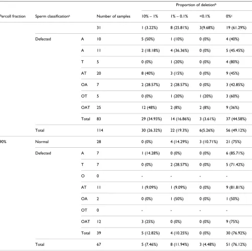
Regarding the proportion of the "common" deletion, our results indicate that the majority of men with normal sperm parameters carried fewer ΔmtDNA4977 molecules compared to the "defective" group (P = 0.04) (Table 2 and Figure 2A, B). Fisher's exact test of independence also revealed that men with asthenozoospermia, asthenotera-tozoospermia and oligoasthenoteraasthenotera-tozoospermia carried more ΔmtDNA4977 molecules compared to "normal" individuals (P = 0.004).
According to WHO criteria, a "normal" semen sample could carry a number of spermatozoa with abnormal parameters, such us low motility or abnormal morphol-ogy. Fractionation of spermatozoa in percoll gradients separates the highly motile and morphologically normal spermatozoa [27,28], and under this explains the signifi-cantly higher occurrence of the deletion in the "low-motil-ity" 45% fraction compared to the "high-motil"low-motil-ity" 90% fraction. Also, since the majority of spermatozoa with low motility, from "normal" or "defective" samples, congre-gate in the 45% fraction, we may explain the absence of a statistically significant difference in the occurrence of the deletion in this fraction between "normal" and "defec-tive" samples. The same conclusion holds for the 90% fraction. Finally, our observation that "defective" samples bear a higher proportion of deleted molecules can be explained by the fact that "defective" samples carry more abnormal spermatozoa than the "normal" group.
cor-relation with morphology or sperm count, or with the proportion of the deletion. This could be explained under the assumption that mtDNA defects would primarily affect energy production and, thus, movement of sperma-tozoa.
[image:5.612.60.553.99.590.2]From the 67 patients which underwent IVF or ICSI treat-ment, 14 had low fertility rates (<25%). Fisher's test revealed a significant correlation between the proportion of the "common" deletion in the 90% percoll fraction, and low fertilization rates after IVF (Figure 2C, D). Specif-ically, 75% of men that had a deletion proportion above
Table 2: Proportion of the "common" deletion in 45% and 90% percoll fraction of semen samples
Proportion of deletionb
Percoll fraction Sperm classificationa Number of samples 10% – 1% 1% – 0.1% <0.1% 0%c
31 1 (3.22%) 8 (25.81%) 3(9.68%) 19 (61.29%)
Defected A 10 5 (50%) 1 (10%) 0 (0%) 4 (40%)
A 11 2 (18.18%) 4 (36.36%) 0 (0%) 5 (45.45%)
T 5 0 (0%) 1 (20%) 0 (0%) 4 (80%)
AT 20 8 (40%) 3 (15%) 0 (0%) 9 (45%)
OA 7 2 (28.57%) 2 (28.57%) 0 (0%) 3 (42.85%)
OT 5 0 (0%) 1 (20%) 1 (20%) 3 (60%)
OAT 25 12 (48%) 2 (8%) 2 (8%) 9 (36%)
Total 83 29 (34.93%) 14 (16.86%) 3 (3.61%) 37 (44.58%)
Total 114 30 (26.32%) 22 (19.3%) 6(5.26%) 56 (49.12%)
90% Normal 28 0 (0%) 4 (14.29%) 3 (10.71%) 21 (75%)
Defected A 7 1 (14.28%) 0 (0%) 0 (0%) 6 (85.71%)
T 7 0 (0%) 2 (28.57%) 0 (0%) 5 (71.42%)
O 0 - - -
-AT 11 1 (9.09%) 1 (9.09%) 0 (0%) 9 (81.81%)
OA 2 0 (0%) 1 (50%) 0 (0%) 1 (50%)
OT 0 - - -
-OAT 12 3 (25%) 0 (0%) 0 (0%) 9 (75%)
Total 39 5 (12.82%) 4 (10.25%) 0 (0%) 30 (76.92%)
Total 67 5 (7.46%) 8 (11.94%) 3 (4.48%) 51 (76.12%)
a A, Asthenozoospermic; T, Teratozoospermic; O, Oligozoospermic; AT: Asthenoteratozoospermic; OA: Oligoasthenozoospermic; OT:
Oligoteratozoospermic; OAT: Oligoastenoteratozoospermic
b Determined according to supplementary Table 2 [see Additional file].
c No deletion-specific product was detected, or the detected deletion-specific product represents pseudo-deletion (I
d/In ≤ 0.0013).
0.1% showed a low fertilization rate, in contrast to 15.64% with a deletion proportion below 0.1% (P = 0.0026). Nevertheless, no correlation was obtained between this parameter and fertilization rate in ICSI patients (P = 0.47). In IVF cases, sperm is selected by
nat-ural barriers, which means that spermatozoa with signifi-cant numbers of ΔmtDNA4977 molecules do not produce enough energy for their movement and thus oocyte ferti-lization is less likely to occur. ICSI, however, bypasses this natural selection process and allows fertilization by
sper-Comparison of the "common" deletion and semen quality and fertilization ability
Figure 2
Comparison of the "common" deletion and semen quality and fertilization ability. (A-B) Comparison of the inci-dence of the "common" deletion in 45% and 90% percoll fraction from "normal" (A) and "defected" (B) semen samples. Samples were classified in four groups according to the calculated proportion of the deletion (see Additional file). (C-D) Comparison of the fertilization rate after IVF (C) and ICSI (D) from patients with different proportion of the "common" deletion. Samples were classified in two groups according to the calculated proportion of the deletion (above or below 0.1%).
0 10 20 30 40 50
10%-1% 1%-0.1% <0.1% 0%
0 10 20 30 40 50
10%-1% 1%-0.1% <0.1% 0%
45% Percoll fraction 90% Percoll fraction
Proportion of mtDNA4977Δ
%
o
f
individuals
%
o
f
individuals
A
B
“normal” sperm parameters “defected” sperm parameters
Proportion of mtDNA4977Δ
IVF ICSI
Fertilization Rate
%
o
f
individuals
%
o
f
individuals
C
D
Proportion of mtDNA4977 >0.1%Δ Proportion of mtDNA4977 <0.1%Δ
Fertilization Rate 0
10 20 30 40 50 60 70 80 90 100
Fertilization <25% Fertilization >25%
0 10 20 30 40 50 60 70 80 90 100
matozoa with low energy production. No statistically sig-nificant difference was detected between deletion proportion and pregnancy or delivery rates per transfer, after either IVF or ICSI. In other words our observations could imply that the load of common deletion in a semen sample reduces male fertility affecting mainly the fertiliza-tion efficiency of spermatozoa rather than affecting embryonic development. These results suggest, for the first time, a correlation between mtDNA integrity and fer-tilization efficiency of spermatozoa. In vivo studies in transmitochondrial mouse also underline the essential role of mitochondrial respiratory in mammalian sperma-togenesis and demonstrate that mtDNA defects are responsible for male infertility [29].
The power of semen parameter analysis in predicting future fertility is questioned in the past few years [30]. Yet, new molecular tests, based on measurements of sperm DNA quality, aim at the successful treatment of male infertile patients. The mitochondrial genome of sperm has also been proposed as a marker of sperm health. Our investigation resolves several literature inconsistencies and points to a way for the construction of a molecular test for the estimation of sperm integrity and fertility potential, even in cases of idiopathic infertility.
Competing interests
The authors declare that they have no competing interests.
Authors' contributions
FI carried out the experiments and drafted the manuscript. GCR supervised the work, participated in experimental design and helped in manuscript preparation. Both authors contributed to data interpretation and analysis, and have read and approved the manuscript.
Additional material
Acknowledgements
This work was supported by the Greek General Secretariat for Research and Technology (PENED-01ED42) and by the National and Kapodistrian University of Athens (ELKE 70/4/7805). We are grateful to Dr. T. G. Lainas, E. Vourvoulia and Dr. A. Argyriou for providing us with frozen fractionated semen samples, and to Prof. E. Zouros and Dr. A. Papantonis for their com-ments on an early version of the paper.
References
1. Dimauro S, Davidzon G: Mitochondrial DNA and disease. Ann Med 2005, 37(3):222-232.
2. MITOMAP: A Human Mitochondrial Genome Database. 2006 [http://www.mitomap.org].
3. Holt IJ, Harding AE, Morgan-Hughes JA: Deletions of mitochon-drial DNA in patients with mitochonmitochon-drial myopathies. Nature 1988, 331:717-719.
4. Zeviani M, Moraes CT, DiMauro S, Nakase H, Bonilla E, Schon EA, Rowland LP: Deletions of mitochondrial DNA in Kearns-Sayre syndrome. Neurology 1988, 38:1339-1346.
5. Schon EA, Rizzuto R, Moraes CT, Nakase H, Zeviani M, DiMauro S:
A direct repeat is a hotspot for large-scale deletion of human mitochondrial DNA. Science 1989, 244:346-349.
6. Shoffner JM, Lott MT, Voljavec AS, Soueidan SA, Costigan DA, Wal-lace DC: Spontaneous Kearns-Sayre/chronic progressive external ophthalmoplegia plus syndrome associated with a mitochondrial DNA deletion: a slip-replication model and metabolic therapy. Proc Natl Acad Sci USA 1989, 86:7952-7956. 7. Wallace DC: Disease of the mitochondrial DNA. Ann Rev Bioch
1992, 61:1175-1212.
8. Cortopassi GA, Arnheim N: Detection of a specific mitochon-drial DNA deletion in tissues of older humans. Nucleic Acids Res 1990, 18:6927-6933.
9. Ro LS, Lai SL, Chen CM, Chen ST: Deleted 4977-bp mitochon-drial DNA mutation is associated with sporadic amyotrophic lateral sclerosis: a hospital-based case-control study. Muscle Nerve 2003, 28:737-743.
10. Kao SH, Chao HT, Wei YH: Mitochondrial deoxyribonucleic acid 4977 bp deletion is associated with diminished fertility and motility of human sperm. Biol Reprod 1995, 52:729-736. 11. Cummins JM, Jequier AM, Martin R, Mehmet D, Goldblatt J: Semen
levels of mitochondrial DNA deletions in men attending an infertility clinic do not correlate with phenotype. Int J Androl 1998, 21:47-52.
12. St John JC, Jokhi RP, Barratt CL: Men with oligoasthenoteratozo-ospermia harbor higher numbers of multiple mitochondrial DNA deletions in their spermatozoa, but individual dele-tions are not indicative of overall aetiology. Mol Hum Reprod 2001, 7:103-111.
13. O'Connell M, McClure N, Lewis SE: A comparison of mitochon-drial and nuclear DNA status in testicular sperm from fertile men and those with obstructive azoospermia. Hum Reprod 2002, 17:1571-15717.
14. Reynier P, Chretien MF, Savagner F, Larcher G, Rohmer V, Barriere P, Malthiery Y: Long PCR analysis of human gamete mtDNA suggests defective mitochondrial maintenance in spermato-zoa and supports the bottleneck theory for oocytes. Biochem Biophys Res Commun 1998, 252:373-377.
15. World Health Organization: Laboratory Manual for the Exami-nation of Human Semen and Semen-Cervical Mucus Interac-tion. 2nd edition. Cambridge University Press; 1992:3-4.
16. Kessopoulou E, Tomlinson MJ, Barratt CL, Bolton AE, Cooke ID:
Origin of reactive oxygen species in human semen: sperma-tozoa or leucocytes? J Reprod Fertil 1992, 94:463-470.
17. Miller D, Tang PZ, Skinner C: Differential RNA fingerprinting as a tool in the analysis of spermatozoa gene expression. Hum Reprod 1994, 9:864-869.
18. Douris V, Giokas S, Lecanidou R, Mylonas M, Rodakis GC: Phyloge-netic analysis of mitochondrial DNA and morphological characters suggest a need for taxonomic re-evaluation within the Alopiinae (Gastropoda: Clausiliidae). J Moll Stud 1998, 64:81-92.
19. Soong NW, Arnheim N: Detection and quantification of mito-chondrial DNA deletions. Methods Enzymol 1996, 264:421-431. 20. von Wurmb-Schwark N, Oehmichen M, Meissner C:
Demonstra-tion of the 4977 bp deleDemonstra-tion in human mitochondrial DNA from intravital and postmortem blood. Mutat Res 1998,
422(2):247-254.
21. Meissner C, von Wurmb N, Oehmichen M: Detection of the age-dependent 4977 bp deletion of mitochondrial DNA. Int J Legal Med 1997, 110:288-291.
22. Pääbo S, Irwin DM, Wilson AC: DNA damage promotes jumping between templates during enzymatic amplification. J Biol Chem 1990, 265:4718-4721.
Additional file 1
Supplementary data. Supportive information for the long-PCR experi-ments, Southern analysis and the quantitative PCR assay.
Click here for file
Publish with BioMed Central and every scientist can read your work free of charge "BioMed Central will be the most significant development for disseminating the results of biomedical researc h in our lifetime."
Sir Paul Nurse, Cancer Research UK
Your research papers will be:
available free of charge to the entire biomedical community
peer reviewed and published immediately upon acceptance
cited in PubMed and archived on PubMed Central
yours — you keep the copyright
Submit your manuscript here:
http://www.biomedcentral.com/info/publishing_adv.asp
BioMedcentral 23. Odelberg SJ, Weiss RB, Hata A, White R: Template-switching
during DNA synthesis by Thermus aquaticus DNA polymer-ase I. Nucleic Acids Res 1995, 23:2049-2057.
24. Patel R, Lin M, Laney M, Kurn N, Rose S, Ullman EF: Formation of chimeric DNA primer extension products by template switching onto an annealed downstream oligonucleotide.
Proc Natl Acad Sci USA 1996, 93:2969-2974.
25. Judo MS, Wedel AB, Wilson C: Stimulation and suppression of PCR-mediated recombination. Nucleic Acids Res 1998,
26:1819-1825.
26. Ferlin A, Raicu F, Gatta V, Zuccarello D, Palka G, Foresta C: Male infertility: role of genetic background. Reprod Biomed Online 2007, 14:734-745.
27. Lessley BA, Garner DL: Isolation of motile spermatozoa by den-sity gradient centrifugation in percoll. Gamete Res 1983, 7:49. 28. Dravland JE, Mortimer D: A simple discontinuous percoll
gradi-ents procedure for washing human spermatozoa. IRCS Med Sci 1985, 13:16.
29. Nakada K, Sato A, Yoshida K, Morita T, Tanaka H, Inoue S-I, Yonekawa H, Hayashi J-I: Mitochondria-related male infertility.
Proc Natl Acad Sci USA 2006, 103:15148-15153.