City, University of London Institutional Repository
Citation
:
Fealy, N., Aitken, L. M., du Toit, E., Lo, S. and Baldwin, I. (2017). Faster Blood Flow Rate Does Not Improve Circuit Life in Continuous Renal Replacement Therapy: A Randomized Controlled Trial. Critical Care Medicine, 45(10), e1018-e1025. doi:10.1097/CCM.0000000000002568
This is the accepted version of the paper.
This version of the publication may differ from the final published
version.
Permanent repository link:
http://openaccess.city.ac.uk/19069/Link to published version
:
http://dx.doi.org/10.1097/CCM.0000000000002568Copyright and reuse:
City Research Online aims to make research
outputs of City, University of London available to a wider audience.
Copyright and Moral Rights remain with the author(s) and/or copyright
holders. URLs from City Research Online may be freely distributed and
linked to.
1 Faster blood flow rate does not improve circuit life in Continuous Renal Replacement
Therapy: A randomized controlled trial
Nigel Fealy RN, MN1,2,3, Leanne Aitken RN, PhD, FACCCN2,4,5,6, Eugene du Toit PhD7, Serigne
Lo PhD, AStat8, Ian Baldwin RN, PhD, FACCCN1,3
1. Department of Intensive Care Medicine, Austin Hospital, Melbourne, Australia
2. School of Nursing and Midwifery, Griffith University, Brisbane, Australia
3. School of Nursing and Midwifery, Deakin University, Melbourne, Australia
4. Centre for Health Practice Innovation, Griffith Health Institute, Griffith University, Brisbane, Australia
5. Intensive Care Unit, Princess Alexandra Hospital, Brisbane, Australia
6. School of Health Sciences, City, University of London, United Kingdom
7. School of Medical Science, Griffith University, Gold Coast, Australia
8. Melanoma Institute Australia, Research and Biostatistics group
This work was performed at the Austin Hospital, Melbourne, Victoria, Australia
A/Prof Fealy, Prof Aitken, Dr du Toit, Dr Lo and Prof Baldwin have disclosed that they do not have any potential conflicts of interest.
Corresponding author
A.Prof. Nigel Fealy
Department of Intensive Care
Austin Hospital
Studley Rd, Heidelberg, Victoria 3084, Australia.
2 Abstract
Objective: To determine whether blood flow rate influences circuit life in continuous renal replacement therapy.
Design: Prospectiverandomized controlled trial.
Setting: Single centre tertiary level intensive care unit.
Patients: Critically ill adults requiring continuous renal replacement therapy.
Interventions: Patients were randomized to receive one of two blood flow rates:150 mL/min or 250 mL/min.
Measurements and Main Results: The primary outcome was circuit life measured in hours. Circuit and patient data were collected until each circuit clotted or was ceased electively for non-clotting reasons. Data for clotted circuits are presented as median (Inter-quartile range) and compared using the Mann Whitney U test. Survival probability for clotted circuits was compared using log-rank test. Circuit clotting data were analysed for repeated events using hazards ratio (HR). One hundred patients were randomised with 96 completing the study (150 mL/min, n=49; 250 mL/min, n=47) using 462 circuits (245 run at 150 mL/min and 217 run at 250 mL/min). Median circuit life for 1st circuit (clotted) was similar for both groups (150 mL/min:
9.1 [5.5, 26] hrs vs. 10 [4.2, 17] hrs; p=0.37). CRRT using blood flow rate set at 250 mL/min was not more likely to cause clotting compared to 150 mL/min (HR, 1.00 [0.60, 1.69]; p=0.68). Gender, Body Mass Index, weight, vascular access type, length, site, mode of CRRT or International Normalised Ratio had no effect on clotting risk. CRRT without anticoagulation was more likely to cause clotting compared to use of heparin strategies (HR 1.62, p=0.003). Longer Activated Partial Thromboplastin Time (HR 0.98, p=0.002] and decreased platelet count (HR 1.19 p=0.03] was associated with a reduced likelihood of circuit clotting.
4 Introduction
Acute kidney injury (AKI) is a complication of critical illness that affects up to 50% of
intensive care patients.1-3 The use of Renal Replacement Therapy (RRT) has evolved as the
treatment for severe AKI and is required in 5-6% of all critically ill patients in intensive care
units (ICUs).4 Continuous Renal Replacement Therapies (CRRT) relies on the maintenance of
extracorporeal circuit (EC) patency for as long as possible, however premature circuit failure
due to clotting may cause blood loss, reduced therapeutic efficacy and increased workload
and treatment costs.5-8
Clogging and clotting of the hemofilter membrane is the major mechanism of
premature failure and circuit loss in CRRT. 9,10 It has been suggested that increasing blood flow
rate (BFR) through the EC to speeds greater than 200 mL/min may reduce premature
clotting.11 One recent report demonstrated a reduction in filter lifespan when BFR was less
than 200 mL/min concluding that the optimal blood flow rate during CRRT is between
250-300 mL/min.12
The impact of BFR on membrane clotting rate is potentially important and has not
been examined in controlled studies. Despite suggestions to increase blood flow rates in the
EC11,12, there remains great variability in the prescription of this setting worldwide. Although
a recent survey of Australian and New Zealand ICUs indicated a BFR of 150-200 mL/min was
the dominant setting, a faster rate of 200-250 mL/min was also commonplace in ICU’s
surveyed.13 Observational studies and recent worldwide practice surveys of CRRT also
5 be an important determinant of circuit life in CRRT, the most suitable speed to reduce clotting
and optimise membrane life has not been identified. To address this question, we conducted
a prospective randomized controlled trial (RCT). We tested the hypothesis that a faster blood
flow rate (250 mL/min) would be superior to a slower blood flow rate (150 mL/min) in
maintaining circuit patency in CRRT.
Materials and Methods
Trial design and setting
This study was a prospective, parallel group RCT conducted in a 24 bed, adult, tertiary
intensive care in Melbourne, Victoria, Australia. The study was registered at the Australian
New Zealand Clinical Trials Registry (ACTRN:12615001353583) and approved by Austin Health
Human Research Ethics Committee (HREC project No. H2012/04772). Written informed
consent was obtained from the patient or their next of kin prior to, or soon after enrolment.
Eligibility criteria
Critically ill patients in ICU were eligible for the study if they fulfilled three criteria: 1)
age ≥ 18 yrs, 2) AKI (RIFLE classification F)16 requiring CRRT and 3) vascular access was via the
femoral vein for standardisation. Patients were considered ineligible for the study if they
fulfilled any of the following exclusion criterion: 1) required citrate anticoagulation (citrate
protocol requires set blood flow rate of 150 mL/min), 2) expected stay in the ICU was less than
6 Interventions
The study compared two blood flow rate settings on circuit life in CRRT. The
intervention was a set BFR of 250 mL/min and the control was a set BFR of 150 mL/min. CRRT
was performed using Continuous VenoVenous Hemofiltration (CVVH) or Continuous
VenoVenous Hemodiafiltration (CVVHDF) modalities. Vascular access was via either Niagara
13.5 catheter (24cm) (Bard, Murray Hill, NJ, USA) or Gamcath Dolphin Protect 13.0
Fr-catheter (25 cm) (Gambro, Hechingen, Germany) dual lumen Fr-catheters. Treatment modality
and choice of vascular access was at the discretion of the treating physician. Prismaflex with
AN69ST (ST100) 1.0 m2 membrane (Gambro Nephral TM, Lund, Sweden) or Infomed HF 440
with DF 140 Polyethersulfone 1.4 m2 membrane(Infomed, Geneva, Switzerland) was used for
all treatments. Bicarbonate buffered replacement and dialysis fluid (Baxter, Castlebar, Co.
Mayo, Ireland) was used. In CVVH, replacement fluid was delivered into the EC before and
after the filter (pre and post-dilution), with a ratio of 50% predilution and 50% postdilution.
Dose in CVVH was standardised at 2000 mL/hour. In CVVHDF, the replacement fluid was all
delivered postdilution. Dose in CVVHDF was standardised at 1000 mL/hour replacement and
1000 mL/hour dialysate.
Anticoagulation was provided according to a predefined ICU protocol and mandates
no anticoagulation in patients at risk of bleeding from a coagulopathy or thrombocytopaenia.
Options for anticoagulation when used included regional heparinisation with unfractionated
heparin (1000 IU/hour) delivered prefilter and protamine (10 mgs/hour) delivered in the
7 delivered pre-filter at 5-10 IU/kg/hour. CRRT was prescribed by the treating intensivist and
delivered by ICU nurses. The decision to start or stop CRRT, and determining the reason for
stopping, was done by ICU doctors and nurses respectively as is usual protocol for the ICU.
Data collection
Baseline data relating to age, gender, weight, BMI, source of admission, severity of
illness (Acute Physiology and Chronic Health Evaluation score II, III; Simplified Acute
Physiology Score II), diagnostic group, presence of sepsis, mechanical ventilation,
inotropes/vasopressors, and basic laboratory variables pertaining to renal function was
collected.
Outcomes
The primary outcome was circuit life (measured in hours) and recorded as clotted
when; 1) Transmembrane pressure across the circuit exceeded 300 mmHg, 2) pre-filter
pressure > 200 mmHg, 3) visible clot obstructing flow through the circuit and 4) the blood
pump was unable to rotate due to clot obstruction in the membrane or for ‘elective’ reasons.
e.g. CT, MRI, surgical intervention or cessation prior to clotting for native renal assessment.
Sample size
Without supportive data to inform a power calculation for this study, 100 patients
were chosen to ensure a sample that was sufficient to reflect usual ICU patient characteristics
and allow recruitment completion in one year. Patients stayed in the BFR treatment group
8 patient (CVVH or CVVHDF) which was maintained/retained for all subsequent treatments. All
circuits used for these patients were included and analysed accordingly.
Randomization
Patients were screened and entered the study by ICU clinical staff. Patients were
randomly assigned with stratification for mode (CVVH or CVVHDF). Once the treating
physician prescribed CRRT and the mode of therapy, patients were randomized using a web
based central randomization service. A variable block randomization with parallel allocation
was used to allocate to each study group (150mL/min vs. 250 mL/min).
Statistical methods
The primary outcome (circuit life) was analysed in a two-step process. First analysis:
this excluded all electively removed or non-clotted circuits from the data. The distribution of
data for all circuits meeting the defined clotting criteria was then assessed. As expected study
variables were not normally distributed and non-parametric statistics were used with circuit
life reported as median and IQR. Circuit life for the two groups (150 mL/min vs. 250 mL/min)
was compared using Mann-Whitney U-test. Analysis of the two groups was then assessed for
survival probability and presented graphically using Kaplan-Meier survival plots. A log-rank
test was used to compare circuit life between the two groups. This analysis was not adjusted
for any confounding variables.
Second analysis: included all circuits (clotted and those electively removed). Circuit
9 frailty model17 (an extension of cox regression) was used to test for within patient
dependence. It was expected that there would be heterogeneity among individual trial
patients. In addition, individual trial patients may contribute one or multiple circuits. It was
assumed to be a correlation between an individual trial patient contributing multiple circuits
and circuit life. The frailty model was used to test event dependence (where the event is a
clotted circuit) within the trial patients. Event dependence in this study meant that an
occurrence of one event (time to clotting of the circuit) may make further events (additional
circuit clotting times) more or less likely. The advantage of this model is that it considers any
within-cluster correlation of circuit life. Proportions were compared using a chi-square test.
First analysis was carried out using IMB SPSS statistics for Windows (v2, IBM Corp.,
Armonk, NY). Second analysis was performed using SAS (Enterprise Guide v5.1; SAS Institute
Inc., Carey, NC).
Results
Participants and recruitment
One hundred patients were randomized between June 2013 and August, 2014. Two
patients from each group were randomized but did not receive CRRT. The CONSORT diagram
for patient enrolment is shown in Figure 1. Overall, 96 patients (49 in the 150 mL/min group
and 47 in the 250 mL/min group) contributed a total of 463 study circuits: 245 in the 150
mL/min group and 218 in the 250 mL/min group. Median study circuits per patient was four
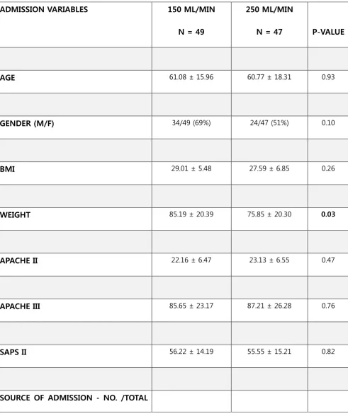
10 At randomization, patients were similar with respect of age, sex, severity of illness
scores (APACHE II, III, SAPS II), admission source and diagnosis (Table 1.). There was a nine kg
difference in patient weight between the two groups (p= 0.03); however, BMI was similar for
both groups. Pre-randomization renal laboratory variables were also similar for both groups.
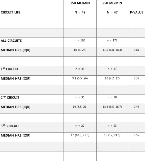
Primary outcomes - Circuit Life
The first analysis incorporated 369 defined clotted circuits. The median circuit life for
these circuits (n=369) was similar for both groups (150 mL/min, n=196 [10 hrs IQR 6.0, 24])
vs. (250 mL/min, n = 173 [11.5 hrs IQR 6.8, 18.3] p = 0.81). For first analysis, there were 81
clotted first circuits. The median circuit life of these circuits was 9.1 hrs (IQR, 5.5, 26 hr) in the
150 mL/min group compared to 10 hrs (IQR, 4.2, 17 hr) in the 250 mL/min group, p=0.37.
Second and third median circuit lives for those deemed to have clotted were also similar
(Table 2). The probability of the first study circuit from each patient failing due to clotting did
not differ between BFR groups (150 ml/min vs. 250 mL/min; HR, 0.85, log rank test = 0.46)
(Figure 2).
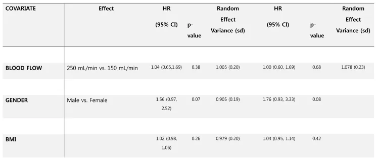
The second analysis involved evaluation of all circuit terminations (clotted and
electively removed) and revealed that a BFR of 250 mL/min was not more likely to cause
clotting compared to 150 mL/min (HR, 1.00 [0.60, 1.69]; p=0.68, variance of the random
effect, 1.078 [0.23]) (Table 3). There were no differences in likelihood of clotting for: gender,
BMI, weight, vascular access type, length or site, mode of CRRT or INR. CRRT without use of
anticoagulation was more likely to cause clotting compared to use of heparin or
11 p=0.002) was associated with a lower likelihood of circuit clotting. Probability of clotting was
higher in those patients with higher platelet counts (HR 1.19, [1.01, 1.40], p=0.03) (Table 3).
Discussion
This is the first known prospective study to examine the effect of blood flow rate on
circuit duration in both CVVH and CVVHDF. In a cohort of 100 ICU patients requiring CRRT
three key findings have been identified: first, when blood flow rate is increased to 250 mL/min
it does not increase circuit life during CRRT. Second, the use of an anticoagulation strategy and
longer APTT’s extends CRRT circuit life. Third, patients with higher platelet counts were more
prone to premature circuit clotting in this study.
Relationship to previous studies
The maintenance of circuit patency by prevention of clotting is the greatest challenge
associated with providing CRRT for critically ill patients. As a result, many studies have focused
on anticoagulation strategies to extend circuit life,7,18-27 while many aspects of treatment and
prescription setting have not been investigated. One RCT has included blood flow rate in the
assessment of circuit clotting in CRRT, indicating that blood flow rates >125 mL/min did not
improve circuit survival.28 This study was conducted in CVVHD mode which is rarely used in
current practice.13,15,29,30 Pure diffusive modes of hemofiltration such as CVVHD have been
shown to be associated with decreased procoagulatory activity in the dialyser membrane
when compared to convective modes31,32 and makes comparisons to CVVH and CVVHDF
12 One single centre study assessing 1332 treatments from 355 patients concluded that
BFR did indeed affect circuit life.12 In this retrospective audit, the authors suggest a BFR less
than 200 mL/min significantly decreased circuit life compared to rates greater than 200
mL/min. They also determined that BFR greater than 300 mL/min led to lower median circuit
lives and recommended an optimal BFR of between 250-300 mL/min.
Implications of study findings
Our data provides evidence to suggest that a faster blood flow rate does not influence
circuit life and prescription of rates greater than 150 mL/min makes no difference to the
likelihood of clotting in either CVVH or CVVHDF. It has previously been suggested that blood
flow should be maintained at 200 mL/min33-35 and always be greater than 100 mL/min to avoid
premature clotting.36 The ability to maintain consistent and constant flow may be more
critical, with the flow and resistance balance being more important. We have previously
reported these mechanical factors and their adverse effect on circuit life.10,37,38
Despite many authors suggesting blood flow rates for CRRT of 200-250 mL/min12,33-35
and international surveys indicating practical prescriptions of >200 mL/min; there has been
no endorsements for this setting. The Acute Dialysis Quality Initiative (ADQI) consensus
guidelines for operational characteristics from 2002 indicate that blood flow may be increased
to augment solute clearance but do not include a recommendation for this prescription.39 The
more recent Kidney Disease Improving Global Outcomes (KDIGO) consensus guidelines
outline settings for different RRT modalities indicating 150-250 mL/min is typically prescribed
13 on evidence.40
The use of anticoagulants to prevent extracorporeal clotting and extend circuit life in
CRRT has been used for decades.41 Unfractionated heparin (UFH) remains the most commonly
prescribed anticoagulant13,42 worldwide and remains the standard against which other
anticoagulant regimens are compared.7,21,22,24,25,43. A regional heparin technique favours
patient safety and anticoagulant free CRRT is used for a high risk of bleeding.44,45
We have previously reported on circuit life in CVVH when no anticoagulation was used
compared to low dose UFH and a regional heparin technique.45 This study of 300 filters
described similar circuit lives for all three methods and has similarities to our findings which
indicate the strong association between higher platelet counts and premature circuit clotting.
Strengths and Limitations
This RCT of 100 patients presents for the first time the outcomes of an investigation
into the effect of blood flow rate on circuit life in two commonly used modes of CRRT. This
analysis is based on a large number of circuits and for 8206 hours of treatment time. This
number of patients and treatment time is representative of a tertiary level intensive care unit
and signifies important findings for current CRRT practice. The presentation of our analysis for
first circuit (clotted) and the analysis of all circuits using repeated events survival analysis
should be the new standard for studies reporting circuit life in CRRT where previously an all
circuits analysis may have drawn conclusions not valid due to repeated measures effect. The
14 to make the appropriate group size calculation. One further limitation may be the defined
blood flow rates used in this study. Blood flow rates less than 150 mL/min or greater than 250
mL/min may have yielded a different outcome. Circuit life (hrs) in both groups may be shorter
due to the large proportion of patients enrolled have a diagnosis of liver failure and liver
transplantation making comparisons to other ICU’s difficult. Two membrane compositions
were used and anticoagulation according to an established local policy. These two factors may
have some influence on our findings.
Conclusions
A blood flow rate of 250 mL/min does not improve CRRT circuit life compared with a
blood flow rate of 150 mL/min. Independent factors that may extend circuit life include
anticoagulation strategies, higher APTT and lower platelet counts.
Acknowledgements
We thank the medical and nursing staff of the Austin intensive care unit for their
support of this study.
Figure 1. Flow diagram of participants showing assessment of eligibility, enrollment, treatment allocation and follow-up in the trial. CRRT = Continuous Renal Replacement Therapy, CVVH = Continuous VenoVenous Hemofiltration, CVVHDF = Continuous VenoVenous Hemodiafiltration
16 References
1. Hoste EAJ, Bagshaw SM, Bellomo R, et al: Epidemiology of acute kidney injury in critically
ill patients: the multinational AKI-EPI study. Intensive Care Med 2015; 41:1411-1423
2. Ostermann M, Chang RW: Acute kidney injury in the intensive care unit according to RIFLE.
Crit Care Med 2007; 35: 1837-1843
3. Uchino S, Kellum J, Bellomo R et al: Acute renal failure in critically ill patients: a multinational
multicentre study. JAMA 2005; 294: 813-818
4. Dennen P, Douglas IS, Anderson R: Acute Kidney Injury in the intensive care unit: an update
and primer for the intensivist. Crit Care Med 2010; 38: 261-275
5. Bellomo R, Mårtensson J, Lo S, et al: Femoral access and delivery of continuous renal
replacement therapy dose. Blood Purif 2016; 41:11-17
6. Fealy N, Baldwin I, Bellomo R: The effect of circuit “down-time” on uraemic control during
continuous veno-venous hemofiltration. Crit Care Resusc 2002; 4:266-270
7. Gattas DJ, Rajbhandari D, Bradford C, et al: A randomized controlled trial of regional citrate
versus regional heparin anticoagulation for continuous renal replacement therapy in critically
ill adults. Crit Care Med 2015; 43(8):1622-1629
8. The RENAL Replacement Therapy Study Investigators, Bellomo R, Cass A, Cole L, et al:
Intensity of continuous renal replacement therapy in critically ill patients N Engl J Med 2009;
17 9. Ward D. Principles of extracorporeal circulation. In Ronco C, Bellomo R, Kellum JA. editors.
Critical Care Nephrology, 2nd ed. Philadelphia Elsevier Saunders 2009; p. 1141-1145
10. Baldwin I, Bellomo R, Koch B: Blood flow reductions during continuous renal replacement
therapy and circuit life. Intensive Care Med 2004; 30:2074-2079
11. Ronco C, Ricci Z, De Backer D, et al: Renal Replacement therapy in acute kidney injury:
controversy and consensus. Crit Care 2015; 19:146
12. Dunn WJ, Sriram S: Filter lifespan in critically ill adults receiving continuous renal
replacement therapy: the effect of patient and treatment related variables Crit Care Resusc
2014; 16(3): 225-231
13. Fealy N, Aitken L, du Toit E, et al: Continuous renal replacement therapy: current practice
in Australian and New Zealand intensive care units. Crit Care Resusc 2015; 17:83-91
14. Uchino S, Toki N, Ohnuma T, et al: Validity of low intensity continuous renal replacement
therapy. Crit Care Med 2013; 41(11):2584-2591
15. Jones S, Devonald MAJ: How acute kidney injury is investigated and managed in UK
intensive care units – a survey of current practice. Nephrol Dial Transplant 2013; 28:
1186-1190
16. Bellomo R, Ronco C, Kellum JA, et al: Acute renal failure – definition, outcome measures,
animal models, fluid therapy and information technology needs: the Second International
18 8:R204-212
17. Box-Steffensmeier JM, De Bouf S: Repeated events survival models: The conditional frailty
model. Stat Med 2004; 25:3518-3533
18. Oudemans-van Straaten HM, Bosman RJ, Koopmans M et al: Citrate anticoagulation for
continuous veno-venous hemofiltration. Crit Care Med 2009; 37:545-552
19. Tolwani A, Wille KM: Advances in continuous renal replacement therapy: citrate
anticoagulation update Blood Purif 2012; 34(2): 88-93
20. Oudemans-van Straaten HM, Wester JP, de Pont AC, et al: Anticoagulation strategies in
continuous renal replacement therapy: can the choice be evidence based? Intensive Care Med
2006; 188-202
21. Bai M, Zhou M, He L et al: Citrate versus heparin anticoagulation for continuous renal
replacement therapy: an updated meta-analysis of RCTs. Intensive Care Med 2015;
41:2098-2110
22. Monchi M, Berghmans D, Ledoux D, et al: Citrate vs. heparin for anticoagulation in
continuous venovenous hemofiltration: a prospective randomized study. Intensive Care Med
2004; 30(2): 260-265
23. van de Wetering J, Westendorp RG, van der Hoeven JG, et al: Heparin use in continuous
renal replacement procedures: the struggle between circuit coagulation and patient
19 24. Joannidis M, Kountchev J, Rauchenzauner M et al: Enoxaparin versus unfractionated
heparin for anticoagulation during continuous veno-venous hemofiltration – a randomized
controlled crossover study. Intensive Care Med 2007; 33: 1571-1579
25. Schilder L, Nurmohamed SA, Bosch FH et al. CASH study group: Citrate anticoagulation
versus systemic heparinisation in continuous venovenous hemofiltration in critically ill
patients with acute kidney injury: a multi-center randomized clinical trial. Crit Care 2014;
18(4): 472
26. Lee YK, Lee HW, Choi KH, et al: Ability of nafamostat mesilate to prolong filter patency
during continuous renal replacement therapy in patients of high risk of bleeding: A
randomized controlled study. PLos One 2014; 9(10): e108737
27. Gainza FJ, Quintanilla N, Pjoan Jl, et al: Role of prostacyclin (epoprostenol) as an
anticoagulant in continuous renal replacement therapies: efficacy, security and cost analysis.
J Nephrol 2006; 19(5): 648-655
28. Ramesh Prasad GV, Palevsky P, Burr R, et al: Factors affecting system clotting in continuous
renal replacement therapy: results of a randomized controlled trial. Clin Nephrol 1998; 53(1):
55-60
29. Legrand M, Darmon M, Joannidis M, et al: Management of renal replacement therapy in
ICU patients: an international survey Intensive Care Med 2013; 39: 101-108
30. Jamal JA, Mat-Nor MB, Mohamad-Nor FS, et al: A national survey of renal replacement
20 2014; 19:507-512
31. Klingel R, Schaefer M, Schwarting A, et al: Comparative analysis of procoagulatory activity
of haemodialysis, hemofiltration and haemodiafiltration with a polysulfonemembrane (APS)
and with different modes of enoxaparin anticoagulation. Nephrol Dial Transplant 2004; 19(1):
164-170
32. Ricci Z, Ronco C, Bachetoni A, et al: Solute removal during continuous renal replacement
therapy in critically ill patients: convection versus diffusion. Crit Care 2006; 10:R67
33. Baldwin I: Factors affecting circuit patency and circuit life. Contrib Neph 2007; 156:
178-184
34. Baldwin I, Bridge N, Elderkin T: Nursing issues, practices and perspectives for the
management of continuous renal replacement therapy in the intensive care unit. In: Ronco C
& Bellomo R (Eds). Critical Care Nephrology (1st Ed). Dordecht: Klower Academic Publishers
1998; p. 1309-1325
35. Davies H, Leslie G: Maintaining the CRRT circuit: non-anticoagulant alternatives. Australian
Crit Care 2006; 19(4): 133-138
36. Kox WJ, Rohr U, Waurer H: Practical aspects of renal replacement therapy. Int J Artif Organs
1996; 19: 100-105
37. Baldwin I, Fealy N, Carty P, et al: Bubble chamber clotting during continuous renal
34:213-21 218
38. Inbyung K, Fealy N, Baldwin I, et al: Premature circuit clotting due to likely mechanical
failure during continuous renal replacement therapy. Blood Purif 2010; 30: 79-83
39. Palevsky P, Bunchman T, Tetta C: The Acute Dialysis Quality Initiative – part V: Operational
characteristics of CRRT. Adv Ren Replace Ther 2002; (9)4: 268-272
40. Kidney Disease: Improving Global Outcomes (KDIGO) Acute Kidney Injury Work Group.
KDIGO Clinical Practice Guideline for Acute Kidney Injury. Kid Int Supp 2012; 2: 1–138
41. George CRP: Hirudin, heparin and Heinrich Necheles. Nephrol 1998; 4: 225-228
42. Uchino S, Bellomo R, Morimatsu H et al: Continuous renal replacement therapy: a
worldwide practice survey. The beginning and ending supportive therapy for the kidney (BEST
kidney) investigators. Intensive Care Med 2007; 33: 1563-1570
43. Mei-Yi W, Yung-Ho H, Chyi-Huey B, et al: Regional citrate versus heparin anticoagulation
for continuous renal replacement therapy: A meta-analysis of randomized controlled trials.
Am J Kidney Dis 2012; 59(6): 810-818
44. Nongnuch A, Tangsujaritvijit V, Davenport A: Anticoagulation for renal replacement
therapy for patients with acute kidney injury. Minerv Urol Neph 2016; 68: 87-104
45. Uchino S, Fealy N, Baldwin I, et al: Continuous venovenous hemofiltration without
22
TABLE 1.
Baseline demographic and clinical characteristics
ADMISSION VARIABLES 150 ML/MIN
N = 49
250 ML/MIN
N = 47 P-VALUE
AGE 61.08 ± 15.96 60.77 ± 18.31 0.93
GENDER (M/F) 34/49 (69%) 24/47 (51%) 0.10
BMI 29.01 ± 5.48 27.59 ± 6.85 0.26
WEIGHT 85.19 ± 20.39 75.85 ± 20.30 0.03
APACHE II 22.16 ± 6.47 23.13 ± 6.55 0.47
APACHE III 85.65 ± 23.17 87.21 ± 26.28 0.76
SAPS II 56.22 ± 14.19 55.55 ± 15.21 0.82
23 NO. (%)
ED 13 (27.7%) 12 (25.5%)
WARD 17 (34.7%) 17 (36.2%)
POST OP (ELECTIVE) 7 (14.3%) 6 (12.8%)
POST OP (EMERGENCY) 5 (10.2%) 4 (8.5%)
TRANSFER OTHER ICU 5 (10.2%) 5 (10.6%)
TRANSFER OTHER HOSPITAL 2 (4.1%) 3 (6.4%)
ADMISSION DIAGNOSIS - NO. /TOTAL
NO. (%)
CARDIOVASCULAR 6 (12.2%) 5 (10.6%)
CARDIAC SURGERY 11 (22.4%) 8 (17.0%)
RESPIRATORY 0 1 (2.1%)
GASTROINTESTINAL 6 (12.2%) 6 (12.8%)
LIVER FAILURE 5 (10.2%) 6 (12.8%)
LIVER TRANSPLANT 10 (20.4%) 13 (27.7%)
ACUTE RENAL/GENITOURINARY 5 (10.2%) 5 (10.6%)
24
INFECTION/ABSCESS 2 (4.1%) 2 (4.3%)
MECHANICAL VENTILATION – NO. (%) 41 (83.7%) 36 (76.6%) 0.44
VASOPRESSOR/INOTROPE – NO. (%) 41 (83.7%) 41 (87.2%) 0.77
SEVERE SEPSIS – NO. (%) 24/49 (49%) 26/47 (55.3%) 0.55
LAB DATA PRIOR TO RANDOMISATION
SERUM CREATININE 317.20 ± 171.61 297 ± 181.54 0.16
SERUM UREA 23.62 ± 14.94 21.19 ± 10.03 0.33
25
TABLE 2.
Circuit life duration – Clotted circuits only
CIRCUIT LIFE
150 ML/MIN
N = 49
250 ML/MIN
N = 47 P-VALUE
ALL CIRCUITS n = 196 n = 173
MEDIAN HRS (IQR) 10 (6, 24) 11.5 (6.8, 18.3) 0.81
1ST CIRCUIT n = 49 n = 47
MEDIAN HRS (IQR) 9.1 (5.5, 26) 10 (4.2, 17) 0.37
2ND CIRCUIT n = 35 n = 38
MEDIAN HRS (IQR) 14 (8.5, 21) 13.8 (8.5, 16.7) 0.45
3RD CIRCUIT n = 22 n = 23
MEDIAN HRS (IQR) 17 (10.5, 28.5) 16 (12, 21.5) 0.52
26
TABLE 3.
Univariate and Multivariate analysis for all circuits
UNIVARIATE MODEL MULTIVARIATE MODEL
COVARIATE Effect HR
(95% CI)
p-value
Random
Effect
Variance (sd)
HR
(95% CI)
p-value
Random
Effect
Variance (sd)
BLOOD FLOW 250 mL/min vs. 150 mL/min 1.04 (0.65,1.69) 0.38 1.005 (0.20) 1.00 (0.60, 1.69) 0.68 1.078 (0.23)
GENDER Male vs. Female 1.56 (0.97,
2.52)
0.07 0.905 (0.19) 1.76 (0.93, 3.33) 0.08
BMI 1.02 (0.98,
1.06)
27
WEIGHT for every 5kg increase 1.04 (0.98,
1.11)
0.19 0.957 (0.20) 0.96 (0.82, 1.12) 0.59
VASCATH SITE Right Femoral vs. Left femoral 0.75 (0.55,
1.03)
0.07 1.075 (0.22) 0.94 (0.66, 1.34) 0.73
VASCATH LENGTH 20 cm vs. 15 cm 0.83 (0.31,
2.19)
0.10 1.091 (0.22) 0.92 (0.34, 2.50) 0.18
24 cm vs. 15 cm 0.53 (0.22,
1.27)
0.17 (0.03, 1.16)
VASCATH DIAMETER 13 Fr vs. 13.5 Fr 1.41 (0.96,
2.09)
28
CRRT MODE CVVHDF vs. CVVH 1.10 (0.68,
1.77)
0.70 0.993 (0.20)
ANTICOAGULATION None vs. Heparin/regional
heparin
1.65 (1.21,
2.24)
0.001 1.148 (0.23) 1.62 (1.18, 2.23) 0.003
PLATELETS (X 1011) 1.13 (0.97,
1.31)
0.11 0.928 (0.19) 1.19 (1.01, 1.40) 0.03
INR (RATIO) 0.84 (0.65,
1.09)
0.19 0.993 (0.20) 1.05 (0.77, 1.42) 0.77
APTT (SECONDS) 0.98 (0.97,
0.99)