R E S E A R C H A R T I C L E
Open Access
Serum complement C4b, fibronectin, and
prolidase are associated with the pathological
changes of pulmonary tuberculosis
Chong Wang
1, Yan-Yuan Li
2, Xiang Li
3, Li-Liang Wei
4, Xiu-Yun Yang
5, Dan-Dan Xu
1, Ting-Ting Jiang
1, Zhong-Jie Li
1,
Zhong-Liang Chen
1, Xing Zhang
1, Ji-Yan Liu
1, Ze-Peng Ping
1and Ji-Cheng Li
1*Abstract
Background:Mycobacterium tuberculosisinfection can activate the immune system, leading to characteristic pathological changes such as inflammatory granuloma, caseous necrosis, and cavity formation.
Methods:Clinical data of 187 cases of pulmonary tuberculosis (PTB) were analyzed using statistical methods, while serum levels of complement C4b (C4b), fibronectin (FN), and prolidase (PEPD) were detected using the ELISA method among the control, minimal PTB, moderate PTB, and advanced PTB groups.
Results:We found significantly higher levels of serum C4b and PEPD (P= 0.018,P= 0.003), and significantly lower levels of serum FN (P< 0.001) in PTB patients. Furthermore, the serum levels of 3 proteins were significantly different among 3 PTB groups. FN level was significantly higher in the moderate PTB group, compared with patients in the minimal and advanced PTB groups (P< 0.05,P< 0.01). PEPD level was significantly higher in the moderate PTB group, compared with the minimal PTB group (P< 0.05). Analysis of clinical data showed that serum albumin, C-reactive protein (CRP), prealbumin, and C4 were significantly higher (P< 0.05), while serum globulin was significantly lower in patients with PTB (P< 0.001). A significant negative correlation was found between C4b and albumin, prealbumin. On the other hand, a significant positive correlation was found between C4b and globulin, CRP, PEPD, as well as between PEPD and CRP (P< 0.05).
Conclusions:Our study showed that C4b, FN, and PEPD are associated with tissue damage, granuloma formation, and cavity formation, respectively, in patients with PTB. The present study provides a new experimental basis to understand the pathogenesis and pathological changes of PTB.
Keywords:Serum, Pulmonary tuberculosis, Complement C4b, Fibronectin, Prolidase, Granuloma, Cavity
Background
China has the world’s second largest tuberculosis (TB) epidemic with 0.9 to 1.1 million new cases and 45,000 to 49,000 death cases in 2011 [1]. The fifth national TB epi-demiological survey data showed that China’s active TB population is up to 4.99 million, including 0.72 million sputum smear positive patients and 1.29 million sputum culture positive patients [2]. We found that the number of active TB patients was even larger due to the increase in the total population compared to 2000, indicating that TB is still a major infectious disease in China.
Pulmonary tuberculosis (PTB) is caused by infection of Mycobacterium tuberculosis (MTB). It is one of the most common infectious diseases in the world. Innate immunity provides the first line of defense against MTB infection. In addition, complement proteins also act as a functional bridge between innate and adaptive immunity, thereby participating in many complex immune re-sponses [3]. Several previous studies have shown that de-fects in innate immunity could lead to PTB progression [4,5] and defensins could cause increased tissue damage [6] while, the Toll-like receptor, CD14 is required for MTB-cell recognition [7,8]. As a part of complement system, the mannose-binding lectin (MBL) pathway can cause cytolysis by identifying mannose residues on the * Correspondence:[email protected]
1Institute of Cell Biology, Zhejiang University, Hangzhou 310058, P.R. China
Full list of author information is available at the end of the article
surface of MTB, and complement C4b (C4b) is a prod-uct of activated complement C4 (C4) in the early stage of MBL pathway [9]. So, we hypothesized that the C4b levels may be associated with MTB infection and tissue damage. It is well known that PTB undergo many char-acteristic changes such as granuloma formation, caseous necrosis, and cavity formation, but the molecular mech-anisms underlying these changes remain unclear. Cur-rently, many proteins have been demonstrated to participate in the pathogenesis and pathological changes of PTB, including a large amount of extracellular matrix proteins such as matrix metalloproteinase 9 (MMP-9) [10], tissue inhibitor of metalloproteinases-2 (TIMP-2) [11], and osteopontin [12]. MMP-9 has been shown to be involved in the recruitment of macrophages and tis-sue remodeling at the early stage of granuloma forma-tion in PTB [10]. Fibronectin (FN) is a type of extracellular matrix proteins, which binds to β1 integrin on the cell surface, leading to cellular adhesion to the extracellular matrix. Considering this, there is a big chance that FN could be involved in granuloma forma-tion. TB cavity is formed by liquid discharge through the bronchial tree after the hard caseum softens [13]. Kumar et al. [14] attributed granuloma formation, caseous ne-crosis, and liquefaction to host proteases disorder. Prote-ase (PEPD) is a type of proteProte-ases that hydrolyzes peptides with proline or hydroxyproline at the carboxy terminus. All together, we hypothesized that serum C4b (Swiss-Prot: P20851), FN (Swiss-Prot: P02751), and PEPD (Swiss-Prot: P12955) levels may be associated with MTB infection, tissue damage, granuloma formation, cavity formation and other pathological changes in PTB patients.
In this study, we explored the serum C4b, FN, and PEPD levels in patients with PTB and healthy controls. We divided PTB patients according to the standard of the modified classification of the National Tuberculosis Association (NTA) of the USA and revealed the relation-ship between the three proteins and pathological changes in order to clarify the role of these proteins in the pathogenesis of clinical TB.
Methods
Patients and control subjects
A total of 187 subjects with pulmonary tuberculosis were recruited from the Sixth Hospital of Shaoxing. A total of 115 subjects, aged 18–70 years (mean age 41.6 ± 17.2 years) were tested by ELISA. The control group comprised 39 healthy subjects, aged 23–58 years (mean age 39.9 ± 9.9 years), and unrelated blood donors with no history of TB or other immune diseases. Females constituted 31.3% of the PTB patients, and 38.5% of healthy controls (Table 1). This study was approved by the Ethics Committee of the Faculty of Medicine (Zhejiang University, China), and informed consent was obtained from all subjects before blood sampling. Blood was drawn into regular bottles in the morning from each patient before the anti-TB therapy. Similarly, fasting blood samples were drawn from healthy controls. The samples were stored at - 70°C for further analysis.
Patients were diagnosed according to the diagnostic criteria for PTB of Ministry of Health of the People’s Republic of China [15]. All patients meet one of the following PTB diagnostic criteria: (1) positive sputum examination (smear or culture); (2) negative sputum examination, chest X-ray, and CT revealing evidence of typical active TB; (3) pathological diagnosis of TB in lung specimens; (4) suspected of having PTB after clin-ical follow-up and X-ray observations, and excluding other lung diseases; (5) clinically ruling out other causes of pleural effusion, and diagnosis of tuberculous pleurisy. All patients were classified as having minimal, moderate or advanced PTB using a modified classification of the NTA [16,17]. The study group comprised 115 PTB pa-tients classified as minimal (N = 39), moderate (N = 41), or advanced (N = 35) PTB. There was no significant dif-ference in the age and gender distribution among the three groups.
ELISA methods
[image:2.595.57.539.604.717.2]Human C4b ELISA Kit (Cusabio Biotech Co., LTD, China), with a detection limit of 15.6 ng/mL, was used to detect C4b in serum. Human FN ELISA Kit (Abnova
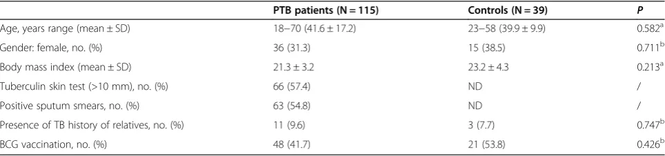
Table 1 Characteristics of pulmonary tuberculosis patients and healthy controls
PTB patients (N = 115) Controls (N = 39) P
Age, years range (mean ± SD) 18−70 (41.6 ± 17.2) 23−58 (39.9 ± 9.9) 0.582a
Gender: female, no. (%) 36 (31.3) 15 (38.5) 0.711b
Body mass index (mean ± SD) 21.3 ± 3.2 23.2 ± 4.3 0.213a
Tuberculin skin test (>10 mm), no. (%) 66 (57.4) ND /
Positive sputum smears, no. (%) 63 (54.8) ND /
Presence of TB history of relatives, no. (%) 11 (9.6) 3 (7.7) 0.747b
BCG vaccination, no. (%) 48 (41.7) 21 (53.8) 0.426b
AP-value of less than 0.05 indicates statistical significance. PTB: pulmonary tuberculosis; N: number of subjects; ND: not determined.aP
Co., Taipei, Taiwan), with a minimum detection limit of 0.31 ng/mL, was used to detect FN in serum. Human PEPD ELISA kit (Cusabio Biotech. Co., LTD, China), with a minimum detection limit of 93.75 mU/mL, was used to detect PEPD in serum. The protein concentra-tion of 39 healthy controls and 115 PTB patients were measured according to the manufacturer’s instructions. Briefly, serum samples were diluted with dilution factors of 1:2,000, 1:1,000,000 and 1:10 for C4b, FN and PEPD, respectively. Diluted samples were incubated in microti-ter wells coated with antibodies of proteins. Afmicroti-ter incu-bation and washing, the biotinylated tracer antibody conjugated with streptavidin-peroxidase was added to the wells. Substrate tetramethylbenzidine (TMB) was added to the wells after a second incubation and wash-ing, and then the oxalic acid was added to stop the enzyme reaction. Absorbance was read on xMark micro-plate spectrophotometer (Bio-Rad, Inc., USA) at a wave-length of 450 nm. The concentration of the protein was estimated using a four-parameter logistic curve (Micro-plate Manager 6 software, Bio-Rad, Inc., USA) based on the measured standard values.
Statistical analysis
Parametric data were presented as mean ± SD while non-parametric data were presented as median ± IQR, and
P< 0.05 was considered as statistically significant by the SPSS software, version 16.0 (SPSS, Chicago, IL). The One-samplet-test was used to investigate the difference between PTB patients and normal reference range after taking the logarithm. Two groups’ data were tested using the chi-square test for composition ratio andt-test for means. Non-parametric analysis was carried out using the Mann– Whitney U-test. Spearman correlation method was performed to determine association between two different parameters. The study sample provided 88.93% power to identify significant differences between whole PTB patients and healthy controls at a statistical support level ofα= 0.05 with an d of 0.6 applying a two tails model, and provided 75.47% power when identifying significant differences be-tween minimal, moderate, advanced PTB groups and healthy controls.
Results
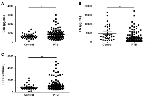
[image:3.595.60.540.418.699.2]Clinical data analysis of 187 PTB cases showed various significant differences between PTB patients and healthy controls of different parameters (Table 2). Comparison of 115 PTB patients and 39 healthy controls by using the ELISA method showed significant higher levels of serum C4b and PEPD (P= 0.018, P= 0.003), and signifi-cant lower level of FN (P< 0.001) in PTB patients (Figure 1).
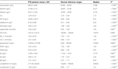
Table 2 Clinical data of pulmonary tuberculosis patients (N = 187) and normal reference ranges
PTB patients (mean ± SD) Normal reference ranges Median Pa
Total protein (g/L) 68.32 ± 6.66 64.00−83.00 72.88 < 0.001**
Albumin (g/L) 37.94 ± 5.13 36.00−52.00 43.27 < 0.001**
Globulin (g/L) 30.27 ± 5.60 22.00−34.00 27.35 < 0.001**
A/G 1.29 ± 0.32 1.20−2.50 1.73 < 0.001**
CRP (mg/L) 29.69 ± 38.72 0.00−0.82 0.41 < 0.001**
Prealbumin (g/L) 0.18 ± 0.08 0.15−0.36 0.23 < 0.001**
Fibrinogen (g/L) 5.69 ± 2.23 1.90−5.60 3.26 < 0.001**
Triglyceride (mmol/L) 1.06 ± 0.67 0.56−1.69 1.05 0.001**
LDH (U/L) 167.55 ± 54.74 100.00−240.00 154.92 0.064
HDL-C (mmol/L) 1.03 ± 0.34 1.03−1.55 1.26 < 0.001**
LDL-C (mmol/L) 2.31 ± 0.81 0.00−3.10 1.55 < 0.001**
Lipoprotein (mg/L) 270.60 ± 275.90 0.00−300.00 150.00 0.004**
APOA1 (g/L) 1.05 ± 0.23 1.20−1.60 1.39 < 0.001**
APOB (g/L) 0.76 ± 0.20 0.80−1.05 0.92 < 0.001**
IgG (g/L) 14.05 ± 3.60 11.50−14.22 12.79 0.007**
IgA (g/L) 2.84 ± 1.37 1.70−3.25 2.35 0.044*
IgM (g/L) 1.21 ± 0.57 0.73−1.17 0.92 < 0.001**
Complement 4 (mg/L) 377.78 ± 80.38 100.00−400.00 200.00 < 0.001**
Complement 3 (g/L) 1.04 ± 0.24 0.83−1.77 1.21 < 0.001**
AP-value of less than 0.05 indicates statistical significance. PTB: pulmonary tuberculosis; N: number of subjects; A/G: Albumin/ Globulin; CRP: C-reactive protein; LDH: Lactate dehydrogenase; HDL-C: High-density lipoprotein cholesterol; LDL-C: Low-density lipoprotein cholesterol; APOA1: apolipoprotein A1; APOB: apolipoprotein B;*P
< 0.05;**P
< 0.01.aP
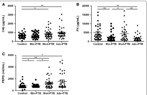
Then, we compared the control group with the min-imal, moderate, and advanced PTB groups. Our results indicated that the levels of serum C4b were different among groups (P= 0.024). There were significant differ-ences in serum C4b levels of the moderate and advanced PTB groups, compared to the control group (P< 0.05, P< 0.01) (Figure 2A). We also found different levels of FN (P< 0.001) among groups. FN level was found to be significantly different in the moderate PTB group, com-pared with patients in the minimal and advanced PTB groups (P< 0.05, P< 0.01), not to mention the control and minimal (P< 0.01), and the control and advanced PTB groups (P< 0.01) (Figure 2B). PEPD also showed different expression levels among the groups (P= 0.011). There were significant differences in serum PEPD levels between all three PTB groups and the control group (P< 0.05, P< 0.01, P< 0.05). In addition, a significant higher PEPD level was found in the moderate PTB group than the minimal PTB group (P< 0.05) (Figure 2C).
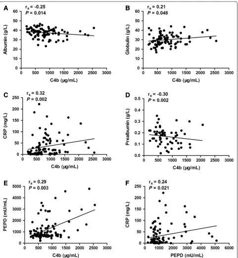
By using the Spearman correlation analysis, we found a significant negative correlation between C4b and albu-min, prealbumin. A significant positive correlation was observed between C4b and globulin, CRP, PEPD, as well
as PEPD and CRP (P< 0.05) (Figure 3). After that, we separated all 115 subjects based on age, gender, current smoker, single/double lung lesion, cavity/non-cavity, sputum smear results (−/+/++/+++/++++). Significant difference of C4b level was found between smokers and non-smokers (P= 0.012), as well as PEPD level between males and females (P= 0.011) (Table 3). In addition, FN was related to sputum smear positive results (rs= 0.22, P= 0.028) (Table 4).
Discussion
[image:4.595.60.538.90.396.2]Innate immunity is the body’s first line of defense against MTB infection. As a part of the complement system, the MBL pathway can cause cytolysis by identifying mannose residues on the surface of MTB. C4b is a product of acti-vated C4 in the early stage of MBL pathway [9]. Clinical data analysis showed liver dysfunction (decreased albumin), immune system activation (increased globulin), and com-plement system activation (increased C4, C3) in PTB pa-tients (Table 2). Similar findings were observed by ELISA method (Figure 1A). Correlation analysis demonstrated a weak but significant correlation between C4b and albumin, globulin (P< 0.05) (Figure 3A, 3B), indicating that the re-duction of liver synthetic function, and the activation of
immune and complement systems occurred in the early stage of PTB. Kingery et al. [18] revealed that increased plasma C4b may enhance the tissue damage by inflamma-tory response, indicating C4b as a prognostic biomarker of type I diabetes. In our study, a significant difference in C4b expression was observed in the moderate and advanced PTB patients, compared to the control group (P< 0.05), suggesting that a larger amount of bacterial infection may cause complement system activation, tissue damage, and cavity formation, which is also proven by Yoon et al. [19].
PTB is characterized by necrotizing granulomatous in-flammation of the lung tissue. FN is an extracellular glyco-protein that binds toβ1 integrin on the cell surface, leading to cellular adhesion to the extracellular matrix. There are two types of FN: plasma fibronectin (pFN), a soluble dimer present in the body fluid, and cellular fibronectin (cFN), an insoluble oligomer present in the extracellular matrix [20]. Fibroblasts could secrete proteases, such as metalloprotein-ase to digest pFN at first, and then secrete cFN to form extracellular matrix during the tissue injury. This may be the reason for the decreased serum FN levels (P< 0.001) in PTB patients in our study. Moreover, Kim et al. [21] re-vealed increased serum pFN in retinoic acid-deficient mice, which could be reversed by adding exogenous retinoic acid.
Retinol-binding protein 4 (RBP4) is responsible for binding and transporting blood retinol into the cells to become ret-inoic acid, and RBP4 reduction was observed previously in PTB patients [22]. Therefore, we hypothesized that reduced RBP4 protein may be related to the FN reduction in PTB patients. RBP4 transport retinol into the cell, resulting in increased intracellular retinoic acid and decreased serum FN.
[image:5.595.58.540.87.396.2]Interestingly, there was a higher level of FN in the moderate PTB group, compared to both minimal and advanced PTB groups (P< 0.05, P< 0.01). FN is thought to be the earliest biomarker of Schistosoma haemato-biumandSchistosoma mansoniinfested granuloma [23]. Rojas et al. [24] demonstrated that phosphatidylinositol mannoside of MTB can bind to α5β1integrin on CD4+ T cells, resulting in T cell adhesion to FN in granuloma. So, we suggested that a higher FN level in moderate PTB patients is related to the granuloma formation. In addition, correlation between FN and sputum smear positive results (rs= 0.22, P= 0.028) supported our sug-gestion. As the disease progresses, advanced PTB patients had a reduced level of serum FN, which may be caused by the increased expression of proteases, result-ing in increased FN digestion.
Host proteases disorders can cause granuloma for-mation, caseous necrosis, and liquefaction, leading to collagen degradation and tissue damage. PEPD is a type of proteases that hydrolyzes peptides with
[image:6.595.58.540.87.611.2]proline or hydroxyproline at the carboxy terminus. Myara et al. [25] demonstrated a higher plasma PEPD activity in the early stage of chronic liver dis-ease, indicating its role in the extracellular matrix
Figure 3Correlations between serum C4b, PEPD and some biochemical parameters.AP-value of less than 0.05 indicates statistical significance using the Spearman correlation method. The range of rsfrom−0.3 to−0.1 or from 0.1 to 0.3 means weak correlation, while the
Table 3 Average serum levels of proteins according to the clinical characteristics of the pulmonary tuberculosis patients
Clinical characteristics (Cases) C4b (μg/mL) Pa FN (
μg/mL) Pa PEPD (mU/mL)
Pa
Age 0.651 0.139 0.499
18−34 (51) 663.83 ± 482.42 1,931.50 ± 3,389.50 784.47 ± 492.87
35−49 (19) 610.22 ± 539.22 837.00 ± 1,338.00 764.53 ± 724.93
≥50 (45) 772.48 ± 508.49 1,567.00 ± 2,292.00 931.92 ± 647.87
Gender 0.698 0.545 0.011*
Male (79) 694.83 ± 525.59 1,637.50 ± 3,085.25 878.56 ± 628.99
Female (36) 747.14 ± 417.058 1,274.00 ± 2,198.00 672.24 ± 341.35
Current smoker 0.012* 0.838 0.205
Yes (50) 614.25 ± 464.86 1,582.50 ± 3,446.25 925.33 ± 635.58
No (65) 818.59 ± 471.98 1,511.00 ± 2,285.50 778.75 ± 557.74
Lung lesion 0.320 0.692 0.357
Single (53) 645.45 ± 444.61 1,550.00 ± 1,705.00 778.75 ± 380.34
Double (62) 808.42 ± 454.55 1,582.50 ± 3,508.75 845.27 ± 856.62
Chest X-ray 0.555 0.739 0.878
Non-cavity (70) 663.83 ± 664.71 1,567.00 ± 1,675.00 833.59 ± 405.02
Cavity (45) 729.10 ± 389.30 1,637.50 ± 3,566.75 755.98 ± 871.74
Sputum smear 0.258 0.121 0.441
Negative (52) 645.45 ± 526.00 1,509.00 ± 1,925.75 771.10 ± 326.47
1+ (13) 587.64 ± 326.10 1,189.00 ± 1,348.00 1,079.22 ± 810.94
2+ (18) 976.66 ± 500.99 1,725.00 ± 2,333.00 819.06 ± 674.19
3+/4+ (32) 791.67 ± 409.65 2,504.00 ± 3,974.50 925.33 ± 833.83
NTA classification 0.024* < 0.001** 0.011*
Minimal PTB (39) 567.63 ± 518.38 1,528.00 ± 1,605.00 795.95 ± 258.84
Moderate PTB (41) 729.10 ± 472.71 2,894.00 ± 4,915.00 968.67 ± 759.27
Advanced PTB (35) 846.87 ± 402.95 856.00 ± 1,609.00 761.75 ± 1,895.57
All the data were presented as median ± IQR. AP-value of less than 0.05 indicates statistical significance.aPvalue among groups, for Mann–Whitney U-test. *P
< 0.05;**P
< 0.01.
Table 4 Correlations for clinical characteristics and serum protein levels of the pulmonary tuberculosis patients (N = 115)
Sputum smear NTA classification C4b FN PEPD
Sputum smear rs 1 0.19 0.11 0.22 0.10
Pa . 0.043* 0.244 0.028* 0.287
NTA classification rs 1 0.19 −0.18 0.08
Pa . 0.045* 0.086 0.420
C4b rs 1 0.10 0.29
Pa . 0.354 0.003**
FN rs 1 −0.05
Pa . 0.659
PEPD rs 1
Pa .
The rsin [−0.3, -0.1] or [0.1, 0.3] means weak correlation, in [−0.5, -0.3] or [0.3, 0.5] means moderate correlation, and in [−0.7, -0.5] or [0.5, 0.7] means significant
[image:7.595.58.539.562.716.2]formation. Gumus et al. [26] found a higher serum PEPD activity in patients with PTB, especially in pa-tients with the lung cavity. It was suggested that in-creased PEPD level might be related to tissue damage, enhanced fibroblast activity, and increased levels of immunoglobulin and complement C1q (C1q). However, our previous unpublished results, using iTRAQ 2D LC-MS/MS technique, showed no significant difference in serum C1q level between PTB patients and the control group. Clinical data analysis showed a higher levels of IgG, IgA, and IgM in patients with PTB (P< 0.05) (Table 2), supporting the speculation that high immunoglobulin levels can lead to increased PEPD level. Therefore, we hypothe-sized that increased serum PEPD level might be re-lated to tissue damage, enhanced fibroblastic activity, and increased immunoglobulin. Significantly higher levels of PEPD in moderate PTB patients, compared to minimal PTB patients (P< 0.05) indicated that PEPD may be involved in the granuloma substrate hydrolysis process leading to the cavity formation.
Furthermore, there was a significant difference in
C4b level between smokers and non-smokers
(P= 0.012), and a significant difference was observed in PEPD level between males and females (P= 0.011). We suggested that the lower level of C4b in current smokers may be caused by a reactive inhibition in-duced by the activation of complement system than non-smokers before MTB infection. The reason for higher level of PEPD in males may be due to the higher ratio of hepatitis B infection among males leading to liver fibrosis [27], as PEPD is an early in-dicator of liver fibrosis [25].
Conclusion
In conclusion, we revealed increased serum C4b and PEPD levels, and decreased FN level in PTB patients. C4b may be associated with tissue damage, while FN and PEPD are associated with granuloma and cavity formation, respectively. The study provides a new experimental basis to understand the pathogenesis and pathological changes of PTB.
Competing interests
The authors declare they have no competing financial interests.
Authors’contributions
LJC conceived the study and designed the experiments. WC, LYY, LX, WLL, YXY, LZJ, CZL, ZX, LJY and PZP collected the serum samples and clinical data. WC, XDD and JTT analyzed the data with suggestions by LJC. WC and LJC wrote the manuscript. All authors read and approved the final manuscript.
Acknowledgements
This work was supported by grants from National Natural Science Foundation of China (No. 81273882, No. 81072724) and National Special Sci-Tech Projects (No. 2012ZX10005001-006).
Author details
1
Institute of Cell Biology, Zhejiang University, Hangzhou 310058, P.R. China.
2Department of Pathology, First Affiliated Hospital, Zhejiang University,
Hangzhou 310003, P.R. China.3Key Laboratory of Gastroenteropathy, Zhejiang Province People’s Hospital, Hangzhou 310012, P.R. China.4The Sixth
Hospital of Shaoxing, Shaoxing 312000, P.R. China.5Department of Respiratory Medicine, Tongde Hospital of Zhejiang Province, Hangzhou 310012, P.R. China.
Received: 24 June 2013 Accepted: 30 January 2014 Published: 31 January 2014
References
1. World Health Organization:Global tuberculosis report 2012.http://www. who.int/tb/publications/global_report/gtbr12_main.pdf.
2. Technical Guidance Group of the Fifth National TB Epidemiological Survey, The Office of the Fifth National TB Epidemiological Survey:The fifth national tuberculosis epidemiological survey in 2010.Chin J Antituber2012, 34(8):485–508.
3. Dunkelberger JR, Song WC:Complement and its role in innate and adaptive immune responses.Cell Res2010,20(1):34–50.
4. Kleinnijenhuis J, Oosting M, Joosten LA, Netea MG, Van Crevel R:Innate immune recognition of Mycobacterium tuberculosis.Clin Dev Immunol
2011,2011:405310.
5. Vankayalapati R, Barnes PF:Innate and adaptive immune responses to human Mycobacterium tuberculosis infection.Tuberculosis (Edinb)2009, 89(Suppl 1):S77–S80.
6. Hernandez-Pando R, Orozco H, Aguilar D:Factors that deregulate the protective immune response in tuberculosis.Arch Immunol Ther Exp (Warsz)2009,57(5):355–367.
7. Means TK, Wang S, Lien E, Yoshimura A, Golenbock DT, Fenton MJ:Human toll-like receptors mediate cellular activation by Mycobacterium tuberculosis.J Immunol1999,163(7):3920–3927.
8. Constantoulakis P, Filiou E, Rovina N, Chras G, Hamhougia A, Karabela S, Sotiriou A, Roussos C, Poulakis N:In vivo expression of innate immunity markers in patients with Mycobacterium tuberculosis infection.
BMC Infect Dis2012,10:243.
9. Lachmann PJ:Microbial immunology: a new mechanism for immune subversion.Curr Biol1998,8(3):99–101.
10. Taylor JL, Hattle JM, Dreitz SA, Troudt JM, Izzo LS, Basaraba RJ, Orme IM, Matrisian LM, Izzo AA:Role for matrix metalloproteinase 9 in granuloma formation during pulmonary Mycobacterium tuberculosis infection.
Infect Immun2006,74(11):6135–6144.
11. De Groote MA, Nahid P, Jarlsberg L, Johnson JL, Weiner M, Muzanyi G, Janjic N, Sterling DG, Ochsner UA:Elucidating novel serum biomarkers associated with pulmonary tuberculosis treatment.PLoS One2013, 8(4):e61002.
12. O’Regan AW, Chupp GL, Lowry JA, Goetschkes M, Mulligan N, Berman JS: Osteopontin is associated with T cells in sarcoid granulomas and has T cell adhesive and cytokine-like properties in vitro.J Immunol1999, 162(2):1024–1031.
13. Grosset J:Mycobacterium tuberculosis in the extracellular compartment: an underestimated adversary.Antimicrob Agents Chemother2003,47(3):833–836. 14. Kumar V, Abbas AK, Fausto N, Aster JC:Acute and chronic inflammation.In
Robbins and Cotran Pathologic basis of disease.7th edition. Philadelphia, PA: Elsevier Saunders; 2005:82.
15. Ministry of Health of the People’s Republic of China:Diagnostic criteria for pulmonary tuberculosis.http://www.moh.gov.cn/publicfiles///business/ cmsresources/zwgkzt/cmsrsdocument/doc3242.pdf.
16. Falk A, O’Connor JB, Pratt PC:Classification of pulmonary tuberculosis.In
Diagnostic standards and classification of tuberculosis. 12, vol.6th edition. New York: National Tuberculosis and Respiratory Disease Association; 1969:68–76.
17. Hussain R, Hasan R, Khurshid M, Sturm AW, Ellner JJ, Dawood G:Pulmonary tuberculosis in a BCG vaccinated area: relationship of disease severity with immunological and hematological parameters and drug resistance patterns.
Southeast Asian J Trop Med Public Health1996,27(2):257–262.
19. Yoon SH, Lee NK, Yim JJ:Impact of sputum gross appearance and volume on smear positivity of pulmonary tuberculosis: a prospective cohort study.BMC Infect Dis2012,12:172.
20. Lucena S, Arocha Pinango CL, Guerrero B:Fibronectin. Structure and functions associated to hemostasis. Review.Invest Clin2007, 48(2):249–262.
21. Kim HY, Wolf G:Vitamin A deficiency alters genomic expression for fibronectin in liver and hepatocytes.J Biol Chem1987,262(1):365–371. 22. Tanaka T, Sakurada S, Kano K, Takahashi E, Yasuda K, Hirano H, Kaburagi Y,
Kobayashi N, Hang NT, Lien LT,et al:Identification of tuberculosis-associated proteins in whole blood supernatant.BMC Infect Dis2011, 11:71.
23. Al Adnani MS:Concomitant immunohistochemical localization of fibronectin and collagen in schistosome granulomata.J Pathol1985, 147(2):77–85.
24. Rojas RE, Thomas JJ, Gehring AJ, Hill PJ, Belisle JT, Harding CV, Boom WH: Phosphatidylinositol mannoside from Mycobacterium tuberculosis binds alpha5beta1 integrin (VLA-5) on CD4+ T cells and induces adhesion to fibronectin.J Immunol2006,177(5):2959–2968.
25. Myara I, Myara A, Mangeot M, Fabre M, Charpentier C, Lemonnier A:Plasma prolidase activity: a possible index of collagen catabolism in chronic liver disease.Clin Chem1984,30(2):211–215.
26. Gumus S, Yaman H, Ozcan O, Deniz O, Karaman B, Cakir E, Tozkoparan E, Ozkan M, Bilgic H:Serum prolidase activity in patients with pulmonary tuberculosis.Scand J Clin Lab Invest2011,71(6):467–472.
27. Custer B, Sullivan SD, Hazlet TK, Iloeje U, Veenstra DL, Kowdley KV:Global epidemiology of hepatitis B virus.J Clin Gastroenterol2004,
38(10 Suppl 3):S158–S168.
doi:10.1186/1471-2334-14-52
Cite this article as:Wanget al.:Serum complement C4b, fibronectin, and prolidase are associated with the pathological changes of pulmonary tuberculosis.BMC Infectious Diseases201414:52.
Submit your next manuscript to BioMed Central and take full advantage of:
• Convenient online submission
• Thorough peer review
• No space constraints or color figure charges
• Immediate publication on acceptance
• Inclusion in PubMed, CAS, Scopus and Google Scholar
• Research which is freely available for redistribution