R E S E A R C H
Open Access
CD160 isoforms and regulation of CD4 and CD8
T-cell responses
Mohamed El-Far
1,3*, Charles Pellerin
1†, Louise Pilote
1†, Jean-Francois Fortin
1, Ivan A D Lessard
1, Yoav Peretz
2,
Elizabeth Wardrop
1, Patrick Salois
1, Richard C Bethell
1, Michael G Cordingley
1and George Kukolj
1,4*Abstract
Background:Coexpression of CD160 and PD-1 on HIV-specific CD8+T-cells defines a highly exhausted T-cell subset. CD160 binds to Herpes Virus Entry Mediator (HVEM) and blocking this interaction with HVEM antibodies reverses T-cell exhaustion. As HVEM binds both inhibitory and activatory receptors, our aim in the current study was to assess the impact of CD160-specific antibodies on the enhancement of T-cell activation.
Methods:Expression of the two CD160 isoforms; glycosylphosphatidylinositol-anchored (CD160-GPI) and the transmembrane isoforms (CD160-TM) was assessed in CD4 and CD8 primary T-cells by quantitative RT-PCR and Flow-cytometry. Binding of these isoforms to HVEM ligand and the differential capacities of CD160 and HVEM specific antibodies to inhibit this binding were further evaluated using a Time-Resolved Fluorescence assay (TRF). The impact of both CD160 and HVEM specific antibodies on enhancing T-cell functionality upon antigenic stimulation was performed in comparativeex vivostudies using primary cells from HIV-infected subjects stimulated with HIV antigens in the presence or absence of blocking antibodies to the key inhibitory receptor PD-1.
Results:We first show that both CD160 isoforms, CD160-GPI and CD160-TM, were expressed in human primary CD4+and CD8+T-cells. The two isoforms were also recognized by the HVEM ligand, although this binding was less pronounced with the CD160-TM isoform. Mechanistic studies revealed that although HVEM specific antibodies blocked its binding to CD160-GPI, surprisingly, these antibodies enhanced HVEM binding to CD160-TM, suggesting that potential antibody-mediated HVEM multimerization and/or induced conformational changes may be required for optimal CD160-TM binding. Triggering of CD160-GPI over-expressed on Jurkat cells with either bead-bound HVEM-Fc or anti-CD160 monoclonal antibodies enhanced cell activation, consistent with a positive co-stimulatory role for CD160-GPI. However, CD160-TM did not respond to this stimulation, likely due to the lack of optimal HVEM binding. Finally,ex vivoassays using PBMCs from HIV viremic subjects showed that the use of CD160-GPI-specific antibodies combined with blockade of PD-1 synergistically enhanced the proliferation of HIV-1 specific CD8+ T-cells upon antigenic stimulation.
Conclusions:Antibodies targeting CD160-GPI complement the blockade of PD-1 to enhance HIV-specific T-cell responses and warrant further investigation in the development of novel immunotherapeutic approaches.
Keywords:CD160, PD-1, HVEM, HIV, Immunopotentiation
* Correspondence:[email protected]; [email protected]
†Equal contributors
1Boehringer Ingelheim Ltd., 2100 Rue Cunard, Laval, Quebec, Canada 4
Boehringer Ingelheim (Canada) Ltd., 5180 South Service Road, Burlington, Ontario L7L 5H4, Canada
Full list of author information is available at the end of the article
Introduction
Negative immune regulators such as Programmed Death-1 (PD-1) and Cytotoxic T Lymphocyte Antigen 4 (CTLA-4) are part of a large network of immune checkpoints that are tightly regulated in order to limit exaggerated immune re-sponses and prevent autoimmunity [1-4]. However, in some instances such as persistent antigenic stimulation during chronic HIV or other viral infections, these negative regulators accumulate progressively on the cell surface of total and Ag-specific T and B cells [5-9]. Ex-pression and engagement of these negative regulators with their cognate ligands down modulate cell functions in a hierarchical manner with cell proliferation and IL-2 production being lost at earlier stages whereas IFNγand TNFαare lost at later stages in what is referred to as im-mune exhaustion [10,11].
PD-1, a central negative regulatory molecule was one of the early studied mediators of immune exhaustion in chronic infectious diseases, particularly HIV-1 infection [6,7] and in animal viral chronic infectious models [12]. A large body of evidence indicates that loss of function is not simply associated with PD-1 expression alone. Other characteristics such as the level of PD-1 expression and/or its co-expression with other negative modulators may better identify functionally impaired T-cells [13,14]. Co-expression of CD160 with PD-1, 2B4 and KLRG1 on HCV-specific CD8+T-cells was associated with dimin-ished cell functions and an intermediate differentiation stage [15]. Similarly, co-expression of CD160 and PD-1 was also shown to define a subset of HIV-specific CD8+ T-cells with advanced dysfunction characterized by up-regulation of different inhibitory pathways and down-regulation of the NF-ΚB transcriptional node [14].
CD160 is a glycosylphosphatidylinositol (GPI)-anchored protein member of the Ig superfamily with a restricted expression profile that is limited to CD56dimCD16+NK cells, NKT-cells,γδT-cells, cytotoxic CD8+T-cells lack-ing the expression of CD28, a small fraction of CD4+ T-cells and all intraepithelial lymphocytes [16-18]. Binding of CD160 to both classical and non-classical MHC I enhances NK and CD8+CTL functions [19-22]. However, engagement of CD160 by the Herpes Virus Entry Medi-ator (HVEM) was shown to mediate inhibition of CD4+ T-cell proliferation and TCR-mediated signaling [23].
HVEM protein is a bimolecular switch that binds both co-stimulatory LT-α/LIGHT and co-inhibitory receptors BTLA/CD160 (Reviewed by del Rio et al., [24]). The bind-ing of LIGHT on T-cells to HVEM, a co-stimulatory cell surface protein expressed by immature DCs and activated T-cells, induces potent inflammatory signals and a Th1-mediated response [25]; in turn, the binding of LIGHT to HVEM on T-cells elicits activation and survival signals through the induction of NF-ΚB and AP1 [26,27]. In
con-trast, binding of HVEM to BTLA expressed by T-cells
engages a potent negative signaling pathway involving both SHP-1 and SHP-2 phosphatases and effectively at-tenuates TCR activation [28,29]. During chronic HIV infection,ex vivoblockade of the HVEM network with polyclonal antibodies to HVEM enhances HIV-specific CD8+ T-cell functions, such as cell proliferation and cytokine production [14]. The functional effects of HVEM binding is probably influenced by several fac-tors in addition to the interacting partner, such as cell types, strength of stimulation and expression kinetics of the receptor/ligand pairs. Consequently, the interpret-ation of results based exclusively on HVEM-directed block-ade may benefit from additional exploration involving the interacting ligand(s).
As CD160 expression was shown to be specifically up-regulated on CD8+T-cells during the chronic phase of HIV infection, we aimed in the current study to assess the tar-geting of CD160 receptor on HIV-specific responses. We evaluated the interaction of the two CD160 isoforms CD160-GPI and CD160-TM with HVEM ligand, as well as the impact of targeting CD160, in combination with anti-PD-1, to provide a beneficial pharmacological effect on HIV-specific CD8+T-cells in response.
Materials and methods
Cloning of human CD160-GPI and CD160-TM isoforms
The complete CD160 cDNA sequence was synthesized in vitro (DNA2.0) and codon-optimized for human ex-pression. To generate the CD160-GPI and the CD160-TM expression plasmids, the CD160 sequence was first PCR amplified using the following oligonucleotides: GATTGC AGATCTGCCACCATGCTTCTTGAACCTGGTCGCGG TTG (sense), CTGACGCTCGAGCTACAAAGCCTGCA ACGCGACCAGCGAAGTTACC (antisense, CD160-GPI), CTGACGCTCGAGCTAGTGGAACTGATTCGAGGACT CTTG (antisense, CD160-TM). The PCR fragments were then digested with BglII and XhoI and inserted into the BamHI/XhoI digested pcDNA3.1/neo(+) vector (Invitro-gen), downstream of the CMV promoter. Note thatBglII andBamHI produce compatible ends.
Production of stable cell lines
CHO-K1 (ATCC, CCL-61) stable cell lines expressing hu-man CD160-GPI or CD160-TM were generated by lipofec-tion of the CD160 expression vectors (pcDNA3.1) into naïve CHO-K1 cells using Lipofectamine 2000 (Invitrogen). Transfected cells were incubated at 37°C-5% CO2in
pres-ence of 800μg/ml Geneticin and, after a selection of 10–14 days, resistant T-cell colonies were isolated and transferred into 48-well tissue culture plate. Following incubation at 37°C-5% CO2to allow for cell growth, cell surface
(Perkin Elmer, AD0124). Cell clones expressing high levels of CD160-GPI or CD160-TM were expanded.
Jurkat stable cell lines expressing GPI or CD160-TM were also generated by transfecting Jurkat-NFAT-Luc cells (stably transfected with pGL4.30 NFAT-luciferase with NFAT enhancer element, Promega, and maintained with hygromycin selection) with pcDNA3.1/neo(+) vector encoding the respective CD160 isoform. The CD160-GPI form was amplified with the following PCR primers; sense: CTAGCTAGCGAGCCATGCTTCTTGAACCTGGTCG CGGTTG, anti-sense: ATAGTTTAGCGGCCGCTCAC AACGCCTGCAACGCGACCAGCGAAGTTACC, and inserted into the compatible plasmid vector via the under-scored NheI and NotI restriction sites. The CD160-TM form was PCR amplified using the CD160-GPI forward primer in combination with the following NotI-encoding anti-sense primer: ATAGTTTAGCGGCCGCTCACTAG TGGAACTGATTCG, and inserted into an NheI-NotI restricted pcDNA3.1 vector. Jurkat-CD160 positive clones were selected with Geneticin as described above.
Time-Resolved Fluorescence (TRF) assay
A TRF assay was used to evaluate the capacity of differ-ent antibodies to inhibit the binding of recombinant hu-man HVEM-Fc fusion protein (R&D systems, 356-HV/ CF) to cells expressing either GPI or CD160-TM. In this assay, naïve CHO-K1 cells (used for back-ground controls) or CHO-K1 cells expressing CD160 were trypsinized and diluted in F-12 media (Invitrogen) containing 10% FBS (Hyclone). Cells (40,000 per well) were then aliquoted in poly-D-lysine treated white 384-well tissue culture plates and incubated for 20 h at 37°C-5% CO2. After incubation, supernatant was removed and
cells were washed once with 100 μl of TRF wash buffer (50 mM Tris pH 7.5, 0.05% Tween, 0.2% BSA, 150 mM NaCl). Ten μl of either CD160 or HVEM antibodies di-luted in NaPO4buffer (50 mM NaPO4pH 6.6, 150 mM
NaCl, 2% FBS) were added to each well, except for the background and the no-inhibition controls which re-ceived 10μl of NaPO4buffer, followed by the addition of
40 μl of 1.25 μg/ml HVEM-Fc, also diluted in NaPO4
buffer. The plate was then incubated for 1 h at RT and the wells were washed 3 times with 100μl of TRF wash buffer. Following this wash step, 50 μl of 0.25 μg/ml anti-human Eu-N1 (Perkin Elmer, 1244–330) diluted in DELFIA assay buffer (Perkin Elmer, 1244–111) was added to each well and the plate was incubated for 1 h at RT. The wells were then washed as above (3 times 100μl TRF wash buffer) and 50μl of DELFIA enhance-ment solution (Perkin Elmer, 1244–105) were added. After an incubation of 20 min at RT, the fluorescence signal was monitored using a Wallac Victor microplate reader (excitation at 340 nm and emission at 615 nm). The antibodies tested in this assay included CD160 mAb
clone CL1-R2 (MBL International), CD160 mAb clone 688327 (R&D), polyclonal anti-HVEM (R&D) and mono-clonal anti-HVEM clone 94801 (R&D).
RNA isolation from cells and quantification
The“RNeasy Kit”(Qiagen) was used to isolate RNA from cells. The total RNA concentration was determined using the “Quant-iT RiboGreen® RNA Assay Kit” from Invi-trogen. The RNA concentration of the samples was de-termined from the standard curve generated using the ribosomal RNA standards.
Real-time qRT-PCR assays
were used to generate a standard for gene-specific expres-sion analysis and to determine changes in transcript levels.
Antibodies
FACS analyses used anti-CD3 (V-500), anti-CD4 (BV-605), anti-CD8 (APC-H7), anti-CD25 (A700), anti-CD134 (FITC), anti-PD-1 (eFlour 605), anti-CD45RA (ECD) anti-CCR7 (PE-Cy7), anti-CD27 (eFluor 780) and anti-CD160 clone BY55 (A647) from BD. Blocking assays used mouse monoclonal anti-CD160 clone CL1-R2 (custom purified from MBL International), mouse monoclonal anti-CD160 clone 688327, mouse monoclonal anti-HVEM clone 94801 and goat polyclonal anti-HVEM (R&D systems). PD-1 monoclonal antibody clone 5C4 (human IgG4 back-ground) was obtained from sequence ID in patent applica-tion US20090217401; binding specificity for PD-1 and functional capacity of this antibody was characterized and confirmed (data not shown).
Subjects
HIV-negative and HIV-1-infected subjects provided written informed consent and studies were approved by the Royal Victoria Hospital (Montreal, QC, Canada) and Boehringer-Ingelheim Institutional Review Boards. The study popula-tion of HIV subjects is shown in Table 1.
Primary cell preparation
PBMCs from subjects were obtained by leukapheresis and isolated by density gradient centrifugation (Lymphocyte Separation Medium; Wisent, St-Bruno, QC) and cryo-preserved in 10% dimethyl sulfoxide (Hybri-Max DMSO; Sigma-Aldrich, St Louis, MO); 90% Heat-Inactivated Fetal Bovine Serum (HI-FBS) (PAA Laboratories, Etobicoke, ON).
HLA typing
DNA for molecular HLA-typing was prepared from whole blood using the QIAamp DNA blood kit (Qiagen Inc., Mississauga, ON, Canada). Subjects were typed for HLA class I antigen expression (A, B, and C alleles) by sequence-based typing using kits from Atria Genetics (South San Francisco, CA). Assign software was used to
interpret sequence information for allele typing (Conexio Genetics, Perth, Australia).
Stimulation of primary CD4+and Jurkat cells
Primary CD4+ T-cells were isolated from total PBMCs by magnetic bead separation using EasySep CD4 nega-tive selection kit (StemCell). Purity of isolated CD4+cells was consistently > 98%. Primary CD4+ cells were stimu-lated with plate-bound anti-CD3 clone UCHT1 (BD) at 1 μg/ml and anti-CD28 clone CD28.2 (BD) at 0.5μg/ml and either human HVEM-mouse Fc fusion (R&D Sys-tems) at a concentration of 0.2 μg/ml or its matched mouse isotype control antibody. Jurkat T-cells were acti-vated with Dynal beads (according to the supplier’s proto-col, Pan Mouse IgG, Invitrogen) coated with anti-CD3 clone UCHT1 and anti-CD28 clone CD28.2 and either anti-CD160 monoclonal antibody clone CL1-R2 (MBL International), human HVEM-mouse Fc fusion, or their matched isotype control mouse IgGs. Stimulation was per-formed at a ratio of 4 beads/cell.
Tetanus toxoid stimulation assay
Total PBMCs from healthy donors were thawed in RPMI-1640 medium containing 10% heat-inactivated human serum (GemCell). Cells were washed twice with medium and suspended at a final concentration of 1.5 × 10⁶cells/ml. Tetanus toxoid (List Biological Laboratories) was added at a concentration of 2.5 μg/ml. Blocking monoclonal antibodies against CD160, custom-purified clone CL1-R2 (MBL International) and polyclonal HVEM antibodies (R&D) or their matched isotype controls were used at 10μg/ml. Cells were incubated for 5 to 7 days and then IFNγ was measured in the supernatant by ELISA using OptEIA Kit (BD) according to the supplier’s protocol.
Design of peptide-pool matrices and IFNγELISPOT assay
[image:4.595.57.539.634.725.2]The HIV peptide sets used for the CFSE and IFNγ ELI-SPOT assays were 15 amino acids (aa) with 11 aa overlaps. The peptides were obtained from the NIH AIDS Research and Reference Reagent Program (NARRRP, Rockville, MD). Lyophilized peptides (n = 769) spanning all HIV-1 gene products were dissolved at a concentration of 10 mg/mL in DMSO and stored at−80°C. These included 123 Gag,
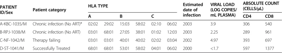
Table 1 Study population and clinical characteristic of each individual HIV infected subject
PATIENT
ID/Sex Patient category
HLA TYPE Estimated
date of infection
VIRAL LOAD (LOG COPIES/ mL PLASMA)
ABSOLUTE COUNT (CELLS/μL)
A B C CD4 CD8
A-KBC-1035/M Chronic infection (No ART)* 02:02 29:02 15:03 58:02 02:10 06:02 2003 3.9 306 540
B-RPJ-1038/M Chronic infection (No ART) 03:01 68:01 27:05 38:01 01:02 12:03 2003 2.25 289 961
C-NF-1042/M Therapy failing 03:01 03:01 40:01 40:02 02:02 03:04 2002 4.97 393 697
D-ST-1041/M Successfully Treated 68:01 68:01 53:01 58:02 04:01 06:02 2000 <1.7 597 1377
249 Pol, 49 Nef, 27 Rev, 23 Tat, 46 Vif, 22 Vpr, 19 Vpu and 211 Env 15-mers corresponding to consensus clade B sequence. Pools containing 1 to 16 peptides were prepared and organized into matrices of Gag, Pol, Nef, Env and accessory (Acc) gene peptide-pools such that each peptide was present in two pools within each matrix. IFNγ secre-tion by HIV-specific cells was quantified using the stand-ard ELISPOT assay. Spots were counted with the CTL ImmunoSpot 6 Analyzer (Immunospot, Cleveland, OH) and results were expressed as spot forming cells per mil-lion PBMCs (SFCs/106PBMCs) following subtraction of negative controls. The threshold for IFNγELISPOT posi-tivity was set to a minimum of 50 SFC/106PBMCs follow-ing background subtraction with a minimum of 10 spots and at least two fold over background values.
5,6-carboxyfluorescein diacetate succinimidyl ester (CFSE) dilution assay
Thawed PBMC were resuspended in PBS 1X and labeled with 0.6μM CFSE (Molecular Probes, Eugene, Oregon). CFSE labeled PBMCs were stimulated with 2 μg/mL of HIV consensus B peptides identified in the ELISPOT assay; Gag7876 (EKIRLRPGGKKKYKL) for subjects NF-1042 and KBC-1035, Gag937 (IYKRWIILGLNKIVR) for subject RJP-1038 and Pol5683 (TAVQMAVFIHNFKRK) for sub-ject ST-1041, in RPMI-1640 containing 10% human AB serum (Gemini, Burlington, ON). Stimulation with media alone served as a negative control, whereas stimulation with 25 ng/ml of Staphylococcol enterotoxin B (SEB) (Sigma-Aldrich) and 2μg/mL of CEFT (CMV, EBV, Influenza and Tetanus peptides) were used as positive control stimula-tions. Monoclonal antibodies directed against immune checkpoint molecules (PD-1, CD160 or HVEM) along with their corresponding isotype controls were added to the culture conditions at 5μg/mL. All stimulatory condi-tions were tested in quadruplicates. Following six days of incubation at 37°C and 5% CO2, cells were monitored for
viability with the Trypan blue exclusion test and further stained for cell surface markers using Live/Dead (Molecu-lar Probes), αCD3,αCD8 (ebioscience), andαCD4 mAbs (BD Biosciences, Mississauga, ON). PBMCs were acquired using a BD LSRII flow cytometer and analyzed with FlowJo software version 9.4.11 (FlowJo LLC, Ashland, Oregon).
Statistical analysis
Statistical analysis and graphical presentation was per-formed using GraphPad Prism 4 (GraphPad software, San Diego, CA), FlowJo 9.1 (Treestar) and FACSDiva V6 (BD Biosciences). Two-tailed paired t test was used to assess differences in the relative frequency of CD4+CD160+ T-cells before and after TCR stimulation from the same donors and in the IL-2 production following triggering with HVEM-Fc. The non-parametric Kruskal-Wallis and
Dunn’s tests were used to analyze data on the enhance-ment of T cell activation as shown in Figure legends.
Results
Expression of CD160 isoforms on primary T-cells and binding to HVEM
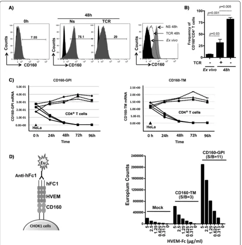
One aim of this study was to develop screening assays to evaluate the impact of CD160 antibodies on the enhance-ment of HIV-specific CD8 T-cell responses. CD160 was previously reported to mediate a co-stimulatory role on CD8+ T-cell activation upon binding to MHC-I, or a co-inhibitory role on CD4+T-cell activation upon binding to HVEM. Our first aim was to establish an inhibitory assay to test anti-CD160 antibody candidates with potential blocking capacity on T-cell activation, herein CD4+T-cells. To this end, we assessed the expression of CD160 on CD4+ T-cells before and after TCR activation to select the optimal time point for CD160 triggering. Levels of CD160 surface expression were determined using the BY55 clone of anti-CD160 that preferentially recognizes the GPI isoform [18]. Consistent with earlier reports [23], we observed that CD160 was expressed on a small fraction (2-8%) ofex vivo CD4+ T-cells at baseline (Figure 1A & B). CD160 expres-sion on cells stimulated with anti-CD3 and anti-CD28 monoclonal antibodies was higher at 48 h post-stimulation (p= 0.03) compared to theex vivobaseline levels. Notably, T-cells which remained un-stimulated for 48 hr showed the highest levels of CD160 compared to TCR-stimulated andex vivostained cells from matching individual donors (n = 3, p= 0.005 and p= 0.001, respectively) (Figure 1A, middle and right panels). Similar results were also obtained with CD8+T-cells (data not shown). The up-regulation of CD160 on resting cells ex vivo and its down-regulation following TCR stimulation thus contrasted observations by Cai et al. [23] who showed that CD160 is upregulated on CD4 T-cells following TCR stimulation. Therefore, we assessed whether this discrepancy was attributable to the expression of the newly identified isoform of CD160, the full-length trans-membrane isoforme (CD160-TM). The CD160-TM isoform is induced on NK cells upon stimula-tion with a panel of cytokines including IL-2, IL-12, IL-15 and IL-18 [18]. Our data in Figure 1C showed that the CD160-TM isoform was indeed clearly detectable at the transcriptional level in CD4+T-cells as measured by quan-titative RT-PCR. However, following TCR stimulation, both CD160-TM and CD160-GPI transcripts decreased gradually with time and became undetectable by 72–96 h post-TCR stimulation. Of note, we could not confirm the specific expression of CD160-TM at the protein level due to the lack of specific antibodies capable of distinguishing between the two isoforms (note that CD160-GPI anti-bodies poorly recognize the CD160-TM isoform [18]).
Figure 1Expression of CD160 isoforms in primary CD4+T-cells and binding to HVEM. A) Left panel:Representative FACS analysis of CD160 on primary CD4+T-cells isolated from total PBMCs of a healthy donor (ex vivoat baseline), gated on CD3+CD4+CD8−cells.Middle panels: CD160 surface expression following 48 h of resting (non-stimulated, NS) or TCR activation (plate-bound anti-CD3 and soluble anti-CD28).Right panel: overlapping histograms showing CD160 surface expression from TCR-stimulated CD4+T-cells (dotted empty histogram) in comparison to 48 h rested CD4 (filled grey histogram) and freshly isolated CD4 cells (filled black histograms) all from the same individual donor.B)Frequency of CD160
+
T-cells to elicit a potent inhibitory signal [23]. In order to study the details of HVEM binding to CD160 and examine its binding to the newly identified isoform of CD160-TM, we established CHO-K1 cell lines that over-expressed either CD160-GPI or CD160-TM to assess the binding of soluble HVEM-Fc. The assay was based on the highly sensitive dissociation-enhanced lanthanide fluor-escent immunoassay” (DELFIA; Perkin Elmer) with time-resolved fluorescence (Figure 1D, left panel). HVEM-Fc specifically but differentially bound to the CD160-GPI and CD160-TM isoforms with a signal to background ratio (S/B) of 11 and 3, respectively (Figure 1D, right panel). Together, CD160-TM, similar to CD160-GPI, was expressed by T-cells and recognized by HVEM, albeit with a lower level of binding compared to CD160-GPI. However, this lower level of binding could be due in part to a lower sur-face expression of CD160-TM.
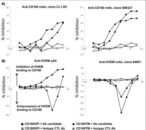
Antibody-mediated specific blockade of CD160/HVEM binding
We next screened benchmark antibodies directed against CD160 and HVEM to evaluate their potential capacity to block CD160/HVEM interaction and to select candidates for functional rescue of antigen-specific T-cells. Previous studies have shown that binding of HVEM to CD160 can be inhibited by the CD160 monoclonal antibody (mAb) CL1-R2 [30], an antibody with antiangiogenic activity [31]. We used the TRF CD160/HVEM binding assay to confirm these observations and to further evaluate other CD160 and HVEM antibodies (some of which were previously shown to enhance HIV-specific responses, [14]). The TRF assay consisted of a fixed concentration of soluble HVEM-Fc (1μg/ml) and serial dilutions of either CD160 mAbs (clones CL1-R2 and clone 688327) or HVEM poly-clonal and monopoly-clonal (clone 94801) Abs. CD160 Abs readily inhibited the binding of HVEM to either CD160-GPI or CD160-TM isoforms (Figure 2A) in the TRF assay. In contrast, the polyclonal HVEM antibody, which inhib-ited the binding of HVEM to CD160-GPI, enhanced HVEM binding to CD160-TM. Furthermore, the mono-clonal HVEM Ab (clone 94801) enhanced the binding of HVEM to both CD160 isoforms (Figure 2B). Together, these results showed that CD160 or HVEM antibodies had differential capacities to inhibit (or augment) the interaction between HVEM and specific CD160 isoforms.
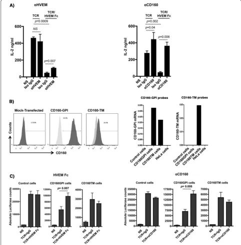
Triggering of CD160-GPI is consistent with positive regulation of CD4+T-cells
An inhibitory assay with primary CD4+T-cells was estab-lished whereby TCR and CD160 were simultaneously trig-gered in the presence or absence of specific antibodies to either HVEM or CD160 (CL1-R2 clone). CD4+ T-cells were isolated from total PBMCs of healthy donors and rested overnight to up-regulate CD160 as described earlier
(expression was monitored by flow cytometry). As shown in Figure 3A (left and right panels), addition of HVEM-Fc significantly reduced IL-2 production from CD4+ T-cells that were stimulated with CD3 and CD28 bodies for 24 h. Blockade of HVEM with specific anti-HVEM monoclonal Ab (Figure 3A left panel) partially restored IL-2 production from cells triggered with TCR/ HVEM-Fc compared to treatment with matched isotype control Ab (p= 0.007). Similarly, treatment of TCR/ HVEM-Fc triggered cells with CD160 specific monoclo-nal antibodies increased IL-2 production to reach levels equal to or higher than cells stimulated with TCR alone (Figure 3A right panel) (p= 0.006). Interestingly, IL-2 production by TCR-stimulated cells in the absence of the HVEM-Fc ligand was also enhanced by the CD160 mAb (p= 0.04). Meanwhile CD160 antibody had no im-pact on CD4+ T-cell activation in the absence of TCR stimulation, thus suggesting that the CD160 antibody-mediated enhancement of cell activation is TCR-dependent. These results are consistent with earlier reports showing that targeting CD160 with monoclonal antibodies may en-hance TCR-mediated signaling in T-cells [32,33].
Whereas the other set of probes were CD160-TM specific and confirmed the exclusive expression of the different CD160 isoforms in these two cell lines (Figure 3B right panels).
The effect of HVEM-mediated CD160 triggering on TCR activation was assessed by measuring the NFAT-responsive luciferase activity of Jurkat cells expressing either CD160-GPI or CD160-TM isoforms. Dynal Beads coated with anti-CD3, anti-CD28 and either HVEM-Fc or matched isotype control were used to perform these experiments. HVEM-Fc specifically activated Jurkat cells that expressed
[image:8.595.61.539.90.514.2]Figure 3Triggering of CD160-GPI is consistent with a positive co-stimulation role. A)Triggering of primary CD4+T-cells with either plate-bound
but not Jurkat-CD160-TM (Figure 3C, right 3 panels). Identical results were also obtained with anti-CD160 clone 688327 (data not shown).
Altogether, the engagement of CD160-GPI, but not CD160-TM, by either HVEM-Fc or specific mAb en-hanced the Jurkat T-cell activation as measured by the higher NFAT-responsive luciferase activity. The lack of any significant impact of HVEM-Fc on the CD160-TM isoform together with the positive co-stimulation medi-ated by HVEM-Fc triggering of CD160-GPI in the Jurkat assay suggested that the HVEM-Fc mediated inhibition of IL-2 production that we observed with primary CD4+ T-cells (Figure 3A) is likely mediated by HVEM inter-action with BTLA, which is constitutively expressed on CD4+T-cells (data not shown and [34]).
CD160 and HVEM antibodies specifically enhance CD4+ T-cell responses to a recall antigen
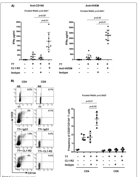
As a first line high throughput assay capable of identify-ing antibodies that modulate antigen specific T-cell acti-vation, we analyzed memory T-cell responses to Tetanus toxoid (TT) recall antigen. This assay allowed us to compare the potency of anti-CD160 (CL1-R2) mAb and the polyclonal anti-HVEM antibodies to enhance T-cell response. Both antibodies increased the production of IFNγby PBMCs from healthy responders upon stimula-tion with suboptimal concentrastimula-tions of TT (2.5 μg/ml) in a 5-day culture assay (Figure 4A). No IFNγ produc-tion was observed in the absence of antigenic stimula-tion (data not shown).
Tetanus toxoid is known to elicit a CD4+T-cell response [35]. In order to confirm the assay specificity, we tested the CD4 response by monitoring the frequency of TT-specific CD4+ T-cells as determined by the surface expression of IL-2Rα(CD25) and OX40 (CD134). This method was pre-viously shown to identify Ag-specific CD4+T-cells without the need for HLA class II multimers [35]. Analogous to the experimental conditions described above, PBMCs from healthy responders were stimulated with the Tetanus gen (TT) for 5 days in the presence or absence of anti-CD160 antibodies. As shown in Figure 4B, only CD4+ T-cells responded to TT-stimulation by up-regulating both CD25 and CD134 [the frequency of CD25+CD134+ DP cells increased from an average of 0.3% to 1.7% (n = 5)]. In contrast, no significant impact was observed on the CD8+ T-cell population. Interestingly, addition of CD160 mAb increased the frequency of CD25+CD134+ DP frac-tion to an average of 3.8% (n = 5), whereas no change in frequency was observed with isotype control antibodies. These results showed that CD160 and HVEM antibodies specifically enhanced memory CD4+ T-cell responses (both qualitatively and quantitatively) against a recall anti-gen upon re-stimulation.
Combined targeting of CD160 and PD-1 enhances HIV-specific CD8+T-cell proliferation
The impact of targeting CD160, with CD160-specific anti-bodies on HIV antigen-specific exhausted T-cells from HIV-infected subjects was studiedex vivo to evaluate its therapeutic potential. We first comprehensively screened HIV-1 epitopes by IFNγ ELISPOT to map the different T-cell responses to HIV-1 peptides from infected subjects. The primary objective of this comprehensive analysis was to determine the baseline responses to HIV-1 peptide stimulations from subjects with different categories/stages of disease and in turn to characterize the change in re-sponses upon targeting CD160 and/or other key cell sur-face regulators, herein PD-1. Study samples were obtained from both cART-treated aviremic and cART-naïve viremic subjects, with one of four subjects having the protective HLA allele B27 (Table 1). As shown in Additional file 2, the breadth of ex vivo responses was higher in samples from the viremic subjects compared to samples from the successfully treated one. Samples from the HLA-B27 sub-ject displayed the highest response values.
Since CD160+PD-1+double positive (DP) populations of HIV-1-specific CD8+ T-cells were previously shown to represent a highly exhausted cell subset [14], we measured the co-expression of CD160 and PD-1 on both total and selected antigen-specific cells based on the CD8+ T-cell epitopes defined by the IFNγELISPOT assay. As shown in Figure 5A, although the frequency of this DP population on total CD8+ T-cells was modest, the DP frequency was higher in CD8+T-cells from HIV viremic subjects relative to the cART treated and virus-suppressed subject or healthy donors (Left panel). Most notably, the DP popula-tion comprised a relatively high proporpopula-tion (3-45%, de-pending on the multimer used) of the HIV-specific CD8+ T-cells in the viremic subjects when compared to the A*02 CMV Ag-specific population from a HIV-uninfected donor (Figure 5A, right panel).
combination with anti-PD-L1 [13]. Interestingly, we did not observe any significant enhancement of CD4+ T-cell proliferation in response to the p24 antigen in the pres-ence of these antibodies (data not shown), which suggests that the observed functional enhancement was specific to CD8+T-cells. Co-targeting of PD-1 with either CD160 or HVEM showed very low levels of enhancement when peptide pools specific to other infectious agents (CEFT: CMV, EBV, Influenza and Tetanus) were used as controls (Additional file 3A & B). Of note, no significant enhance-ment was obtained with the CD160 and PD-1 combined antibody treatment in samples from subjects with low viral load (ST-1041 and RJP-1038), whereas HVEM spe-cific antibodies diminished the frequency of proliferating cells (compared to stimulation in the absence of antibody candidates) in these samples (Figure 5c). No activation-induced cell death (AICD) was observed with HVEM antibodies (data not shown).
Discussion
CD160 belongs to the broad family of T-cell co-regulators. In our efforts to generate a screening assay for selecting antibody candidates with the capacity to block HVEM binding to CD160 and to functionally impact T-cell activa-tion, we over-expressed the two known isoforms of CD160 (GPI and TM) in Jurkat cells harboring a luciferase reporter gene. HVEM ligand enhanced TCR-mediated ac-tivation only in cells expressing the CD160-GPI isoform and not the CD160-TM isoform. The lack of HVEM-mediated activation of CD160-TM may, in part, be due to the weak interaction between these proteins as suggested by our binding assays. However, as we could not confirm equal surface expression of CD160-TM, compared to CD160-GPI, due to the lack of CD160-TM specific antibodies, we cannot exclude the possibility that the low binding of HVEM-Fc to the CD160-TM expressing cells is due, at least in part, to a lower CD160-TM expres-sion at the cell surface. Yet, similar levels of transcription were observed for both CD160-GPI and CD160-TM iso-forms in the CHO-K1 cells, used for the binding assays, and in Jurkat cells, used for the functional assays. Further-more, monoclonal and polyclonal antibodies to HVEM enhanced the binding of HVEM-Fc to the CD160-TM in the CHO-K1 cells, which suggests that CD160-TM was expressed to significant levels at the cell surface. Similar to
antibody-mediated enhancement of HVEM-Fc binding to CD160, earlier observations were also reported for the binding of CD160 to MHC class I molecules [19]. The anti-MHC I monomorphic antibody W6/32 mAb en-hanced interaction between cells expressing CD160 and cells expressing the class I molecules suggesting that lig-and multimerization may promote binding to CD160-TM (20). However, multimerization of HVEM may not be the only possible mechanism to induce HVEM binding to CD160-TM as potential antibody-mediated changes in the HVEM protein conformation may also play a role The distinction between CD160-GPI and CD160-TM with re-gard to the need for HVEM multimerization or antibody-mediated conformational change might explain the lack of HVEM-mediated effect on Jurkat-CD160-TM with bead-bound monomeric HVEM-Fc fusion. How the MHC I or HVEM ligands localize/multimerize or change their con-formational structure under physiological conditions in order to promote binding to CD160, requires further in-vestigations. HVEM is expressed as a monomer and upon binding to the homotrimeric LIGHT forms a trimeric multimer [36,37]. Gonzalezet al.[37] suggest that BTLA is likely to bind to HVEM in the presence of LIGHT or LTα, whereby these latter receptors favor the formation of a trimeric HVEM. The regulation of HVEM association with CD160-TM through multimerization or conform-ational change and its impact on T-cell activation remains to be elucidated.
Triggering of CD160-GPI isoform over-expressed by the CD4+Jurkat T-cell line with monoclonal antibodies in our study was consistent with a positive co-stimulatory role. Similarly, CD160 stimulation was previously shown to enhance CD3-induced activation and proliferation of peripheral blood CD160+T cells [33] and also CD4+CD160+ T cells isolated from inflammatory skin lesions [32]. Though these results are in accordance with earlier reports that used the anti-CD160 CL1-R2 (IgG1) or the BY55 (IgM) [33] clones, they contrast with recent work by Cai et al., [23] showing that triggering of CD160 on primary CD4+ T-cells with the CD160 monoclonal antibody 5D.10A11 inhibits cell activation and cytokine production. These ap-parently discordant observations suggest that CD160 may differentially regulate either activating or inhibitory signal-ing pathways, which may depend on the type/clone of antibody or cognate ligand used to engage the target.
(See figure on previous page.)
Furthermore, the existence of two isoforms of CD160 (GPI and TM) in CD4+ and CD8+ T-cells with a pos-sible differential expression and regulation of ligand binding may also account for the divergent reports on CD160 functions as the selectivity of 5D.10A11 anti-body [23] for the various CD160 isoforms and the resulting effect on TCR signaling have not been char-acterized. Of note, in our Jurkat-NFAT-Luciferase assay with CD160-TM expressing cells, HVEM-Fc did not elicit either a negative or positive effect and may reflect a requirement for HVEM multimerization or in-duced conformational changes to promote CD160-TM binding.
Our study also showed that the GPI isoform was up-regulated on rested T-cells (both CD4+and CD8+)ex vivo likely due to the culture conditions. This apparent up-regulation of CD160 on resting cells and the contribution of ex vivo culture conditions such as the use of human serum require more investigation. Yet CD160 was down-regulated by TCR activation, which indicates that expres-sion of CD160 on primary T-cells is more complex than initially thought. CD160-GPI is likely to undergo receptor shedding upon T-cell stimulation similar to the previously described mechanism for CD160 on NK cells stimulated with IL-15 [38]. Although CD160-GPI and CD160-TM share the same extracellular domains, the GPI isoform does not contain a transmembrane domain. The two iso-forms have differential binding characteristics for CD160 antibodies [18] and they may also differ in their signaling capacity. The presence of these two isoforms of CD160 and their potential differential expression in T-cells re-quires further studies, particularly in the context of im-mune exhaustion. Indeed, our results showed that HVEM antibodies function differently in ex vivoT-cell assays on samples isolated from HIV-infected subjects with higher viral loads compared to aviremic subjects. These anti-bodies restore HIV-specific CD8+ T-cell proliferation in lymphocytes isolated from viremic subjects, but in con-trast dampen the response in CD8+T-cells from aviremic subjects. This difference may be related to potential differ-ential expression of the CD160 isoforms in viremic and aviremic subjects, meanwhile assuming that CD160-TM
mediates a negative regulatory role in this context. An-other potential setting could also be that the anti-HVEM antibodies may enhance binding of HVEM to the negative regulator BTLA that might be differentially expressed in aviremicversus viremic subjects. However these different regulatory mechanisms need more investigations.
Our functional analyses suggest that a pharmacologic effect in HIV viremic subjects may be elicited through the co-targeting of both CD160 (through Ab-mediated activation) and PD-1 (through Ab-mediated blockade). In one notable instance where the CD160+PD-1+ DP HIV-specific CD8+T-cell subset was significantly higher in the HLA-B*2705 chronic infected subject compared to the HIV-uninfected control, the combined targeting of CD160 and PD-1 did not enhance response to HIV antigens. However, this subject had the largest breadth and magnitude of response to HIV peptides in agree-ment with earlier reports associating the HLA-B*2705 allele with protection from disease progression in HIV [39,40] and virus clearance in HCV [41]. In contrast to the B*2705 subject, the successfully treated subject showed low frequencies of the CD160+PD-1+ DP HIV-specific CD8+T-cell, which is likely associated with low levels of viremia (less than 40 RNA copies/ml) and con-sequently reduced immune activation [14]. Similar to the B*2705 subject, combined targeting of CD160 and PD-1 in the successfully treated subject did not enhance HIV-specific T-cell proliferation and surprisingly, HVEM antibodies decreased cell proliferation likely by enhancing binding of HVEM to CD160-TM or BTLA [28,29]. This finding shows that functional T-cells may lose their cap-acity to proliferate and suggest that chronicity of infection and viral load levels may be used as predictive markers to identify patients who may benefit from immunotherapeu-tic intervention that target immune checkpoint molecules.
Conclusions
In this study we usedin vitro andex vivocellular assays to evaluate the targeting of CD160, relative to HVEM, as a co-target with PD-1 in immunopotentiating a response to HIV infection. Antibodies against CD160 and PD-1, used in combination, significantly enhanced HIV-specific
(See figure on previous page.)
CD8+ T-cell proliferation in response to HIV antigens from viremic subjects but showed no impact on CD8+ T-cell response from aviremic subjects. Therapeutic immu-nopotentiation through the specific targeting of negative and positive immune regulators on T-cells represents an interesting approach to complement current treatment regimens in HIV infection. To further our understanding on the HVEM/BTLA/LIGHT/CD160 network during dis-ease, and to identify new correlates or predictive bio-markers in patients who may benefit from the combined Ab treatment with other targets, it would be interesting to analyze the differential expression of these molecules, in-cluding the two isoforms of CD160, in a longitudinal study that spans acute, chronic and treatment phases.
Additional files
Additional file 1:Stimulation of Jurkat-CD160-GPI with decreasing concentrations of anti-CD3 in the presence or absence of HVEM-Fc. A)Dynal Beads (4 × 10⁷beads) were loaded with 80, 40, 20 or 10 ng of
anti-CD3, a fixed concentration of anti-CD28 (1μg) and 3.2μg of either HVEM-Fc or the Isotype control antibody IgG2a. Stimulation was performed for 24 h at a ratio of 4 beads/cell.Pvalues were calculated by non-parametric two-tailttest (Mann–Whitney).B)A representative Loading control for activator beads (set #4: 10 ng of anti-CD3) monitored by FACS. Beads loaded with anti-CD3, anti-CD28 and either HVEM-Fc or Isotype control were stained with the secondary antibody Goat anti-mouse (GAM-FITC).
Additional file 2:Breadth of responses to HIV-1 clade B consensus peptides measured by IFNγELISPOT assay. Absolute numbers of recognized peptides to HIV-1, calculated as the sum of all responses to peptides from the same protein.Responses are derived from four HIV-1 infected subjects described in Table 1. A larger breadth was observed in the B*027-expressing subject.
Additional file 3:CFSE lymphoproliferation assays on total PBMCs from the four subjects stimulated with the control peptide pools CEFT (4 replicates for each condition).Pvalues were generated using the nonparametric Kruskal-Wallis and Dunn’s post-test. * Represents a significantpvalue <0.05.
Competing interests
All authors were employees of Boehringer Ingelheim Canada when this work was performed.
Authors’contributions
ME designed the study, performed the experiments, analyzed the data and wrote the manuscript. CP, LP, PS and EW helped with the Jurkat assays and RNA quantification. YP, helped with the CFSE assays and writing of the methods section. J-FF, ILR, RCB and MGC helped with data interpretation and study design. GK designed the study, analyzed the data, supervised the work and wrote the manuscript. All authors read and approved the final manuscript.
Acknowledgements
We would like to thank Dr. Gordon J. Freeman (Dana Farber Cancer Institute, USA) for fruitful discussions and comments on the data presented in the manuscript. Thanks to Dr. Jean-Pierre Routy (Royal Victoria Hopspital, Canada) for discussions and recruitment of HIV subjects. We would like also to thank Kishanda Vyboh for careful reading of the manuscript.
Funding
This work was supported by Boehringer Ingelheim Canada.
Author details
1
Boehringer Ingelheim Ltd., 2100 Rue Cunard, Laval, Quebec, Canada. 2Caprion/ImmuneCarta Services, Montreal, Québec, Canada.3Centre de
Recherche du CHUM, Montreal, Quebec H2X 0A9, Canada.4Boehringer Ingelheim (Canada) Ltd., 5180 South Service Road, Burlington, Ontario L7L 5H4, Canada.
Received: 6 May 2014 Accepted: 21 July 2014
References
1. Sharpe AH, Wherry EJ, Ahmed R, Freeman GJ:The function of programmed cell death 1 and its ligands in regulating autoimmunity and infection.Nat Immunol2007,8:239–245.
2. Keir ME, Butte MJ, Freeman GJ, Sharpe AH:PD-1 and its ligands in tolerance and immunity.Annu Rev Immunol2008,26:677–704. 3. Tivol EA, Borriello F, Schweitzer AN, Lynch WP, Bluestone JA, Sharpe AH:
Loss of CTLA-4 leads to massive lymphoproliferation and fatal multiorgan tissue destruction, revealing a critical negative regulatory role of CTLA-4.Immunity1995,3:541–547.
4. Ueda H, Howson JM, Esposito L, Heward J, Snook H, Chamberlain G, Rainbow DB, Hunter KM, Smith AN, Di Genova G, Herr MH, Dahlman I, Payne F, Smyth D, Lowe C, Twells RC, Howlett S, Healy B, Nutland S, Rance HE, Everett V, Smink LJ, Lam AC, Cordell HJ, Walker NM, Bordin C, Hulme J, Motzo C, Cucca F, Hess JF,et al:Association of the T-cell regulatory gene CTLA4 with susceptibility to autoimmune disease.Nature2003,
423:506–511.
5. Barber DL, Wherry EJ, Masopust D, Zhu B, Allison JP, Sharpe AH, Freeman GJ, Ahmed R:Restoring function in exhausted CD8 T cells during chronic viral infection.Nature2006,439:682–687.
6. Trautmann L, Janbazian L, Chomont N, Said EA, Gimmig S, Bessette B, Boulassel MR, Delwart E, Sepulveda H, Balderas RS, Routy JP, Haddad EK, Sekaly RP:Upregulation of PD-1 expression on HIV-specific CD8+ T cells leads to reversible immune dysfunction.Nat Med2006,12:1198–1202. 7. Day CL, Kaufmann DE, Kiepiela P, Brown JA, Moodley ES, Reddy S, Mackey
EW, Miller JD, Leslie AJ, DePierres C, Mncube Z, Duraiswamy J, Zhu B, Eichbaum Q, Altfeld M, Wherry EJ, Coovadia HM, Goulder PJ, Klenerman P, Ahmed R, Freeman GJ, Walker BD:PD-1 expression on HIV-specific T cells is associated with T-cell exhaustion and disease progression.Nature
2006,443:350–354.
8. Kaufmann DE, Kavanagh DG, Pereyra F, Zaunders JJ, Mackey EW, Miura T, Palmer S, Brockman M, Rathod A, Piechocka-Trocha A, Baker B, Zhu B, Le Gall S, Waring MT, Ahern R, Moss K, Kelleher AD, Coffin JM, Freeman GJ, Rosenberg ES, Walker BD:Upregulation of CTLA-4 by HIV-specific CD4+ T cells correlates with disease progression and defines a reversible immune dysfunction.Nat Immunol2007,8:1246–1254.
9. El-Far M, Halwani R, Said E, Trautmann L, Doroudchi M, Janbazian L, Fonseca S, van Grevenynghe J, Yassine-Diab B, Sekaly RP, Haddad EK:T-cell exhaustion in HIV infection.Curr HIV/AIDS Rep2008,5:13–19.
10. Zajac AJ, Blattman JN, Murali-Krishna K, Sourdive DJ, Suresh M, Altman JD, Ahmed R:Viral immune evasion due to persistence of activated T cells without effector function.J Exp Med1998,188:2205–2213.
11. Wherry EJ, Blattman JN, Murali-Krishna K, van der Most R, Ahmed R:Viral persistence alters CD8 T-cell immunodominance and tissue distribution and results in distinct stages of functional impairment.J Virol2003,
77:4911–4927.
12. Velu V, Titanji K, Zhu B, Husain S, Pladevega A, Lai L, Vanderford TH, Chennareddi L, Silvestri G, Freeman GJ, Ahmed R, Amara RR:Enhancing SIV-specific immunity in vivo by PD-1 blockade.Nature2009,
458:206–210.
13. Blackburn SD, Shin H, Haining WN, Zou T, Workman CJ, Polley A, Betts MR, Freeman GJ, Vignali DA, Wherry EJ:Coregulation of CD8+ T cell exhaustion by multiple inhibitory receptors during chronic viral infection.Nat Immunol2009,10:29–37.
14. Peretz Y, He Z, Shi Y, Yassine-Diab B, Goulet JP, Bordi R, Filali-Mouhim A, Loubert JB, El-Far M, Dupuy FP, Boulassel MR, Tremblay C, Routy JP, Bernard N, Balderas R, Haddad EK, Sekaly RP:CD160 and PD-1 co-expression on HIV-specific CD8 T cells defines a subset with advanced dysfunction. PLoS Pathog2012,8:e1002840.
15. Bengsch B, Seigel B, Ruhl M, Timm J, Kuntz M, Blum HE, Pircher H, Thimme R:
CD8+ T cells is linked to antigen recognition and T cell differentiation. PLoS Pathog2010,6:e1000947.
16. Maiza H, Leca G, Mansur IG, Schiavon V, Boumsell L, Bensussan A:A novel 80-kD cell surface structure identifies human circulating lymphocytes with natural killer activity.J Exp Med1993,178:1121–1126.
17. Anumanthan A, Bensussan A, Boumsell L, Christ AD, Blumberg RS, Voss SD, Patel AT, Robertson MJ, Nadler LM, Freeman GJ:Cloning of BY55, a novel Ig superfamily member expressed on NK cells, CTL, and intestinal intraepithelial lymphocytes.J Immunol1998,161:2780–2790. 18. Giustiniani J, Bensussan A, Marie-Cardine A:Identification and
characterization of a transmembrane isoform of CD160 (CD160-TM), a unique activating receptor selectively expressed upon human NK cell activation.J Immunol2009,182:63–71.
19. Agrawal S, Marquet J, Freeman GJ, Tawab A, Bouteiller PL, Roth P, Bolton W, Ogg G, Boumsell L, Bensussan A:Cutting edge: MHC class I triggering by a novel cell surface ligand costimulates proliferation of activated human T cells.J Immunol1999,162:1223–1226.
20. Le Bouteiller P, Barakonyi A, Giustiniani J, Lenfant F, Marie-Cardine A, Aguerre-Girr M, Rabot M, Hilgert I, Mami-Chouaib F, Tabiasco J, Boumsell L, Bensussan A:Engagement of CD160 receptor by HLA-C is a triggering mechanism used by circulating natural killer (NK) cells to mediate cytotoxicity.Proc Natl Acad Sci U S A2002,99:16963–16968.
21. Barakonyi A, Rabot M, Marie-Cardine A, Aguerre-Girr M, Polgar B, Schiavon V, Bensussan A, Le Bouteiller P:Cutting edge: engagement of CD160 by its HLA-C physiological ligand triggers a unique cytokine profile secretion in the cytotoxic peripheral blood NK cell subset.J Immunol2004,
173:5349–5354.
22. Tsujimura K, Obata Y, Matsudaira Y, Nishida K, Akatsuka Y, Ito Y, Demachi-Okamura A, Kuzushima K, Takahashi T:Characterization of murine CD160+ CD8+ T lymphocytes.Immunol Lett2006,106:48–56. 23. Cai G, Anumanthan A, Brown JA, Greenfield EA, Zhu B, Freeman GJ:CD160
inhibits activation of human CD4+ T cells through interaction with herpesvirus entry mediator.Nat Immunol2008,9:176–185.
24. del Rio ML, Lucas CL, Buhler L, Rayat G, Rodriguez-Barbosa JI:HVEM/LIGHT/ BTLA/CD160 cosignaling pathways as targets for immune regulation. J Leukoc Biol2010,87:223–235.
25. Tamada K, Shimozaki K, Chapoval AI, Zhai Y, Su J, Chen SF, Hsieh SL, Nagata S, Ni J, Chen L:LIGHT, a TNF-like molecule, costimulates T cell proliferation and is required for dendritic cell-mediated allogeneic T cell response. J Immunol2000,164:4105–4110.
26. Marsters SA, Ayres TM, Skubatch M, Gray CL, Rothe M, Ashkenazi A:
Herpesvirus entry mediator, a member of the tumor necrosis factor receptor (TNFR) family, interacts with members of the TNFR-associated factor family and activates the transcription factors NF-kappaB and AP-1. J Biol Chem1997,272:14029–14032.
27. Harrop JA, McDonnell PC, Brigham-Burke M, Lyn SD, Minton J, Tan KB, Dede K, Spampanato J, Silverman C, Hensley P, DiPrinzio R, Emery JG, Deen K, Eichman C, Chabot-Fletcher M, Truneh A, Young PR:Herpesvirus entry mediator ligand (HVEM-L), a novel ligand for HVEM/TR2, stimulates proliferation of T cells and inhibits HT29 cell growth.J Biol Chem1998,273:27548–27556. 28. Watanabe N, Gavrieli M, Sedy JR, Yang J, Fallarino F, Loftin SK, Hurchla MA,
Zimmerman N, Sim J, Zang X, Murphy TL, Russell JH, Allison JP, Murphy KM:
BTLA is a lymphocyte inhibitory receptor with similarities to CTLA-4 and PD-1.Nat Immunol2003,4:670–679.
29. Sedy JR, Gavrieli M, Potter KG, Hurchla MA, Lindsley RC, Hildner K, Scheu S, Pfeffer K, Ware CF, Murphy TL, Murphy KM:B and T lymphocyte attenuator regulates T cell activation through interaction with herpesvirus entry mediator.Nat Immunol2005,6:90–98.
30. Kojima R, Kajikawa M, Shiroishi M, Kuroki K, Maenaka K:Molecular basis for herpesvirus entry mediator recognition by the human immune inhibitory receptor CD160 and its relationship to the cosignaling molecules BTLA and LIGHT.J Mol Biol2011,413:762–772.
31. Chabot S, Jabrane-Ferrat N, Bigot K, Tabiasco J, Provost A, Golzio M, Noman MZ, Giustiniani J, Bellard E, Brayer S, Aguerre-Girr M, Meggetto F, Giuriato S, Malecaze F, Galiacy S, Jais JP, Chose O, Kadouche J, Chouaib S, Teissie J, Abitbol M, Bensussan A, Le Bouteiller P:A novel antiangiogenic and vascular normalization therapy targeted against human CD160 receptor.J Exp Med
2011,208:973–986.
32. Abecassis S, Giustiniani J, Meyer N, Schiavon V, Ortonne N, Campillo JA, Bagot M, Bensussan A:Identification of a novel CD160+ CD4+ T-lymphocyte
subset in the skin: a possible role for CD160 in skin inflammation.J Invest Dermatol2007,127:1161–1166.
33. Nikolova M, Marie-Cardine A, Boumsell L, Bensussan A:BY55/CD160 acts as a co-receptor in TCR signal transduction of a human circulating cytotoxic effector T lymphocyte subset lacking CD28 expression.Int Immunol2002,
14:445–451.
34. Otsuki N, Kamimura Y, Hashiguchi M, Azuma M:Expression and function of the B and T lymphocyte attenuator (BTLA/CD272) on human T cells. Biochem Biophys Res Commun2006,344:1121–1127.
35. Zaunders JJ, Munier ML, Seddiki N, Pett S, Ip S, Bailey M, Xu Y, Brown K, Dyer WB, Kim M, de Rose R, Kent SJ, Jiang L, Breit SN, Emery S, Cunningham AL, Cooper DA, Kelleher AD:High levels of human antigen-specific CD4+ T cells in peripheral blood revealed by stimulated coexpression of CD25 and CD134 (OX40).J Immunol2009,183:2827–2836.
36. Mauri DN, Ebner R, Montgomery RI, Kochel KD, Cheung TC, Yu GL, Ruben S, Murphy M, Eisenberg RJ, Cohen GH, Spear PG, Ware CF:LIGHT, a new member of the TNF superfamily, and lymphotoxin alpha are ligands for herpesvirus entry mediator.Immunity1998,8:21–30.
37. Gonzalez LC, Loyet KM, Calemine-Fenaux J, Chauhan V, Wranik B, Ouyang W, Eaton DL:A coreceptor interaction between the CD28 and TNF receptor family members B and T lymphocyte attenuator and herpesvirus entry mediator.Proc Natl Acad Sci U S A2005,102:1116–1121.
38. Giustiniani J, Marie-Cardine A, Bensussan A:A soluble form of the MHC class I-specific CD160 receptor is released from human activated NK lymphocytes and inhibits cell-mediated cytotoxicity.J Immunol2007,
178:1293–1300.
39. Kiepiela P, Leslie AJ, Honeyborne I, Ramduth D, Thobakgale C, Chetty S, Rathnavalu P, Moore C, Pfafferott KJ, Hilton L, Zimbwa P, Moore S, Allen T, Brander C, Addo MM, Altfeld M, James I, Mallal S, Bunce M, Barber LD, Szinger J, Day C, Klenerman P, Mullins J, Korber B, Coovadia HM, Walker BD, Goulder PJ:Dominant influence of HLA-B in mediating the potential co-evolution of HIV and HLA.Nature2004,432:769–775.
40. Harari A, Cellerai C, Enders FB, Kostler J, Codarri L, Tapia G, Boyman O, Castro E, Gaudieri S, James I, John M, Wagner R, Mallal S, Pantaleo G:
Skewed association of polyfunctional antigen-specific CD8 T cell populations with HLA-B genotype.Proc Natl Acad Sci U S A2007,104:16233–16238. 41. Neumann-Haefelin C, McKiernan S, Ward S, Viazov S, Spangenberg HC,
Killinger T, Baumert TF, Nazarova N, Sheridan I, Pybus O, von Weizsacker F, Roggendorf M, Kelleher D, Klenerman P, Blum HE, Thimme R:Dominant influence of an HLA-B27 restricted CD8+ T cell response in mediating HCV clearance and evolution.Hepatology2006,43:563–572.
doi:10.1186/s12967-014-0217-y
Cite this article as:El-Faret al.:CD160 isoforms and regulation of CD4 and CD8 T-cell responses.Journal of Translational Medicine201412:217.
Submit your next manuscript to BioMed Central and take full advantage of:
• Convenient online submission
• Thorough peer review
• No space constraints or color figure charges
• Immediate publication on acceptance
• Inclusion in PubMed, CAS, Scopus and Google Scholar
• Research which is freely available for redistribution