Open Access
Methodology
Evaluation and optimization of membrane feeding compared to
direct feeding as an assay for infectivity
Mouctar Diallo*
1, Abdoulaye M Touré
1,6, Sekou F Traoré
1, Oumou Niaré
1,
Lalla Kassambara
1, Awa Konaré
2, Mamadou Coulibaly
1, Magaran Bagayogo
3,
John C Beier
4,7, Richard K Sakai
1, Yéya T Touré
5and Ogobara K Doumbo
1Address: 1Malaria Research and Training Center (MRTC), University of Bamako, Bamako, Mali, 2Department of Epidemiology of parasitic
Diseases, currently at care Mali, Bamako, Mali, 3World Health Organization, WHO/AFRO, Gabon, 4Department of Tropical Medicine, Tulane
University, New Orleans, LA, USA, 5World Health Organization (WHO), Geneva, Switzerland, 6Currently at Millennium Development Goal
Center for West & Central Africa (MDG Center WCA), Bamako, Mali and 7Currently at Miller School of Medicine, University of Miami, Florida,
USA
Email: Mouctar Diallo* - [email protected]; Abdoulaye M Touré - [email protected]; Sekou F Traoré - [email protected]; Oumou Niaré - [email protected]; Lalla Kassambara - [email protected]; Awa Konaré - [email protected];
Mamadou Coulibaly - [email protected]; Magaran Bagayogo - [email protected];
John C Beier - [email protected]; Richard K Sakai - [email protected]; Yéya T Touré - [email protected]; Ogobara K Doumbo - [email protected]
* Corresponding author
Abstract
Background: Malaria parasite infectivity to mosquitoes has been measured in a variety of ways and setting, includind direct feeds of and/or membrane feeding blood collected from randomly selected or gametocytemic volunteers.
Anopheles gambiae s.l is the main vector responsible of Plasmodium falciparum transmission in Bancoumana and represents about 90% of the laboratory findings, whereas Plasmodium malariae and Plasmodium ovale together represent only 10%.
Materials and methods: Between August 1996 and December 1998, direct and membrane feeding methods were compared for the infectivity of children and adolescent gametocyte carriers to anopheline mosquitoes in the village of Bancoumana in Mali. Gametocyte carriers were recruited twice a month through a screening of members of 30 families using Giemsa-stained thick blood smears. F1 generation mosquitoes issued from individual female wild mosquitoes from Bancoumana were reared in a controlled insectary conditions and fed 5% sugar solution in the laboratory in Bamako, until the feeding day when they are starved 12 hours before the feeding experiment. These F1 generation mosquitoes were divided in two groups, one group fed directly on gametocyte carriers and the other fed using membrane feeding method.
Results: Results from 372 Plasmodium falciparum gametocyte carriers showed that children aged 4–9 years were more infectious than adolescents (p = 0.039), especially during the rainy season. Data from 35 carriers showed that mosquitoes which were used for direct feeding were about 1.5 times more likely to feed (p < 0.001) and two times more likely to become infected, if they fed (p < 0.001), than were those which were used for membrane feeding. Overall, infectivity was about three-times higher for direct feeding than for membrane feeding (p < 0.001).
Conclusion: Although intensity of infectivity was lower for membrane feeding, it could be a surrogate to direct feeding for evaluating transmission-blocking activity of candidate malaria vaccines. An optimization of the method for future trials would involve using about three-times more mosquitoes than would be used for direct feeding.
Published: 2 December 2008
Malaria Journal 2008, 7:248 doi:10.1186/1475-2875-7-248
Received: 9 April 2008 Accepted: 2 December 2008
This article is available from: http://www.malariajournal.com/content/7/1/248
© 2008 Diallo et al; licensee BioMed Central Ltd.
Background
Studies of vector-parasite relationships under field condi-tions have helped to identify human reservoirs of infec-tion[1,2] host-, parasite-, and vector-based determinants of infection[3]; the natural efficiency of sporogonic devel-opment in the mosquito vector Anopheles gambiae[4]; and naturally occurring transmission-blocking antibodies [1,5-7]. Most of these important studies have been per-formed with colony-reared mosquitoes or F1 progeny from field-collected mosquitoes which fed either directly on naturally-infected hosts or using a membrane-feeding apparatus through parafilm or 'baudruche' membranes on blood drawn from infected humans. Although mos-quitoes can be infected using either feeding method, as determined by dissection of mosquito midguts for oocysts 7–8 days after feeding, the two methods have never been compared for efficiency of transmission. The degree to which the two methods reflect the natural transmission-blocking activity of host antibodies in the mosquito is unclear, because of potential differences in infectivity between Plasmodium falciparum gametocytes from human hosts and those from in vitro cultures[8], and because of potential differences in infectivity between F1 An. gambiae and laboratory strains of Anopheles freeborni or Anopheles stephensi.
Studies to date have examined human infectiousness to mosquitoes at three distinct levels [9]: (1) infectiousness of the individual according to gametocyte density, (2) the age group reservoir of infection within a site, and, most recently, (3) across populations of differing intensity.
Direct feeding has been the standard laboratory method for measuring transmission-blocking activity (the ability to block transmission from infected human hosts to mos-quitoes) among candidate malaria vaccines. However, the direct feeding method could expose subjects to malaria infection if the mosquitoes are not reared under sterile conditions, raising ethical concerns – especially when children are involved in this type of study. Here are reported the results of a study conducted from 1996 to 1998 to evaluate the dynamics of transmission in a rural area of Mali using direct feeding and to then determine differences in infectivity rates between direct feeding and membrane feeding from the blood of young volunteers naturally-infected with P. falciparum. Results of this study, conducted as part of a larger epidemiological study of malaria in the area, could be used to inform study design for future vaccine trials using membrane feeding.
Materials and methods
Study design and recruitment of volunteers
Children and adolescent volunteers were recruited to con-duct a direct feeding experiment in Bancoumana to evalu-ate the dynamics of transmission between seasons and
between age groups (4 to 9 years versus 10 to18 years). Comparison of infectivity between direct feeding and membrane feeding was also performed with a subset of these volunteers, in Bamako. Bancoumana, a village of approximately 10,000 inhabitants, is located 60 km from Bamako and 5 km from the Niger River in south-western Mali (12°20'N, 80°20'W). (Figure 1). The climate is sou-danian with two seasons: a rainy season from June to October and a dry season from November to May. Trans-mission of malaria is intense during the rainy season and for a month or two thereafter. The major vectors are An.
gambiae s.s (about 95.5%) and Anopheles arabiensis (about
4.5%) [10].
Twice a month during our study period, from August 1996 to December 1998, volunteers between four and 18 years of age were recruited from 30 families in Bancou-mana. All volunteers were screened for P. falciparum by staining thick blood smears with Giemsa and counting gametocytes and parasites using a microscope. The 10 vol-unteers with the highest gametocyte levels were selected for the direct feeding experiment in Bancoumana. In 1998, two volunteers with the highest gametocyte levels at each time point were transported to Bamako for compar-ison of infectivity between direct feeding and membrane feeding.
Written informed consent was obtained from all volun-teers or their guardians before enrollment in the study. Human subject's research conducted in our experiments was reviewed and approved by the Institutional Ethical Committee of the National School of Medicine and Phar-macy, Mali.
Laboratory-reared An. gambiae mosquitoes
F1 progeny of wild-caught An. gambiae from Bancoumana were used for all experiments. The mosquitoes were reared in the insectary in Bamako, under controlled conditions of 28°C and 80% relative humidity. Because they were reared in a sterile laboratory environment, they remained uninfected before the experiment began. The mosquitoes were reared to the adult stage (three to six days of age) before being used in feeding experiments. After emerging, female mosquitoes were fed daily with a 5% sugar solu-tion, up to 12 hr before blood feedings.
Direct feeding method
above. In both locations, we did not clean off the feeding site on the legs of the volunteers. Volunteers with local reactions such as pruritus were treated with an antihista-minic cream.
Membrane feeding method
For the two volunteers selected at each time point for the comparison experiment, membrane feeding was per-formed using fresh venous blood. Briefly, 3 ml of blood was drawn from each volunteer and mixed with citrate phosphate dextrose. Then, two 1.5-ml aliquots were placed directly into a mosquito feeder within 5 min.
Standard medium-sized membrane feeders were used, that used circulating water to maintain a temperature of 37°C. For each volunteer, two cups of 30 mosquitoes each were placed in the feeder, and the 60 mosquitoes were allowed to feed for 10–15 min. All membrane feedings were conducted in Bamako. After participating in the feed-ing experiments, volunteers were transported back to Ban-coumana.
Infectivity assessment among mosquitoes
After feeding on the blood of infected volunteers, non engorged mosquitoes (those with clear abdomens) were Figure 1
400
Kilometer
0 200
Bancouma
Bamako
Ségou
Gao
Tombouctou
Mopti
Kidal
Kayes
Koulikoro
discarded, and engorged mosquitoes (those with red, dis-tended abdomens) were retained under the insectary con-ditions in Bamako, described above, for 7–8 days to permit parasite maturation to the oocysts stage. Then mosquitoes were dissected and oocysts counted from the stomach using microscopy. By definition an infected mos-quito is any mosmos-quito with oocysts detected after dissec-tion.
Data management and statistical analysis
All data collected during screening were entered into Vis-ual dBASE, SPSS 10.1 for Windows and Microsoft Excel. Parasite and gametocyte prevalence were then determined according to volunteers age. Mosquito data were analysed from information collected during feeding experiments. Effects of age and season on infectivity were determined from the direct feeding experiment using data from 1997 and 1998 only. Then, a comparison of infectivity was made between direct feeding and membrane feeding methods using data from 1998. For that, the comparison was done for the mean percentages of exposed mosqui-toes that were engorged; the mean percentages of engorged mosquitoes that were infected; and the mean percentages of exposed mosquitoes that were infected. Mean percentages were calculated by averaging the per-centages determined twice each month. All were calcu-lated using regular means. Student t tests were used for all comparisons. A p value of 0.05 was considered significant.
Results
Malaria parameters
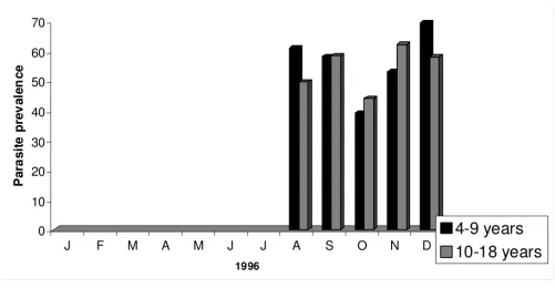
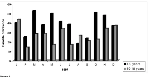
From August 1996 to December 1998, 4,826 children between four and 18 years were screened to determine parasite and gametocyte prevalence in Bancoumana. Fig-ure 2 shows the monthly variation in parasite prevalence according to volunteer's age. In 1996, mean parasite prev-alences were comparable between the two age groups (56.2% for the 4–9 year old versus 54.5% for the 10–18 year old; p = 0. 198). However, mean parasite prevalences were significantly higher in the younger age group for both 1997 (38.8%) and 1998 (27.1%) p = 0.004 for both years. Analogous results were obtained when we exam-ined gametocyte prevalence according to age (Figure 3). The mean gametocyte prevalence was 14.5% in 1996, 11.8% in 1997, and 9.8% in 1998. No statistically signif-icant difference was observed between these percentages. In contrast to mean parasite prevalence, mean gametocyte prevalence were significantly higher in the younger age group in all three years (p < 0.001).
Direct feeding experiment
Among the 4,826 children and adolescents screened, 485 (10.0%) were identified as gametocyte carriers. Of these, 372 (76.7%) were included in the direct feeding experi-ment to determine an age or season effect on the infectiv-ity of mosquitoes. While no significant differences were found in the mean percentages of mosquitoes that became engorged between the two age groups, no
signifi-Figure 2
0
10
20
30
40
50
60
70
P
a
ra
s
ite
p
rev
al
en
c
e
J
F
M
A
M
J
J
A
S
O
N
D
1996
[image:4.612.55.556.440.700.2]cant age effect on infectivity was find (Table 1). The mean percentage of engorged mosquitoes that became infected was significantly higher in the younger group than in the older group (38.3% versus 23.0%; p = 0.039).
When, infectivity according to season was compared, no statistically significant difference in the mean percentages of mosquitoes that became stuffed in the dry season ver-sus the rainy season was observed. However, a higher mean percentage of engorged mosquitoes did become infected during the rainy season than during the dry sea-son, but this difference also was not significant (Table 2).
Direct feeding versus membrane feeding experiment Of the 372 volunteers in the direct feeding experiment, 35 (9.4%) were included in the comparison of infectivity between direct feeding and membrane feeding. Results showed that mosquitoes using the direct feeding method were significantly more likely to become engorged than
were those using the membrane feeding method (mean percentages, 76.0% versus 53.5%). In addition, those that became engorged via direct feeding were significantly more likely to become infected than were those engorged via membrane feeding (mean percentages, 28.4% versus 15.0%). Of all mosquitoes in the experiment, those used in the direct feeding were almost three times more likely to become infected than were those used in the mem-brane feeding (Table 3). Applying the Student t test, there was no statistically significant difference between the per-centage of gametocyte carriers, who were infectious to mosquitoes using the direct feeding technique [(94.2%) and the membrane feeding technique (85.7%); p = 0.5].
Discussion
Because of study implementation difficulties, data from 1996 were not included in this analysis. In the first com-ponent of the study the direct feeding was used to evaluate the dynamics of transmission among children aged 4–18 Figure 3
0 10 20 30 40 50 60
P
a
ra
si
te
p
rev
ale
n
c
e
J F M A M J J A S O N D
1997
[image:5.612.57.561.82.345.2]4-9 years 10-18 years
Table 1: Age effect of gametocyte carriers on infectivity by direct feeding in Bancoumana, Mali
Age group (y) N Mean % engorged/all mosquitoes Mean % infected/engorged mosquitoes Mean % infected/all mosquitoes
4–9 236 55.8 38.3 19.7
10–18 87 55.4 23.0 12.1
p = 0.525 p = 0.039 p < 0.001
years in rural Mali. Results showed that among 372 game-tocyte carriers analyzed, those aged 4–9 years were highly infectious to mosquitoes when compared with those aged 10–18 years, particularly during the rainy season.
In the 1950s, Muirhead-Thompson [11,12] was the first to publish an attempt to determine the proportion of indi-viduals in a malaria-endemic area, who are infectious to mosquitoes. He found that 10–11% of all individuals in West Africa were infectious to mosquitoes by direct feed-ing. Because the infectivity rates in children were signifi-cantly higher than those in adults (34% of children aged 2–4 years and 37% of children aged 5–9 years infected mosquitoes in that study), he proposed that children rep-resented the major infectious reservoir of malaria para-sites. Also, Bousema et al [13] found that the estimated mean duration of gametocytaemia was 9.4 days (range 2.5 – 23.5) for children below five years of age, 7.8 days (range 2.5 – 23.5) for children aged five to nine years and 4.1 days (range 2.5 – 16.5) for children aged ten years and above. A subsequent study by Githeko et al [2], in a holoendemic area of western Kenya, confirmed and extended the previous results. In that study, 72% of the infectious reservoir was in children younger than 10 years. Moreover, Boudin et al [14] found that neither the propor-tions of reducers nor the medians of transmission block-ing immunity (TBI), intensity, or TBI/TI (transmission inhibition) were significantly different between age groups. And thus, age was not predictive for any of the TBI indicators.
The second component of the study, which included 35 gametocyte carriers, compared infectivity rates via direct versus membrane feeding of mosquitoes. A statistically significant difference was observed between the mean per-centage of mosquitoes that fed by direct feeding (76.0%)
and the mean percentage of mosquitoes that fed by mem-brane feeding (53.5%), suggesting that mosquitoes pre-ferred to use direct feeding. These results are comparable to those found by Graves et al [5,6] in 1988. That research also found that for both techniques, 82.82% of all carriers were infectious to mosquitoes. Again, this is similar to our findings that 94.2% and 85.7% of children were infec-tious by direct and membrane feeding, respectively (with no significant difference between the two values).
However, a statistically significant difference was found between the mean of percentages of engorged mosquitoes infected by direct feeding (28.4%) versus membrane feed-ing (15.0%). In Cameroon, Mulder et al [1] observed that a mean of 18.9% of engorged mosquitoes were infected by membrane feeding. Overall, this study shows that, membrane feeding is about half as effective as direct feed-ing for infectfeed-ing mosquitoes. And about three times more mosquitoes in the direct feeding group than in the mem-brane feeding group actually became infected during feed-ing; this finding can be explained, at least in part, by the fact that mosquitoes preferred direct feeding.
Conclusion
[image:6.612.52.561.100.185.2]Based on these results, the conclusion is that the mem-brane feeding method could be used as an alternative to the direct feeding method in biological studies or field tri-als of transmission-blocking activity among candidate malaria vaccines. Although the intensity of infection is considerably higher when the direct feeding method is used, the difference in infectivity between the two meth-ods could be compensated for by including more mosqui-toes in trials using membrane feeding. Using this study as an example, if three-times more mosquitoes had been exposed to membrane feeding, it is expected that the number of mosquitoes infected would have been similar Table 2: Seasonal effect on infectivity of mosquitoes by direct feeding in Bancoumana, Mali (1997–1998)
Season N Mean % engorged/all mosquitoes Mean % infected/engorged mosquitoes Mean % infected/all mosquitoes
Dry Season 159 56.1 29.0 16.7
Rainy Season 164 55.3 39.2 18.6
P = 0.738 p = 0.120 p = 0.402
Total 322 55.7 34.2 17.7
Table 3: Comparison of infectivity between direct and membrane feeding in Bancoumana, Mali
Method Mean % engorged/all mosquitoes Mean % infected/engorged mosquitoes Mean % infected/all mosquitoes
Direct feeding 76.0 28.4 20.6
P < 0.001 P < 0. 001 P < 0.001
[image:6.612.54.552.656.730.2]for both methods. The lower mean percentage of mosqui-toes infected by membrane feeding may result from dam-age during blood drawing and handling, or from loss of infectivity while the parasites are in the membrane feed-ers. Nevertheless, the membrane feeding technique is eas-ily applicable, and its use would resolve the ethical concerns surrounding the direct feeding technique, espe-cially among children.
Competing interests
The authors declare that they have no competing interests.
Authors' contributions
MD conducted parsitotolgic aspects of the study in the field and in laboratory, and initiated the draft zero; AMT conducted entomologic aspects of the study in the field and in the laboratory, and contributed to writing all of the drafts; SFT supervised mosquitoes feeding in the tory; ON supervised mosquitoes breeding in the labora-tory (insectary) and dissection; LK conducted the blood sample collection from volunteers; AK conducted the blood sample collection from volunteers; MC contributed to mosquitoes dissection; MB contributed to mosquitoes dissection, entomology studies, and direct feeding; OkD, SK, JCB, and YTT reviewed the final version
Financial support
This investigation received financial support from Tropi-cal Medicine and Research Center (TMRC) project of Mali-Tulane, TMRC AI 95002.
Acknowledgements
We are grateful to the villagers of Bancoumana, Mali, who kindly partici-pated as volunteers in these studies. We would like to thank Elizabeth Higgs and her team for their facilitating role in writing this paper. We would also like to thank Richard Sakai (Mali) and Louis Miller of LPD/NIH (USA) for their scientific and technical support.
References
1. Mulder B, Tchuinkam T, Dechering K, Verhave JP, Carnevale P, Meu-wissen JH, Robert V: Malaria transmission-blocking activity in experimental infectious of Anopheles gambiae from naturally infected Plasmodium falciparum gametocyte carriers. Trans R Soc Trop Med Hyg 1992, 88:121-125.
2. Githeko AK, Brandling-Bennett AD, Beier M, Atieli F, Owaga M, Col-lins FH: The reservoir of Plasmodium falciparum malaria in a holoendemic area of western Kenya. Trans R Soc Trop Med Hyg
1992, 86:355-358.
3. Beier JC: Malaria parasite development in mosquitoes. Ann Rev Entomol 1998, 43:519-543.
4. Gouagna LC, Mulder B, Noubissi E, Tchuinkam T, Verhave JP, Boudin C: The early sporogonic cycle of Plasmodium falciparum in laboratory infected Anopheles gambiae: an estimation of par-asite efficacy. Trop Med Int Health 1998, 3:21-28.
5. Graves PM, Burkot TR, Carter R, Cattani JA, Lagog M, Parker J, Brabin BJ, Gibson FD, Bradley DJ, Alpers MP: Measurement of malarial infectivity of human populations to mosquitoes in the Madang area, Papua New Guinea. Parasitology 1988, 96:251-263. 6. Graves PM, Carter R, Burkot TR, Quakyi IA, Kumar NK: Antibodies to Plasmodium falciparum gamete surface antigens in Papua New Guinea sera. Parasite Immunol 1988, 10:208-215.
7. Roeffen W, Mulder B, Teelen K, Bolmer M, Eling W, Targett GA, Beckers PJ, Sauerwein R: Association between anti-Pfs48/45 reactivity and P. falciparum transmission-blocking activity in Cameroon. Parasite Immunol 1996, 18:103-109.
8. Lensen A, Mulder L, Tchuinkam T, Willemsen L, Eling W, Sauerwein R: Mechanisms that reduce transmission of Plasmodium falci-parum malaria in semiimmune and nonimmune individuals.
J Infect Dis 1998, 177:1358-1363.
9. Rieckman KH, Beaudoin RL, Cassells JS, Sell KW: Use of attenu-ated sporozoites in the immunization of human volunteers against falciparum malaria. Bull World Health Organ 1979, 57:261-266.
10. Touré YT: Bioécologie des anophèles dans une zone rurale de savanne soudanienne au Mali (village de Banambani). Incidence sur la transmission du paludisme et la filariose de Bancroft Thèse de 3ème cycle, Entomol-ogie mèdicale, Bamako; 1979.
11. Muirhead-Thomsen RC: Factors determining the true reservoir of infection of Plasmodium falciparum and Wucherreria ban-crofti in a West African village. Trans R Soc Trop Med Hyg 1954, 48:208-225.
12. Muirhead-Thomsen RC: The malarial infectivity of an African village population to mosquitoes (Anopheles gambiae): a ran-dom xenodiagnostic survey. Am J Trop Med Hyg 1957, 6:971-979. 13. Bousema T, Gouagna J, Bakeley CJ, Meutstege AM, Okech BA, Akim INJ, Beier JC, Githure JI, Sauerwein RW: Plasmodium falciparum
gametocyte carriage in asymptomatic children in western Kenya. Malar J 2004, 3:18.
14. Boudin C, Kolk M Van Der, Tchuinkam T, Gouagna C, Bonne S, Safeukui I, Mulder B, Meunier JY, Verhave JP: Plasmodium falci-parum transmission blocking immunity under conditions of low and high endemicity in Cameroon. Parasite Immunol 2004, 26:105-110.
15. Mendis C, Gamage-Mendis AC, De Zoysa AP, Abhayawardena TA, Carter R, Mendis KN: Characteristics of malaria transmission in Kataragama, Sri Lanka: a focus for immuno-epidemiolog-ical studies. Am J Trop Med Hyg 1990, 42:298-308.
16. Carter R, Kumar N, Quakyi I, Good M, Mendis K, Graves P, Miller L: Immunity to sexual stages of malaria parasites. Prog Allergy
1988, 41:193-214.
17. Lines JD, Wilkes TJ, Lyimo EO: Human malaria infectiousness measured by age-specific sporozoite rates in Anopheles gam-biae in Tanzania. Parasitology 1991, 102:167-177.
18. Kaslow DC, Isaacs SN, Quakyi IA, Gwadz RW, Moss B, Keister DB: Induction of Plasmodium falciparum transmission-blocking antibodies by recombinant vaccinia virus. Science 1991, 252:1310-1312.
19. Kaslow DC: Transmission-blocking vaccines: uses and current status of development. Int J Parasitol 1997, 27:183-189. 20. Ponnudurai T, Lensen AH, Van Gemeri GJ, Bensink MP, Bolmer M,
Meuwissen JH: Infectivity of cultured Plasmodium falciparum
gametocytes to mosquitoes. Parasitology 1989, 98:165-173. 21. Rieckman KH, Carson PE, Beaudoin RL, Cassells JS, Sell KW:
Sporo-zoites induced immunity in man against an Ethiopian strain of P. falciparum. Trans R Soc Trop Med Hyg 1974, 68:258-259. 22. Paul RE, Bonnet S, Boudin C, Tchuinkam T, Robert V:
Age-struc-ture gametocyte allocation links immunity to epidemiology in mlaria parasites. Malar J 2007, 6:123.