Int. J. Electrochem. Sci., 10 (2015) 8607 - 8629
International Journal of
ELECTROCHEMICAL
SCIENCE
www.electrochemsci.org
Review
An Overview of Fabricating Nanostructured Electrode
Materials for Biosensor Applications
Rasu Ramachandran1, Shen-Ming Chen2,*, George peter Gnana kumar3, Pandi Gajendran1, Natrajan Biruntha Devi4,
1
The Madura College, Department of Chemistry, Vidya Nagar, Madurai – 625 011, Tamil Nadu, India.
2
Electroanalysis and Bioelectrochemistry Lab, Department of Chemical Engineering and
Biotechnology, National Taipei University of Technology, No.1, Section 3, Chung-Hsiao East Road, Taipei 106.Taiwan (ROC).
3
Department of Physical Chemistry, School of Chemistry, Madurai Kamaraj University, Madurai-625 021,Tamil Nadu, India.
4
Department of Chemsitry, S.Vellaichamy Nadar College, Nagamalai pudukkottai, Madurai – 625021, Tamil Nadu, India.
*
E-mail: [email protected]
Received: 17 May 2015 / Accepted: 13 August 2015 / Published: 26 August 2015
Over past few decades different types of nanostructured electrode materials have been reported for the electrochemical analysis and biosensor applications. This article will give overview on the electrode materials based on carbon fiber, carbon nanotube (Single-walled carbon nanotubes and multi-walled carbon nanotubes), graphene oxide, metal oxide, conducting polymer and nanocomposites. The different electrochemical methods such as cyclic voltammetry, square wave voltammetry, differential pulse voltammetry, amperometry, electrochemical impedance spectroscopy and analytical methods such as UV-visible spectroscopy have also been overviewed. The biosensing applications of these nanostructured electrodes to the determination of biologically important analytes such as glucose, cholesterol, dopamine, uric acid, ascorbic acid, chitosan, cytochrome c, paraxon, acetylcholinesterase and hypoxanthine have also been reviewed. There are good prospects to fabricate low cost electrode materials, new conception and attractive aspect for the development of future biosensor applications.
Keywords: Nanostructured materials, Electroanalytical methods, Electrochemistry, Biosensor, Enzyme electrodes.
1. INTRODUCTION
application of different modified electrode materials used in biosensors [2-9] and energy storage [10, 11] device are available in reported literature. Nanocomposite modified electrodes display fascinating electrochemical properties in pesticide sensor [12] and non-enzymatic sensor [13] applications. The discovery of two-dimensional graphene based electrode material has been used for direct electron transfer from cyctochrome C and reduction of nitric oxide [14]. The highly ordered mesoporous carbon-fullerene based modified electrode facilitated direct quantifications of β-nicotinamide adinine dinucleotide (NADH), ascorbic acid (AA), uric acid (UA), dopamine (DA) and epinphrine (EP) [15]. A novel polyvinylpyrrolidone protected graphene/polyethylenimine-functionalized/ionic liquid/glucose oxidase electrode has shown excellent biocompatibility with glucose biosensors [16]. Much electrochemical biosensor detection has been demonstrated on graphene based modified electrodes, i.e., graphene-chistosan/haemoglobin/graphene/ionic liquid [17]. Among the graphene based materials, chemically reduced graphene oxide modified glassy carbon (CR-GO/GC) electrode has significantly improved catalytic performance in biosensors [18]. The immobilized acetylcholinesterase based caroboxyphenylboronic/reduced graphene oxide-gold nanocomposite enhanced the electrochemical sensitivity of biomolecular reaction and presents rapid response with low detection limit [19]. Graphene oxide and sheet-mediated silver composite exhibit excellent electrochemical biosensing ability and sensitively detects the pathogenic bacteria, protein and DNA [20]. Water-soluble polyhydroxylated fullerene derivatives have been extensively exploited for haemoglobin biosensor and to improve the anti-oxidant properties [21]. The most described immobilized acetylcholinesterase on a novel TiO2-decorated graphene nanohybrid can be monitored by measuring a good performance of
organophosphate pesticide sensor analysis [22]. On the other hand, a disposable biosensor based on tyrosinase-protected gold nanoparticle (Tyr-Au) was immobilized with 1-pyrenebutanoic acid, succinimidyl ester and graphene oxide (PASE-GO) forming a biocompitable nanocomposite and it was coated with screen-printed electrode (SPE) for the determination of phenolic compounds [23]. Electrochemical DNA sensor has been evaluated by carboxyl functionalized graphene oxide and it was electropolymerized with poly-L-lysine on glassy carbon electrode. The polymerized composite (GO-COOH/PLLY/GCE) immobilized with ssDNA for the detection of vibrio parahemolyticus (tlh gene) [24]. The human live cell secretion of H2O2 has been detected by ultrasonic-electrodeposited of triple
component designed with Pt-MnO2/graphene nanohybrid electrode [25]. Graphene oxide and nafion
based electrode with incorporated with myoglobin (Mb-GO-nafion) have also been exploited as electrode in biosensors. In this Mb film electrode exhibited good electrocatalytic activities towards H2O2, nitrite and oxygen [26].
2. ELECTRODE MATERIALS FOR BIOSENSORS
2.1. Carbon fiber
Carbon fiber electrode materials are readily available and it act multi-properties like high electrochemical stability, low electrical resistivity and high mechanical strength. The carbon fibre electrode modified with poly β-aminoanthraquinone (pAAQ) for the study of amperometric micro sensor of haemoglobin (HB) [27]. Voltammetric technique can be used for the optimization of electrochemical stability of modified electrode. Biosensing molecule of HB, the amperometric response range of 0.5 M to 340 M and the average current response value of about 23.2 nA. Carbon fiber electrode can be modified with co-deposition of ruthenium (Ru) and rhodium (Rh) nanoparticles. The modified electrode has been immobilized with glutamate dehydrogenase (GLUD) and it can be analyzed for the evaluation of α-ketoglutarate (α-KG) [28]. The amperometric response current range between 100 to 600 M and the lowest limit of detection (LOD) value is 20 M. The unmodified electrode of tyrosinase based carbon fiber paper suggests the monitoring of phenolic compounds like catechol, phenol, bisphenol and 3-aminophenol. The LOD value of catechol (2 nM), phenol (5 nM), bisphenol (5 nM) and 3-aminophenol (12 nM) [29]. A stable carbon fibre electrode modified during the co-immobilisation of acetylcholinesterase (AChE) and chonline oxidase (ChOx) in bovin serum albumin (BSA) membrane for the development of acetylcholine and choline. A convensional three electrode optimized potential value of 800 – 1100 mV vs Ag/AgCl, exhibited LOD value of 1 M for acetylcholine and choline. The biosensor exhibited good sensitivity and selectivity for acetylcholine and choline [30]. Deng et al [31] used a silk derived carbon fibre modified with Au@Pt urchilike nanoparticles (Au@Pt NPs) for the development of Escherichia coli (E-Coli)-based electrochemical sensor. The reported LOD value of 0.09 mg L-1 and it possesses high conductivity, high electrocatalytic activity and biocompatibility.
2.2. Carbon nanotube
[34]. In this sensor study, a linear relationship between biosensor and D-alanine concentration range from 0.001 to 0.7 mM, the LOD of 0.2 mM and the sensitivity of 54.85 mA cm-2 mM-1. Apetric et al [35] have used carboxyl functionalized single-walled carbon nanotube electrode immobilized with Tyrosinase to employ an amperometric biosensing analysis of tyramine. This modified CNT electrode is suitable for environment for the study of bioactivity of tyrosinase. This bioactive reaction has a good linearity with the concentration of tyramine in the range from 5 – 180 M and exhibited limit of detection of 0.62 M.
2.3. Fullerene
A fullerene-C60/glassy carbon electrode (FLR/GCE) have used for the interactions study of
carbidopa (CD) with double standard calf thymus DNA (dsDNA) by using phosphate buffer solution (pH = 4.0) [36]. Cyclic voltammetry (CV), linear sweep voltammetry (LSV) and square wave voltammetry (SWV) techniques have used in the interaction study of CD with dsDNA. Similarly, UV-visible and fluerescence spectroscopy have monitored the interaction between CD and dsDNA. Apply SWV in a linear response of dsDNA concentration range between 0.1 to 25 nM and the reported LOD value of 0.03 nM, however the fabricated electrode expressed good repeatability, reproducibility and long term stability. A tentative application of fullerene and gold nanoparticle have examined for the development of nanostructured enzyme based biosensor and evaluation of polyphenol [37]. Surface Plasmon resonance spectroscopy (SPR), CV and chronoamperometry have been used for the characterization of modified electrode surface area and their biocatalytic activity. The biosensor showed fast amperometric response to gallic acid and a standard for poly phenol analysis of wine, the amperometry linear range from 0.03 – 0.30 m mol L-1 and the reported LOD value of 1.1 mg L-1. Sheng et al [38] used a haemoglobin (Hb) immobilized fullerent-nitrogen doped carbon nanotube and chitosan (C60-NCNT/CHIST) composite for sensing H2O2. The composite showed a well defined redox
peak values of E0 = -335 mV vs SCE and assigned the redox reaction of Hb (FeIII/FeII). The
immobilized Hb have been optimized the fast electron transfer rate order processes of ks = 1.8 s-1, the LOD value of 1 and the sensitivity of 438 mA mM-1. An air-plasma activated fullerene impregnated to screen printed electrode was immobilized with DNA for the detection of Escherichia coli 16S rDNA [39]. The fullerene modified lipid bilayer membrane (s-BLMS) electrode have been used an electrochemical sensor for the detection of neutral odorant molecules [40], by using voltmmetry analysis, the two systems (I2/I- and ferrocene/ferrocinium) were studied with C60 lipid
bilayer modified electrode. The results are clearly indicated that, C60 I2/I- exhibit more sensitivity than
C60 ferrocene/ferrocinium.
2.4. Graphene oxide
the real sample of glucose obtained from human blood serum. Electrodeposition of hybrid film composed with laccase, tyrosinase, gold nanoparticles and chitosan on graphene doped carbon paste (LACC-TYR-AuNPs-CS/GPE) electrode used for the development of carbamate pesticide sensor collected from citrus fruits [42]. In this (LACC-TYR-AuNPs-CS/GPE) composite have used for the evaluation of Michaelis-Menten kinetic constant (km) value (26.9 ± 0.5 M). A titania nanosheet modified reduced graphene oxide (TiO2NS-rGO) composite was immobilized with haemoglobin and it
can be used for electrochemical analysis of H2O2 and nitrite [43]. The morphological and structural
analysis has been characterized by SEM, XRD and Raman spectroscopy. The immobilized TiO2
NS-rGO electrode displayed good electrochemical biosensor performance for the detection of H2O2 and
nitrite, i.e. a wide linear range of 0.1 – 145 M for H2O2 and 1 – 15,000 M for nitrite, the LOD of 10
nM for H2O2 and 0.21 for nitrite. Peng et al [44] have used gold nanoparticle/toludine
blue-graphene oxide (AuNPs/TB-GO) composite, a facile and sensitive labile-free electrochemical DNA biosensor of multidrug resistance (MDR) gene. The sensor part has been analyzed by two different electrochemical techniques, such as electrochemical impedance spectroscopy (EIS) and differential pulse voltammetry (DPV), by employing the DPV hybridization of DNA measured from the peak current of TB. The currents were proportional to the logarithmic of the concentration of DNA, the optimized range from 1.0 x 10⁻¹¹ - 1.0 x 10⁻⁹ M and the LOD of 2.95 x 10⁻¹² M. A new type of electrochemical biosensor has been analyzed by used human serum albumin/graphene oxide/3-amino propyl triethoxy silane modified with indium tin oxide (ITO/APTES/GO/HSA) electrode used for the determination of enentiomers D and L – tryptophan (Trp) [45]. The composite electrodes were investigated by CV method, the exhibited oxidation peak potential of D and L trp occur at 860 and 1260 mV vs SCE respectively.
2.5. Metal oxides
Recently, metal oxides can offer promising electro active materials due to enhancement of the electrochemical reversible redox reaction, wide potential window and large surface-volume ratio. A nanostructure of metal oxide (CeO2)-chitosan (CH) made electrode used as biosensor for sensing
cholesterol molecule [46]. The fabricated ITO-CeO2-CH nanocomposite immobilized with cholesterol
oxidase (ChOx). A novel core-shell zinc oxide based (ZnO/chistosan-graft-poly(vinyl alcohol)) (ZnO/CHIT-g-PVAL) nanocomposite led to the sensing of glucose, the composite reported LOD value of 0.2 M [47]. Nanoparticle of titanium oxide modified with reduced graphene oxide (TiO2
/NPs-rGO) composite has been immobilized to haemoglobin (Hb) for a mediator-free biosensor of hydrogen peroxide (H2O2), the LOD value of 10 nM [48]. Glucose biosensor for millimolar level determination
of glucose generated with a glucose oxidase immobilized with cellulose tin-oxide (SnO2) hybrid
nanocomposite electrode [49]. A three layer of magnetic core-shell Au-Fe3O4@SiO2 metal oxide based
electrode (AChE/Fe3O4-CH/GCE) electrode can served as an excellent biosensor detection value of 3.6
x 10-9 M.
2.6. Conducting polymers
Conducing polymers (polythiophene, polyaniline and polypyrrole) have considerable application in optical, electrical and electrochemical properties. A number of authors have reviewed for the preparation, properties and applications of conducing polymers [52-54]. Electrodeposition of conducting polymers of poly(4,7-di(2,3)-dihydrothenol) [3,4-b] [1,4] dioxin-5-yl-benzo[1,2,5] thiazole (PBDT) and poly(4,7-di(2,3)dihydrothienol[3,4-b] [1,4] dioxin-5-yl-2,1,3-benzoselenadiazole (PESeE) on graphite electrode, the modified electrodes were immobilized with glucose oxidase (GO) and the biosensor’s LOD value of 0.05 mM for PBDT and 0.01 mM for PESeE [55]. Gokoglan et al [56] have electrochemically deposited a conducing polymer of poly(4,7-bis(thieno[3,2-b] thiophene-2-yl) benzo[c] [1,2,5] selenadiazole) poly(BSeTT) on gold electrode. The immobilized conducting polymers which can be apply for the biosensor detection of pyranose, the LOD value is 0.229 mM. There has been explosive growth of conducting polymer poly(4-(2,5-di(thiophene-2-yl)-1H-pyrrole-1-yl)-benzenamine) (poly(SNS-NH2)) on graphite electrode for acetylcholine biosensor. The electrode leads
to the LOD value of 0.11 mM [57]. The attractiveness of conducting polymers of 2-Heptyl-4,7-di(thiopehene-2-yl)-1H-benzo[d] imidazole) (BImTh) were electrocopolymerised on graphite electrode for cholesterol biosensor development[58]. One of the most interesting developments of conducting polymer (Poly(4-(2,5-di(thiophene-2-yl)-1H-pyrrole-1-yl)benzamine) (Poly(SNS-NH2)) electrode
modified with multi-walled carbon nanotube could be used for the biosensor for the detection of acetylcholinesterase [59].
2.7. Nanocomposite
composite has been used as an excellent electrocatalytic studies of H2O2 [64]. The exhaustive
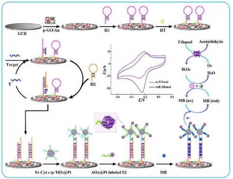
explanation, electrode fabrication and the LOD of electrochemical biosensors illustrated in Fig.1.
Figure 1. Schematic representation of the proposed strategy for miRNA-155 detection. ("Reprinted with permission from (ACS Appl. Mater. Interfaces 7 (2015) 713-720). Copyright (2015) American Chemical Society”).
A highly sensitive detection of an DPV electrochemical biosensor of microRNA (miRNA) using cyctochrome C (Cyt C) and alcohol oxidase (AOx) on Pt based metal oxide based nanocomposite (Cyt C-p-TiO2@PtNPs) electrode [65]
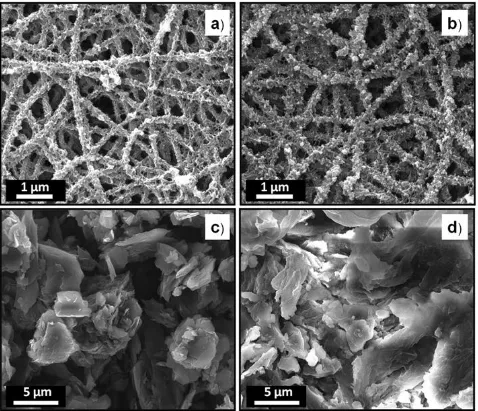
3. ROLE OF MORPHOLOGY
[image:7.596.63.541.139.506.2][image:8.596.59.537.264.675.2]
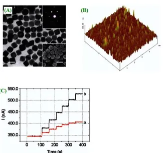
RF magnetron sputtering. The surface morphology and roughness electrode surface area of ZnO modified electrode exhibits cavities of nano porous film as an effective biosensing area of enzyme by used field emission-scanning electron microscope (FE-SEM) analysis [66]. The environmental scanning electron microscope (ESEM) was used for the analysis of a smooth nature-like structure of carbon nanotubes without aggregation observed on CNTs-chitosan (CNTs-CS) film surface. In this type of smooth like composite provides a biosensing matrix of enzyme based studies due to good biocompatibility, good conductivity and long durability [67]. Ge et al [68] have used a linker-free connected graphene oxide/Au nanocluster (GO-Au NCs) composite for biosensor of L-cystein analysis. In this composite, a single layer of GOs was obtained smooth surface and Au NCs showed a uniform surface tophography, the measured diameter of about 6 nm.
Figure.2.shows a layer-by-layer graphene oxide (GOx) has been synthesized by electro spinning method and it was mixed with MWCNT and Nylon 6,6. In this carbon nanotube composite (Nylon 6,6/MWCNT/PBIBA/GOx), which was uniformly wrapped with the nanofiber [69].
4. EFFECT OF pH
All electrocatalytic reactions have been evaluated by acidic to basic (4 to 14) medium, because optimization pH is one of the most important parameter for the study of bio sensing molecules. Electrochemical deposition of gold based laccase electrode used for sensing of enzymatic biosensor analysis. This enzymatic catalyzed reactions are mainly depends on pH and also maintained the maximum peak currents are clearly occur at pH = 5 [70]. Tsopela et al [71] studied two different (tungsten/tungsten oxide and platinum/iridium oxide) electrode used for the reduction of O2 and H2O2.
The detection of O2 and H2O2, Pt/IrO2 exhibited more sensitive pH measurement than W/WO2. This
type of electrodes were almost demonstrated a linear response wide range of 2.0–12.0. Ascorbic acid is an essential nutrient component for human diet and it can be obtained from fruits. The ascorbic acid biosensors are mainly studied in acidic medium, the optimized pH range between 5.5 and 7.5. The study of as-fabricated poly(3,4-ethylenedioxythiophene)-laurolsarcosinate film biosensor electrode between 5.5 and 8.0 [72]. Zou et al [73] have evolved a haemin functionalized graphene oxide electrode for simultaneous determination of ascorbic acid, dopamine and uric acid. The exhibited peak currents of AA, DA and UA at the modified electrode were affected by the pH values of Britton-Robinson buffer solution. The optimized pH oxidation peak currents values of AA, DA and UA were obtained in the range of 3.0 to 8.0. The maximum oxidation peak current values were exhibited at pH = 6. Carbon and silica based electrode modified with niobium oxide, alumina and DNA, exhibited, a potential application in electrochemical biosensor for amitriptyline. The optimized UV-visible results showed a maximum peak current value obtained at pH = 7.5 [74]. The electrochemical response of H2O2 at poly(N-isopropylacrylamide)-g-poly(N-isopropylacrylamide-co-styrene)
(PNIPAM-g-P(NIPAM-co-St)) with MWCNT (Hb/PNNS/MWCNTs/GCE) composite was examined in 0.1M phosphate buffer solution of pH range from 5.0 to 9.0. [75].
5. ELECTROANALYTICAL TECHNIQUES
5.1. Cyclic voltammetry
[image:10.596.194.418.309.629.2]
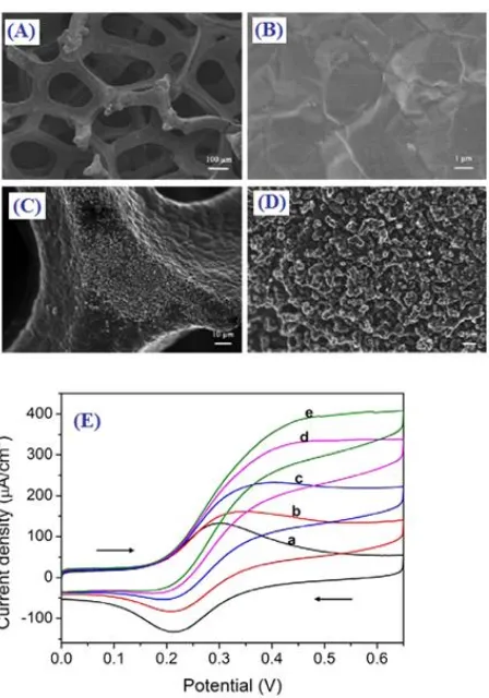
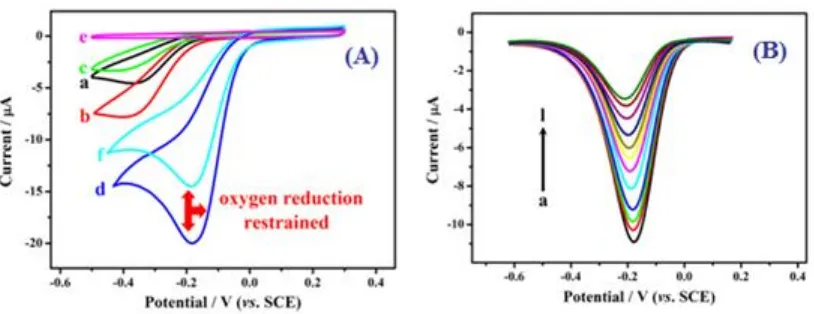
enzyme catalyzed anodic oxidation reaction of thiocholine [77]. Reddy et al [78] showed the possibility of electrochemical characterization of p-Nitrophenol by lipase enzyme inhibition method. From this voltmmetry analysis, the anodic peak current increased with an increase of scan rate. The plot of peak current against square root of scan rates, a linear coefficient value of 0.9997. Arya et al [79] made a significant contribution in that a polyaniline protected electrode fabricated on gold electrode (PPAuNp/Au) and the modified electrode immobilized with cortisol specific monoclonal antibody (C-Mab). Voltommogaram studies have confirmed the PPAuNp/Au composite electrode exhibited repeatable redox behaviour and the LOD of cortisol in the range of 1 pM – 100 nM. In Fig.3.shows the SEM morphological (Skeleton) structure (A,B,C and D) and electrochemical analysis of glucose with chitosan (CS) modified with ferrocene (Fc) and 3D graphene foam (Fc-CS/SWCNT/GOD/3DG) electrode (Fig.3A). Fig.3(B) shows the various concentrations ((a) 0, (b) 10, (c) 15, (d) 20 and (e) 25 nM) of glucose in PBS (0.1 M, pH = 7). The electrode catalyst was displayed the LOD glucose of 1.2 M [80].
5.2. Square wave voltammetry
[image:11.596.72.535.226.433.2]Square wave voltammetry is one of the sensitive techniques than CV i.e the measurement of accuracy and the limit of detection value up to nano molar (nM) to pico molar (pM). Yadav et al [81] reported the detection of chloramphenicol (CAP) using a immobilized poly(4-amino-3-hydroxynapthalene sulfonic acid) (p-AHNSA) modified with pyrolytic graphite electrode. The square wave voltammetry method was applied for the determination of CAP with a detection limit of 0.02 nM.
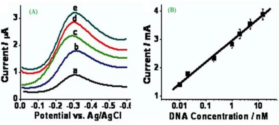
Figure 4. The target DNA concentration shown in the (A) was 0 (a), 0.04 nM (b), 0.2 nM (c), 1 nM (d), and 2 nM (e), separately. (B) Variance of the redox current with the target DNA concentration measured by the E-DNA biosensor with an adjunct probe. ("Reprinted with permission from (Anal. Chem. 82 (2010) 9500-9505). Copyright (2010) American Chemical Society”).
Figure.4 (A) and (B) illustrated the electrochemical biosensor detection of nucleic acid response exhibited different concentrations using square wave voltammety at adjunct probe E-DNA. The E-DNA based electrode provide with a limit of detection of 2.0 pM [82]. The use of square wave voltammetry biosensor, the double stranded calf thymus DNA (dsCT-DNA) entrapped polyaniline-polyvinyl sulfonate/indium-tin oxide (PANI-PVS/ITO) composite for chlorpyrifos and malathion [83]. Yola et al [84] developed a high sensitive method for the detection (2.0 x 10⁻¹⁵ M) of DNA (5’-TA CCG CGT GCT CGA GCT-(CH2)3-SH-3’ single-stranded probe) hybridization on modified with
5.3. Differential pulse voltammetry (DPV)
Several methods have been extensively applied for the development of biosensor applications, like CV, amperometry and square wave voltammetry. Among these methods, DPV exhibit an excellent compatibility, high sensitivity and easily handled. Noaradrenalin (NA) and acetaminophen (AC) are electroactive compound, they can be detected by hematoxylin modified glassy carbon electrode. The electrocatalytic oxidation of NA and AC simultaneous measurement by using DPV [86]. The recent surge of flower-like morphology of zinc oxide (ZnO) nanostructure has been synthesized by hydrothermal method. The DNA immobilizations are mainly focused on interaction of physically immobilized single stranded thiolate DNA (SS th-DNA) and the nanostructure of ZnO. The assembled immobilized electrode can quantify the target molecule of ss th-DNA [87]. Hu et al [88] employed a simple and irreversible electrochemical biosensor of glucose by Cv and DPV. Fig.5.shows that the glucose oxidation concentrations increases with increasing of peak current. The development of biosensors glucose oxidation LOD value of 0.015 mM.
Figure 5. (A) Cyclic voltammograms in air-saturated 0.1 M, pH 7.4 PBS at (a) an electrochemically pre-treated bare Au electrode, (b) after formation of Pt@BSA layer, (c) after cross-linking reaction with GA, after covalent immobilization of GOD in the (d) presence and (e) absence of dissolved oxygen, and (f) with the addition of 6.55 mM glucose to air-saturated buffer. (Scan rate: 50 mV s−1). (B) Differential pulse voltammograms of GOD/GA/Pt@BSA-modified Au electrode in air-saturated 0.1 M, pH 7.4 PBS buffer (under the optimized conditions) with the glucose concentration increased from a to l (0.05, 0.55, 1.55, 3.55, 5.05, 6.05, 6.55, 7.55, 9.55, 11.05, 12.05 mM glucose injection, respectively). ("Reprinted with permission from (ACS Appl. Mater. Interfaces 6 (2014) 4170-4178). Copyright (2014) American Chemical Society”).
The electrochemical biosensor of vitamin B1 on pre-treated multi-walled carbon nanotube paste
[image:12.596.98.505.328.485.2]
on electrochemical entrapped dsDNA immobilization on screen-printed electrode has been used for detection of quanine [91].
5.4. Amperometry technique (i vs t curve)
Amperometry technique has a powerful tool for the determination of biosensor analysis up to low level and it has highly sensitivity, selectivity and simplicity. In a typical amperometric experiment mainly explained by cottrel equation (Eq. 1)
t π D nFAC
i(t) (Eq.1) The main parameter of i-is current and t-is time. A novel multi-walled carbon nanotube modified with redox dye nile blue (NB) and co-immobilized with horseradish peroxide (MWCNT/NB/NAF/HRP) composite served as an excellent hot matrices for a second generation of amperometric biosensor.
[image:13.596.148.473.366.672.2]
The impregnated composite were used as an electrode material in electrocatalytic activity for the reduction of hydrogen peroxide [92]. Similarly, the other interesting co-immobilization horseradish and glucose oxidase on silver nano cube with chitosan (Gox-CS-HRP/AgNCs-CS) nanocomposite film electrode has been also reported for the oxidation of glucose biosensor applications [93]. Carbonic acid immobilization of a core-shell ceria oxidase-polyaniline (GCE/CeO2-PANI/CA) nanocomposite have
been shown to exhibit better electrocatalytic activity of carbonic acid in human blood [94 ]. A third generation of biosensor detection of uric acid by using ferrocene (Fc) induced electro active uricase (UOx) deposited with nafion on glassy carbon electrode (Naf/UOx/Fc/GCE) composite has been also reported [95].
The biosensors for the amperometric detection of glucose response range from 0.125 to 12 mM (Fig.6) and the sensitivity of 0.07 A mM-1. Moreover, this type of glucose oxidation has been developed on the parallelogram shape (SEM) and layered structure (AFM) on gold based (Au nanooctrahedra/GOx) composite [96]. Rahman et al [97] have studied poly-5,2’:5,2’-tetrathiophene-3’-carboxylic acid (poly-TTCA) layer electrode immobilized with hydrazine and horseradish peroxide (HRP) for the development of amperometric biosensor.
5.5. Electrochemical impedance spectroscopy (EIS)
This technique has been widely used in many electrochemical studies (To monitor the electrode surface properties) and it may apply to investigate the change in charge transfer resistance (RCT) value.
Such as electrochemical sensors, corrosion, super capacitors and batteries.
The sulfite oxidase (SOx) immobilized with nano composite of Prussian blue nanoparticle/polypyrrole film electrodeposited on gold electrode (SOx/PBPNPs/PPY/Au). The composite electrode has been investigated the RCT value by using EIS analysis. The reported RCT value
of 650 Ω for PBNPs/PPY/Au composite and 1300 Ω for SOxX/PBNPs/PPY/Au electrode. In this results are clearly explained the binding of SOx onto the PBNPs/PPY/Au electrode exhibited poor electrical conduction at low frequency (˂10 kHz) value [98]. The EIS of deoxy ribonucleic acid (DNA) biosensor based multi-walled carbon nanotube deposited on Ag-TiO2 composite for labile-free
phosphinothricin acetyltransferase gene [99]. The nano molar detection of aptamar based thrombin by pyrolyzed carbon film electrode, the electrochemical biosensor exhibited high sensitivity and selectivity of thrombin [100]. The glucose oxidase biosensor has been detected, the limit of detection value of 15.6 M and the sensitivity of 9.66 x 10⁻⁷ Ω-1 mM-1 by used immobilized glucose oxidase gold mercaptopropionic acid self-assembled monolayer (Au-MPA-GOx SAMS) electrode [101]. Recently, ultrasensitive impedometric biosensors of bovinserum albumin (BSA) modified with MWCNT on GCE (BSA/MWCNT/GCE) composite studied for the detection of buprenorphine hydrochloride (BN).
Figure 7. Nyquist plots of impedance spectra obtained by the DNA sensor after incubation with different concentrations of target DNA for 1.5 h and 10 nM GNPs for another 0.5 h: (a) 1 pM; (b) 10 pM; (c) 100 pM; (d) 1 nM; (e) 10 nM; (f) 100 nM; (g) 500 nM. ("Reprinted with permission from (ACS Appl. Mater. Interfaces 6 (2014) 7579-7584). Copyright (2014) American Chemical Society”).
6. ANALYTICAL TECHNIQUE (UV-VISIBLE SPECTROPHOTOMETER)
Figure 8. Low-voltage ADF-STEM imaging of nanocomplexes at MPS/AuCl4− ratios of (A) 0.25, (B)
0.5. UV-visible spectra of nanocomplexes synthesized with the different thiol/AuCl4− ratios
tested in this work. VP6-Au nanocomplexes without coating are denoted as 0. (a) MPS-coated
nanocomplexes. (b) GlcC5SH-coated nanocomplexes. ("Reprinted with permission from
[image:15.596.171.431.82.269.2] [image:15.596.163.431.407.650.2]
The UV-visible spectrum is plotted the graph of absorbance against wavelength, the molecules are commonly exposed to light having possible energy electronic transitions occur to n → π* or π→ π*. The electrochemical biosensor of DNA/hemin/nafion-graphene/GCE modified electrode have been used to investigate the DNA damage attributed to the benzo(a)pyrene enzyme and also study the feasibility of hemine/H2O2 system mimicking the benzo(a)pyrene enzymatic effect by UV-visible
spectrophotometer [103]. LaF3 doped CeO2 (LaF3-DP-CeO2) composite immobilized with myoglobin
for the electrochemical determination of nitrite biosensor. By using UV-visible spectrum is an important tool, which gives sufficient structural information on the possible denaturation of heme protein [104]. Zhong et al [105] have compared two different electrode materials for glucose amperometric biosensor. By using UV-visible spectrophotometry, PANI absorption peak exhibited at 328 and MWCNT-PANI occur at 630 nm.
This is due to clearly indicated that a strong interaction between PANI and MWCNT-PANI electrode. A typical advantages of ultraviolet visible absorption spectroscopy has been used to detect blue shift (520 and 510 nm) of thiol/AuCl4- ratio by used a linear ligand of sodium
3-mercapto-1-propane sulfonate (MPS)-coated complex (Fig.8) [106].
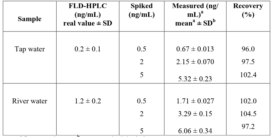
[image:16.596.80.516.462.682.2]7. REAL SAMPLE ANALYSIS
Table 1. Determination of the concentration of BPA of the water samples using the proposed sensor. ("Reprinted with permission from (ACS Appl. Mater. Interfaces 7 (2015) 7492-7496). Copyright (2015) American Chemical Society”).
Sample
FLD-HPLC (ng/mL) real value ± SD
Spiked (ng/mL)
Measured (ng/ mL)a meana ± SDb
Recovery (%)
Tap water 0.2 ± 0.1 0.5
2 5
0.67 ± 0.013 2.15 ± 0.070 5.32 ± 0.23
96.0 97.5 102.4
River water 1.2 ± 0.2 0.5
2 5
1.71 ± 0.027 3.29 ± 0.15 6.06 ± 0.34
102.0 104.5 97.2
a
The mean of five experiments. bSD = standard deviation.
used to determine the content of phenformin obtained from the commercial tablet. DPV and HPLC techniques measured the obtained product of phenformin [107]. The electrochemical DNA nano-biosensor on water sample has been tested with genotoxicity method, Ames test which is the main tool for the analysis of genotoxicity of environmental pollution [108]. Similarly, bioluminescent bacteria has been found in waste water sample by immobilized double stranded calf thymus DNA modified screen printed electrode [109]. A new modified luteolin (Lu) immobilized on functionalized multi-walled carbon nanotube modified glassy carbon (f MWCNT/GCE) electrode for real sample analysis, especially Lu was studied in pharmaceutical sample, diary products and urine sample [110]. High performance liquid charomotography (HPLC) can be used as an analytical technique for the evaluation of real sample (Tap and river water) analysis; the evaluated samples are listed in Table.1 [111].
8. KINETIC STUDIES (MICHAELIS-MENTEN)
Michaelis and Menten have proposed a simple model of reversible reaction of enzyme substrate complex and the complex split into product. The rate of kinetic catalytic reaction Km has been evaluated by using Lineweaver-Burk equation (Eq.2) [112].
c I
K
I
I Max
M app Max
SS
1
1
(Eq.2) Where ISSsteady-state is current after the addition of substrate, C- is concentration of the bulk substrate, IMaxis the maximum current and KappM value obtained from slope and intercept of the plot of reciprocal of the steady-state current. Dong et al [113] have used a new porous nanomaterials of zirconium phytate (Zr-IP6) modified with horse radish peroxide (HRP) and nafion
(Nafion/HRP/Zr-IP6) composite electrode was employed to measure the
M app
K value of 0.306 mM. On the other hand, horseradish peroxide (HRP) immobilization on Ag@C core-shell modified with indium-tin oxide (HRP-Ag@C/ITO) electrode exhibited higher bioactivity, greater affinity to H2O2 and the calculated
biosensor of KappM value of 3.75 x 10⁻⁵ M [114]. The immobilization of glucose oxidase with chitosan-gold nanoparticle film was prepared via electrodeposition method directly through electrochemical glucose biosensor application and to estimate KappM value of 3.5 mM [115]. Recently the electrochemical biosensor analysis of lactase dehydragenase (LDH) immobilized with nano zinc rod and gold (Au/Nano ZnO/LDH) has been studied using amperometry method to evaluate the KappM value [116].
9. ANALYTICAL PARAMETERS
for 5-10 minutes in 5 ml standard solution of MP or AChCl. This kind of inhibited/immobilized electrode, the electrochemical parameter of I(%)can be calculated from the amperometric biosensor equation (Eq.3).
100 (%)
0 1 0
I I I
I (Eq.3) Similarly, the reactive efficiency (R%) has been estimated the following equation (Eq.4)
100 (%) 1 0 1 I I I I
R r (Eq.4) By using sensor analysis, Δ is change of biosensors peak current recorded in square wave voltammetry method. This is another kind of electrochemical parameter has been identified the calculated by the applied equation (Eq.5).
0 0 I I I
(Eq.5) Where I0 is the biosensor current before the addition of sensor sample and I is the biosensors peak current after the addition of sensor sample [118]. Chen et al [119] studied graphene oxide-dopamine complex to estimate the kd (dissociation constant) value form Langmuir adsorption isotherm equation is as follows (Eq.6).
Max d Max I k I DA I DA ] [ ] [ (Eq.6) In order to maintain fragile dopamine bioactivity during the electrochemical measurement studies.
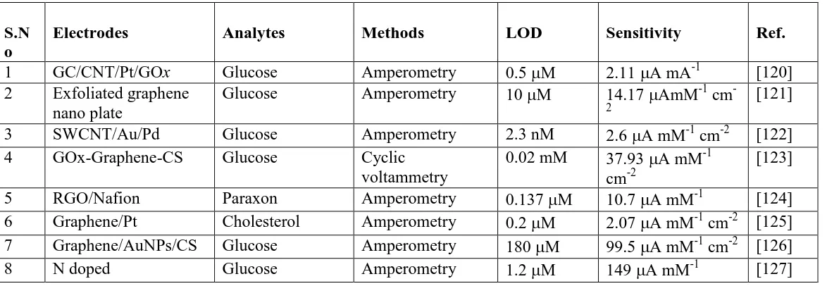
The comparisons of different electrodes, biomolecules, techniques, lower limit of detection and accuracy values have been discussed in Table. 2. From the over all studies, glucose biosensors exhibited maximum cited literatures have been optimized by amperometric method.
Table 2. Comparison of electroanalytical parameters for various films modified electrode towards different analytes
S.N o
Electrodes Analytes Methods LOD Sensitivity Ref.
1 GC/CNT/Pt/GOx Glucose Amperometry 0.5 M 2.11 A mA-1 [120]
2 Exfoliated graphene nano plate
Glucose Amperometry 10 M 14.17 AmM-1 cm
-2
[121]
3 SWCNT/Au/Pd Glucose Amperometry 2.3 nM 2.6 A mM-1 cm-2 [122]
4 GOx-Graphene-CS Glucose Cyclic
voltammetry
0.02 mM 37.93 A mM-1
cm-2
[123]
5 RGO/Nafion Paraxon Amperometry 0.137 M 10.7 A mM-1 [124]
6 Graphene/Pt Cholesterol Amperometry 0.2 M 2.07 A mM-1 cm-2 [125] 7 Graphene/AuNPs/CS Glucose Amperometry 180 M 99.5 A mM-1 cm-2 [126]
[image:18.596.9.596.568.771.2]
CNT/MWCNT
9 RGO/PPy/PSS-g-PPy Hypoxanthane Amperometry 10 nM 673 A mM-1
cm-2 [128]
10 GOx/C60/Fc/CS Glucose Chronoamperomet
ry
3 nM 234.67 A mM-1 cm-2
[129]
11 CS-Fc/GO/GOx Glucose Amperometry 7.6 M 10 A mM-1 cm-2 [130]
12 Graphene/CNT/Nafio n/AuPtNPs
H2O2 SWV 0.17 M 3.7 x 10² A mM-1 [131]
13 Graphene oxide/PB Glucose Amperometry 122 nM 408.7 A mM-1
cm-2
[132] 14 Graphene-Fe3O4 H2O2 Amperometry 0.6 M 132 A mM-1 cm-2 [133]
15 RGO/PB Glucose Amperometry 8.4 M 59 mA M-1 cm-2 [134]
*GOx– Glucose oxidase; SWCNT-Single walled carbon nanotubes; CS – Chitosan; Ppy – Polypyrrole; Fc – Ferrocene; PB - Prussian blue; RGO – Reduced graphene oxide; PSS-g-PPy – Poly(styrenesulfonic acid g- polypyrrole); C60 – Fullerene; SWV- Square wave voltammetry.
10. CONCLUSIONS
Nanostructured electrodes are an important and promising tool for the analysis of electrochemical applications. Especially, in this review we have highlighted to odorize electrode materials and the applied electrochemical technique for application in biosensor field. We found varying reported literatures have been tabularized with different nanostructured electrodes and its bioactive detectivity and sensitivity decorated. The amperometric biosensors of glucose oxidase reaction was one of the most cited article of electrochemical biosensor studies. Most of the authors are mainly focused on cost effective electrode, lowest limit of detection, high sensitivity and long term stability for the evaluation of biosensors applications.
ACKNOWLEDGEMENTS
The research work was supported by the Ministry of Science and Technology, Taiwan and India-Taiwan Science and Technology Cooperation program, DST, India.
References
1. L. Wang, X. Zhang, H. Xiong, S. Wang, Biosens. Bioelectron. 26 (2010) 991-995. 2. J. Wang, G. Rivas, X. Cai, E. Palecek, P. Nielsen, H. Shiraishi, N. Dontha, C. Parrdo, M.
Chicharro, P.A.M. Farias, F.S. Valera, D.H. Grant, M. Ozsoz, M.N. Flair, Anal. Chim. Acta 347 (1997) 1-8.
3. J. Wang, Anal. Chim. Acta, 469 (2002) 63-71.
4. B.P. Simon, M. Cortina, M. Campus, C.C. Blanchard, Sens. Actuators, B 129 (2008) 459-466. 5. M. Campas, B.P. Siman, J.L. Marty, Seminars in cell & development biology 20 (2009) 3-9. 6. E.H. Asl, I. Palchetti, E. Hasheminejed, M. Mascini, Talanta 115 (2013) 74-83.
7. S. Prakash, T. Chaterabarty, A.K. Sing, V.K. Shani, Biosens. Bioelectron.41 (2013) 43-53. 8. L. Ding, A.M. Bond, J. Zhai, J. Zhang, Anal. Chim. Acta 797 (2013) 1-12.
10. R. Ramachandran, V. Mani, S.M. Chen, R. Saraswathi, B.S. Lou, Int. J. Electrochem. Sci., 8 (2013) 11680-11694.
11. S.M. Chen, R. Ramachandran, V. Mani, R. Saraswathi, Int. J. Electrochem. Sci., 9 (2014) 4072-4085.
12. R. Ramachandran, V. Mani, S.M. Chen, G. Gnanakumar, M. Govindasamy, Int. J. Electrochem. Sci., 10 (2015) 859-869.
13. K.J. Babu, A. Zahoor, K.S. Nahm, R. Ramachandran, M.A.J. Rajan, G. Gnanakumar, J. Nanopart. Res., 16 (2014) 2250.
14. J.F. Wu, M.Q. Xu, G.C. Zhao, Electrochem. Commun., 12 (2010) 175-177. 15. M. Zhou, J. Guo, L.P. Guo, J. Bai, Anal. Chem., 80 (2008) 4642-4650.
16. C. Shan, Y. Yang, J. Song, D. Han, A. Ivaska, L. Niu, Anal. Chem., 81 (2009) 2378-2382. 17. L. Wang, X. Zhan, H. Xiong, S. Wang, Biosens. Bioelectron.26 (2010) 991-995.
18. M. Zhou, Y. Zhai, S. Dong, Anal. Chem., 81 (2009) 5603-5613.
19. T. Liu, H. Su, X. Qu, P. Ju, L. Cui, S. Ai, Sens. Actuators, B 160 (2011) 1255-1261. 20. Y. Wan, Y. Wang, J. Wu, D. Zhang, Anal. Chem., 83 (2011) 648-653.
21. X. Guo, S. Yang, R. Cui, J. Hao, H. Zhang, J. Dong, B. Sun, Electrochem. Commun., 20 (2012) 44-47.
22. K. Wang, H.N. Li, J. Wu, C. Ju, J.J. Yan, Q. Liu, B. Qui, Analyst 136 (2011) 3349-3354. 23. W. Song, D.W. Li, Y.T. Li, Y. Li, Y.T. Long, Biosens. Bioelectron.26 (2011) 3181-3186. 24. W. Sun, Y. Zhang, X. Ju, G. Li, H. Gao, Z. Sun, Anal. Chim. Acta 752 (2012) 39-44. 25. F. Xiao, Y. Li, X. Zan, K. Liao, R. Xu, H. Duan, Adv. Funct. Mater., 22 (2012) 2487-2494. 26. C. Guo, H. Sun, X.S. Zhao, Sens. Actuators, B 164 (2012) 82-89.
27. H. Ju, H. Sun. H. Chen, Anal. Chim. Acta 327 (1996) 125-132.
28. S. Poorahong, S. Santhosh, G.V. Ramirez, T.F. Tseng, J.I. Wang, P. Kanatharana, P. Thavarunkum, J. Wang, Biosens. Bioelectron.26 (2011) 3670-3673.
29. C.J. Yuan, C.L. Wang, T.Y. Wu, K.C. Hwang, W.C. Chao, Biosens. Bioelectron.26 (2011) 2858-2863.
30. Q.N. Schuvaico, S.V. Dzyadevych, A.V.E. Skaya, S.G. Sauvigne, E. Csoregi, R. Cespuglio, A.P. Soldatkin, Biosens. Bioelectron.21 (2005) 87-94.
31. L. Deng, S. Guo, M. Zhou, L. Liu, C. Liu. S. Dong, Biosens. Bioelectron.25 (2010) 2189-2193. 32. N. Sato, H. Okuma, Sens. Actuators, B 129 (2008) 188-194.
33. G. Li, Y. Lin, Anal. Chem., 78 (2006) 835-843.
34. S. Lata, B. Batra, P. Kumar, C.S. Pundir, Analtical biochemistry 437 (2013) 1-9. 35. I.M. Apetrei, C. Apetrei, J. Food engnieering 149 (2015) 1-8.
36. M.B. Gholivand, A.R. Jalavand, H.C. Goicoechea, Int. J. Biol. Macromol. 69 (2014) 369-381. 37. C. Lanzellotto, F. Favero, M.L. Antonelli, C. Tortolini, S. Cannistro, E. Coppari, F. Mazzei,
Biosens. Bioelectron.55 (2014) 430-437.
38. Q. Sheng, R. Liu, J. Zheng, Bioelectrochemistry 94 (2013) 39-46.
39. H. Shiraishi, T. Itoh, H. Hayashi, K. Takagi, M. Sakane, T. Mori, J. Wang, Bioelectrochemistry 70 (2007) 481-487.
40. I. Szymanska, H. Radecka, J. Radecki, D.K. Ligaj Biosens. Bioelectron.16 (2001) 911-915. 41. V. Mani, B. Devadas, S.M. Chen, Biosens. Bioelectron.41 (2013) 309-3015.
42. T.M.B.F. Oliveira, M.F. Barroso, S. Morais, M. Araujo, C. Freire, P.D.L. Neto, A.N. Correia, M.B.P.P. Oliveira, C.D. Mates, Bioelectrochemistry 98 (2014) 20-29.
43. H. Liu, C. Duan, X. Su, X. Dong, Z. Huang, W. Shen, Z. Zhu, Sens. Actuators, B 203 (2014) 303-310.
44. H.P. Peng, Y. Hu, P. Liu, Y.N. Deng, P. Wang, W. Chen, A.L. Liu, Y.Z. Chen, X.H. Lin, Sens. Actuators, B 207 (2015) 269-276.
47. S.K. Sukla, S.R. Deshpande, S.K. Shukla, A. Tiwari, Talanta 99 (2012) 283-287. 48. H. Liu, C. Duan, X. Su, X. Dong, W. Shen, Z. Zhu, Ceram. Int. 40 (2014) 9867-9874. 49. S.K. Mahadev, J. Kim Sens. Actuators, B 157 (2011) 177-182.
50. X. Chen, J. Zhu, Z. Chen, C. Xu, Y. Wang, C. Yao, Sens. Actuators, B 159 (2011) 220-228. 51. T. Jeyapragasam, R. Saraswathi, Sens. Actuators, B 191 (2014) 681-687.
52. R.J. Waltman, J. Bargon, Can. J. Chem., 64 (1986) 76.
53. A. Mohammadi, O. Inganas, I. Lundstrom, J. Electrochem. Soc., 133 (1986) 947. 54. S. Bhadra, D. Khastgin, N.K. Singha, J.H. Lee, Prog. Polym. Scie 34 (2009) 783.
55. F.B. Emre, F. Ekiz, A. Bala, S. Emre, S. Timur, L. Toppare, Sens. Actuators, B 158 (2011) 117-123.
56. T.C. Gokoglan, S. Soylemez, M. Kesik, S. Toksabay, L. Toppare, Food Chem. 172 (2015) 219-224.
57. F.E. Kanik, M. Kolb, S. Timur, M. Bahadir, L. Toppare, Int. J. Biol. Macromol. 59 (2013) 111-118.
58. S. Soylemez, F.E. Kanik, A.G. Nurioglu, H. Akpinar, L. Toppere, Sens. Actuators, B 182 (2013) 322-329.
59. M. Kesik, F.E. Kanik, J. Turan, M. Kolb, S. Timur, M. Bahadir, L. Toppare, Sens. Actuators, B 205 (2014) 39-49.
60. Y. Fang, Y. Ni, G. Zhang, C. Mao, X. Huang, J. Shen, Bioelectrochemsitry 88 (2012) 1-7. 61. J. Li, X. Lin, Biosens. Bioelectron.22 (2007) 2898-2905.
62. K. Xue, S. Zhou, H. Shi, X. Feng, H. Xin, W. Song, Sens. Actuators, B 203 (2014) 412-416. 63. M.M. Rahman, X.B. Li, J. Kim, B.O. Lim, A.J.S. Ahammad, J.J. Lee, Sens. Actuators, B 202
(2014) 536-542.
64. X. Jiang, Y. Wu, X. Mao, X. Cui, L. Zhu, Sens. Actuators, B 153 (2011) 158-163. 65. X. Wu, Y. Chai, P. Zhang, R. Yuan, ACS Appl. Mater. Interfaces 7 (2015) 713-720.
66. S.A. Mozaffari, R. Rahmanian, M. Abedi, H.S. Amoli, Electrochim. Acta 146 (2014) 538-547. 67. Y. Liu, X. Qu, H. Guo, H. Chen, B. Liu, S. Dong, Biosens. Bioelectron.21 (2001) 2195-2201. 68. S. Ge, M. Yan, J. Lu, M. Zhang, F. Yu, J. Yu, Biosens. Bioelectron.31 (2012) 49-54.
69. S.D. Uzun, F. Kayaci, T. Uyar, S. Timur, L. Toppare, ACS Appl. Mater. Interfaces 6 (2014) 5235-5243.
70. F.W.P. Ribeio, M.F. Barroso, S. Morais, S. Viswanathan, P.D.L. Neto, A.N. Correia, M.B.P.P. Oliveira, C.D. Matos, Bioelectrochemistry 95 (2014) 7-14.
71. A. Tsopela, A. Lale, E. Vanhov, O. Reynes, I. Seguy, Biosens. Bioelectron.61 (2014) 290-297. 72. Y. Wen, J. Xu, M. Liu, D. Li, L. Lu, R. Yue, H. He, J. Electroanal. Chem. 674 (2012) 71-82. 73. H.L. Zou, B.L. Li, H.Q. Luo, N.B. Li, Sens. Actuators, B 207 (2015) 535-541.
74. J.P. Marco, K.B. Borges, C.R.T. Tarley, E.S. Ribeiro, A.C. Pereira, J. Electroanal. Chem. 704 (2013) 159-168.
75. G. Zhang, N. Yang, Y. Ni, J. Shen, W. Zhao, X. Huang, Sens. Actuators, B 158 (2011) 130-137. 76. N. Nasirizadeh, H.R. Zare, Talenta 80 (2009) 656-663.
77. P. Raghu, T.M. Reddy, K. Reddaiah, B.E.K. Swamy, M. Sreedhar, Food Chem. 142 (2014) 188-196.
78. K.G. Reddy, G. Madhavi, B.E.K. Swamy, J. Mol. Liq. 198 (2014) 181-186. 79. S.K. Arya, A. Dey, S. Bhansali, Biosens. Bioelectron.28 (2011) 166-173.
80. J. Liu, X. Wang, T. Wang, D. Li, F. Xi, J. Wang, E. Wang, ACS Appl. Mater. Interfaces 6 (2014) 19997-20002.
81. S.K. Yadav, B. Agarwal, P. Chandra, R.N. Goyal, Biosens. Bioelectron.55 (2014) 337-342. 82. K. Yang, C.Y. Zhang, Anal. Chem. 82 (2010) 9500-9505.
83. N. Prabhakar, G. Sumanna, K. Arora, H. Sing, B.D. Malhotra, Electrochimica Acta 53 (2008) 4344-4350.
85. M. Lin, X. Hu, Z. Ma, L. Chen, Anal. Chim. Acta 746 (2012) 63-69. 86. N. Nasirizadeh, H.R. Zare, Talanta 80 (2009) 656-663.
87. M. Tak, V. Gupta, M. Tomar, Biosens. Bioelectron.59 (2014) 200-207.
88. C. Hu, D.P. Yang, F. Zhu, F. Jiang, S. Shen, J. Zhang, ACS Appl. Mater. Interfaces 6 (2014) 4170-4178.
89. P.K. Brahman, R.A. Dar, K.S. Pitre, Sens. Actuators, B 177 (2013) 807-812. 90. R.P. Talemi, M.H. Mashhadizhah, Talenta 131 (2015) 460-466.
91. L.D. Mallo, S. Hernandez, G. Marrazza, M. Mascini, L.T. Kubota, Biosens. Bioelectron.21 (2006) 1374-1382.
92. A.K. Upadhyay, Y.Y. Peng, S.M. Chen, Sens. Actuators, B 141 (2009) 557-565. 93. P. Yang, L. Wang, Q. Wu, Z. Chen, X. Lin, Sens. Actuators, B 194 (2014) 71-78.
94. M. Sing, N. Nesakumar, S. Sethuraman, U.M. Krishnan, J.B.B. Rayappan, J. Colloid Interface Sci. 425 (2014) 52-58.
95. T. Ghosh, P. Sarkar, A.P.F. Turner, Bioelectrochemistry 102 (2015) 1-9.
96. X.J. Huang, C.C. Li, B. Gu, J.H. Kim, S.O. Cho, Y.K. Choi, J. Phys. Chem. C 112 (2008) 3605-3611.
97. M.A. Rahman, M.S. Won, Y.B. Shim, Biosens. Bioelectron.21 (2005) 257-265. 98. R. Rawal, C.S. Pandir, Biochem. Eng. J. 71 (2013) 30-37.
99. Z. Na, Y. Tao, J. Kui, S.C. Xia, Chin. J. Anal. Chem. 38 (2010) 301-306.
100. J.A. Lee, S.H. Wang, J. Kwak, S.I. Park, S.S. Lee, K.C. Lee, Sens. Actuators, B 129 (2008) 372-379.
101. R.K. Shervedani, A.H. Mehrjardi, N. Zamiri, Bioelectrochemistry 69 (2006) 201-208.
102. Y. Yang, C. Li, L. Yin, M. Liu, Z. Wang, Y. Shu, G. Li, ACS Appl. Mater. Interfaces 6 (2014) 7579-7584.
103. Y. Ni, P. Wang, H. Song, X. Lin, S. Kokot, Anal. Chim. Acta 821 (2014) 34-40. 104. S. Dong, N. Li, T. Huang, H. Tang, J. Zheng, Sens. Actuators, B 173 (2012) 704-709. 105. H. Zhong, R. Yuan, Y. Chai, W. Li, X. Zhong, Y. Zhang, Talenta 85 (2011) 104-111.
106. L.C. Fuentes, G.P. Villa, L.A. Palomares, S.E. Moya, O.T. Ramirez, Langmuir 30 (2014) 14991-14998.
107. L. Zeng, R. Wang, L. Zhu, J. Zhong, Colloids and surfaces B: Biointerfaces 110 (2013) 8-14. 108. H.B. Xu, R.F. Ye, S.Y. Yang, R. Li, X. Yang, Chin. Chem. Lett. 25 (2014) 29-34.
109. F. Lucarelt, A. Kicela, I. Palchetti, G. Marrazza, M. Mascini, Bioelectrochemistry 58 (2002) 113-118.
110. M. Baghayeri, M. Namadchian, Electrochim. Acta 108 (2013) 22-31.
111. Y. Zhou, Y. Cai, L. Xu, L. Zheng, L. Wang, B. Qi, C. Xu, ACS Appl. Mater. Interfaces 7 (2015) 7492-7496.
112. R. Kamin, G. Wilson, Anal. Chem., 52 (1980) 1198-1205.
113. J. Dong, Y. Wen, Y. Miao, Z. Xie, Z. Zhang, H. Yang, Sens. Actuators, B 150 (2010) 141-147. 114. S. Mao, Y. Long, W. Li, Y. Tu, A. Deng, Biosens. Bioelectron.48 (2013) 258-262.
115. Y. Du, X.L. Luo, J.J. Xu, H.Y. Chen, Bioelectrochemistry 70 (2007) 342-347.
116. N. Nesakumar, K. Thandavan, S. Sethuraman, U.M. Krishnan, J.B.B. Rayappan, J. Colloid Interface Sci. 414 (2014) 90-96.
117. M. Jarczewska, R. Ziolkowski, L. Gorski, E. Malinowska, Bioelectrochemistry 96 (2014) 1-6. 118. J.M. Nugent, D. Du, X. Huang, J. Cai, A. Zheng, Biosens. Bioelectron.23 (2007) 285-289. 119. J.L. Chen, X.P. Yan, K. Meng, S.F. Wang, Anal. Chem., 83 (2011) 8787-8793.
120. S. Hrapovic, Y. Liu, K.B. Male, J.H.T. Luong, Anal. Chem., 76 (2004) 1083-1088. 121. J. Lu, L.T. Drazal, R.M. Worden, I. Lee, Chem. Mater., 19 (2007) 6240-6246.
122. J.C. Claussen, A.D. Franklin, A.U. Haque, D.M. Portefield, T.S. Fisher, ACS Nano 3 (2009) 37-44.
124. B.G. Choi, H.S. Park, T.J. Park, M.H. Yang, J.S. Kim, S.Y. Jang, N.S. Heo, S.Y. Lee, J. Kong, W.H. Hong, ACS Nano 4 (2010) 2910-2918.
125. R. Sundar, C.R. Raj, J. Phy. Chem. C 114 (2010) 21427-21433.
126. S. Shan, H. Yang, D. Han, Q. Zhang, A. Ivaska, L. Niu, Biosens. Bioelectron.25 (2010) 1070-1074.
127. X. Xu, S. Jiang, Z. Hu, S. Liu, ACS Nano 4 (2010) 4292-4298.
128. J. Zhang, J. Lei, R. Pan. Y. Xue, H. Ju, Biosens. Bioelectron.26 (2010) 371-376.
129. W. Zhilei, L. Zaijun, S. Xiulan, F. Yinjun, L. Junkang, Biosens. Bioelectron.25 (2010) 1434-1438.
130. J.D. Qiu, J. Huang, R.P. Liang, Sens. Actuators, B 160 (2011) 287-294. 131. Q. Zhang, M. Wang, J. Zheng, Sens. Actuators, B 160 (2011) 1070-1077.
132. Y. Zheng, X. Sun, L. Zhu, H. Shen, N. Jia, Electrochim. Acta 56 (2011) 1239-1245. 133. K. Zhou, Y. Zhu, X. Yang, C. Li, Electroanalysis 23 (2011) 862-869.
134. X. Bai, G. Chen, K.K Shiu, Electrochim. Acta 89 (2013) 454-460.