INVESTIGATING VENOM SYNTHESIS: EXPLORING
THE COMPOSITION, VARIATION AND GENE
EXPRESSION DYNAMICS OF
BITIS ARIETANS
VENOM
Thesis submitted in accordance with the requirements of the
University of Liverpool for the degree of Doctor of Philosophy
By Rachel Beth Currier
ABSTRACT
Snake venom is a critical evolutionary innovation enabling venomous snakes to become successful limbless predators; it is therefore vital that snakes possess a highly efficient venom production system to maintain their predatory arsenal. The dynamics of venom synthesis and the regulatory mechanisms by which the expression of venom protein-encoding genes is controlled are little understood. The overarching aim of the work described in this thesis was to investigate the dynamics of venom synthesis in terms of the production of venom in juvenile snakes from birth and in the immediate replenishment of depleted venom stores using the African Puff Adder (Bitis arietans) as a model viperid species. We also aimed to investigate the underlying control mechanisms which regulate venom production
Initial studies revealed a remarkable degree of intra-species variation in the protein profile, immunoreactivity and enzyme activity of venom between B. arietans specimens originating from different geographical origins across sub-Saharan Africa and Arabia, and within the same geographical origin. Variation was most evident in the snake venom metalloproteinases (SVMPs); toxins with a primary role in the haemorrhagic and tissue-necrotic pathologies suffered by envenomed victims. Our findings are of therapeutic importance as observations could translate into variations in the clinical manifestation of B. arietans envenoming and affect the patient response to antivenom treatment.
To monitor the synthesis of venom proteins, we exploited the unusual stability of messenger RNA in lyophilised snake venoms as an alternative source of transcriptionally active mRNA to venom gland tissues, thus avoiding the requirement to sacrifice specimens for transcriptome analysis. Our optimised approach was used to quantitatively track changes in expression of venom protein-encoding genes. Our results showed that the gene expression, protein composition and functional activity of juvenile B. arietans venom did not appear to significantly change over time from birth to four years indicating that some aspects of venom are genetically hard-coded. We also showed that venom resynthesis triggered by venom expulsion peaked between days 3-7 following depletion of venom, with different protein families expressed in parallel. It appeared that venom production in both adult and juvenile specimens occurs very rapidly, presumably to ensure that venomous snakes retain their ability to efficiently predate and remain defended from predators.
Table of Contents
ABSTRACT ... ACKNOWLEDGEMENTS ... I AUTHOR DECLARATION ... I PUBLICATIONS AND PRESENTATIONS ARISNG FROM THIS WORK ... II ABBREVIATIONS ... III LIST OF FIGURES ... V LIST OF TABLES ... IX
1. INTRODUCTION ... 1
1.1. Taxonomy and global distribution of venomous snakes ... 1
1.2. Medical importance of snakebite ... 2
1.3. Origin and evolution of the venom arsenal ... 5
1.4. The venom delivery apparatus ... 8
1.4.1. Morphological adaptations of venomous snakes ... 8
1.4.2. Fang dentition ... 10
1.4.3. Cellular structure of the venom gland ... 11
1.4.4. The accessory gland ... 12
1.5. The venom arsenal: Protein families ... 13
1.5.1. Snake venom metalloproteinases (SVMPs) ... 13
1.5.2. Serine proteases ... 16
1.5.3. Phospholipase A2s (PLA2s) ... 18
1.5.5. Hyaluronidase ... 21
1.5.6. Acetylcholinesterase (AChE) ... 23
1.5.7. Nucleases, nucleotidases and phosphomonoesterases ... 24
1.5.8. C-type lectins (CTLs) ... 25
1.5.9. Disintegrins ... 27
1.5.10. Bradykinin potentiating peptides (BPPs) ... 29
1.5.11. Inhibitory peptides (QKW tripeptides) ... 29
1.5.12. Kunitz serine proteinase inhibitors... 31
1.5.13. Vascular endothelial growth factors (VEGFs) and nerve growth factor (NGFs) ... 31
1.5.14. Cysteine rich secretory proteins (CRISPs) ... 33
1.5.15. Purine and pyrimidine nucleosides ... 34
1.6. Venom protein synthesis and storage ... 35
1.7. The venom glandular environment; protecting against self-proteolysis ... 37
1.8. Analysis of venom composition; venomics and transcriptomics ... 39
1.9. Variation in venom composition ... 41
1.10. Aims of this work ... 43
2. MATERIALS AND METHODS ... 46
2.1. Venom extraction, preparation and storage ... 46
PROTEIN ANALYSIS ... 47
2.2.1. Venom sample preparation for SDS-PAGE ... 47
2.2.2. SDS-PAGE gel preparation and electrophoresis... 47
2.3. Immunoblotting ... 49
2.4. Substrate zymography ... 50
2.4.1. Venom sample preparation for substrate zymography... 50
2.4.2. Zymogram gel preparation, electrophoresis and activation ... 51
2.5. Mass spectrometry: ... 51
2.5.1. In-gel trypsin digestion ... 51
2.5.2. HPLC – tandem mass spectrometry (HPLC-MS/MS) and protein identification using bioinformatics tools ... 52
MOLECULAR BIOLOGY ... 53
2.6. Extraction of Poly(A) mRNA from venom using oligo (dT) magnetic Dynabeads® ... 53
2.7. First strand complementary DNA (cDNA) synthesis ... 55
2.8. Plasmid purification of venom gland library cDNA for PCR positive control ... 56
2.9. Polymerase chain reaction (PCR) ... 57
2.9.1. PCR primer design ... 57
2.9.2. Conventional PCR ... 57
2.9.3. Purification of PCR products ... 59
2.9.4. Quantitative PCR (qPCR) ... 60
2.10.1. Genomic DNA isolation ... 61
2.10.2. Quality assessment of genomic DNA ... 62
2.10.3. Genomic library construction using GenomeWalkerTM Kit... 62
2.10.4. Genome walking PCR primer design ... 64
2.10.5. Long range PCR amplification of genomic DNA and purification of PCR products ... 65
2.10.6. Sub-cloning and transformation of E. coli with purified genomic PCR products ... 67
2.10.7. Selection and amplification of recombinant E. coli colonies ... 68
2.10.8. Purification and analysis of recombinant plasmids by restriction enzyme digestion ... 68
2.10.9. Selection and submission of recombinant plasmids for DNA sequencing 69 2.10.10. Bioinformatics interrogation of genomic DNA sequences ... 69
2.11. Ethical declaration ... 70
3. INTRA-SPECIFIC VARIATION IN VENOM OF THE AFRICAN PUFF ADDER (BITIS ARIETANS); DIFFERENTIAL EXPRESSION AND ACTIVITY OF THE SNAKE VENOM METALLOPROTEINASES (SVMPs) ... 71
3.1. Abstract ... 71
3.2. Introduction ... 72
3.3. Materials and Methods: ... 75
3.3.2. Venom protein composition profiling by 1D SDS-PAGE ... 76
3.3.3. Antibody cross-reactivity of venoms by immunoblotting ... 76
3.3.4. Venom enzyme activity profiling by substrate zymography ... 77
3.3.5. Identification of venom proteins by LC-MS/MS ... 77
3.4. Results: ... 78
3.4.1. Variation in venom composition between B. arietans specimens from different geographical origins: ... 78
3.4.2. Variation in venom between individual B. arietans specimens from the same geographical origin (Nigeria):... 81
3.4.3. Comparison with variation in venom of Ghanaian B. arietans ... 92
3.4.4. Comparison with variation in venom of B. arietans with other species, Echis ocellatus and Cerastes cerastes ... 93
3.5. Discussion ... 97
4. OPTIMSATION OF QUANTITATIVE POLYMERASE CHAIN REACTION (PCR) PROTOCOLS; EXPLOITING VENOM AS A SOURCE OF MESSENGER RNA FOR GENE EXPRESSION ANALYSES. ... 101
4.1. Abstract ... 101
4.2. Introduction ... 102
4.3. Materials and Methods: ... 105
4.3.1. Venom sample preparation ... 105
4.3.2. Extraction of Poly(A) mRNA from venom using Dynabeads® ... 105
4.3.4. Design of PCR primer pairs for conventional PCR and quantitative PCR
reactions ... 106
4.3.5. Amplification of venom transcripts by conventional PCR ... 110
4.3.6. Experimental optimisation of annealing temperature for qPCR
amplification ... 110
4.3.7. Optimising cDNA concentration for qPCR ... 112
4.3.8. Standard curve analysis to determine the amplification reaction efficiency
in qPCR ... 112
4.3.9. Melt curve analysis to determine the specificity of primer pairs in qPCR 113
4.4. Results: ... 116
4.4.1. Quantity of mRNA recovered from venom ... 116
4.4.2. Testing the ability of primer pairs to specifically amplify a range of venom
protein targets from venom gland and venom cDNA ... 117
4.4.3. Assessing DNA contamination of mRNA samples ... 120
4.4.4. Experimental optimisation of annealing temperature for qPCR
amplification ... 122
4.4.5. Optimising cDNA concentration for qPCR amplification ... 123
4.4.6. Standard curve analysis to determine amplification reaction efficiency in
qPCR ... 124
4.4.7. Melt curve analysis to determine qPCR primer specificity ... 129
4.6. Discussion ... 132
COMPOSITION AND ENZYME ACTIVITY OF VENOM FROM BIRTH TO
MATURITY. ... 135
5.1. Abstract ... 135
5.2. Introduction ... 136
5.3. Materials and Methods: ... 139
5.3.1. Venom sample collection and preparation ... 139
5.3.2. Extraction of Poly(A) mRNA from venom using Dynabeads® ... 140
5.3.3. Monitoring gene expression levels using relative quantitative PCR (qPCR): ... 140
5.3.4. Venom protein composition profiling 1D SDS-PAGE: ... 142
5.3.5. Identification of venom proteins by LC-MS/MS ... 142
5.3.6. Venom enzyme activity profiling by substrate zymography: ... 142
5.4. Results: ... 142
5.4.1. Juvenile Bitis arietans survival, growth and venom yield rate ... 142
5.4.2. Quantitative PCR (qPCR) toxin expression profiles of juvenile B. arietans siblings from birth ... 145
5.4.3. Venom protein profiles of juvenile B. arietans siblings from birth ... 149
5.4.4. Mass spectrometry identification of proteins of interest from juvenile B. arietans samples ... 151
6. UNUSUAL STABILITY OF MESSENGER RNA IN SNAKE VENOM REVEALS GENE EXPRESSION DYNAMICS OF VENOM
REPLENISHMENT. ... 166
6.1. Abstract ... 166
6.2. Introduction ... 167
6.3. Methods ... 169
6.3.1. Venom samples and standards: ... 169
6.3.2. Extraction of Poly(A) mRNA from venom using Dynabeads®: ... 170
6.3.3. cDNA synthesis: ... 170
6.3.4. Quantitative PCR (qPCR): ... 170
6.3.5. One-dimensional SDS-PAGE and gelatin zymography: ... 171
6.3.6. HPLC separation of venom and mass spectrometry protein identification ... 172
6.4. Results: ... 172
6.4.1. Quantity and quality of mRNA recovered from venom: ... 172
6.4.2. Venom protein expression profiles during venom re-synthesis: ... 172
6.4.3. Protein profiles and venom activity during venom re-synthesis: ... 179
6.5. Discussion ... 184
7. INVESTIGATION OF THE GENETIC ORGANISATION AND MECHANISMS RESPONSIBLE FOR REGULATING THE EXPRESSION OF VENOM PROTEIN-ENCODING GENES ... 189
7.2. Introduction ... 190
7.3. Materials and methods ... 193
7.3.1. Isolation, quality control and PCR amplification of venom transcripts from Bitis arietans genomic DNA ... 193
7.3.2. Construction of ‘Genome Walker libraries’ ... 195
7.3.3. Genome walking by long-range PCR ... 195
7.3.4. Primer design ... 196
7.3.5. Sub-cloning and sequencing ... 197
7.3.6. Bioinformatics interrogation of sequences for promoter and transcription factors ... 197
7.4. Results: ... 198
7.4.1. Isolation and quality control of genomic DNA from B. arietans liver tissue ... 198
7.4.2. Amplification of venom protein targets from B. arietans genomic DNA using long-range PCR ... 199
7.4.3. Sub-cloning and sequencing of PCR products amplified from B. arietans genomic DNA ... 201
7.4.4. Sequence analysis of PCR products amplified from B. arietans genomic DNA ... 202
7.4.5. Amplification of gene targets from ‘Genome walker’ libraries ... 207
7.5. Discussion ... 213
9. FUTURE WORK ... 223 APPENDICES ... 226 APPENDIX I: Recipes for buffers and stock solutions ... 226
APPENDIX II: The ‘Minimum information for Publication of Quantitative Real-time
PCR Experiments’ (MIQE) guidelines. ... 230
APPENDIX III: Optimisation of annealing temperature for amplification by
quantitative PCR. ... 233
APPENDIX IV: Optimisation of starting cDNA quantity for amplification by
quantitative PCR. ... 235
APPENDIX V: Optimisation of quantitative PCR by standard and melt curve
analysis. ... 236
APPENDIX VI: Juvenile Bitis arietans specimens ... 239
APPENDIX VII: Optimisation of relative real-time gene expression analysis with
reference to the MIQE guidelines. ... 244
APPENDIX VIII: Sequence alignments of Bitis arietans serine protease and vascular
endothelial growth factor gene sequences with homologous genes... 248
APPENDIX IX: Sequences and BLAST analysis of PCR products amplified and
cloned from genome walker libraries. ... 252
I
ACKNOWLEDGEMENTS
Firstly, I wish to thank my supervisors Dr Simon Wagstaff and Dr Rob Harrison for
their consistent support, enthusiasm and encouragement throughout the course of this
work. I would also like to thank Paul Rowley for imparting his expert knowledge of
venomous snakes and for extracting venom samples used in this project. Without
their help and guidance, none of this work would have been possible. I would like to
thank students in the Alistair Reid Venom Research Unit, old and new; Darren, Nick,
Camila, Maimonah and Fiona, for being great friends and making my time in the lab
thoroughly enjoyable. Finally, I would like to thank my friends and family for their
endless support and encouragement, and for always believing in me.
AUTHOR DECLARATION
The work presented in this thesis was performed entirely by myself with the
exception of the high performance liquid chromatography (HPLC) separation and
mass spectrometry identification of venom proteins described in Chapter 6 which
was performed by Professor Juan J. Calvete (Laboratorio de Proteinomica Estructual
at the Instituto de Biomedicina de Valencia, Valencia, Spain). All venom extractions
were performed by Paul Rowley and Dr Robert Harrison in the Alistair Reid Venom
II
PUBLICATIONS AND PRESENTATIONS ARISNG FROM THIS WORK
1. Currier, R.B., Calvete, J.J., Sanz, L., Harrison, R.A., Rowley, P.D., Wagstaff,
S.C. (2012) Unusual stability of messenger RNA in snake venom reveals gene
expression dynamics of venom replenishment. PLoS ONE 7(8): e41888.
doi:10.1371/journal.pone.0041888.
2. Currier, R.B., Harrison, R.A., Rowley, P.D., Laing, G.D., Wagstaff, S.C. (2010)
Intra-specific variation in venom of the African Puff Adder (Bitis arietans):
Differential expression and activity of snake venom metalloproteinases
(SVMPs). Toxicon 55: 864-873.
This work has been presented as oral and poster presentations at The International
Society on Toxinology (IST) World Congress (Recife, Brazil) in 2009 and European
Congress (Valencia, Spain) in 2011, the University of Bangor Herpetological Society
Venom Day in 2010 and 2012 and the British Herpetological Society Annual
III
ABBREVIATIONS
ADAM A disintegrin and metalloproteinase
APS Ammonium persulphate
bp Base pairs
BLAST Basic Local Alignment Search Tool
°C Degrees centigrade
cDNA Copy deoxyribonucleic acid
CTL C-type lectin
CLP CTL-like proteins
DAB 3, 3'-diaminobenzidine
dH2O Distilled water
DNA Deoxyribonucleic acid
DTT Dithiothreitol
E. coli Escherichia coli
EDTA Ethylenediaminetetraacetic acid
EST Expressed sequence tag
HPLC High performance liquid chromatography
IAN Iodoacetamide
IPTG Isopropyl-β-D-thio-galactoside
kb Kilo base
kDa Kilo dalton
KTI Kunitz inhibitors
LAO L-amino acid oxidase
LB Luria-bertani
M Molar concentration
IV
MS Mass spectrometry
Min Minutes
MMP Matrix metalloproteinase
ORF Open reading frame
PAGE Polyacrylamide gel electrophoresis
PBS Phosphate buffered saline
PCR Polymerase chain reaction
PDI Protein disulphide isomerase
PLA2 Phospholipase A2
PLOB Protein loading buffer
Poly(A) Polyadenylated
qPCR Quantitative PCR
RGD Arginine-glycine-aspartate sequence
RNA Ribonucleic acid
rpm Revolutions per minute
RT-PCR Reverse transcriptase PCR
SDS Sodium dodecyl sulphate
SP Serine protease
SVMP Snake venom metalloproteinase
TBST Tris-buffered saline-Tween 20
TEMED Tetramethylethylenediamine
TGE Tris-glycine-EDTA
UV Ultraviolet
V Voltage
VEGF Vascular endothelial growth factor
V
LIST OF FIGURES
Figure 1.1 Global medical importance of snakebite.
Figure 1.2 Cladogram of evolutionary relationships of the Toxicofera showing the recruitment timing of different protein-scaffold types for use as toxins.
Figure 1.3 The Viper venom delivery system.
Figure 1.4 Fine structure of the viperid venom gland.
Figure 1.5 Fang dentition of venomous snake families.
Figure 1.6 Structural classifications of the snake venom metalloproteinases.
Figure 1.7 Snake venom hyaluronidase sequence analysis.
Figure 1.8 Snake venom disintegrin sequence analysis.
Figure 1.9 Sequence analyses of QKW tripeptides from viper venoms.
Figure 1.10 The VEGF protein family and receptor selectivity.
Figure 1.11 Secretory cycle of the venom gland during venom synthesis.
Figure 2.1 Venom extracted from an adult Bitis arietans specimen.
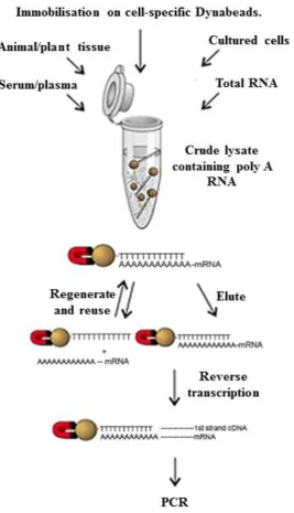
Figure 2.2 Extraction of poly(A) mRNA from venom using Dynabeads®.
Figure 2.3 The GenomeWalker adaptor.
Figure 3.1 Adult Bitis arietans coloration and geographical distribution.
Figure 3.2 Analysis of pooled B. arietans venom samples from six different geographical origins.
VI
Figure 3.4 Reproducibility of venom protein profile of Nigerian Bitis arietans specimens.
Figure 3.5 Full and partial-length peptide sequences identified by LC-MS/MS from adult Bitis arietans specimens.
Figure 3.6 Analysis of individual Ghanaian Bitis arietans venom samples.
Figure 3.7 Analysis of other African viper venoms, Nigerian E. ocellatus (A) and Egyptian C. cerastes (B).
Figure 3.8 Immunoreactivity of Bitis arietans venoms to polyspecific antivenom.
Figure 3.9 Analysis of low molecular weight protein components of Bitis arietans venom.
Figure 4.1 qPCR plate and thermocycling for optimisation of annealing temperature.
Figure 4.2 qPCR plate and thermocycling for standard and melt curve validation.
Figure 4.3 Identifying the optimal venom quantity for mRNA and cDNA yield.
Figure 4.4 Amplification of venom targets from venom vs. venom gland cDNA.
Figure 4.5 Amplification of venom targets from different quantities of venom.
Figure 4.6 Assessing DNA contamination of mRNA samples using reverse transcriptase negative controls (conventional PCR primers).
Figure 4.7 Assessing DNA contamination of mRNA samples using reverse transcriptase negative controls and DNase treatment (qPCR primers).
VII
Figure 4.9 Optimisation of initial cDNA concentration for amplification by quantitative PCR.
Figure 4.10 Standard curves generated by qPCR using purified PCR products as a standard cDNA sample.
Figure 4.11 Standard curves generated by qPCR using purified PCR products as a standard cDNA sample.
Figure 4.12 Melt curves generated by qPCR following amplification using purified PCR products as a standard cDNA sample.
Figure 4.13 Melt curves generated by qPCR following amplification using mature venom cDNA as a standard cDNA sample.
Figure 5.1Bitis arietans juvenile specimen.
Figure 5.2 Juvenile Bitis arietans growth rate and venom production.
Figure 5.3 Juvenile Bitis arietans toxin gene expression profiles during venom production.
Figure 5.4 Juvenile Bitis arietans venom protein profile.
Figure 5.5 Mass spectrometry identification of protein bands from juvenile Bitis arietans venom.
Figure 5.6 Full and partial-length peptide sequences identified by LC-MS/MS from juvenile Bitis arietans specimens.
Figure 5.7 Juvenile Bitis arietans venom enzyme activity profiles.
Figure 6.1 Venom gene expression profiles during venom replenishment.
VIII
Figure 6.3 Average gene expression profiles of all Bitis arietans specimens.
Figure 6.4 HPLC-MS/MS protein profiling of Ghanaian and Nigerian Bitis arietans venoms.
Figure 6.5 Protein profiling of individual venom samples by HPLC and 1D SDS-PAGE.
Figure 6.6 Venom enzyme activity profiles during venom replenishment.
Figure 6.7 Stability of venom mRNA during long-term storage of lyophilised venom.
Figure 7.1 Genome walking protocol
Figure 7.2 Identification of DNA upstream of transcription start site by genome walking.
Figure 7.3 Analysis of genomic DNA extracted from Bitis arietans liver tissue.
Figure 7.4 PCR amplification of venom targets from Bitis arietans genomic DNA.
Figure 7.5 Analysis of venom target inserts following sub-cloning.
Figure 7.6 Genomic DNA sequences encoding Bitis arietans venom proteins.
Figure 7.7 Gene structures of venom protein-encoding genes.
Figure 7.8 Primary PCR amplification of targets from Genome Walker libraries.
IX
LIST OF TABLES
Table 2.1 Rapid silver staining protocol for staining SDS-PAGE protein gels.
Table 3.1 LC-MS/MS identification of venom proteins.
Table 4.1 Conventional PCR primer sequences.
Table 4.2 Quantitative PCR primer sequences.
Table 4.3 Quantitative PCR reaction efficiencies.
Table 5.1 Relative qPCR gene expression levels during venom production in juvenile Bitis arietans specimens.
Table 5.2 Statistical analysis of gene expression data from juvenile Bitis arietans venom samples.
Table 5.3 LC-MS/MS identification of venom proteins from juvenile Bitis arietans venom samples.
Table 6.1 Relative qPCR gene expression levels during venom replenishment individual adult Bitis arietans venom samples.
Table 6.3 Statistical significance of relative changes in gene expression during venom replenishment.
Table 7.1 PCR primer sequences for genome walking protocol.
1
1. INTRODUCTION
This PhD project aimed to explore and understand how the toxic array of proteins in
snake venom are synthesised and regulated by the venom production system of
venomous snakes. It is clearly important that this work is viewed in the context of
our current knowledge of snakes and the biology of their venoms. The purpose of
this introduction is to outline the taxonomy, medical importance,
distribution/diversity of venomous snakes, evolution of the venom toxic arsenal,
venom gland delivery system, structure/function of venom proteins, venom
synthesis, the venom glandular microenvironment, the approaches used to analyse
venom composition, reports of variation in venom composition and the aims of this
work.
1.1. Taxonomy and global distribution of venomous snakes
The Serpentes (Ophidia) comprises approximately 2,800 snake species, categorised
into three superfamilies and 16-20 families (O'Shea, 2005). All venomous snakes
(advanced snakes) belong to the Colubroidea (or Caenophidia) superfamily. The
Colubroidea has been traditionally subdivided into four families; Colubridae,
Atractaspididae, Elapidae and Viperidae. The Colubridae are the largest and diverse
family containing around 1,700 species distributed worldwide and the classification
of this group is subsequently undergoing continual evaluation. The colubrids do not
naturally form a monophyletic group, as many species are more closely related to
other groups, such as elapids, than to each other (Lawson et al., 2005). While many
are considered non-venomous, some colubrids such as the African Boomslang
2
humans. The Atractaspididae contain around 30 species which are distributed across
Africa and Arabia. Although venomous, atractaspidids (e.g. burrowing asps) are not
associated with causing a significant number of human fatalities primarily due to
their small size. The Elapidae consists of around 351 species which are widely
distributed across the Americas, Africa, the Middle East, Asia and Australasia. There
are two recognized sub-families; the Elapinae (coral snakes, cobras, mambas and
kraits) and the Hydrophiinae (sea snakes). The elapids are front-fanged snakes, most
of which produce highly potent neurotoxic venom, and are of significant medical
importance to humans. The Viperidae contains around 311 species and have a
worldwide distribution (except Australasia), occupying a range of terrestrial habitats
across the Americas, Africa, Europe and Asia. The Viperidae contains two main
sub-families; the Viperinae (pit-less vipers e.g. Bitis, Cerastes, Echis and Vipera genera)
and the Crotalinae (pitvipers e.g. Agkistrodon, Bothrops and Crotalus genera).
Viperids are also front-fanged snakes capable of producing highly potent hemotoxic
venom and are responsible for a significant number or human fatalities worldwide
(O'Shea, 2005).
1.2. Medical importance of snakebite
Envenoming following snakebite is largely a neglected threat to public health, yet
causes considerable morbidity and mortality throughout the world. There are up to
1.8 million incidences of snakebite worldwide per year leading to up to 94,000
deaths, the majority of which occur in sub-Saharan Africa, South and Southeast Asia
and Latin America (Kasturiratne et al., 2008). In Africa, a recent meta-analytical
3
Figure 1.1 Global medical importance of snakebite: The global distribution of the annual estimates of snakebite-induced deaths. Darker colours denote the highest
numbers of snakebite mortality (Kasturiratne et al., 2008).
occurred in rural environments (Chippaux, 2011). Those most at risk of
death/morbidity from snakebite are subsistence farmers, agricultural workers and
children, where the threat of snakebite is a daily occupational hazard and access to
health services is poor. In addition to the risk of mortality, many victims of snakebite
are also left with permanent disabilities and sequelae due to severe tissue necrosis.
The morbidity of snakebite in Africa is reflected in the high incidence of amputations
of the affected limb (5,900-14,600 per year) (Chippaux, 2011) demonstrating that
snakebite may also have a considerable economic impact. A strong correlation
between snakebite-induced mortality and poverty has been demonstrated through
mapping the global distribution of snakebite with economic factors such as gross
domestic product (GDP) and per capita government expenditure on health,
illustrating that snakebite is a significant health issue of the rural poor (Figure 1.1)
4
Antivenom is the only effective treatment for snakebite. Antivenoms are produced by
immunising animals (most commonly horses) with either a single venom to produce
monospecific antivenoms, or a mixture of venoms to produce polyspecific
antivenoms, with increasing doses over several months. The venom-neutralising
antibodies (IgG) produced by the immunised animal are collected and purified,
resulting in antivenom (Lalloo and Theakston, 2003). Although effective, safe and
affordable antivenoms are available, the lack of a coordinated and constructive
strategy for the delivery of antivenoms in the developing world has resulted in a
global antivenom crisis (Williams et al., 2011). The Global Snakebite initiative is a
worldwide, rational approach developed by toxinologists which aims to reduce the
incidence of death and morbidity due to snakebite through a combination of
community education, surveillance of snakebite, clinical research, medical
management, rehabilitation and implementation of government health policies
(Williams et al., 2010). In current research, there are several strategies which aim to
improve the efficiency and specificity of antivenom treatment and to address the
safety issues with some current antivenoms by improving the dose, purity and
geographic/species efficacy of antivenom (Harrison et al., 2011). One strategy
includes the use of IgGs from alternative venom-immunised animals (e.g. camelids).
Camelid IgGs are less immunogenic and therefore lower the risk of activating
complement, thus reducing the incidence of adverse effects and improving the safety
of antivenom treatment (Herrera et al., 2005). Current research has provided
encouraging support for the potential use of camelid IgG in antivenom production
(Cook et al., 2010a, Cook et al., 2010b, Cook et al., 2010c). Another strategy
involves the research into the design of toxin-specific epitopes, directed to target
5
thus improving the dose-efficacy of antivenom is also providing promising results
(Wagstaff et al., 2006). In combination, these approaches aim to significantly
improve the treatment and management of snakebite worldwide.
1.3. Origin and evolution of the venom arsenal
Venom delivery systems have evolved independently several times throughout
nature, including in the Cnidaria (jellyfish and anemones) (Bloom et al., 1998),
Gastropoda (cone snails) (Olivera et al., 2002), Hymenoptera (bees and wasps),
Arachnida (spiders and scorpions) (Escoubas et al., 2006), Chordata (fish), Reptilia
(snakes and lizards)(Fry et al., 2006) and Mammalia (platypus)(Whittington et al.,
2009). Arguably, one of the most sophisticated, diverse and efficient venom systems
exist among the venomous snakes.
The Colubroidea encompass around 80% of the ~2,900 species of snakes (Vidal et
al., 2007). The Colubroids form a clade with the closely related Iguania and
Anguimorphs, known collectively as the Toxicofera. Members of this group share a
number of basal protein families that were recruited into the venom delivery system
prior to the divergence of these lineages (Fry et al., 2009). Although evidence
supports the presence of venom as a basal evolutionary characteristic in the
Serpentes (Fry et al., 2006), it is currently thought that only ~450 venomous species
of medical importance exist, which are found exclusively within the Colubroidea,
thus, venomous dependency appears to have been lost over time. The families of
venomous snakes which are the most medically important to humans are the
Viperidae, Elapidae and to a lesser extent, the Atractaspididae, each of which
6
Snake venom has evolved into a highly complex mixture of several hundred unique
constituents including enzymatic and non-enzymatic proteins and peptides,
carbohydrates, lipids, metal ions and organic compounds (Aird, 2002).While the
composition and toxicity of venoms can vary widely between snake taxa, the primary
evolutionary function of venom is to ensure the rapid and efficient immobilisation
and killing of a diverse range of prey species (Kordiš et al., 2002). Venoms may also
serve in a defensive role as a deterrent to predators/aggressors.
Venom toxins have been recruited into the venom arsenal from normal, non-toxic
physiological proteins by adaptive evolution (Fry, 2005). The rapid explosion of
multiple gene duplication and diversification, selective expression in the venom
gland and subsequent structural and functional divergence from non-toxin
homologues has resulted in the formation of multi-isoform, multi-domain protein
families in snake venoms (Fry, 2005, Fry et al., 2009). The first protein families
recruited into a venomous role prior to the divergence of the Colubroidea include the
cysteine-rich secretory proteins, hyaluronidase, kallikrein enzymes and nerve growth
factor (Figure 1.2). Newly recruited toxin groups have evolved to form large,
multi-gene families via the ‘birth-and-death’ model (Fry et al., 2003). This involves the
creation of gene families as a result of repeated gene duplication events and rapid
structural and functional diversifications of genes to form evolutionarily related but
functionally distinct genes; a key process in the process of adaptive evolution. Where
some genes are retained in the genome, over time, other genes are subsequently
deleted from the genome or become non-functional giving rise to pseudogenes(Nei
7
Figure 1.2 Cladogram of evolutionary relationships of the Toxicofera showing the recruitment timing of different protein-scaffold types for use as toxins: Blue X shows independent evolution of hollow front-fanged, high-pressure venom
delivery systems and red lines indicate a toxin recruitment event. 3FTX = three
finger toxin, C3/CVF=ComplementC3/Cobra Venom Factor, CRISP=Cysteine-rich
secretory protein, NGF=Nerve Growth Factor, SVMP = snake venom
metalloproteinase, VEGF = vascular endothelial growth factor (Fry et al., 2009).
Snake venom toxins are continually subjected to adaptive evolutionary pressures,
[image:28.595.112.524.78.515.2]8
Figure 1.3 The viper venom delivery system.
substrate specificities and consequent diverse pathologies of venoms generate great
interest in both biological and therapeutic research.
1.4. The venom delivery apparatus
1.4.1. Morphological adaptations of venomous snakes
The highly complex and functionally diverse mixture of proteins present in venom is
synthesised by venom glands which are modified parotid glands with an elongated
and specialised structure. Front-fanged venomous snakes (viperids, elapids and
atractaspidids) possess a large post-orbital venom-producing apparatus lying along
the upper jaw, whereas the venom glands of venomous lizards run along the lower
jaw and glands along the upper jaw have been apparently lost. The venom delivery
system of venomous snakes comprises of the specialised secretory glands, a toxic
venom arsenal, compressor muscles, fangs and a predatory behaviour (Kardong,
9
In front-fanged venomous snakes, the venom gland apparatus is a highly pressurized,
closed system which employs extremely efficient mechanisms to result in the rapid
expulsion and injection of venom into the envenomed prey tissues. The venom gland
structure consists of four discrete regions; the main glandular lumen, the accessory
gland, the primary duct and the secondary duct. Venom is synthesised and stored in
the main glandular lumen. During expulsion, venom is transported, via the ducts,
from the lumen to the tubular fangs which inject venom into the envenomed prey.
However, the structural features of glands can vary between different families of
snakes (Kardong, 1980, Kochva, 1987, Weinstein et al., 2010). The Viperidae family
possess the most morphologically specialised and efficient venom delivery systems
of all venomous snakes. Figure 1.4 shows the detailed structure of the viperid venom
glands. Viperid venom glands have a complex tubular structure with a highly folded
glandular secretory epithelium divided into several ductules enabling maximal
venom synthesis and storage (Jackson, 2003). Viper venom glands also have
capacious glandular lumen enabling storage of significant volumes of venom
resulting in the accumulation of potentially large quantities of venom that can be
injected into the victim following snakebite. The glandular lumen is connected, via
the primary duct, to the accessory gland, a separate region of the apparatus which
may contribute to venom composition. The secondary duct connects the accessory
gland to the fangs which inject the venom. Venom glands of elapid snakes share
similarities to those of viperids but have a more simplified structure with a smaller
lumen consisting of branching tubules converging into the accessory gland and
primary/secondary ducts. The venom glands of atractaspidid snakes differ from those
10
Figure 1.4 Fine structure of the viperid venom gland: Venom glandular structure showing compartmental sections and features of the glands including the secretory
epithelium, main lumen, ducts and accessory gland. Figure adapted from Mackessey
and Baxter (Mackessy and Baxter, 2006).
with characteristic unbranched tubules and do not possess a distinct, discrete
accessory gland (Kochva, 1987).
1.4.2. Fang dentition
The dentition of fangs/teeth in non-venomous and venomous snakes varies greatly
between different families of snakes (Jackson, 2002, Jackson, 2003, Jackson, 2007).
Aglyphous snakes, such as pythons, are mostly non-venomous and have teeth lacking
any specialisation (e.g. grooves). Opisthoglyphous snakes (Colubridae) are
rear-fanged snakes possessing rear-grooved elongate fangs at the back of the mouth. Most
opisthoglyphous snakes are considered non-venomous or weakly venomous and
therefore harmless to humans due to their inefficient venom delivery systems, with
exceptions including the Boomslang (Dispholidus typus) which are capable of
delivering haemotoxic venom following snakebite. Proteroglyphous snakes
11
Figure 1.5 Fang dentition of venomous snake families: Variation in fang dentition of the most medically important front-fanged venomous snakes A) Viperidae and B)
Elapidae in comparison to the rear-fanged Colubridae (C). (Photographs courtesy of
Juan M. Renjifo).
channel through which venom is directed. Proteroglyphous fangs are relatively short,
and therefore elapids are required to maintain contact with their prey during injection
of venom. Solenoglyphous snakes (Viperidae) are front-fanged snakes with hollow,
tubular hyperextendable fangs resembling hypodermic needles. Solenoglyphs
possess the most efficient venom delivery systems as the fangs are connected to a
moveable maxillary bone allowing for maximal fang length. Their incredibly long
fangs are indicative of the manner by which they predate, typically adopting a ‘strike
and release’ behaviour whereby large quantities of venom can be injected deep into
the prey tissues to exert rapid pharmacological effects. Figure 1.5 shows variation in
fang dentition between the most clinically important families of venomous snakes,
the viperids and elapids, in comparison to the predominantly non-venomous family,
the colubrids, of lesser medical importance.
1.4.3. Cellular structure of the venom gland
Venom glands comprise of a highly folded secretory epithelium consisting of several
12
(Oron and Bdolah, 1978, Mackessy, 1991, Mackessy and Baxter, 2006). An
abundance of glandular secretory cells (79% of the proportion of cells in the
epithelium) are required to synthesise and secrete venom proteins and other
components including physiological proteins, mucus and saliva into the glandular
lumen. In lower abundance (10%) are horizontal cells, interdigitating between the
basement membrane and base of the secretory cells, which function to phagocytose
cellular debris and replace dead secretory cells. ‘Dark’ cells (9%) possess long
dendritic processes which interdigitate between secretory cells and may play a role in
cellular communication between cells. Mitochondria-rich cells (2%), which are
densely packed with mitochondria, are thought to sustain the high energetic costs of
venom production by the venom gland (Mackessy, 1991). The venom gland
epithelium is basally supported by a connective tissue capsule through which pass a
network of capillaries and nerves processes. The venom gland is surrounded by
compressor muscle tissue which contracts during venom extraction in order to expel
the venom glandular contents in a highly pressurized system.
1.4.4. The accessory gland
Currently, very little is known about the characteristics and function of the accessory
glands in the venom delivery system. In viperids, the accessory glands are separate
from but connected to the main gland via the primary duct. In contrast, in elapids, the
accessory gland surrounds the duct. There is currently little understanding of the
input of accessory gland secretions to the main venom bolus. Although there have
been no significant biochemical or toxic contribution detected, it is thought that
13
expelled from the main gland before release into the secondary duct and fang to be
injected into prey tissue during envenomation (Mackessy and Baxter, 2006), but this
is yet to be confirmed by experimental data.
1.5. The venom arsenal: Protein families
Snake venom proteins have been extensively studied and characterised. Studies have
shown significant structural and functional diversity among proteins, but the majority
of toxin isoforms belong to a few key protein groups. A description of the structure,
biological function and pathological role of the key components of snake venoms
(categorised as enzymatic or non-enzymatic) are outlined as follows:
Enzymatic components:
1.5.1. Snake venom metalloproteinases (SVMPs)
Zinc-dependent metalloproteinases in snake venom are members of the M12
reprolysin subfamily of metalloproteinases, which also contains the ADAMs (a
disintegrin and metalloproteinase) group of proteins which share some structural
features such as the homologous metalloproteinase domain (Takeda et al., 2012). The
snake venom metalloproteinases (SVMPs) are a group of enzymes which
demonstrate relatively broad proteolytic specificity and have been the focus of much
research due to their association with severe pathologies including local and systemic
haemorrhage (Gutiérrez et al., 2005b). The structure and function of the SVMPs
shows a significant level of diversity resulting in a complex, multi-isoform family.
14
Figure 1.6 Structural classifications of the snake venom metalloproteinases: Schematic diagram to show the domain structure of each class of SVMPs ranging
from PI to PIIID/PIV with examples of SVMPs purified from snake venoms (right).
Figure taken from Fox and Serrano (Fox and Serrano, 2008). P = signal peptide, Pro
= prodomain, S = spacer domain.
function, ranging from PI class to the more complex and multi domain-containing
PIV SVMPs shown in Figure 1.6 (Fox and Serrano, 2008). The domain structure of
SVMPs is described in detail by Fox and Serrano (Fox and Serrano, 2008).
The PI class SVMPs represent the structurally simplest form and consist only of the
propeptide and metalloproteinase domains in the nascent form. In the mature
enzyme, the propeptide is proteolytically processed. All SVMPs share the highly
conserved metalloproteinase domain characterized by a canonical zinc-binding motif
(HEX box). The PII class SVMPs contain an additional disintegrin domain. There
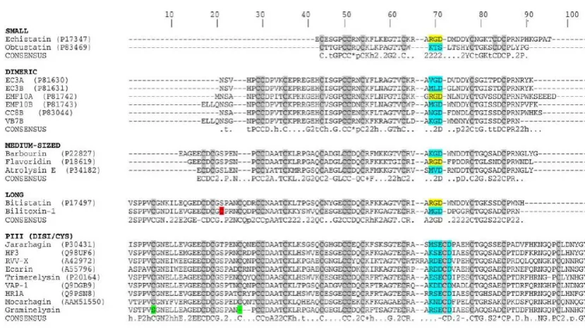
[image:35.595.120.536.168.450.2]15
disintegrin domain (usually characterised by the presence of an RGD motif) is
proteolytically cleaved from the metalloproteinase domain, giving rise to a ‘free’
disintegrin. In the PIIb class, the disintegrin domain is also proteolytically processed
but remains part of the enzyme structure. The PIIc SVMPs represent a dimeric form
of the PIIb class. The PIId and PIIe classes are precursor enzyme forms which give
rise to homodimeric and heterodimeric RGD-containing disintegrins.
The sub-classifications of PIII SVMPs all contain additional disintegrin-like and
cysteine-rich domains in their nascent form (Fox and Serrano, 2005, Fox and
Serrano, 2008). The disintegrin domain here shares some sequence homology to the
disintegrin domains of PII SVMPs but is said to be structurally distinct due to
differences in the sequence homology of the RGD integrin-binding site. In the PIIIa
subclass, the disintegrin-like and cysteine-rich domains are not proteolytically
processed from the metalloproteinase domain, whereas in the PIIIb subclass,
proteolytic processing occurs. The PIIIc subclass represents a dimeric form of the
PIIIa. The PIV (recently classified as PIIId) SVMPs contain, in addition to the
domains described for PIII SVMPs, disulphide-bonded CTL-like domains (Fox and
Serrano, 2005, Fox and Serrano, 2008).
SVMPs have been shown to exhibit a diverse array of biological activities. The
proteolytic activities of the SVMPs are largely associated with the metalloproteinase
domain and characteristically include haemorrhage, apoptosis, myonecrosis,
proteolysis, fibrinolysis, inhibition of platelet aggregation and activation of
coagulation factors such as prothrombin, extensive tissue necrosis, oedema and
inflammation (Baramova et al., 1989, Gutiérrez and Rucavado, 2000, Gutiérrez et al.,
2005b, Kamiguti et al., 1996). This is due to the wide range of cellular targets of
16
coagulation factors (Escalante et al., 2006, Franceschi et al., 2000, Rucavado et al.,
1998). The non-proteolytic activities of the SVMPs are mostly associated with the
disintegrin and cysteine-rich domains contained in the PII and PIII classes.
SVMPs are one of the most abundant enzyme families in viper venoms. Recent
transcriptomic and proteomic investigations have estimated that the SVMP
composition of most viperid venoms is at least 32%, (Bazaa et al., 2005, Juárez et al.,
2006, Sanz et al., 2008), with the most SVMP-rich venom identified from the
saw-scaled viper, Echis ocellatus of which SVMPs account for approximately 67% of the
total protein composition (Wagstaff et al., 2009), whereas SVMPs constitute very
little of the composition of elapid venoms (Li et al., 2004a, Nawarak et al., 2003).
The high proportion of SVMPs in venoms, and their broad range of highly
destructive toxic properties, gives an indication of the functional and pathological
importance of this group of enzymes during viper envenomation.
1.5.2. Serine proteases
Snake venom serine proteases (SPs) are a group of enzymes present in snake venoms
which affect reactions involved in the blood coagulation cascade. SPs can be
categorised into thrombin-like or kallikrein-like proteases. The thrombin-like
subgroup contains enzymes functionally related to thrombin, defined by their ability
to cleave fibrinogen, releasing fibrinopeptides following cleavage the Arg-Lys bonds
on the α- and β-chains of fibrinogen, and subsequently converting fibrinogen to
fibrin (Pirkle, 1998). Many thrombin-like SPs in venoms mimic other catalytic
17
coagulation cascade involved in platelet aggregation (e.g. factor V) (Siigur et al.,
1999).
Kallikrein-like SPs in snake venoms are similar to the mammalian kallikrein
enzymes which initiate the release of bradykinin through the proteolytic cleavage of
kininogen (Matsui et al., 2000). Bradykinin is a potent vasodilator which increases
vascular permeability, resulting in hypotensive symptoms in victims following
envenomation with venom containing kallikrein-like SPs (Warrell, 2010). Some SPs
in venom show both kallikrein-like and thrombin-like protease activities such as the
SPs halytase isolated from Agkistrodon halys blomhoffii (Matsui et al., 1998).
Halytase showed sequence homology to thrombin-like snake venom SPs (66-72%),
mammalian tissue kallikrein enzymes (42%) and thrombin (26%) and was shown to
specifically cleave both fibrinogen and kininogen, although this enzyme appeared to
be devoid of coagulant activity (Matsui et al., 1998). Other enzymes such as
crotalase, also show both kinin-releasing and coagulant activities (Markland, 1976).
In addition to the SVMPs, SPs are also highly pathologically important in viper
envenoming. The abundance of SPs in viper venom ranges from 2 to 31% of the total
protein composition of venom (Gutiérrez et al., 2009). Although SPs in snake venom
exert a wide range of biological activities affecting the coagulation cascade and
haemostatic system, individual SPs usually catalyse a specific reaction in the
coagulation cascade with macromolecular specificity to their protein target
18
1.5.3. Phospholipase A2s (PLA2s)
Phospholipase A2s (PLA2s) are esterolytic enzymes and are occur abundantly in nature. Snake venoms are also a rich source of PLA2s. The primary action of these
enzymes is in the hydrolysis glycerophospholipids at the sn-2 position of the glycerol
backbone. Snake venom PLA2s can be classified into two groups based on amino acid sequence, three-dimensional structure and disulphide bond patterns. Group I
PLA2s are found in the mammalian pancreas and in the venoms of elapid and colubrid snakes. Group I PLA2s are typically 115-120 amino acid residues in length
with 7 disulphide bridges. Elapid venom PLA2s contain a characteristic loop that connects the catalytic α-helix and the β-wing known as the elapid loop, whereas
mammalian PLA2s contain an additional extension of 5 amino acids, called the
pancreatic loop. Group II PLA2s enzymes are present in viper venoms and typically contain 120-125 amino acid residues with 7 disulphide bridges. This group lack the
pancreatic or elapid loop which is characteristic of group I PLA2s, but contain an additional C-terminal extension. Group II PLA2s can be further divided into groups based on the amino acid residue at the 49th position. Those with an aspartic acid
residue are known as D49 enzymes; aspartic acid plays an important role in catalysis
and is therefore conserved in most group II PLA2s (Scott et al., 1990). Amino acid substitutions at the 49th position can occur; where the amino acid residue is replaced
by lysine, serine, asparagine or arginine, enzymes are identified as K49 (Maraganore
et al., 1984), S49 (Polgár et al., 1996), N49 (Tsai et al., 2004) or R49 (Chijiwa et al.,
2006). Enzymes in which the aspartic acid residue has been substituted show low or
19
by elapids predominantly responsible for the cause of death by paralysis of prey and
are therefore vital for the envenomation strategy of this family of venomous snakes.
Snake venom PLA2s show remarkable functional diversity and can exert a wide variety of pharmacological effects including pre- and post-synaptic neurotoxicity,
myotoxicity, cardiotoxicity, coagulopathy, hypotension, haemolysis and
proinflammatory, in addition to the potential role they play in digestion of prey (Kini,
2003, Kini, 2006, Montecucco et al., 2008). Some PLA2s exhibit pre- or post-synaptic neurotoxicity. These enzymes belong to the group I PLA2s and are abundant
components of elapid venoms. Pre-synaptic PLA2s disrupt neurotransmitter release at the neuromuscular junction, the effects of which are irreversible. Pre-synaptic
neurotoxins have been shown to specifically target voltage-sensitive K+ channels (e.g. β-bungarotoxin (Black et al., 1988)), brain synaptic membrane proteins (e.g.
ammodytin (Kriz a et al., 5)) and neuronal Ca2+ binding proteins (e.g. taipoxin
(Kirkpatrick et al., 2000)). Post-synaptic neurotoxins extracellularly target the
neuromuscular junction by competitively blocking acetylcholine receptors, the
effects of which can be reversed by antivenom treatment.
Viper venoms primarily contain group II PLA2s. Some group II PLA2s such as crotoxin and notexin have myotoxic properties and can induce myonecrosis or
system myotoxicity (Gopalakrishnakone et al., 1984, Mebs and Ownby, 1990).
Others have been demonstrated to evoke inflammatory events such the PLA2 isolated from Naja naja atra venom which promotes inflammatory cell infiltration (Zhang
20
1.5.4. L-amino acid oxidases (LAOs)
L-amino acid oxidases (LAOs) are homodimeric flavoenzymes which catalyse the
oxidative deamination of L-amino acids to form α-ketoacids, in addition to the
production of ammonia and hydrogen peroxidase. LAOs occur frequently in nature
and are present in the venom of most snakes, occurring most abundantly in the
venom of Crotaline snakes. The proportion of LAO in venoms as a percentage of the
total weight of dried venoms ranges from 1-4% up to 30% in venom from the
Malayan pitviper, Calloselasma rhodostoma (Ponnudurai et al., 1994). The venoms
from mambas and sea kraits contain trace amounts of LAO and colubrid venoms also
appear to be devoid of LAO activity (Mackessy, 2002).
Several LAOs have been shown to affect platelet aggregation. LAOs purified from
the venoms of Naja naja kaouthia (Tan and Swaminathan, 1992) and Agkistrodon
halys blomhoffii (Takatsuka et al., 2001) demonstrated platelet aggregating inhibiting
properties, whereas LAOs from the venoms of Ophiophagus hannah (Ahn et al.,
1997) and Eristocophis macmahoni (Ali et al., 2000) cause an induction of platelet
aggregation. Other LAOs show additional effects including haemolysis and
oedema-induction (e.g. Trimeresurus flavoviridis (Abe et al., 1998) and E. macmahoni venom
LAOs (Ali et al., 2000)). Other venom LAOs have been reported to demonstrate
cytotoxic effects (e.g. O. hannah (Ahn et al., 1997)) or antibacterial properties (Stiles
et al., 1991, Stábeli et al., 2004, Izidoro et al., 2006). LAOs from snake venoms have
been reported to show moderate lethal toxicity and are therefore not thought to be a
21
1.5.5. Hyaluronidase
Hyaluronidase is an enzyme which specifically degrades hyaluronan, a ubiquitous
component of the extracellular matrix which is essential in maintaining the structure
and viscosity of the extracellular matrix. Hyaluronan has also been implicated in
many biological processes such as wound healing and inflammation (Kreil, 1995,
Stern and Jedrzejas, 2006). Although hyaluronidases are not thought to be toxic
themselves, the resultant effect of hyaluronidase activity leads to an increased
permeability of the extracellular matrix (Kudo and Tu, 2001). This facilitates the
diffusion of toxins into tissues of the envenomed prey and assists the entry of more
destructive venom enzymes such as proteases into the haemostatic system, therefore
contributing to the local and systemic effects of envenomation (Pukrittayakamee et
al., 1988). Thus, hyaluronidase has been termed the ‘venom spreading factor’.
Hyaluronidase is a highly conserved constituent component which is ubiquitously
expressed in all snake venoms. Despite its important role in snake envenomation,
hyaluronidase remains a relatively unstudied venom toxin as the rapid autolysis of
this protein has inhibited its chromatographic isolation preliminary to functional
studies. Hyaluronidases have been isolated and purified from the venoms of
Deinagkistrodon acutus (Xu et al., 1982), Agkistrodon contortrix (Kudo and Tu,
2001) and the Indian Cobra, Naja naja (Girish et al., 2004). A study by Harrison et al
determined the nucleotide sequence of hyaluronidase from several viperid species
(Echis ocellatus, Echis pyramidum leakeyi, Cerastes cerastes cerastes and Bitis
arietans), demonstrating a high level of sequence homology between species (over
95%) (Figure 1.7) (Harrison et al., 2007). This study, for the first time, determined
the sequence structure of a snake venom hyaluronidase which has five conserved
22
Figure 1.7 Snake venom hyaluronidase sequence analysis: Alignment of inferred full length hyaluronidase venom gland cDNA sequences from Echis ocellatus,
Cerastes cerastes, Echis pyramidum leakeyi, Bitis arietans, Echis carinatus
sochureki with EST sequences from Agkistrodon acutus and Lachesis muta from
Harrison et al 2007 (Harrison et al., 2007). The conserved catalytic and positional
residues are identified by the solid and open triangles respectively.
cysteine scaffold regions with a predicted molecular mass of over 50 kDa (Harrison
et al., 2007).
Hyaluronidase activity facilitates the diffusion of venom proteins into prey tissues
and therefore results in the rapid influx of target-specific venom toxins into the
circulating blood system and thus, is an essential component of all snake venoms. It
is also thought that hyaluronidase is also involved in the symptoms of extensive local
tissue destruction observed following snakebite. Current antivenoms are unable to
neutralise the local symptoms and so the resultant uncontrollable tissue destruction
23
hyaluronan, a key structural component of the extracellular matrix, it is thought that
hyaluronidase may be one of several factors involved in causing severe tissue
damage as it has been demonstrated that the inhibition of hyaluronidase presents
local tissue destruction (Yingprasertchai et al., 2003). In addition, the inhibition of
hyaluronidase by injection of inhibitors following envenomation can delay the time
to death (Girish and Kemparaju, 2006) which also implicates the potential
therapeutic use of hyaluronidase inhibitors in the treatment of snakebite.
1.5.6. Acetylcholinesterase (AChE)
Acetylcholinesterase (AChE) is present in all vertebrates and plays a functional role
in the cholinergic system in the rapid hydrolysis and subsequent inactivation of the
neurotransmitter, acetylcholine at the neuromuscular junction (Tougu, 2001). AChE
has been detected at high levels in most elapid venoms, with the exception of the
genus Dendroaspis which instead contains fasciculins, potent inhibitors of AChE
(Karlsson et al., 1984, Cousin and Bon, 1997, Frobert et al., 1997). Studies analysed
the AChE activity of 45 snake venoms and detected an absence of AChE activity in
viperid and crotalid venoms (Frobert et al., 1997). In this study, a variation in the
stability of AChEs among elapid venoms was apparent; AChE isolated from
Bungarus venoms was much more stable than AChE isolated from venoms of
Hemachatus, Ophiophagus and Naja species (Frobert et al., 1997). Venoms from
Bungarus species were also shown to have the richest source of AChE activity with
over twice as much activity as Naja venoms (Frobert et al., 1997). Despite the
common occurrence of AChE in venoms, the biological function of AChE during
24
1.5.7. Nucleases, nucleotidases and phosphomonoesterases
Hydrolytic enzymes, such as nucleases (DNase, RNase and phosphodiesterase),
nucleotidases (5’ nucleotidase, ATPase and ADPase) and phosphomonoesterases
(acid and alkaline phosphomonoesterases) have been found in venoms.
Nucleases are enzymes which act on nucleic acids (DNA/RNA). Venoms have been
shown to contain both endonucleases which degrade either DNA or RNA
specifically, and exonucleases which degrade both DNA and RNA. Currently, there
have been very few studies investigating venom nucleases and so the occurrence,
distribution and function are not fully understood. RNases have been identified from
the venoms of Naja naja oxiana (Vasilenko and Ryte, 1975) and Naja naja
(Mahalakshmi and Pandit, 1987, Mahalakshmi et al., 2000), and DNases have only
been identified from Bothrops atrox venom (Georgatsos and Laskowski, 1962).
Phosphodiesterases (PDEs) catalyse the phosphodiester bonds in polynucleotides
acting on DNA, rRNA and tRNA. Unlike DNases/RNases, PDEs have been found
ubiquitously in snake venoms across a wide range of taxa including colubrids,
elapids and viperids (Aird, 2002, Aird, 2005, Mackessy, 2002) although few studies
have attributed a biological function to this enzyme group.
Nucleotidases act upon nucleic acid derivative and nucleic acid-related substrates
(e.g. ATP and ADP). 5’ nucleotidases catalyse the hydrolysis of phosphate from the
sugar moiety. These enzymes have also been found ubiquitously in snake venoms
with viper venoms showing a greater 5’ nucleotidase activity than elapids (Aird,
2005). There is, again, a lack of information about the biological role of 5’
nucleotidases in snake venoms, although 5’ nucleotidases in the venoms from
25
(Ouyang and Huang, 1983)were shown to inhibit platelet aggregation either induced
by ADP, collagen, sodium arachidonate, ionophore A-23187 in platelet-rich plasma
and thrombin in platelet poor plasma. More recently, 5’ nucleotidases in the venom
of Naja naja have been shown to be involved in the anticoagulant effect of this
venom by interacting with anticoagulant factors (Dhananjaya et al., 2006).
ATPases and ADPases catalyse the hydrolysis of ATP and ADP respectively.
ATPase and ADPase activity have been observed in several snake venoms (Kini and
Gouda, 1982a, Kini and Gouda, 1982b, Sales and Santoro, 2008). As the ATPases
from venoms have not been purified, little is known about their biological role in
venom. The ADPases, for example in D. acutus venom, have been shown to have
platelet aggregation inhibiting properties (Ouyang and Huang, 1986).
Phosphomonoesterases catalyse non-specific hydrolysis of phosphate esters. Acid
phosphomonoesterases are most active at pH 5.0 whereas alkaline
phosphomonoesterases are most active at pH 9.5. Both acid and alkaline
phosphomonoesterases have been found in venoms, although the alkaline enzymes
appear more widespread and abundant, appearing in venoms from a range of snake
taxa (Mackessy, 2002, Aird, 2002). However, phosphomonoesterases in venoms are
yet to be characterised or assigned a biological role.
Non-enzymatic components:
1.5.8. C-type lectins (CTLs)
C-type lectins (CTLs) are non-enzymatic proteins found in many animals which bind
26
are two types of CTLs; CTL-like proteins (CLPs) and the classic sugar binding snake
lectins. CLPs are found only in snake venoms and show a level of sequence
homology with classic CTLs, but lack the Ca2+ binding loop which recognises
carbohydrates (Ogawa et al., 2005). The basic structure of snake CTLs is a
homodimeric form, whereas CLPs are heterodimers composed of homologous α- and
β-subunits, interacting with a loop linked by a disulphide bond.
Sugar binding CTLs are not highly toxic components of snake venoms. They have
been shown to initiate various immunological processes including adhesion,
endocytosis and pathogen neutralisation (Weis et al., 1998). They have also
demonstrated the ability to inhibit tumour cell lines and endothelial cell growth (de
Carvalho et al., 2001), thus their role in snake venom is currently not fully
understood. CLPs, on the other hand, show a diverse array of pharmacological
activities affecting coagulation factors, platelets, haemostasis and thrombosis and can
be divided into three subgroups based on biological activity; these are coagulant
proteins, platelet aggregation agonists and platelet aggregation antagonists (Morita,
2005, Ogawa et al., 2005).
Firstly, anticoagulant proteins, which target the coagulation factors X and factor IX,
have been isolated from several venoms. These bind to the γ-carboxyglutamic acid
(Gla) domain of factor X and IX in the presence of Ca2+ (Atoda et al., 1994). Anticoagulant CTLs have been identified from the venoms of Bothrops jararaca
(Sekiya et al., 1993), Trimeresurus flavoviridis (Atoda et al., 1994), Echis carinatus
leucogaster (Chen and Tsai, 1996), Deinagkistrodon acutus (Atoda et al., 1998) and
Agkistrodon halys brevicaudus (Koo et al., 2002). Secondly, platelet aggregation
agonists, which have been isolated from the venoms of Trimeresurus albolabris