October 8-11, 2006, Taipei, Taiwan
Abstract—The purpose of this work is to develop an interactive tool which helps botanists to extract the vein system with its hierarchical properties with as little user interaction as possible. In this paper, we present a new venation extraction method using independent component analysis (ICA). The popular and efficient FastICA algorithm is applied to patches of leaf images to learn a set of linear basis functions or features for the images and then the basis functions are used as the pattern map for vein extraction. In our experiments, the training sets are randomly generated from different leaf images. Experimental results demonstrate that ICA is a promising technique for extracting leaf veins and edges of objects. ICA, therefore, can play an important role in automatically identifying living plants.
I. INTRODUCTION
lant is one of the most important forms of life on earth. Plant recognition is very demanding in biology and agriculture as new plant discovery and the computerization of the management of plant species become more popular. The recognition is a process resulting in the assignment of each individual plant to a descending series of related plants in terms of their common characteristics. The process is very time-consuming as it has been mainly carried out by botanists. Computer-aided plant recognition is still very challenging task in computer vision as the lack of proper models or representation schemes, a large number of variations of the plant species, and imprecise image preprocessing techniques, such as edge detection and contour extraction. The focus of computerized living plant recognition is on stable feature’s extraction of plants. The information of leaf veins, therefore, play an important role in identifying living plants.
Another advantage of vein feature extraction is that a botanical interpretation of leaves needs the data to be in a form that allows comparisons between different tree leaves.
This work was supported by research grants from the Hong Kong Polytechnic University, Hong Kong (project no: A/Cs G-T851 and B-Q590).
Yan Li is with the Department of Mathematics and Computing, The University of Southern Queensland, QLD 4350, Australia. (corresponding author: phone: 61 7 46315533 fax: 61 7 4631 5550; e-mail: liyan@ usq.edu.au).
Zheru Chi is with the Department of Electronic and Information Engineering, The Hong Kong Polytechnic University, Hong Kong. (e-mail: enzheru@polyu.edu.au).
Dagan Feng is with the Department of Electronic and Information Engineering, The Hong Kong Polytechnic University, Hong Kong. (e-mail: enfeng@polyu.edu.au).
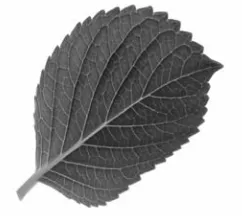
[image:1.612.376.497.249.361.2]Therefore the results of the growth analysis have to be transformed into a coordinate system affixed to the leaf [1]. The obvious axes of this coordinate system are the leaf veins. The leaf vein system is a hierarchical entity with a main vein and side veins of possibly several orders. Plant leaves are well structured objects consisting of line-like veins and areas in between as shown in Fig.1.
Fig. 1. Vein structure in a tree leaf.
Recently, researchers started to investigate on the extraction of venation and vein-like objects. Gouveia et al [2] proposed a two-step solution to segment the veins of a chestnut-tree leaf whose secondary venations are approximately straight and have the same inclination. Sollie [3] applied morphological filters to extract leaf veins. Fu and Chi [4] developed an efficient two-stage approach for leaf vein extraction. At the first stage of their method, a preliminary segmentation based on the intensity histogram of the leaf image was carried out to estimate the rough regions of vein pixels. This was followed at the second stage by a fine checking using a trained artificial neural network classifier. They demonstrated that the approach was capable of extracting more precise venation modality of the leaf than the conventional edge detection methods.
The purpose of this work is to develop an interactive tool which helps botanists to extract the vein system with its hierarchical properties with little user interaction. In this paper, we present a new venation extraction technique by using Independent Component Analysis (ICA). To date, ICA has been successfully applied to blind signal separation problem in both speech signal processing and medical signal processing (such as EEG signals) areas. Recently, some researchers applied ICA to learn efficient codes of natural images that utilize a set of linear basis functions or features. Olshausen and Field [5] used a sparseness criterion and found codes that were similar to localized and oriented receptive fields. Similar results were also obtained by Bell
Leaf Vein Extraction Using Independent Component Analysis
Yan Li, Zheru Chi, Member, IEEE, and David D. Feng, Fellow, IEEE
and Sejnowski [6] and Lewicki and Olshausen [7] using the Infomax ICA algorithm and Bayesian approach, respectively.
In this paper, we apply the FastICA algorithm to patches of leaf images to learn the basis functions and then the basis functions are used as the pattern map for vein detection. A gray-scale image is transformed into a pattern map (feature map) in which the leaf, edge, background and other pixels are classified into different classes by pattern matching. Compared with conventional mathematics-based templates such as Harr transform [8] and Gabor functions [9], the proposed method is based on the statistics of images. High accuracy of vein detection can be achieved, and it is free of the influences of illumination and there is no need to pre-processing. ICA algorithms attempt to find sparse linear codes for images, and result in a complete family of localized, oriented, bandpass receptive fields, similar to those found in the primary visual cortex [5-7]. The resulting sparse image code provides a more efficient representation for leaf images because it possesses a higher degree of statistical independence among its outputs.
The rest of the paper is organized as follows. The fundamentals of independent component analysis techniques are briefly introduced in Section II. In Section III, a popular ICA algorithm, FastICA, is summarized. We, then, apply the FastICA algorithm to a set of leaf images for vein extraction. Experimental results are presented in Section IV. Finally, Section V concludes the paper.
II. INDEPENDENT COMPONENT ANALYSIS REPRESENTAION OF NATURAL IMAGES
Independent Component Analysis (ICA) is a signal processing method to extract independent sources given only observed data that are mixtures of the unknown sources. ICA was originally developed for the blind signal separation (BSS) problem [10]. Recently, there have been a considerable amount of papers presenting the applications of ICA algorithms on image data, such as [8, 11-13].
In ICA, the data variables, x1, x2, …, xN, are assumed to be
the mixtures of unknown statistically independent variables, s1, s2, …, sN, which can be expressed as
∑
= = n j j ij i a s X1
(1)
Where the coefficients, aij (1≤i,j≤N), are the unknown
mixing system. In a matrix form, it is represented as
X=AS (2)
Here, X=[x1 x2, … xN]T is the observations, S=[s1 s2, … sN]T is
the independent sources, and A is an NxN mixing matrix. For image processing application, a perceptual system is exposed to a series of small image patches, taken from one or more large images. The pixel gray-scale intensity (or
contrast) in an image, or in practice, a small image patch is an observation, xi. We consider each image patch as a linear
superposition of some features or basis vectors aij that are
constant for an image. The S are stochastic coefficients, different from patch to patch. In a neuronscientific interpretation, the variables si(i=1…n) model the responses
of simple cells, and the columns of A are closely related to their receptive fields. The basis functions form the columns of the constant matrix, A. The weighting of this linear combination is given by a vector, S. Each component of this vector has its own associated basis function, and represents an underlying “cause” of the image.
The goal of a perceptive system is to linearly transform the images, X, with a matrix of filters, W, so that the resulting vector:
Y=WX (3)
recovers the underlying causes, S, possibly permuted or rescaled. The estimation of the model is also equivalent to determine the values of W=A-1. This is a case of unsupervised learning as there is no ‘teacher’ to give the right output values si(i=1,2,…n) to the system.
III. THE FASTICA ALGORITHM
We use the FastICA training algorithm to learn the unmixing filters, W, in this paper. FastICA is an efficient and popular algorithm for independent component analysis invented by Aapo Hyvärinen [14]. The algorithm is based on a fixed-point iteration scheme maximizing non-gaussianity as a measure of statistical independence. It can be also derived as an approximative Newton iteration.
Hyvarinen and his co-workers have introduced a family of fixed-point algorithms. The members of this family are differentiated firstly by the algorithmic approach and secondly by the contrast function used. The key to all the variations is to find independent components by separately maximising the negentropy of each mixture [14]. There are mainly two algorithmic approaches, the symmetric approach and deflation approach, in the fixed-point algorithm class. The symmetric approach uses a modified rule for the update of the unmixing matrix W that enables simultaneous separation of all independent components, whereas the deflation approach updates the columns of W individually, finding the independent components once at a time. Either of these approaches is able to use some non-quadratic contrast function to provide estimates of negentropy [14]. The original algorithm use kurtosis, but more recent versions use the hyperbolic tangent, exponential or cubic functions.
The update rule for the deflation method is given by:
) 1 ( )] ) 1 ( ( ' [ )] ) 1 ( ( [ ) ( 1 * − − − −
=C−E xg wk x Eg wk x wk
k
w T T
) ( ) (
) ( )
(
* *
*
k Cw k w
k w k
w
T
= (5)
where E[.] is the expectation operation, w*(k) is the complex conjugate of w(k), g can be any suitable non-linear contrast function, with derivative g’, and C is the covariance matrix of the mixtures, X.
IV. LEAF VEIN EXTRACTION USING ICA
In this section, we present the experimental results of leaf vein extraction using the FastICA algorithm. In our experiments, the training set consisted of randomly generated 50,000 12x12 pixel patches from 12 sub-images, taken from 21 kinds of tree leaf images. For any two points p(i,j) and p(i’,j’), their gray-scale values (contrasts) were uncorrelated over time as we sampled them randomly. The training set was considered as the observation data in the ICA model. Before the learning, the means of the data components were subtracted and the components were scaled to unit variance. This implies that there is no need to estimate the bias vectors in case of the image applications presented here.
Based on ICA, each image patch can be represented by a linear combination of ‘basis’ patches [15]. Therefore, the image X is the linear combination of basis images aj the
[image:3.612.323.549.73.303.2]columns of A as shown in Fig. 2.
Fig. 2. The linear image synthesis model.
Each patch is represented as a linear combination of basis patches. In sparse coding, one attempts to find a representation such that the coefficients sj are as ‘sparse’ as
possible, meaning that for most image patches only a few of them are significantly active. In ICA, the purpose is to find a representation such that they are mutually as statistically independent as possible.
Obviously, for different images, its matrix A is different. So is the inverse W. In other words, W reflects the features of the image. In this paper, W is a 12x12 matrix and contains 144 parameters.
Fig. 3 shows the basis functions trained by FastICA, which are the columns of coefficient matrix, A. Each row of W can be considered as a filter for the image edge detection or other image processing tasks. The filters resemble Gabor filters from mathematics formulae [9], which are used to enhance edge features. Olshausen and Field got this similar results by sparseness and maximization network and argued that this is a family of localized, oriented, and bandpassed receptive fields [5]. The most features are corresponded to the veins in the leaf images.
Fig. 3 Basis Functions for 12x12 pixel patches.
Several original leaf subimages (a) and their corresponding extracted veins by using the proposed algorithms (b) are shown in Fig. 4.
We also applied the ICA basis functions to several whole leave images and compared the results with the popular edge detection operator, Prewitt operator. Fig. 5 shows two samples of the results. It is noted that the results using the FastICA algorithm for the whole leaves are not as good as those shown in Fig. 4, but comparable to the results by using Prewitt edge detection operator. Prewitt edge detection operator is a powerful feature detection algorithm used in computer vision. The performance of FastICA algorithm is not better than those of Prewitt because FastICA is a linear technique and was originally developed for the blind signal separation problem. Following up this study, we will apply our nonlinear ICA algorithm [16] to extract leaf venation and we consider the vein extraction as a very finer case of edge detection. Another task in the future is to compare the methods with other existing approaches.
V. CONCLUSION
[image:3.612.57.297.402.447.2](a) The original leaf subimages
[image:4.612.58.295.65.505.2](b) their extracted veins
Fig. 4 The extracted veins and their original leaf images.
a. original images
b. the vein extraction using ICA
[image:4.612.73.285.564.681.2]c. the vein extraction using Prewitt operator
Fig.5 The vein extraction of whole leaves using the ICA and Prewitt and their original leaf images.
ACKNOWLEDGMENT
The work reported in this paper was supported by research grants from The Hong Kong Polytechnic University, Hong Kong. (Project No: A/Cs G-T851 and B-Q590).
REFERENCES
[1] D. Schmundt and U. Schurr, “Plant leaf growth studies by image sequence analysis, “, In B. Jahne, H. Haubecker, and P. Geibler, editor, Computer Vision and Applications Volume 2, Signal Processing and Pattern Recognition, Academic Press, San Diego, New York, Boston, 1990.
[2] F. Gouveia, V. Filipe, M. Reis, C. Couto and J. Bulas-Cruz, ‘Biometry: the characterization of chestnut-tree leaves using computer vision,” ISIE’97, pp. 757-760, 1997.
[3] P. Sollie, “Morphological image analysis applied to crop field mapping,” Image and Vision computing, pp. 1025-1032, 2000. [4] H. Fu and Z. Chi, “A two-stage approach for leaf vein extraction,” Proceedings of International Conference on Neural Networks and Signal Processing (ICNNSP2003), Vol. I, pp. 208-211, Nanjing, Jiangsu, China, December 12-15, 2003.
[5] B. Olshausen and D. Field, “Emergence of simple-cell receptive field properties by learning a sparse code for natural images,”
Nature, 381, 1996, pp. 607-609.
[6] A. Bell and Tony Sejnowski, “The independent components of nature scenes are edge filters,” Vision Research, 37, 1997, pp. 3327-3338.
in Neural Information Processing Systems, 10, 1998, pp. 556-562.
[8] W. B. Park, E. Ryu and Y. J. Song, “Visual feature extraction under wavelet domain for image retrieval,”, Key Engineering Materials, Vol. 277., pp. 206-211, 2005.
[9] B. S. Manjunath, W. Y. Ma, “Texture features for browsing and retrieval of iamge data,”, IEEE Transactions on Pattern Analysis and Machine Intelligence, Vol. 18(8), pp. 837-842, 1996.
[10] P. Comon, “Independent component analysis - a new concept?”, Signal Processing, vol. 36, pp. 287-314, 1994. [11] T. W. Lee and M. Lewicki, ‘Unsupervised Image
Classification, Segmentation and Enhancement Using ICA Mixture Models,” IEEE Transactions on Image Processing, Vol. 11, No. 3, 2002, pp. 270-279.
[12] A. Hyvarinen and P. Hoyer and J. Hurri, “Extensions of ICA as Models of Natural Images and Visual Processing,” 4th International Symposium on Independent Component Analysis and Blind Signal Separation (ICA2003), Nara, Japan, pp. 963-974.
[13] A. Hyvärinen, M. Gutmann and P.O. Hoyer, “Statistical model of natural stimuli predicts edge-like pooling of spatial frequency channels in V2,”BMC Neuroscience, Vol. 6, pp. 6-12, 2005.
[14] A. Hyvarinen, “A family of fixed-point algorithms for independent component analysis”, In Proceedings of IEEE International Conference on Acoustics, Speech and Signal Processing (ICASSP’97), pp. 3917-1920, Munich, Germany, 1997.
[15]P. O. Hoyer and A. Hyv¨arinen, “Independent component analysis applied to feature extraction from color and stereo images,” Network: Computation in Neural Systems, Vol. 11, pp. 191-210, Mar 2000. [16] Y. Li, D. Powers and K. Pope, “Blind signal separation using

