Altered resting-state EEG source
functional connectivity in schizophrenia:
the effect of illness duration
The Harvard community has made this
article openly available. Please share how
this access benefits you. Your story matters
Citation
Di Lorenzo, G., A. Daverio, F. Ferrentino, E. Santarnecchi, F.
Ciabattini, L. Monaco, G. Lisi, et al. 2015. “Altered resting-state EEG
source functional connectivity in schizophrenia: the effect of illness
duration.” Frontiers in Human Neuroscience 9 (1): 234. doi:10.3389/
fnhum.2015.00234. http://dx.doi.org/10.3389/fnhum.2015.00234.
Published Version
doi:10.3389/fnhum.2015.00234
Citable link
http://nrs.harvard.edu/urn-3:HUL.InstRepos:16121121
Terms of Use
This article was downloaded from Harvard University’s DASH
repository, and is made available under the terms and conditions
applicable to Other Posted Material, as set forth at
http://
Edited by: Zafiris J. Daskalakis, University of Toronto, Canada Reviewed by: Tamer Demiralp, Istanbul University, Turkey Qingbao Yu, The Mind Research Network, USA *Correspondence: Giorgio Di Lorenzo, Via Nomentana, 1362, 00137 Rome, Italy [email protected]
Received:02 September 2014 Accepted:11 April 2015 Published:05 May 2015 Citation: Di Lorenzo G, Daverio A, Ferrentino F, Santarnecchi E, Ciabattini F, Monaco L, Lisi G, Barone Y, Di Lorenzo C, Niolu C, Seri S and Siracusano A (2015) Altered resting-state EEG source functional connectivity in schizophrenia: the effect of illness duration. Front. Hum. Neurosci. 9:234. doi: 10.3389/fnhum.2015.00234
Altered resting-state EEG source
functional connectivity in
schizophrenia: the effect of illness
duration
Giorgio Di Lorenzo1, 2*, Andrea Daverio1, 2, 3, Fabiola Ferrentino2, 3,
Emiliano Santarnecchi4, 5, Fabio Ciabattini1, 2, 3, Leonardo Monaco1, 2, Giulia Lisi2, 3,
Ylenia Barone2, 3, Cherubino Di Lorenzo6, Cinzia Niolu2, 3, Stefano Seri7 and
Alberto Siracusano1, 2, 3
1Laboratory of Psychophysiology, Chair of Psychiatry, Department of Systems Medicine, University of Rome “Tor Vergata”,
Rome, Italy,2Chair of Psychiatry, Department of Systems Medicine, University of Rome “Tor Vergata”, Rome, Italy, 3Psychiatric Clinic, Fondazione Policlinico “Tor Vergata”, Rome, Italy,4Department of Medicine, Surgery and Neuroscience,
University of Siena, Siena, Italy,5Berenson-Allen Center for Non-Invasive Brain Stimulation, Beth Israel Medical Center,
Harvard Medical School, Boston, MA, USA,6Don Carlo Gnocchi Onlus Foundation, Milano, Italy,7School of Life and Health
Sciences, Aston Brain Centre, Aston University, Birmingham, UK
Despite the increasing body of evidence supporting the hypothesis of schizophrenia as a disconnection syndrome, studies of resting-state EEG Source Functional Connectivity (EEG-SFC) in people affected by schizophrenia are sparse. The aim of the present study was to investigate resting-state EEG-SFC in 77 stable, medicated patients with schizophrenia (SCZ) compared to 78 healthy volunteers (HV). In order to study the effect of illness duration, SCZ were divided in those with a short duration of disease (SDD;
n = 25) and those with a long duration of disease (LDD; n = 52). Resting-state EEG
recordings in eyes closed condition were analyzed and lagged phase synchronization (LPS) indices were calculated for each ROI pair in the source-space EEG data. In delta and theta bands, SCZ had greater EEG-SFC than HV; a higher theta band connectivity in frontal regions was observed in LDD compared with SDD. In the alpha band, SCZ showed lower frontal EEG-SFC compared with HV whereas no differences were found between LDD and SDD. In the beta1 band, SCZ had greater EEG-SFC compared with HVs and in the beta2 band, LDD presented lower frontal and parieto-temporal EEG-SFC compared with HV. In the gamma band, SDD had greater connectivity values compared with LDD and HV. This study suggests that resting state brain network connectivity is abnormally organized in schizophrenia, with different patterns for the different EEG frequency components and that EEG can be a powerful tool to further elucidate the complexity of such disordered connectivity.
Keywords: schizophrenia, psychosis, brain oscillations, disconnectivity, synchronization, excitatory/inhibitory dysfunction, neural plasticity, brain network
Introduction
This conclusion is based on neuroimaging evidence on structural, functional, and effective brain connectivity (Friston, 1998; Ribolsi et al., 2009; Schmitt et al., 2011). Structural Connectivity (SC) refers to the anatomical substrate of defined fiber pathways con-necting different brain regions (Koch et al., 2002), whereas Func-tional Connectivity (FC) is defined as the temporal correlation of the activities of different cortical/brain regions (Fingelkurts et al., 2005). The definition of Effective Connectivity (EC) has been more contentious (Horwitz, 2003), referring to direct or indi-rect influence that one neural system exerts over another (Friston, 2011).
FC impairment in schizophrenia has been extensively inves-tigated. Studies using functional Magnetic Resonance Imaging (fMRI) have shown abnormal FC both in first episode/early stage (Begre and Koenig, 2008) and in chronic schizophrenic patients (Lynall et al., 2010; Fitzsimmons et al., 2013; Wang et al., 2014). EEG studies, capitalizing on its high temporal resolution, have shown impaired FC in all frequencies bands of the EEG spectrum (Stephan et al., 2006). Transcranial magnetic stimulation (see for reviewRogasch et al., 2014) and transcranial direct current stim-ulation (Hasan et al., 2013; Ribolsi et al., 2013) studies have iden-tified altered inhibitory/excitatory properties of brain networks and of interhemispheric connectivity in schizophrenia. Finally, confirmation of the central role of disordered connectivity has also emerged from animal models of schizophrenia (Dickerson et al., 2010; Gruber et al., 2010).
In schizophrenic patients, altered FC in response to cog-nitive and sensory paradigms has been demonstrated in all the traditional EEG frequency bands (Pachou et al., 2008; Fujimoto et al., 2013) supporting the hypothesis that elec-troencephalographic indices of FC may be a useful marker of specific impairment in higher-order processing (see the review of Uhlhaas et al., 2008). Furthermore, EEG-FC in the rest-ing state reflects the activity of interneuron connections and cortical synchronization that are temporally interrupted dur-ing specific tasks (Cabral et al., 2014) and has the poten-tial to reveal an a priori intrinsic dysfunctionality which may represent either a state or trait-marker of the schizophrenic condition.
An important limiting factor in some EEG-FC studies is that the majority of connectivity measures are based on scalp sensors, which may cause spurious connectivity patterns due to the vulnerability of EEG signal to volume conduction phe-nomena (Nolte et al., 2004). To address this issue, a num-ber of electrical source imaging methods have been proposed as a valid alternative to channel-related FC (Thatcher et al., 2007; Schoffelen and Gross, 2009; Sakkalis, 2011). In this con-text, an index of physiological “lagged connectivity” between pairs of brain sources capable of minimizing non-cerebral arti-facts and that is not affected by active reference electrodes was recently proposed (Pascual-Marqui et al., 2011). This method has been successfully applied to investigate EEG Source Func-tional Connectivity (EEG-SFC) between cortical regions in health (Stein et al., 2013) and disease (Canuet et al., 2011, 2012; Clemens et al., 2011; Pagani et al., 2012; Olbrich et al., 2014).
Using this approach, Lehmann and colleagues showed aber-rant resting-state EEG cortical connectivity pattern in first episode, drug-naïve schizophrenic patients (Lehmann et al., 2014).
The primary aim of this study was to investigate differences in resting-state EEG SFC between a group of clinically stable, med-icated schizophrenic patients, and healthy controls. A secondary aim was to investigate the effect of disease duration on source connectivity patterns in the group of schizophrenic patients.
Materials and Methods
Subjects
From November 2007 to October 2012, patients between 18 and 65 years of age from the outpatient program of “Tor Vergata” University Psychiatry Clinic with a diagnosis of schizophrenia (SCZ) were considered for the study. The inclusion criterion was a stable clinical and pharmacological condition from at least 3 months. The exclusion criteria were history of head trauma, mental retardation, and history in the last 6 months of alcohol, and/or substance abuse. In order to study the effect of illness duration patients were divided in those with short disease dura-tion (SDD, with less than 5 years from the first psychotic episode) and those with long disease duration (LDD, more than 5 years from the first psychotic episode). During the same period, healthy volunteers (HV) of comparable age and socio-economical status without a personal and familiar history of psychiatric disorders were enrolled as controls. The MINI-Plus (Sheehan et al., 1998) was used to confirm the diagnosis of SCZ according to DSM-IV criteria in the clinical sample and the absence of psychiatric diagnosis in the control group. The Positive and Negative Syn-drome Scale (Kay et al., 1987) was used to assess the severity of psychopathology. Based on the recent literature (Van den Oord et al., 2006; van der Gaag et al., 2006; Wallwork et al., 2012; Liemburg et al., 2013) the 30 items (7 for Positive symptoms, 7 for Negative symptoms and 16 for General psychopathology) were rearranged in six dimensions: Positive (POS; P1+P3+P5 +P6+G9); Negative – Expressive (NEG-EXP; the core
nega-tive symptoms characterized by expressive deficits; N1+N3+
N6+ G7); Negative - Social (NEG-SOC; social emotive
with-drawal/social amotivation; N2 + N4 +G16); Disorganization
(DIS; disorganized/concrete/cognitive items; P2+N7+N5+
G10+G11+G12+G5+G13); Excitement (EXC; P4+P7+
G8+G14); Emotional Distress (EMO-D; anxiety and
depres-sion; G1+G2+G3+G4+G6+G15). The total score of the
Calgary Depression Scale for Schizophrenics (CDSS;Addington et al., 1990) was used to measure the depression severity in SCZ sample, independently from confounding factor as the positive and negative symptoms of SCZ and any drug effects. The pre-morbid Intelligence Quotient (IQ) was investigated, in the HV and SCZ, through a reading task, theTest di Intelligenza Breve
Social and Occupational Functioning Assessment Scale (SOFAS;
American Psychiatric Association, 2000). All subjects enrolled in the study were right-handed on the basis of the Edinburgh Handedness Inventory (Oldfield, 1971). All participants pro-vided written informed consent. The study was approved by the “Tor Vergata” University Hospital ethics committee and carried out in accordance with “Ethical Principles for Medical Research Involving Human Subjects” (Declaration of Helsinki, 1964).
EEG Recordings and Data Acquisition
The EEG was recorded from 37 scalp locations using a pre-cabled electrode cap (Bionen, Florence, Italy), with Ag/AgCl disk electrodes located at the following positions: FP1, FPz, FP2, AF7, AF3, AF4, AF8, F7, F3, Fz, F4, F8, CP5, CP1, CP2, CP6, T7/T3, C3, Cz, C4, T8/T4, CP5, CP1, CP2, CP6, P7/T5, P3, Pz, P4, P8/T6, PO7, PO3, PO4, PO8, O1, Oz, O2. Recording channels were connected to a reference electrode located in the AFz position and the ground electrode was in POz. Electrode impedances were kept to less than 10 K. The signal was ampli-fied by 40-channel EEG device (Galileo MIZAR-sirius, EBNeuro, Florence, Italy) and acquired with GalNT software. Data were collected with a sampling rate (SR) of 1024, hardware EEG High-Pass filter at 0.099 Hz and Low-Pass at 0.45/SR (0.45×1024
Hz=460.8 Hz).
Participants were instructed not to drink coffee, tea, or any other beverage containing stimulants and to refrain from smok-ing cigarettes in the 2 h before the beginnsmok-ing of the recordsmok-ing session. Information on the quality of sleep during the night prior to the recording was collected and the EEG session was postponed if the subject reported a non-restoring sleep. Women were recorded during the follicular phase of menstrual cycle. EEG was continuously recorded for 3 min while the subjects were at rest with closed eyes. Participants sat comfortably in an armchair, in a sound attenuated room and were instructed to relax while staying awake with eyes closed and to limit eye movements.
EEG Preprocessing
EEG data was exported to EDF from the native format using NPX Lab 2012 (publicly available software at www.brainterface. com) and analyzed in the EEGLAB environment (http://sccn. ucsd.edu/eeglab/index.html;Delorme and Makeig, 2004), a col-lection of analytical tools running under Matlab 7.7.0 R2010a (Mathworks Inc., Natick, MA). EEG signal was digitally band-pass filtered between 1 and 100 Hz (with a FIR filter) and re-referenced to the average reference. After visual inspection and manual removal of segments characterized by gross artifacts, non-cerebral source activities (eye blinks and movements, car-diac and electromyographic activity) were identified and rejected using a semiautomatic procedure (Medaglia et al., 2009; Porcaro et al., 2009). The EEG signal was first decomposed into inde-pendent components (ICs) using FastICA version 2.5 ( Hyvari-nen and Oja, 2000; http://www.cis.hut.fi/projects/ica/fastica). ICs corresponding to artifactual sources and brain activity were sep-arated with a manual procedure (Medaglia et al., 2009; Porcaro et al., 2009). The electrical power line noise was removed on ICs
using the CleanLine plug-in of EEGLab. After removal of arti-factual non-cerebral ICs, the “cleaned” signal was reconstructed by retro-projecting only the ICs containing cerebral signal. Cleaned data were segmented in 2-s epochs for following analysis steps.
EEG Source Functional Connectivity Analysis
This stage was performed using the exact Low Resolution Brain Electromagnetic Tomography (eLORETA) software (available at http://www.uzh.ch/keyinst/loreta.htm) and consisted of two steps
(1) Region of interests (ROI), to identify intracerebral electrical sources;
(2) EEG-SFC to compute the functional connections between the identified sources.
ROIs
The current implementation of eLORETA (Pascual-Marqui, 2007) uses a realistic head model (Fuchs et al., 2002) based on the MNI152 template, with the three-dimensional solution space restricted to the cortical gray matter and hippocampi, as deter-mined by the probabilistic Talairach atlas (Lancaster et al., 2000). The intracerebral volume (eLORETA inverse solution space) is partitioned in 6239 voxels at 5 mm spatial resolution (i.e., cubic elements of 5×5×5 mm). Anatomical labels corresponding to
Brodmann areas are reported using neuroanatomical Montreal Neurological Institute space (MNI; Montreal, Quebec, Canada), converted to Talairach space (Brett et al., 2002). In order to iden-tify intracerebral electrical sources and reduce the number of esti-mates, we chose a ROI-maker method (available in eLORETA) for the construction of the ROIs. Firstly, we selected 28 ROIs (14 for each hemisphere; seeSupplementary Table 1), starting from 42 Brodmann Areas (BAs) in each hemisphere according to the anatomical definitions of BAs provided by eLORETA software package, based on the Talairach Daemon (http://www.talairach. org/). Secondly, we selected a single voxel as the centroid of each ROI, as this representative voxel is the closest to the center of the ROI mass (seeSupplementary Figure 1, for the complete ROI list and coordinates of each voxel). The reduction to a single ROI voxel was done to avoid potential bias in the analysis due, mainly, to the high correlation among neighboring voxels generated by the relatively low spatial resolution and inherent smoothness of the eLORETA inverse solution.
EEG-SFC
lagged components. While the instantaneous (zero-lag connec-tivity) component in a given frequency band is sensitive to intrin-sic limitations such as the effect of volume conduction and to low spatial resolution, the lagged (non-instantaneous connec-tivity) component has a physiological origin. The physiological lagged connectivity index, namely Lagged Phase Synchronization (LPS), measures the functional connection (a corrected lagged phase synchrony value after the instantaneous zero-lag contri-bution has been excluded) between two intracerebral electrical source signals in the frequency domain based on normalized dis-crete Fourier transforms. For each ROI pair, we calculated LPS indexes for the following six frequency bands: delta (1.5–4), theta (4–8 Hz), alpha (8–12 Hz), beta1 (12–20 Hz), beta2 (20–30 Hz), and gamma (30–80 Hz).
Four contrasts were analyzed by eLORETA Log-F ratio statistics (with 5000 randomizations): HV vs. SCZ; LDD vs. SDD; HV vs. SDD; HV vs. LDD. In each contrast, a unique Bonferroni-corrected two-tailed p-value was produced for all ROI pairs in the six frequency bands (n = 2268).
Significant threshold was set at p < 0.01. The visual-ization of brain networks was performed using BrainNet Viewer (http://www.nitrc.org/projects/bnv/) Matlab toolbox (Xia et al., 2013).
Results
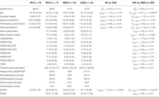
Seventy-seven (25 SDD and 52 LDD) SCZ patients and 78 HV were included in the study. Descriptive and univariate statistics of socio-demographic and clinical characteristics of the sample are summarized inTable 1. HV and SCZ did not differ for gender, age, education, and premorbid IQ scales. As expected SCoRS was higher in SCZ, indicating an impairment in general cognitive functioning. No significant differences were found between SDD and LDD other than for age (younger age of the SDD group) and illness duration (longer by definition for the LDD group).
EEG-SFC
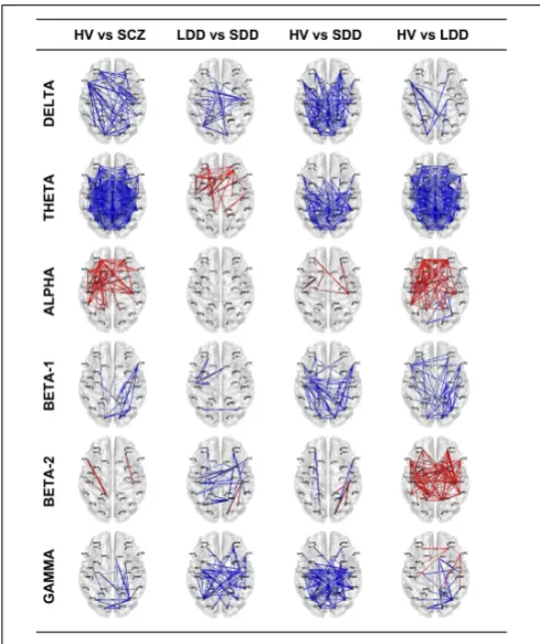
Results of lagged non-linear connectivity differences between HV and SCZ, and within the SCZ group between LDD and SDD in all frequency bands are summarized inFigure 1. Tables with detailed results of all comparisons are available as Supplementary Material.
Delta Band
[image:5.595.46.553.358.668.2]SCZ had significantly higher connectivity than HV; differences where more evident between left prefrontal and right parieto-temporal ROIs (e.g., lLFC-rLTL), left prefrontal and cingulate
TABLE 1 | Descriptive and univariate statistics of sociodemographic and clinical characteristics of sample.
HV (n=78) SCZ (n=77) SDD (n=25) LDD (n=52) HV vs. SCZ (HV vs.) SDD vs. LDD
Gender (w/m) 36/42 26/51 11/14 15/37 χ2(1)=2.48,p=0.12 χ2(2)=4.09,p=0.13
Age 32.78 (10.94) 35.44 (11.05) 25.72 (4.32) 40.12 (10.23) t(153)= −1.51,p=0.13 F(2,152)=19.11,p<0.0001a Education (years) 13.60 (3.57) 12.75 (3.32) 12.64 (3.16) 12.81 (3.42) t(153)=1.53,p=0.13 F(2,152)=1.19,p=0.31 Verbal-premorbid IQ 120.73 (8.85) 119.18 (8.85) 118.02 (9.92) 119.74 (8.33) t(153)=1.09,p=0.28 F(2,152)=0.91,p=0.40 Performance-premorbid IQ 110.64 (3.75) 110.08 (3.45) 109.74 (3.90) 110.25 (3.23) t(153)=0.96,p=0.34 F(2,152)=0.60,p=0.54 Total-premorbid IQ 114.76 (4.86) 113.95 (4.68) 113.00 (4.47) 114.41 (4.75) t(153)=1.06,p=0.29 F(2,152)=1.30,p=0.28
Illness onset (years) 21.13 (4.82) 22.28 (3.93) 20.58 (5.14) t(75)=1.46,p=0.15
Illness duration (years) 14.31 (9.90) 3.44 (1.04) 19.54 (7.74) t(75)= −10.32,p<0.0001
Hospitalization 2.95 (1.34) 2.56 (1.42) 3.14 (1.27) t(75)= −1.79,p=0.08
PANSS POS 11.70 (5.93) 10.20 (4.28) 12.42 (6.49) t(75)= −1.56,p=0.12
PANSS NEG-EXP 10.70 (3.65) 11.64 (3.21) 10.25 (3.79) t(75)=1.58,p=0.12
PANSS NEG-SOC 11.78 (4.22) 12.00 (4.72) 11.67 (2.47) t(75)=0.32,p=0.75
PANSS DIS 15.31 (6.00) 13.96 (5.05) 15.96 (6.35) t(75)= −1.38,p=0.17
PANSS EXC 6.77 (2.32) 6.64 (2.45) 6.83 (2.28) t(75)= −0.33,p=0.74
PANSS EMO-D 15.40 (6.29) 15.36 (6.04) 15.42 (6.46) t(75)= −0.04,p=0.97
CDSS 4.96 (4.21) 5.84 (3.94) 4.54 (4.31) t(75)=1.28,p=0.21
Chlorpromazine equivalent 306.10 (167.27) 330.84 (162.44) 294.21 (169.80) t(75)=0.90,p=0.37
Antipsychotics (SGA/FGA)b 65/21 19/6 37/15
Benzodiazepines (no/yes) 59/18 19/6 40/12
Antidepressants (no/yes) 59/18 20/5 39/13
Anticonvulsant (no/yes) 67/10 21/4 46/6
Anticolinergic (no/yes) 70/7 23/2 47/5
SCoRS 22.56 (1.73) 33.55 (6.13) 32.24 (5.53) 34.17 (6.36) t(153)= −15.21,p<0.0001 F(2,152)=118.90,p<0.0001c
SOFAS 55.61 (15.85) 58.88 (12.60) 54.04 (17.09) t(75)=1.26,p=0.21
HV, Healthy Volunteers; SCZ, people affected by schizophrenia; LDD, people affected by schizophrenia with Long Duration of Disease; SDD, people affected by schizophrenia with Short Duration of Disease. Data are frequencies and means (SDs).
aBonferroni post-hoc: HV vs. SDD, p=0.007; HV vs. LDD, p<0.0002; SDD vs. LDD, p<0.0001. bIn 15 SCZ (6 SDD and 9 LDD; SDD vs. LDD:χ2
FIGURE 1 | Resting-state EEG SFC results of the four comparisons in the six bands.The red color indicates an increase of LPS (functional connectivity) indices and the blue a decrease. The thicker is the line the bigger is the difference. HV, Healthy Volunteers; SCZ, people affected by
schizophrenia; LDD, people affected by schizophrenia with Long Duration of Disease; SDD, people affected by schizophrenia with Short Duration of Disease.
ROIs (e.g., lLFC-rPCC) and left prefrontal and occipital ROIs (e.g, lLFC-rFLC, lLFC-lPVC). Patients in the SDD group showed higher connectivity values than those in the LDD group and than HV.
Theta Band
SCZ showed a widespread higher connectivity between most of ROI pairs compared to HV. Similarly, LDD patients showed diffusely higher connectivity compared with HV. Compared with SDD, LDD had higher connectivity in prefrontal, cingulate, and parieto-temporal ROI pairs (e.g., rLFC-lPHG, lAFC-lPCC). SDD, compared with HV, showed higher connectivity in parieto-temporal, cingulate and occipital ROI pairs with fewer differ-ences in connectivity values of prefrontal ROIs (e.g., lAFC, rAFC, lOFC).
Alpha Band
SCZ had significantly lower connectivity in prefrontal, parieto-temporal, and cingulate ROI pairs compared to HV. No signifi-cant differences were found between LDD and SDD. Decreased connectivity was most evident in LDD compared with HV.
Beta1 Band
SCZ had significant higher connectivity between occipital, pari-etal, and prefrontal ROIs compared with HV. In the comparisons between HV and LDD and HV and SDD, both LDD and SDD showed diffusely higher connectivity in respect of HV.
Beta2 Band
Differences between SCZ and HV in a limited number of ROI pairs (lLFC-lPCC, rOFC-rSMA). SDD had a complex pat-tern characterized by both increased and reduced connectiv-ity between different ROIs compared with LDD and HV. LDD showed a diffuse reduced connectivity compared with HV.
Gamma Band
SCZ had significantly higher connectivity between right occipital-right prefrontal, right occipital-right parieto-temporal, and right occipital-right cingulate ROI pairs (rFLC-rSFC,rFLC-rACC, rFLC-rPHG) compared to HV. SDD had significant diffuse increased connectivity compared with HV and LDD. LDD showed a decreased connectivity between prefrontal and parieto-temporal ROI pairs (e.g., rLFC-rIPL) and increased connectivity between occipital and right cingulate ROIs (rFLC-rACC) and between occipital and right temporal ROIs (e.g., rFLC-rPHG) compared with HV.
Discussion
The results of our study, capitalizing on the additive dimension offered by the functional specificity of the frequency content of EEG, extend prior studies and suggest that brain networks in the resting state are abnormally organized in schizophrenia. The main findings can be summarized as:
• In low frequencies (delta, theta), the SCZ group showed
greater EEG-SFC (in particular a widespread increase in theta band) compared to HV and increased frontal connectivity in LDD in theta band compared with SDD.
• In the alpha band, the SCZ group showed greater frontal
EEG-SFC compared with HV; no differences were found between LDD and SDD.
• In the beta1 band, the SCZ group presented greater EEG-SFC
compared with HV, and in SDD compared with LDD who had the lowest EEG-SFC.
• In beta2 band, the LDD group presented lower frontal and
parieto-temporal EEG-SFC compared with HV and with the SDD group.
• In the gamma band, SCZ had lower occipital and
cingu-late connectivity compared with HV, while SDD had greater connectivity values compared with LDD and HV.
Hyperconnectivity in Low Frequencies
An increase in low-frequency oscillations (power) in delta and theta bands at rest is one of the most consistent findings in schizophrenia (Moran and Hong, 2011). Less is known about resting state FC in the segment of the EEG spectrum; earlier studies (see the review ofLeocani and Comi, 1999) have found increased coherence in low frequencies (Nagase et al., 1992; Mann et al., 1997). Low-frequency power but not coherence can be modified by pharmacological treatment (Merrin et al., 1989), suggesting that impaired low frequency coherence could be the expression of abnormal cortical organization. In first episode, drug-naïve SCZ, a delta band increase was recently shown in EEG-SFC (Lehmann et al., 2014). Increased resting-state theta band connectivity between fronto-temporo-parietal source-pairs is reported in first episode medicated SCZ, with an effect of medi-ation on the verbal memory performance. Moreover, recent evi-dence of abnormal theta connectivity in subjects at high-risk for psychosis has been reported suggesting that impairment in this frequency band could be a trait of the SCZ/psychosis spectrum (Andreou et al., 2015). Our results are largely in line with these findings. Moreover diffuse hyperconnectivity characterizes both early stage (SDD) and chronic (LDD) disease even if only the lat-ter is characlat-terized by a prefrontal involvement. Animal studies have demonstrated that GABAergic inhibition and disinhibition can directly modulate cortical synchrony especially in low fre-quencies (Xiao et al., 2012); GABA neurotransmission is altered in prefrontal cortex (Volk and Lewis, 2002) and its impairment plays a key role in schizophrenic disease (Stan and Lewis, 2012; Schmidt and Mirnics, 2015). Bearing in mind the limitations in translating data from animal models, increased EEG-SFC at rest in low frequencies may be explained as a result of inefficient modulation (impaired inhibition), leading to aberrant synchrony.
Frontal Alpha Hypoconnectivity
As noted above, reduced frontal alpha EEG-SFC is a robust result of our study, independent of the duration of schizophrenic disease course and may represent a valid trait-marker of schizophrenic disease. This finding extends and confirms pre-vious reports on schizophrenic patients (Jetha et al., 2009) and patients with mood disorders (Allen et al., 2004; Vuga et al., 2006). Resting-state EEG deficit in alpha power has been widely reported in schizophrenic patients (see review Boutros et al., 2008) with no difference between first episode and chronic patients (Sponheim et al., 1994). Likewise, reduction of EEG alpha coherence, which is an index of synchronization and connectivity, was noted and related to psychopathological dimension of schizophrenia (John et al., 2002). A recent study investigated resting state alpha band EEG FC in the sensor space and concluded that schizophrenic patients had aberrant ante-rior alpha FC, more pronounced in the left hemisphere (Peng et al., 2013). Finally, an alpha band decrease from resting-state EEG-SFC was recently shown in first episode, drug-naïve SCZ (Lehmann et al., 2014). Due to the key role of alpha oscillations as neural substrates of attention modulation through the suppres-sion/inhibition and selection functions (Klimesch, 2012), impair-ment in alpha resting-state connectivity might be one of the main indices of long lasting and stable cognitive deficit observed in SCZ.
Beta Band Dysfunction
The increased beta1 EEG-SFC in the SCZ group is mainly seen in the posterior regions of the right hemisphere; SDD showed greater EEG-SFC specifically between left temporal, left orbitofrontal, and right anterior cingulate ROI pairs. When compared separately against HV, LDD, and SDD showed different patterns of increased EEG-SFC. An increase of beta1 EEG-SFC was recently described in patients with at-risk men-tal state (ARMS) for psychosis, suggesting that atypical beta-band source synchronization could be an indicator of an increased vul-nerability for SCZ/psychosis spectrum (Ramyead et al., 2014). Furthermore, patients with short disease duration showed greater EEG-SFC in the beta2 band compared with those with longer duration who show widespread reduction in EEG-SFC com-pared with HV. Although results of beta2 FC do not appear as univocal as those of the other frequency bands, they could be explained by hypothesizing that the initial pattern of hyper-connectivity, which has indeed been demonstrated in paradigm-related MEG connectivity studies (Sun et al., 2013), could in time be followed by the diffuse deficit seen in chronic patients. Comparison with other EEG studies (see for review Boutros et al., 2008) is even more complex due to methodological dif-ferences such as beta band having been analyzed as one or more sub-components. It has been postulated that communi-cation within the fronto-parieto-temporal attentional network is characterized by transient long-range phase synchroniza-tion in the beta-band (Schnitzler and Gross, 2005); the dif-fuse alterations seen in LDD (hyperconnectivity in beta1 and hypoconnectivity in beta2) may be different expressions of the same dysfunction in attentional and salience-related net-works. Interestingly at molecular level, beta oscillations and coherence have been linked with GABA-B-R1 Gene Polymor-phism in healthy individuals (Winterer et al., 2003). Since GABAergic transmission is profoundly impaired in schizophre-nia (Schmidt and Mirnics, 2015), future EEG-SFC studies could investigate if beta-band disconnectivity may be considered as an intermediate phenotype of GABAergic dysfunction at system level.
Gamma Band Abnormalities
in glutamate and glutamine concentration in schizophrenia compared to controls (Marsman et al., 2013). Our findings have interesting similarities with those from studies of ani-mal models of psychoses that showed increased gamma oscil-lations after acute administration of ketamine (Pinault, 2008) and decrease after chronic administration (McNally et al., 2013). Recent studies have contributed to better understand the role of gamma oscillations in human brain function (Buzsaki and Wang, 2012) and its implications in schizophrenia (Sun et al., 2011; Uhlhaas, 2011; Uhlhaas and Singer, 2013). In particular, GABAer-gic interneurons with glutamate regulating effect through N-methyl-D-aspartate (NMDA) receptors have been postulated to be responsible for the dysfunction of gamma oscillations observed in schizophrenia (Spencer, 2009; Gonzalez-Burgos and Lewis, 2012). Finally, as showed in basic (Kocsis et al., 2014), computational (Komek et al., 2012; Wang and Wong-Lin, 2013) and human studies (Demiralp et al., 2007; Komek et al., 2012) a relevant role in the generating and modulating gamma activity, via regulation of GABAergic (Andersson et al., 2012b) and glu-tamatergic transmission (Andersson et al., 2012a), is also played by dopamine (Herrmann and Demiralp, 2005; Furth et al., 2013), a key neurotransmitter in the pathophysiology of schizophrenia. Due to action of dopamine receptor antagonists in suppress-ing the gamma activity in humans (Ahveninen et al., 2000) and in animal models (Dejean et al., 2011), we could speculate that prolonged exposition to antipsychotics might account for the decreased gamma connectivity observed in the LDD group.
The Effect of Disease Duration on Aberrant Connectivity of Schizophrenia
Long-term effects of schizophrenic disease on brain structure and function have been extensively documented. These include decreased gray matter volume (Glahn et al., 2008), ventricular enlargement (Vita et al., 2006), focal alteration of white matter tracts (Ellison-Wright and Bullmore, 2009) and reduced oblig-atory auditory cortical responses (Todd et al., 2008). Cognitive abilities as well as affective domains are also affected by disease duration (Fujino et al., 2014). The duration of the schizophrenic illness is strongly associated with long-term treatment with antipsychotic drugs. Recent evidence has shown that exposure to antipsychotic treatment is associated with loss of cortical gray matter (Vita et al., 2012, 2015) and this has generated a debate as to whether the effect of “disease duration” on brain structures and functions in schizophrenia is an expression of a “natural” pro-gression of the illness or a result of a neuro-degenerative effect of antipsychotics. Moreover, early stages of the schizophrenic disease have different clinical and pathophysiological features from those seen in patients with long disease duration (van Os and Kapur, 2009). Furthermore, positive and negative symptoms associated with acute Ketamine administration in HV are asso-ciated with hyperconnectivity (Driesen et al., 2013); this finding has strong analogy with functional changes observed in indi-viduals at high risk for schizophrenia and in the early stages of the disease (Anticevic et al., 2015). The glutamatergic-dependent hyperconnectivity appears to be specific for the early phases of schizophrenia and not a feature present in chronic patients.
Limitations
Some limitations in the current study must be considered. The pharmacological therapy could be a potential confounding factor on connectivity indices of SCZ. Dopamine receptor antago-nists can alter FC and network parameters (Achard and Bull-more, 2007). Our patients had been treated with antipsychotics for several years; however, SDD and LDD patients didn’t show significant differences in antipsychotic dosage (in chlorpro-mazine equivalents) making it unlikely for the reported dif-ferences in EEG-SFC metrics between the two groups to be attributable to current treatment effects. A further potentially limiting factor could be the relatively low spatial resolution allowed by the number of channels (37) used in our EEG record-ings. While there is sufficient evidence to suggest that increased spatial sampling is one of the factors in achieving higher localiza-tion accuracy in EEG studies (Liu et al., 2002), the size of cortical patches used in the present study to define ROIs is well within the resolution allowed by the spatial sampling.
Conclusions
Our study reports for the first time differences in resting-state EEG-SFC functional connectivity within and between brain areas in medicated schizophrenic patients and differences between short duration vs. long duration of the schizophrenic illness. The main findings were a diffuse increase in EEG-SFC in delta and theta bands and a decreased frontal alpha EEG-SFC. Inter-estingly, low frequency EEG-SFC was more evident in patients with long disease duration while decreased EEG-SFC appeared to be a stable phenomenon throughout the disease course, sup-porting the idea of functional specificities of the EEG bands (Uhlhaas and Singer, 2010). Lastly, EEG-SFC in the gamma band showed a complex pattern characterized by increase in SDD and decrease in LDD, which might be partially explained by differ-ent inhibitory/excitatory patterns of dysfunction in early-stage vs. chronic schizophrenia.
Globally, our findings could be interpreted either as the expression of long-term changes in mechanisms of neural plastic-ity of brain networks (neurodegeneration hypothesis), or result-ing from prolonged exposition to antipsychotics and the asso-ciated dysfunctions in inhibitory/excitatory neurotransmission (antipsychotic driven neurodegeneration hypothesis). Current study is not able to discriminate between these two mecha-nisms and the clinical applicability of these findings has to be determined with prospective studies.
Acknowledgments
clinical assessment without any type of reimbursement for the participation to the present study.
Supplementary Material
The Supplementary Material for this article can be found online at: http://journal.frontiersin.org/article/10.3389/fnhum. 2015.00234/abstract
Supplementary Figure 1 | Regions Of Interest (ROIs) based on 6239 voxels of eLORETA inverse matrix. Label, centroid coordinates (in MNI space) and the voxel number were reported for each left and right hemisphere ROI. In axial view of BrainNet Viewer template 28 ROIs are represented according to the ROI centroid MNI coordinates.
Supplementary Table1 | Results of four comparisons (HV vs. SCZ; LDD vs. SDD; HV vs. SDD; HV vs. LDD) for all ROI pairs in the six frequency bands are reported.
References
Achard, S., and Bullmore, E. (2007). Efficiency and cost of economical brain func-tional networks.PLoS Comput. Biol.3:e17. doi: 10.1371/journal.pcbi.0030017 Addington, D., Addington, J., and Schissel, B. (1990). A depression rating scale for
schizophrenics.Schizophr. Res.3, 247–251. doi: 10.1016/0920-9964(90)90005-R Ahveninen, J., Kähkönen, S., Tiitinen, H., Pekkonen, E., Huttunen, J., Kaakkola, S., et al. (2000). Suppression of transient 40-Hz auditory response by haloperidol suggests modulation of human selective attention by dopamine D2 receptors. Neurosci. Lett.292, 29–32. doi: 10.1016/S0304-3940(00)01429-4
Allen, J. J., Urry, H. L., Hitt, S. K., and Coan, J. A. (2004). The stability of resting frontal electroencephalographic asymmetry in depression.Psychophysiology41, 269–280. doi: 10.1111/j.1469-8986.2003.00149.x
American Psychiatric Association, A.P.a.T.F.O.D.S.M.I.V. (2000).Diagnostic and Statistical Manual of Mental Disorders: DSM-IV-TR.Washington, DC: Ameri-can Psychiatric Association.
Andersson, R., Johnston, A., and Fisahn, A. (2012a). Dopamine D4 receptor activation increases hippocampal gamma oscillations by enhancing synchro-nization of fast-spiking interneurons.PLoS ONE7:e40906. doi: 10.1371/jour-nal.pone.0040906
Andersson, R. H., Johnston, A., Herman, P. A., Winzer-Serhan, U. H., Karavanova, I., Vullhorst, D., et al. (2012b). Neuregulin and dopamine modulation of hip-pocampal gamma oscillations is dependent on dopamine D4 receptors.Proc. Natl. Acad. Sci. U.S.A.109, 13118–13123. doi: 10.1073/pnas.1201011109 Andreou, C., Leicht, G., Nolte, G., Polomac, N., Moritz, S., Karow, A.,
et al. (2015). Resting-state theta-band connectivity and verbal memory in schizophrenia and in the high-risk state.Schizophr. Res.161, 299–307. doi: 10.1016/j.schres.2014.12.018
Andreou, C., Nolte, G., Leicht, G., Polomac, N., Hanganu-Opatz, I. L., Lambert, M., et al. (2014). Increased resting-state gamma-band connectivity in first-episode schizophrenia.Schizophr. Bull. doi: 10.1093/schbul/sbu121. [Epub ahead of print].
Anticevic, A., Corlett, P. R., Cole, M. W., Savic, A., Gancsos, M., Tang, Y., et al. (2015). N-Methyl-D-Aspartate receptor antagonist effects on prefrontal corti-cal connectivity better model early than chronic schizophrenia.Biol. Psychiatry 77, 569–580. doi: 10.1016/j.biopsych.2014.07.022
Begre, S., and Koenig, T. (2008). Cerebral disconnectivity: an early event in schizophrenia.Neuroscientist14, 19–45. doi: 10.1177/1073858406298391 Boutros, N. N., Arfken, C., Galderisi, S., Warrick, J., Pratt, G., and Iacono,
W. (2008). The status of spectral EEG abnormality as a diagnostic test for schizophrenia.Schizophr. Res.99, 225–237. doi: 10.1016/j.schres.2007.11.020 Brett, M., Johnsrude, I. S., and Owen, A. M. (2002). The problem of
func-tional localization in the human brain.Nat. Rev. Neurosci.3, 243–249. doi: 10.1038/nrn756
Buzsaki, G., and Wang, X. J. (2012). Mechanisms of gamma oscillations.Annu. Rev. Neurosci.35, 203–225. doi: 10.1146/annurev-neuro-062111-150444 Cabral, J., Kringelbach, M. L., and Deco, G. (2014). Exploring the network
dynam-ics underlying brain activity during rest.Prog. Neurobiol.114, 102–131. doi: 10.1016/j.pneurobio.2013.12.005
Canuet, L., Ishii, R., Pascual-Marqui, R. D., Iwase, M., Kurimoto, R., Aoki, Y., et al. (2011). Resting-state EEG source localization and functional connec-tivity in schizophrenia-like psychosis of epilepsy.PLoS ONE6:e27863. doi: 10.1371/journal.pone.0027863
Canuet, L., Tellado, I., Couceiro, V., Fraile, C., Fernandez-Novoa, L., Ishii, R., et al. (2012). Resting-state network disruption andAPOEgenotype in Alzheimer’s
Disease: a lagged functional connectivity study.PLoS ONE 7:e46289. doi: 10.1371/journal.pone.0046289
Clemens, B., Puskas, S., Bessenyei, M., Emri, M., Spisak, T., Koselak, M., et al. (2011). EEG functional connectivity of the intrahemispheric cortico-cortical network of idiopathic generalized epilepsy. Epilepsy Res. 96, 11–23. doi: 10.1016/j.eplepsyres.2011.04.011
Dejean, C., Arbuthnott, G., Wickens, J. R., Le Moine, C., Boraud, T., and Hyland, B. I. (2011). Power fluctuations in beta and gamma frequencies in rat globus pallidus: association with specific phases of slow oscillations and differential modulation by dopamine D1 and D2 receptors.J. Neurosci.31, 6098–6107. doi: 10.1523/JNEUROSCI.3311-09.2011
Delorme, A., and Makeig, S. (2004). EEGLAB: an open source toolbox for anal-ysis of single-trial EEG dynamics including independent component analanal-ysis. J. Neurosci. Methods134, 9–21. doi: 10.1016/j.jneumeth.2003.10.009 Demiralp, T., Herrmann, C. S., Erdal, M. E., Ergenoglu, T., Keskin, Y. H., Ergen,
M., et al. (2007). DRD4 and DAT1 polymorphisms modulate human gamma band responses.Cereb. Cortex17, 1007–1019. doi: 10.1093/cercor/bhl011 Dickerson, D. D., Wolff, A. R., and Bilkey, D. K. (2010). Abnormal long-range
neu-ral synchrony in a maternal immune activation animal model of schizophrenia. J. Neurosci.30, 12424–12431. doi: 10.1523/JNEUROSCI.3046-10.2010 Driesen, N. R., McCarthy, G., Bhagwagar, Z., Bloch, M., Calhoun, V., D’Souza,
D. C., et al. (2013). Relationship of resting brain hyperconnectivity and schizophrenia-like symptoms produced by the NMDA receptor antagonist ketamine in humans.Mol. Psychiatry18, 1199–1204. doi: 10.1038/mp.2012.194 Ellison-Wright, I., and Bullmore, E. (2009). Meta-analysis of diffusion ten-sor imaging studies in schizophrenia. Schizophr. Res. 108, 3–10. doi: 10.1016/j.schres.2008.11.021
Fingelkurts, A. A., Fingelkurts, A. A., and Kahkonen, S. (2005). Functional connec-tivity in the brain–is it an elusive concept?Neurosci. Biobehav. Rev.28, 827–836. doi: 10.1016/j.neubiorev.2004.10.009
Fitzsimmons, J., Kubicki, M., and Shenton, M. E. (2013). Review of functional and anatomical brain connectivity findings in schizophrenia.Curr. Opin. Psychiatry 26, 172–187. doi: 10.1097/YCO.0b013e32835d9e6a
Friston, K. J. (1998). The disconnection hypothesis.Schizophr. Res.30, 115–125. doi: 10.1016/S0920-9964(97)00140-0
Friston, K. J. (2011). Functional and effective connectivity: a review.Brain Connect. 1, 13–36. doi: 10.1089/brain.2011.0008
Fuchs, M., Kastner, J., Wagner, M., Hawes, S., and Ebersole, J. S. (2002). A standard-ized boundary element method volume conductor model.Clin. Neurophysiol. 113, 702–712. doi: 10.1016/S1388-2457(02)00030-5
Fujimoto, T., Okumura, E., Takeuchi, K., Kodabashi, A., Otsubo, T., Nakamura, K., et al. (2013). Dysfunctional cortical connectivity during the auditory odd-ball task in patients with schizophrenia.Open Neuroimag. J.7, 15–26. doi: 10.2174/1874440001307010015
Fujino, J., Takahashi, H., Miyata, J., Sugihara, G., Kubota, M., Sasamoto, A., et al. (2014). Impaired empathic abilities and reduced white matter integrity in schizophrenia.Prog. Neuropsychopharmacol. Biol. Psychiatry48, 117–123. doi: 10.1016/j.pnpbp.2013.09.018
Furth, K. E., Mastwal, S., Wang, K. H., Buonanno, A., and Vullhorst, D. (2013). Dopamine, cognitive function, and gamma oscillations: role of D4 receptors. Front. Cell. Neurosci.7:102. doi: 10.3389/fncel.2013.00102
Gonzalez-Burgos, G., and Lewis, D. A. (2012). NMDA receptor hypofunction, parvalbumin-positive neurons, and cortical gamma oscillations in schizophre-nia.Schizophr. Bull.38, 950–957. doi: 10.1093/schbul/sbs010
Gruber, A. J., Calhoon, G. G., Shusterman, I., Schoenbaum, G., Roesch, M. R., and O’donnell, P. (2010). More is less: a disinhibited prefrontal cortex impairs cog-nitive flexibility.J. Neurosci.30, 17102–17110. doi: 10.1523/JNEUROSCI.4623-10.2010
Hasan, A., Bergener, T., Nitsche, M. A., Strube, W., Bunse, T., Falkai, P., et al. (2013). Impairments of motor-cortex responses to unilateral and bilat-eral direct current stimulation in schizophrenia.Front. Psychiatry4:121. doi: 10.3389/fpsyt.2013.00121
Herrmann, C. S., and Demiralp, T. (2005). Human EEG gamma oscilla-tions in neuropsychiatric disorders.Clin. Neurophysiol.116, 2719–2733. doi: 10.1016/j.clinph.2005.07.007
Horwitz, B. (2003). The elusive concept of brain connectivity.Neuroimage19, 466–470. doi: 10.1016/S1053-8119(03)00112-5
Hyvarinen, A., and Oja, E. (2000). Independent component analysis: algo-rithms and applications. Neural Netw. 13, 411–430. doi: 10.1016/S0893-6080(00)00026-5
Jetha, M. K., Schmidt, L. A., and Goldberg, J. O. (2009). Long-term stability of resting frontal EEG alpha asymmetry and power in a sample of stable com-munity outpatients with schizophrenia.Int. J. Psychophysiol.72, 228–233. doi: 10.1016/j.ijpsycho.2008.12.011
John, J. P., Khanna, S., Pradhan, N., and Mukundan, C. R. (2002). EEG alpha coher-ence and psychopathological dimensions of schizophrenia.Indian J. Psychiatry 44, 97–107.
Kay, S. R., Fiszbein, A., and Opler, L. A. (1987). The positive and negative syn-drome scale (PANSS) for schizophrenia.Schizophr. Bull.13, 261–276. doi: 10.1093/schbul/13.2.261
Keefe, R. S., Poe, M., Walker, T. M., Kang, J. W., and Harvey, P. D. (2006). The Schizophrenia Cognition Rating Scale: an interview-based assessment and its relationship to cognition, real-world functioning, and functional capacity.Am. J. Psychiatry163, 426–432. doi: 10.1176/appi.ajp.163.3.426
Kegeles, L. S., Mao, X., Stanford, A. D., Girgis, R., Ojeil, N., Xu, X., et al. (2012). Elevated prefrontal cortex gamma-aminobutyric acid and glutamate-glutamine levels in schizophrenia measured in vivowith proton magnetic resonance spectroscopy.Arch. Gen. Psychiatry69, 449–459. doi: 10.1001/archgenpsychi-atry.2011.1519
Klimesch, W. (2012). Alpha-band oscillations, attention, and controlled access to stored information. Trends Cogn. Sci. 16, 606–617. doi: 10.1016/j.tics.2012.10.007
Koch, M. A., Norris, D. G., and Hund-Georgiadis, M. (2002). An investigation of functional and anatomical connectivity using magnetic resonance imaging. Neuroimage16, 241–250. doi: 10.1006/nimg.2001.1052
Kocsis, B., Lee, P., and Deth, R. (2014). Enhancement of gamma activity after selec-tive activation of dopamine D4 receptors in freely moving rats and in a neu-rodevelopmental model of schizophrenia.Brain Struct. Funct. 219, 2173–2180. doi: 10.1007/s00429-013-0607-6
Komek, K., Bard Ermentrout, G., Walker, C. P., and Cho, R. Y. (2012). Dopamine and gamma band synchrony in schizophrenia–insights from computational and empirical studies.Eur. J. Neurosci.36, 2146–2155. doi: 10.1111/j.1460-9568.2012.08071.x
Lancaster, J. L., Woldorff, M. G., Parsons, L. M., Liotti, M., Freitas, C. S., Rainey, L., et al. (2000). Automated Talairach atlas labels for functional brain map-ping.Hum. Brain Mapp.10, 120–131. doi: 10.1002/1097-0193(200007)10:3
<120::AID-HBM30>3.0.CO;2-8
Lehmann, D., Faber, P. L., Pascual-Marqui, R. D., Milz, P., Herrmann, W. M., Koukkou, M., et al. (2014). Functionally aberrant electrophysiological cortical connectivities in first episode medication-naive schizophrenics from three psy-chiatry centers.Front. Hum. Neurosci.8:635. doi: 10.3389/fnhum.2014.00635 Lehmann, D., Faber, P. L., Tei, S., Pascual-Marqui, R. D., Milz, P., and Kochi,
K. (2012). Reduced functional connectivity between cortical sources in five meditation traditions detected with lagged coherence using EEG tomography. Neuroimage60, 1574–1586. doi: 10.1016/j.neuroimage.2012.01.042
Leocani, L., and Comi, G. (1999). EEG coherence in pathological conditions. J. Clin. Neurophysiol.16, 548–555. doi: 10.1097/00004691-199911000-00006 Liemburg, E., Castelein, S., Stewart, R., Van Der Gaag, M., Aleman, A., Knegtering,
H., et al. (2013). Two subdomains of negative symptoms in psychotic disorders:
established and confirmed in two large cohorts.J. Psychiatr. Res.47, 718–725. doi: 10.1016/j.jpsychires.2013.01.024
Liu, A. K., Dale, A. M., and Belliveau, J. W. (2002). Monte Carlo simulation stud-ies of EEG and MEG localization accuracy.Hum. Brain Mapp.16, 47–62. doi: 10.1002/hbm.10024
Lynall, M. E., Bassett, D. S., Kerwin, R., McKenna, P. J., Kitzbichler, M., Muller, U., et al. (2010). Functional connectivity and brain networks in schizophrenia.J. Neurosci.30, 9477–9487. doi: 10.1523/JNEUROSCI.0333-10.2010
Mann, K., Maier, W., Franke, P., Roschke, J., and Gansicke, M. (1997). Intra-and interhemispheric electroencephalogram coherence in siblings discordant for schizophrenia and healthy volunteers.Biol. Psychiatry42, 655–663. doi: 10.1016/S0006-3223(96)00497-0
Marsman, A., van den Heuvel, M. P., Klomp, D. W., Kahn, R. S., Luijten, P. R., and Hulshoff Pol, H. E. (2013). Glutamate in schizophrenia: a focused review and meta-analysis of (1)H-MRS studies.Schizophr. Bull.39, 120–129. doi: 10.1093/schbul/sbr069
McNally, J. M., McCarley, R. W., and Brown, R. E. (2013). Chronic ketamine reduces the peak frequency of gamma oscillations in mouse prefrontal cortex ex vivo.Front. Psychiatry4:106. doi: 10.3389/fpsyt.2013.00106
Medaglia, M. T., Tecchio, F., Seri, S., Di Lorenzo, G., Rossini, P. M., and Porcaro, C. (2009). Contradiction in universal and particular reasoning.Hum. Brain Mapp. 30, 4187–4197. doi: 10.1002/hbm.20838
Merrin, E. L., Floyd, T. C., and Fein, G. (1989). EEG coherence in unmedi-cated schizophrenic patients.Biol. Psychiatry25, 60–66. doi: 10.1016/0006-3223(89)90147-9
Moran, L. V., and Hong, L. E. (2011). High vs low frequency neural oscillations in schizophrenia.Schizophr. Bull.37, 659–663. doi: 10.1093/schbul/sbr056 Mulert, C., Kirsch, V., Pascual-Marqui, R., McCarley, R. W., and Spencer, K.
M. (2011). Long-range synchrony of gamma oscillations and auditory hal-lucination symptoms in schizophrenia.Int. J. Psychophysiol.79, 55–63. doi: 10.1016/j.ijpsycho.2010.08.004
Nagase, Y., Okubo, Y., Matsuura, M., Kojima, T., and Toru, M. (1992). EEG coherence in unmedicated schizophrenic patients: topographical study of predominantly never medicated cases.Biol. Psychiatry32, 1028–1034. doi: 10.1016/0006-3223(92)90064-7
Natsubori, T., Inoue, H., Abe, O., Takano, Y., Iwashiro, N., Aoki, Y., et al. (2014). Reduced frontal glutamate + glutamine and N-Acetylaspartate levels in patients with chronic schizophrenia but not in those at clinical high risk for psy-chosis or with first-episode schizophrenia.Schizophr. Bull.40, 1128–1139. doi: 10.1093/schbul/sbt124
Nelson, H. (1982).The National Adult Reading Test (NART): Test Manual. Windsor: NFER-Nelson Publishing.
Nolte, G., Bai, O., Wheaton, L., Mari, Z., Vorbach, S., and Hallett, M. (2004). Identifying true brain interaction from EEG data using the imaginary part of coherency. Clin. Neurophysiol. 115, 2292–2307. doi: 10.1016/j.clinph.2004.04.029
Olbrich, S., Trankner, A., Chittka, T., Hegerl, U., and Schonknecht, P. (2014). Functional connectivity in major depression: increased phase synchronization between frontal cortical EEG-source estimates.Psychiatry Res.222, 91–99. doi: 10.1016/j.pscychresns.2014.02.010
Oldfield, R. C. (1971). The assessment and analysis of handedness: the Edinburgh inventory.Neuropsychologia9, 97–113. doi: 10.1016/0028-3932(71)90067-4 Pachou, E., Vourkas, M., Simos, P., Smit, D., Stam, C. J., Tsirka, V.,
et al. (2008). Working memory in schizophrenia: an EEG study using power spectrum and coherence analysis to estimate cortical activation and network behavior. Brain Topogr. 21, 128–137. doi: 10.1007/s10548-008-0062-5
Pagani, M., Di Lorenzo, G., Verardo, A. R., Nicolais, G., Monaco, L., Lauretti, G., et al. (2012). Neurobiological correlates of EMDR monitoring–an EEG study. PLoS ONE7:e45753. doi: 10.1371/journal.pone.0045753
Pascual-Marqui, R. (2007).Discrete, 3D Distributed, Linear Imaging Methods of Electric Neuronal Activity. Part 1: Exact, Zero Error Localization [Online]. Available online at: http://arxiv.org/pdf/0710.3341
Peng, H., Hu, B., Li, L., Ratcliffe, M., Zhai, J., Zhao, Q., et al. (2013). A study on validity of cortical alpha connectivity for schizophrenia.Conf. Proc. IEEE Eng. Med. Biol. Soc.2013, 3286–3290. doi: 10.1109/EMBC.2013.6610243
Pinault, D. (2008). N-methyl d-aspartate receptor antagonists ketamine and MK-801 induce wake-related aberrant gamma oscillations in the rat neocortex.Biol. Psychiatry63, 730–735. doi: 10.1016/j.biopsych.2007.10.006
Porcaro, C., Coppola, G., Di Lorenzo, G., Zappasodi, F., Siracusano, A., Pierelli, F., et al. (2009). Hand somatosensory subcortical and cortical sources assessed by functional source separation: an EEG study.Hum. Brain Mapp.30, 660–674. doi: 10.1002/hbm.20533
Ramyead, A., Kometer, M., Studerus, E., Koranyi, S., Ittig, S., Gschwandtner, U., et al. (2014). Aberrant current source-density and lagged phase synchroniza-tion of neural oscillasynchroniza-tions as markers for emerging psychosis.Schizophr. Bull. doi: 10.1093/schbul/sbu134. [Epub ahead of print].
Ribolsi, M., Koch, G., Magni, V., Di Lorenzo, G., Rubino, I. A., Siracusano, A., et al. (2009). Abnormal brain lateralization and connectivity in schizophrenia.Rev. Neurosci.20, 61–70. doi: 10.1515/REVNEURO.2009.20.1.61
Ribolsi, M., Lisi, G., Di Lorenzo, G., Koch, G., Oliveri, M., Magni, V., et al. (2013). Perceptual pseudoneglect in schizophrenia: candidate endophenotype and the role of the right parietal cortex.Schizophr. Bull. 39, 601–607. doi: 10.1093/schbul/sbs036
Rogasch, N. C., Daskalakis, Z. J., and Fitzgerald, P. B. (2014). Cortical inhi-bition, excitation, and connectivity in schizophrenia: a review of insights from transcranial magnetic stimulation. Schizophr. Bull.40, 685–696. doi: 10.1093/schbul/sbt078
Sakkalis, V. (2011). Review of advanced techniques for the estimation of brain con-nectivity measured with EEG/MEG.Comput. Biol. Med.41, 1110–1117. doi: 10.1016/j.compbiomed.2011.06.020
Sartori, G., Colombo, L., Vallar, G., Rusconi, M., and Pinarello, A. (1997). T.I.B.: Test di Intelligenza Breve per la valutazione del quoziente intellettivo attuale e pre-morboso.La Professione di Psicologo1, II–XXIV.
Schmidt, M. J., and Mirnics, K. (2015). Neurodevelopment, GABA system dysfunc-tion, and schizophrenia.Neuropsychopharmacology40, 190–206. doi: 10.1038/ npp.2014.95
Schmitt, A., Hasan, A., Gruber, O., and Falkai, P. (2011). Schizophrenia as a dis-order of disconnectivity.Eur. Arch. Psychiatry Clin. Neurosci.261(Suppl. 2), S150–S154. doi: 10.1007/s00406-011-0242-2
Schnitzler, A., and Gross, J. (2005). Normal and pathological oscillatory commu-nication in the brain.Nat. Rev. Neurosci.6, 285–296. doi: 10.1038/nrn1650 Schoffelen, J. M., and Gross, J. (2009). Source connectivity analysis with MEG and
EEG.Hum. Brain Mapp.30, 1857–1865. doi: 10.1002/hbm.20745
Sheehan, D. V., Lecrubier, Y., Sheehan, K. H., Amorim, P., Janavs, J., Weiller, E., et al. (1998). The Mini-International Neuropsychiatric Interview (M.I.N.I.): the development and validation of a structured diagnostic psychiatric interview for DSM-IV and ICD-10.J. Clin. Psychiatry59(Suppl. 20), 22–33; quiz: 34–57. Spencer, K. M. (2009). The functional consequences of cortical circuit
abnor-malities on gamma oscillations in schizophrenia: insights from computational modeling.Front. Hum. Neurosci. 3:33. doi: 10.3389/neuro.09.033.2009 Sponheim, S. R., Clementz, B. A., Iacono, W. G., and Beiser, M. (1994). Resting
EEG in first-episode and chronic schizophrenia.Psychophysiology31, 37–43. doi: 10.1111/j.1469-8986.1994.tb01023.x
Stan, A. D., and Lewis, D. A. (2012). Altered cortical GABA neurotransmis-sion in schizophrenia: insights into novel therapeutic strategies.Curr. Pharm. Biotechnol.13, 1557–1562. doi: 10.2174/138920112800784925
Stein, M., Egenolf, Y., Dierks, T., Caspar, F., and Koenig, T. (2013). A neurophysiological signature of motivational incongruence: EEG changes related to insufficient goal satisfaction. Int. J. Psychophysiol.89, 1–8. doi: 10.1016/j.ijpsycho.2013.04.017
Stephan, K. E., Baldeweg, T., and Friston, K. J. (2006). Synaptic plastic-ity and dysconnection in schizophrenia. Biol. Psychiatry59, 929–939. doi: 10.1016/j.biopsych.2005.10.005
Sun, L., Castellanos, N., Grutzner, C., Koethe, D., Rivolta, D., Wibral, M., et al. (2013). Evidence for dysregulated high-frequency oscillations during sensory processing in medication-naive, first episode schizophrenia.Schizophr. Res. 150, 519–525. doi: 10.1016/j.schres.2013.08.023
Sun, Y., Farzan, F., Barr, M. S., Kirihara, K., Fitzgerald, P. B., Light, G. A., et al. (2011). Gamma oscillations in schizophrenia: mechanisms and clinical significance.Brain Res.1413, 98–114. doi: 10.1016/j.brainres.2011.06.065
Thatcher, R. W., Biver, C. J., and North, D. (2007). Spatial-temporal current source correlations and cortical connectivity.Clin. EEG Neurosci.38, 35–48. doi: 10.1177/155005940703800109
Todd, J., Michie, P. T., Schall, U., Karayanidis, F., Yabe, H., and Naata-nen, R. (2008). Deviant matters: duration, frequency, and intensity deviants reveal different patterns of mismatch negativity reduction in early and late schizophrenia. Biol. Psychiatry 63, 58–64. doi: 10.1016/j.biopsych. 2007.02.016
Uhlhaas, P. J. (2011). High-frequency oscillations in schizophrenia.Clin. EEG Neurosci.42, 77–82. doi: 10.1177/155005941104200208
Uhlhaas, P. J., Haenschel, C., Nikolic, D., and Singer, W. (2008). The role of oscillations and synchrony in cortical networks and their putative relevance for the pathophysiology of schizophrenia.Schizophr. Bull.34, 927–943. doi: 10.1093/schbul/sbn062
Uhlhaas, P. J., and Singer, W. (2010). Abnormal neural oscillations and synchrony in schizophrenia.Nat. Rev. Neurosci.11, 100–113. doi: 10.1038/nrn2774 Uhlhaas, P. J., and Singer, W. (2013). High-frequency oscillations and the
neuro-biology of schizophrenia.Dialog. Clin. Neurosci.15, 301–313.
Van den Oord, E. J., Rujescu, D., Robles, J. R., Giegling, I., Birrell, C., Bukszar, J., et al. (2006). Factor structure and external validity of the PANSS revisited. Schizophr. Res.82, 213–223. doi: 10.1016/j.schres.2005.09.002
van der Gaag, M., Hoffman, T., Remijsen, M., Hijman, R., De Haan, L., Van Mei-jel, B., et al. (2006). The five-factor model of the Positive and Negative Syn-drome Scale II: a ten-fold cross-validation of a revised model.Schizophr. Res. 85, 280–287. doi: 10.1016/j.schres.2006.03.021
van Os, J., and Kapur, S. (2009). Schizophrenia. Lancet 374, 635–645. doi: 10.1016/S0140-6736(09)60995-8
Vita, A., De Peri, L., Deste, G., Barlati, S., and Sacchetti, E. (2015). The effect of antipsychotic treatment on cortical gray matter changes in schizophrenia: does the class matter? A meta-analysis and meta-regression of longitudinal mag-netic resonance imaging studies.Biol. Psychiatry. doi: 10.1016/j.biopsych.2015. 02.008. [Epub ahead of print].
Vita, A., De Peri, L., Deste, G., and Sacchetti, E. (2012). Progressive loss of cor-tical gray matter in schizophrenia: a meta-analysis and meta-regression of longitudinal MRI studies.Transl Psychiatry2:e190. doi: 10.1038/tp.2012.116 Vita, A., De Peri, L., Silenzi, C., and Dieci, M. (2006). Brain morphology in
first-episode schizophrenia: a meta-analysis of quantitative magnetic reso-nance imaging studies.Schizophr. Res.82, 75–88. doi: 10.1016/j.schres.2005. 11.004
Vita, A., Deste, G., Barlati, S., De Peri, L., Giambra, A., Poli, R., et al. (2013). Interview-based assessment of cognition in schizophrenia: applicability of the Schizophrenia Cognition Rating Scale (SCoRS) in different phases of illness and settings of care.Schizophr. Res.146, 217–223. doi: 10.1016/j.schres.2013. 02.035
Volk, D. W., and Lewis, D. A. (2002). Impaired prefrontal inhibition in schizophre-nia: relevance for cognitive dysfunction.Physiol. Behav. 77, 501–505. doi: 10.1016/S0031-9384(02)00936-8
Vuga, M., Fox, N. A., Cohn, J. F., George, C. J., Levenstein, R. M., and Kovacs, M. (2006). Long-term stability of frontal electroencephalographic asymmetry in adults with a history of depression and controls.Int. J. Psychophysiol.59, 107–115. doi: 10.1016/j.ijpsycho.2005.02.008
Wallwork, R. S., Fortgang, R., Hashimoto, R., Weinberger, D. R., and Dickin-son, D. (2012). Searching for a consensus five-factor model of the Positive and Negative Syndrome Scale for schizophrenia.Schizophr. Res.137, 246–250. doi: 10.1016/j.schres.2012.01.031
Wang, D.-H., and Wong-Lin, K. (2013). Comodulation of dopamine and serotonin on prefrontal cortical rhythms: a theoretical study.Front. Integr. Neurosci.7:54. doi: 10.3389/fnint.2013.00054
Wang, X., Xia, M., Lai, Y., Dai, Z., Cao, Q., Cheng, Z., et al. (2014). Disrupted resting-state functional connectivity in minimally treated chronic schizophre-nia.Schizophr. Res. doi: 10.1016/j.schres.2014.03.033
Winterer, G., Smolka, M., Samochowiec, J., Ziller, M., Mahlberg, R., Gallinat, J., et al. (2003). Association of EEG coherence and an exonic GABA(B)R1 gene polymorphism.Am. J. Med. Genet. B. Neuropsychiatr. Genet.117B, 51–56. doi: 10.1002/ajmg.b.10031
Xiao, Y., Huang, X. Y., Van Wert, S., Barreto, E., Wu, J. Y., Gluckman, B. J., et al. (2012). The role of inhibition in oscillatory wave dynamics in the cortex.Eur. J. Neurosci.36, 2201–2212. doi: 10.1111/j.1460-9568.2012.08132.x
Conflict of Interest Statement:The authors declare that the research was con-ducted in the absence of any commercial or financial relationships that could be construed as a potential conflict of interest.