
An Assembly
Defect
as a
Result
of
anAttenuating
Mutation in the
Capsid Proteins of the
Poliovirus Type 3 Vaccine Strain
ANDREW J. MACADAM,* GERALDINE FERGUSON, CATHERINE ARNOLD,t ANDPHILIP D. MINOR
National Institute for Biological Standards and Control,Blanche Lane, South Mimms, Potters Bar,
Hertfordshire
EN63QG,
UnitedKingdom
Received2 May 1991/Accepted 18 June 1991
The molecular basis of the temperature-sensitive (ts) phenotype ofP3/Sabin, thetype3 vaccine strain of
poliovirus, was investigated in light of the known correlation between ts and attenuation phenotypes. A
phenylalanine at residue 91 of the capsid protein VP3 was a major determinant ofboth phenotypes, and
attenuationandtscould be reverted by thesamesecond-site mutations. Thetsphenotype wasduetoadefect
earlyintheassemblyprocessthat inhibited the formation of 14Spentamers, emptycapsids,andvirions.Itwas
furthershown that capsid proteins thatwerenotincorporated into higher-orderstructureshad shorthalf-lives atthenonpermissivetemperature.
The genetic basis of attenuation of the Sabin type 3
vaccine strain of poliovirus, and that of reversion to
viru-lence, have been extensively studied (1, 4, 15, 32). The
vaccine strain, P3/Sabin, differs from its neurovirulent par-ent,P3/Leon,at10 nucleotide positions (28), but attenuation
is due mainly to just two mutations, a C-U transition at nucleotide 472 in the 5' noncoding region and a Ser-Phe
substitutionatVP3-091 in the capsid proteins(32).The latter change renders growth of the virus in HEp-2C cells sensitive
to elevated temperatures (21), and suppression of this
phe-notypeby second-site mutation results in loss of attenuation (15). This strict correlation between temperature-sensitive (ts) and attenuation phenotypes attributable to the amino acid change in VP3 suggested that an understanding of the
molecular basis of the ts phenotype would give similar insights into the molecular basis of attenuation of the Sabin
type 3 vaccine strain.
Thecrystal structures of P3/Sabin and P1/Mahoney have
beendeterminedto0.24 and0.29nm(5, 9),andthe positions
ofVP3-091 andphenotypic suppressormutations suggested a structural basis for thets phenotype. In P1/Mahoney, and presumably inP3/Leon, the side chain of the SeratVP3-091 pointsintoapocketwhere theserinehydroxyl-group
hydro-gen bonds with a buried water molecule. In P3/Sabin,
however, the large aromatic side chain ofthe Phe residue
points outward, makingunfavorable contacts with the sol-vent(5). This situation might predispose the virion to
ther-mal transitions, resulting in burial ofthe Phe residue such
that the virus particle is rendered unstable or defective in
someway.Suppressormutations have been found in several
locations (5, 21), including protomer-protomer and
penta-mer-pentamerinterfaces wherethey couldactby stabilizing
the interfaces against such transitions. Another mutation
wasfoundataresidue in thehydrophobic pocket analogous
to the binding site in some rhinoviruses of antiviral
com-poundsthat stabilize these virions to heat(6).
These dataareconsistent with the idea that the ts
pheno-typeof P3/Sabinis the resultofthermolabilityofone or more
capsid structures. Here we show that the capsid protein
*Corresponding author.
tPresent address: CRB Ltd., Gadbrook Park, Northwich,
Cheshire CW97RA, UnitedKingdom.
mutation does not affect virion stability itself but rather
inhibits virion assembly, specifically at the
protomer-to-pentamer step.
MATERIALS ANDMETHODS
Cells and viruses. HEp-2Ccellsweregrowninmonolayers
asdescribed by Minor (19).Construction andrecoveryofthe
cDNA-derived recombinant SV3/L have been described
previously (32). SV3/Ldiffersfrom P3/Leon onlyatresidue 91of VP3,which isaPhe inSV3/L, like P3/Sabin,andaSer in P3/Leon. Isolates of P3/Leon and P3/Sabin were those
used for cDNA cloning and recombinant construction (29,
32).
Plaque assays. Cell sheets in six-well plates were
inocu-latedwithdilutions ofvirusfor 30minand then overlaid with
1%agarinminimal essential mediumsupplementedwith 2%
fetal calf serum. Plates were incubated in sealed plastic
boxes submerged in precision water baths (16) at relevant
temperaturesfor 3 days, and then cell sheets were stained
with naphthalene black.
Virus purification and RNA extraction. Viruses were
grown and purified on sucrose gradients before phenol
extraction ofRNAasdescribed by Minor(19).
Antisera.Monoclonalanti-poliovirusreceptor280(22)was
agiftfrom P. Pipkin. At a dilution of1/1,280, antibody 280
prevented poliovirus infection ofHEp-2C cell sheets forat
least 5days (datanotshown). Polyclonal antiserum 119was
raised in rabbitsagainstaderivativeof P3/Sabin.Thisserum
selectively precipitated capsid proteins from sucrose
gradi-entfractionscontaining14Spentamers, emptycapsids,and
virions butnotfrom thosecontaining5Spromoters(datanot
shown).
Shift-up one-step growth. Replica cell sheets in 25-cm2
flasksweretransfected withequalamountsofviral RNA(31)
at room temperaturefor 30 min, and then 5 ml of minimal
essential medium containing 1% fetal calf serum and 1%
anti-poliovirusreceptor monoclonalantibody (MAb)280(22)
was added to each flask. Flasks were submerged in water
bathsat35°Cand then shiftedto40°Cbathsoneatatimeat
hourly intervals until 10 h posttransfection. Flasks were
harvestedat10 or24 hposttransfection by rapid freezingin
C02-ethanol bathsandwere frozen and thawedtwice more
before virus titration.
5225
0022-538X/91/105225-07$02.00/0
Copyright© 1991,American
Society
for Microbiologyon November 10, 2019 by guest
http://jvi.asm.org/
5226 MACADAM ET AL.
Infection at 40°C. Cell sheets in 25-cm2 flasks were incu-bated in a water bath at 40°C for 15 minbefore inoculation at 40°C with SV3/L or P3/Leon at a multiplicity of infection of 0.1. At 1 h postinfection (p.i.), inocula were removed and cell sheets were washed three times at 40°C and then incubated at 40°C in minimal essential medium containing 1% fetal calf serum and 1% MAb 280. At 2 hp.i., one flask pervirus was shifted to35°C andanotherwas harvestedby rapid freezing. One flask was left at 40°C. An additional control flask was inoculated at 35°C andincubated at 35°C throughout the experiment. At 10 h p.i., flasks were har-vested by rapidfreezing and then frozen and thawed twice morebefore virustitration.
In vivo labeling of viral proteins. Cell sheets in 25-cm2 flaskswereinoculated at amultiplicityof infectionof10for 1h at 35°C and then incubated in minimalessential medium containing 1% fetal calf serum. After 2 h, medium was
removed and replaced bymethionine-free medium. After a further 2-h incubation at 35°C, flasks were submerged in waterbaths at 40°C or left at 35°C for 15min beforelabeling with [35S]methionine (10 1iCi/ml; >1,000 Ci/mol; Amer-sham). For labeling experiments, flasks were incubatedfor a further 45 or 90 min at the relevant temperature, medium was removed, still at the relevant temperature, and then cell sheets wererapidly frozen in CO2-ethanol. For pulse-chase experiments, labeling was donefor 10 min before the addi-tion of excess unlabeled methionine. Cell sheets were har-vested atvarioustimes thereafter as described above.
Cells were lysed at 4°C by the addition of 0.5 ml of phosphate-buffered saline containing 0.5% Nonidet P-40 (NP-40), 0.25%deoxycholate, 5 mM EDTA, 1% aprotinin, 0.5 mM phenylmethylsulfonyl fluoride, and 1 mM N-ethyl-maleimide (protease inhibitors were addedjustbefore use). After removal of nuclei by centrifugation, lysates were
stored at -70°Cbefore further analysis.
Sucrose gradients.Cell lysateswerefractionatedon10-ml 5 to 20% or 15 to 30% sucrose gradients prepared in lysis buffer by usingan SW41 rotor (Beckman). Gradients were
centrifugedat35,000 rpm for 16 h (5to20%)or at18,000 rpm for 12 h (15 to 30%); then 0.5-ml fractions were collected after bottom puncture. Pellets from 5to20%gradients were
resuspended in 0.5 ml of lysis buffer. Aliquots of5
[l
weresometimesremoved for scintillationcounting.
Immunoprecipitation. Aliquots of100
F.l
of cell lysates orsucrose gradient fractions were incubated with 5 ,ul of
polyclonal antiserum 119 overnight at 4°C. Samples were
then incubated with 100
RIl
of 10% protein G (Sigma) in sodium dodecyl sulfate(SDS)-NP-40wash (10 mMTris-HCl [pH 7.4], 5 mMEDTA,0.5%SDS,1%NP-40,1% aprotinin)at4°C for2 h andthencentrifuged at 6,000 x gfor 2 min. Pellets were washed once each with 0.5 ml of SDS-NP-40 wash,0.5 M NaCl wash (0.5 M NaCl, 50 mMTris-HCl[pH 7.4], 5mMEDTA, 1 pigof ovalbumin per ml,0.05% NP-40, 1% aprotinin), and 0.05% NP-40 wash (125 mM NaCl, 50
mM Tris-HCl [pH 7.4], 5 mM EDTA, 0.05% NP-40, 1% aprotinin)and then resuspended in 50 to 100 ,ul of gel loading buffer. Samples were boiled for 1 min and centrifuged at 12,000 x gfor 5 min, and supernatants were either analyzed directlyby SDS-polyacrylamide gel electrophoresis (PAGE) (13) orstored frozen.
RESULTS
Temperature sensitivity. The ts phenotype ofP3/Sabin and related viruses is conveniently measured as a reduction in PFU in plaque assays at nonpermissive temperatures. At
0-R
lL
U-0
LO CVI)
LL
0
0)
-J
2-
3-
4-5 ->5.5-~
38.0 38.5 39.0 T(0C)
39.5 40.0
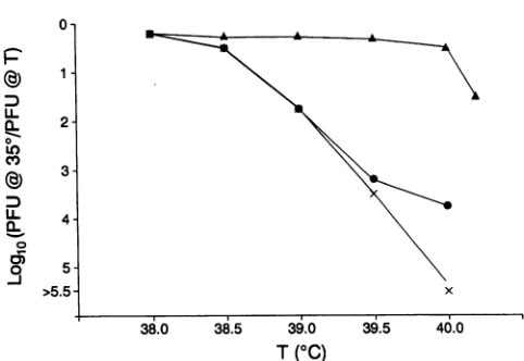
FIG. 1. Temperaturesensitivity ofP3/Sabin (x), SV3/L(0),and P3/Leon (A).
40°C,thetiter of P3/Leon isreduced,relativetothatat35°C, bylessthan1
log1o
unit, whereasthat of P3/Sabin is reduced by >5log1o
units (15). Genetic analysis of this phenotype (21)showed that the Ser-PhechangeatVP3-091was respon-sible fora 3- to4-log1o-unit
reduction at40°C, the residual phenotype being due to one or more differences between P3/SabinandP3/Leonthat haveonlymarginaleffectsonthets phenotype ofa viruswithaSeratVP3-091.
Furtherinvestigationoftheeffectof temperatureonthese virusesshowed that the ts phenotypeofP3/Sabin (definedas a reduction in titer of >1
log1o
unit) was expressed attemperatures above38.7°C (Fig. 1). Titers of P3/Leonwere
hardly affected by temperatures up to 40.1°C. Data for the recombinant SV3/L, which only differs from P3/Leon at
VP3-091, confirmed that the tsphenotype ofP3/Sabin was
mainly duetothe PheatVP3-091. Tostudytheeffectofthis mutationonits own, subsequentstudies,other than thermo-lability assays, compared the phenotypes ofP3/Leon and SV3/L.
Thermolability. Preparations of P3/Leon and P3/Sabin, eitherastissue cultureS10supernatantsorpurifiedon CsCl gradients,wereincubatedat40, 43,45,and50°C forperiods ranging from15 minto 24hwithorwithoutadditional fetal
calfserumanddivalent cations and then titrated. While the
thermolabilities ofpreparationsvaried with each treatment, there was no significant difference between the results for P3/LeonandP3/Sabinin each experiment. Resultsofsome
of the assays are shown in Table 1. The effect ofsample volume, illustrated by comparison of assays a and b, was reproducible.
Temperature shift assays. The stage of the viral growth cycle affected by the presence of a Phe at VP3-091 was
investigated bytemperatureshift one-step growth assays.In oneseriesof one-stepgrowth experiments, cell sheetswere
transfected with viral RNA at room temperature and then incubated in the presence of the anti-poliovirus receptor MAb 280 (22) toprevent a second round of infection. Cell sheets were incubated at 35°C, shifted to 40°C at various times, and then harvestedat 10or 24 hposttransfection by rapid freezingbefore virus titration. Figure2 illustrates the effect of the time of shiftto40°Conthe one-stepgrowth titer
at 10 hposttransfectionexpressed relative totitersobtained after incubation at 35°C. That these were in fact one-step growthtiters wasconfirmed by results for cell sheets
incu-J. VIROL.
on November 10, 2019 by guest
http://jvi.asm.org/
[image:2.612.317.558.73.239.2]TABLE 1. Examples of thermolability assays
Incubation Loglotiterreduction
Assay' temp(°C)
andtime P3/Sabin P3/Leon
a 40, 4 h 2.2 1.8
40, 24 h 3.0 2.8
b 40, 3.5 h 0.1 0.5
40,20 h 0.95 0.8
c 40, 30min 0.1 0.5
43, 30 min 0.2 0.6
45, 30 min 0.6 0.4
50,30 min 4.8 5.2
Inassay a,aliquotsof 0.1 ml of tissueculturesupernatantswere incubated in 1.5-ml Eppendorftubes. In assay b, aliquots of 1 ml of tissue culture supernatantswere incubated in1.5-ml tubes. In assay c,aliquotsof1 ml of tissue culture supernatants diluted 100 times in phosphate-buffered saline wereincubatedin1.5-ml tubes.
batedforatotalof 24 h (in the presence of MAb 280), which were similar to those obtained at 10 h (data not shown).
Incubation at40°C, except after 8 h at35°C, reduced the one-step growth titers of SV3/L to a greater extent than those of P3/Leon. Furthermore, incubation of SV3/L-in-fectedcell sheets at 35°C for up to 4 h posttransfection had little or no alleviating effect on the reduction in final titer obtained. Titers obtained from cell sheets shifted to 40°C at later times probably reflected virion production before the shift. Thus, the tsdefect appeared to act late in the infectious cycle and could not be suppressed by incubation at the permissive temperature during the early period of viral infection.
These results did not rule out an effect of the Phe at VP3-091 onearly stages of infection as well. Therefore, cells were infected at the nonpermissive temperature, the inocu-lumwasremoved,theantireceptorMAb was added, and the cell sheetswereshifteddownto35°Cat2 hp.i. At this time,
no infectious virus could be recovered from infected cell sheets (Table 2). Comparison ofone-step growth titers for P3/Leon and SV3/L showed that, while infection and incu-bationfor 2 hat40°Creduced virusyieldrelative to infection
at 35°C, the effect was similar for both viruses (Table 2). Thus, the Phe at VP3-091 did not seem to affect virus adsorptionat thenonpermissive temperature. Furthermore,
A
75 02
P3/Leon A A A
_ /K
SV3-L
0o1
/
X. -4 (<-4.5 0
0 1 2 3 4 5 6 7 8 9 10
Timeof shiftto400(hoursp.i.)
FIG. 2. Effect oftime ofshiftto40°Conone-stepgrowthtiters,
expressed relativetoone-steptitersreachedafterincubationat35°C
(see MaterialsandMethods).
TABLE 2. Effect of inoculation at 40°C on one-step growth titers
Inoculation Incubation temp Titer/ml(loglo)
temp(°C)
(°C)
and time(h) P3/Leon SV3/L35 35, 10 7.55 7.3
40 40, 10 6.05 2.8
40 40, 2, then 35, 8 6.8 6.55
40 40, 2 -c2.3 <2.3
uncoatingofSV3/Lvirions had occurred at 40°C, at leastto
the stage atwhich particles were no longer infectious. This is consistent with other studies which have shown that inHeLa
cellsat39.5°C, wild-type poliovirus RNA releaseiscomplete by 60 min p.i. (12). Taken together, these experiments showed that the Phe at VP3-091 caused a defect in virion production at the nonpermissive temperature only at a late stagein infection. In viewof the structural data on locations of VP3-091 and suppressor mutations, the effect of thePhe at
VP3-091 on capsid protein synthesis and assembly at the nonpermissive temperature was investigated.
Protein synthesis and assembly. Results of temperature shiftexperiments showed that the process affected by
tem-peraturein SV3/L-infected cells had started by 4 h p.i. but that temperature shiftat any subsequenttime upto 7 h p.i. resulted in reductions in virion production. Thus, [35S]me-thionine labeling of proteins at 5 h p.i. was expected to
illustrate any differences in proteinsynthesis by SV3/Land P3/Leonrelevant to thetsphenotypeof the former. Tostudy proteinsynthesis incellsatboth 35 and 40°Catcomparable stagesofinfection, infected cell sheetswereallincubatedat
35°C until 15 min before the addition of
[35S]methionine.
Shift-up for thisperiod allowed cultures to reach the incu-bation temperature before the addition oflabel.
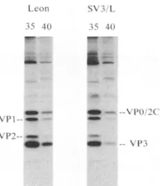
Figure3shows theresults of suchanexperimentinwhich proteinswerelabeledfrom 5 to 6.5 hp.i. The PheatVP3-091 hadno significant effect on labeling orprocessing of capsid proteins at 35°C, but differences were apparent between
extracts from cells infected with SV3/L and P3/Leon when
labeledat 40°C. The totalamountofSV3/L capsidproteins observedattheendof the40°Clabeling periodwasreduced relative to that of P3/Leon capsid proteins (e.g., compare intensitiesof VP3bands). Therewasnosimilar reduction for proteins other than VPO, -1, -2, and -3 (VP4 was not
observedsince it containsnomethionine). Infact, in
Fig.
3,1.Coiln SX
411 i 41
dUb'--= -\1>n
|
_I.
FIG. 3. Effect of temperature on capsid protein levels. Cell sheetswereinfected withP3/LeonorSV3/L,and thenproteinswere labeledat35or40°Cfrom 5to6.5 hp.i. beforecelllysis.Aliquots
of 50p.l of each lysate wereanalyzedbySDS-PAGE.
on November 10, 2019 by guest
http://jvi.asm.org/
[image:3.612.319.563.93.164.2] [image:3.612.63.303.541.700.2] [image:3.612.388.506.556.691.2]5228 MACADAM ET AL.
103CPM
25
20C
15
o0
#,
4 Lear
35°C
Virion
Emrlpty
CaDs,
I ,
10
400C
-SV3 L*L.eon
t-f~~~~~~~
. ,., .,
20 '.0
Fraction Number
b
I,r;i't
I 1.1, ( 1_ .-I !f, 1,s- "!
1'.'I.aon
12--5-1"i''I
.'; 1.
--\T!
--\ ';
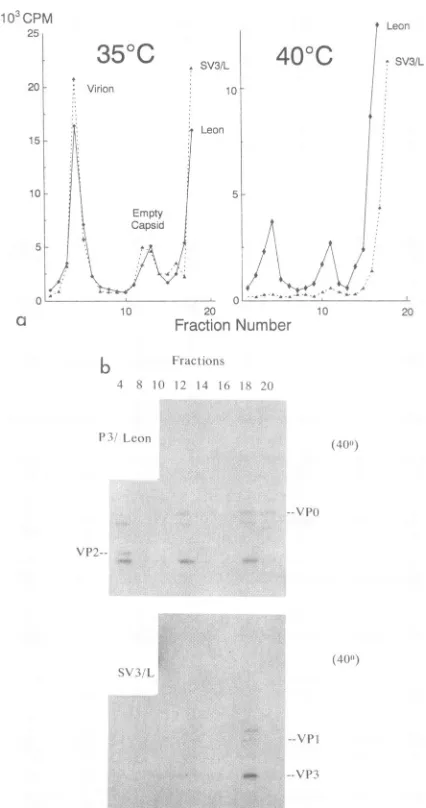
FIG. 4. Sucrose gradient analysis of capsid structures synthe-sized at 35 and 40°C. Lysates shown in Fig. 3 were sedimented
through 15 to 30% sucrose gradients, and0.5-ml fractions were
collected. Aliquots of 5
[lI
were scintillation counted (a), andaliquots of50 p.1 were analyzed by SDS-PAGE (b).
all proteins other than capsid proteins are equally or more
intensely labeled in SV3/L-infected cells than in P3/Leon-infected cellsat
40°C.
This reduction could be duetoadefectin synthesis orprocessingor simplyto ashorter half-life for
capsid structures with a Phe at VP3-091. Also, VP2 was
observed in cells infected with P3/Leon but not in those
infected with SV3/L. This may simply have reflected the
differential intensity of labeling of capsid proteins.
Alterna-tively, since VP2 is produced by the maturation cleavageof
VPOuponRNA encapsidation, this observation could be the
resultofadefect in assembly of SV3/Latthe nonpermissive temperature.
The assembly defect suggested by the lack ofVPO
cleav-age wasinvestigated further by sucrose gradient
centrifuga-tion. Cellextractsshown in Fig. 3 werefractionated on15to
30% sucrose gradients, radioactivity in the fractions was
assayed, and the samples were analyzed by SDS-PAGE
(Fig. 4). Gradient profiles for P3/Leon and SV3/L at 35°C
were almost identical. Afterlabeling at 40°C, P3/Leonvirion production was reduced by about 75% and empty capsid production was reducedby about 50%, whereas forSV3/L, virion production was notdetectable (>99%reduction)and empty capsidproduction was reduced by at least 90%(Fig. 4a). Analysis of thesegradient fractionsby SDS-PAGE(Fig. 4b) showed that SV3/L capsid proteins were present in fractions at the top of thegradient,containing protomers and pentamers, so the reduction in total SV3/L capsidproteins observed relative to P3/Leon was chiefly due to reduced levels of higher-order structures. The band in fraction 20 with the same mobility as VP0 isprobably 2C.
Fractionation of similar extracts on 5 to 20% sucrose
gradients allowed morepreciseidentification of theassembly defect. Figure 5 shows SDS-PAGE analysis of such gradi-ents (the bottomof the gradient is ontherightin this
figure).
SV3/L capsid proteins synthesized at 40°C were found in fractions 14 to 16, fraction 6, andthe pellet. By referenceto
published data (e.g., reference 17), the peaks
corresponded
to 5S protomers and 14S pentamers, respectively, and the pellet contained empty capsids and virions. P3/Leon
capsid
proteins were found mostly in 14Spentamers(fraction 6)and empty capsids and virions (pellet) (Fig. 5a). Comparison of the amounts of VP3 in each peak by densitometry showed that the approximate proportions of protomer/pentamer were 2:1 for SV3/L as opposed to 1:3 forP3/Leon. Thus, at
40°C,
SV3/L was defective in the protomer-to-pentamer assembly step. A similar analysis of35°C extracts(Fig.
Sb)
showed that even at thispermissive temperature, the ratio of protomers to pentamers in SV3/L-infected cells was higher than that in P3/Leon-infected cells (approximately 1:1 as
opposed to 1:3).
In the 14S pentamer peaks (fraction 6), the levels ofVP3 were highrelative to those of the other capsidproteins, even
given that VP3 contains 13 methionine residues while VP0 and VP1 each contain 5. This effect was similar for both SV3/L and P3/Leon. It is possible that VPO and VP1 are
more susceptible than VP3 to mechanical or proteolytic breakdown during extraction orfractionation. Extra bands arevisible below VP1 infraction 6 of eachgradient inwhich pentamers arepresent. It may be relevant that the Ntermini of VPO and VP1, but not VP3, are disordered in empty capsids and, presumably, in pentameric subunits (10).
Theseexperiments showed that the block wasearly inthe assembly process. However, the amount of label
accumu-lating inprotomeric capsid precursors did notcorrespondto
the total amount of labelfound in P3/Leon capsid proteins. Thus, either there was a further defect in synthesis and/or processing or capsid precursors were synthesized normally but, in the absence ofassembly, were degraded.
To distinguish between these possibilities, proteins were
pulse-labeled at 5.5 hp.i. (for 10 min) 15 min after ashift to
40°C and then chased with an excess of unlabeled methio-nine forperiods from 0 to 30 min.
Figure 6 shows the results of one such experiment. It is apparentthat the kinetics of label incorporation into P1 and subsequentprocessing into VPO, -1, and -3 were similar for bothviruses over the labeling period and thefirst 15 min of the chase. Thus, there did not appear to be any defect in synthesis or primary cleavage of the P1 protein ofSV3/L at
40°C. The 30-min time points showed loss of label from capsid proteins of both viruses but to a greater degree for SV3/L. Thisdifference was not only due to virionproduction byP3/Leon (as shown by theappearance of VP2) since VP0 was also longer lived in P3/Leon-infected extracts.
Characterization ofantiserum 119 with sucrose gradient
J. VIROL.
*:1
.11"..
.-\
PHl
on November 10, 2019 by guest
http://jvi.asm.org/
[image:4.612.72.285.74.478.2]FractiOnIs
IS 16 4 12 1) S 6 4 2 P
N'P'
* -\l)P
C.- _
__~dfj "z
-LI-tt4Is
h L Is lbY 42 1)I) 5; 6 4 2 P>
Iwlj,
lL
l.- .w 0
0%,--0
a
L 1s1nT4 12 11) S f 4 2 1P
_ea
- * l_ V 1V\P(Iis 16 14 12 [(I K f -4 2 P
SViL -4
-*.I
5Al -lo
-.A.
_ _ _
FIG. 5. Sucrose gradient analysis ofsubviral particles. Cellsheetswereinfected with P3/LeonorSV3/L,and then proteinswerelabeled
from5to5.75 h p.i. at40°C(a)or35°C(b). Cellswerelysedin0.5mlof lysis buffer,and 0.2-ml aliquots oflysatesweresedimentedthrough
Sto20% gradients. Fractions of 0.5 mlwerecollected. Pellets (lanes P)wereresuspended in 0.5 ml of lysis buffer. Aliquots of 50,llofcell
lysates (lanes L), gradient fractions, and resuspended pellets(lanes P)wereanalyzed by SDS-PAGE.Autoradiogramswereexposedfor 7
days (a) and 5 days(b)exceptfor lysate (lanes L) tracks (3days [a] and 1day[b]).
fractionsshowed that this antiserum selectively precipitated pentameric orlarger structures (and P1, inefficiently) (data not shown). Immunoprecipitation of capsid proteins from lysates shown in Fig. 6 with antiserum 119 showedthatno
such structures were detectable in SV3/L-infected cells
labeled at40°C, whereas the capsid proteinswere
immuno-precipitated from P3/Leon-infected cells (Fig. 7). Whilethe
levels ofcapsid proteins after 15min of chaseweresimilar in
both cases (Fig. 6), the structures in which these proteins
11 Leon
1I 1.0 SV.3 L
AVP
\I,
r\ ^ -a
were found differed. This result was consistent with the
sucrose gradient analysis of intracellular structures in that themajority of SV3/L capsid proteins labeledat40°Cwerein
protomers, whereas thoseof P3/Leonweremainly found in
pentameric or larger structures (Fig. 4 and 5). Thus, the fasterrateofdegradation of SV3/L capsid proteinsat40°C correlated with the block in synthesis ofpentamers, empty
capsids, and virions.
P3 Leon
() 15 3(1
SV .
O1 5 5 30
P
I--V
P)-VP I-VP> -.
V -.
FIG. 6. Pulse-chase labeling. Cell sheets infected withP3/Leon
orSV3/Lwerepulsedat5.5 hp.i. for10min andthenincubatedwith
100-foldexcessofunlabeledmethionine for0,5, 15,or30min before
[image:5.612.122.502.67.342.2]lysis. Aliquotsof50 ,ul of celllysateswereanalyzed by SDS-PAGE.
FIG. 7. Immunoprecipitationofsubviralparticles. Aliquotsof 50 ,uloflysatesshown in Fig.6wereprecipitatedwith antiserum 119
andanalyzed bySDS-PAGE.
a) L
p-; Leon (40)')
I. a
(40"')
U,
_m _w
on November 10, 2019 by guest
http://jvi.asm.org/
[image:5.612.368.512.530.697.2] [image:5.612.104.248.531.685.2]5230 MACADAM ET AL.
DISCUSSION
Mutationsinthe capsidproteins ofpolioviruses have been associated with a number ofdifferent phenotypes and pro-cesses including thermolability (18), polyprotein cleavage
(33), assembly (3, 17), uncoating (12), host range (24),
antiviral drug resistance (8), and antibody reactivity (20). Theresults presented here show that the Ser-Phe
substitu-tion atVP3-091 in P3/Leon that renders viral growth
sensi-tivetoelevatedtemperaturesdoesnot increase thermolabil-ity or result in ts defects in early events such as receptor
bindinganduncoatingorcleavage ofP1 into VPO, VP1, and
VP3. The primary effect of the Ser-Phe substitution at
VP3-091 is to inhibit assembly of protomeric subunits into
14S pentamers, and hence empty capsids and infectious
virions, at the nonpermissive temperature. Even at
permis-sivetemperatures, thePheresidue alteredthe relative levels
of these capsid protein structures (Fig. Sb). Second-site
mutations that revertboth the tsand the attenuation
pheno-typesassociated withthis substitution (15) alsocompensate
for this assembly defect (14a). Thus, it is tempting to
speculate that the assembly defect is also the basis of the attenuation phenotypeofSV3/L.
The location of residue 91 of VP3 in protomeric and pentameric subunits is not known, but the presence of
trapped solvent molecules in this region of virions (5) and empty capsids (8a) suggests that the helix containing this
residue, or the G-H loop of VP3 that folds across it,
undergoes a structural transition during the assembly
pro-cess. The relative ease with which empty capsids and 14S pentamers are interconvertible in vitroargues againstmajor
structural rearrangements at this stage. Furthermore, the locations of VP3-091 in empty capsids and virions of P1/
Mahoney are analogous (8a). Thus, it is possible that a
structural transition occurs during assembly of protomers into pentameric subunits. The environment of the aromatic
ringofVP3-091 PheinP3/Sabin is energeticallyunfavorable
(5). It is possible that this side chain is less exposed to solventinprotomers thaninpentamers. This could resultin a shift in equilibrium between these two forms toward
protomers, similar to that observed here, which could be accentuatedbyincreasesintemperature.Anotherpossibility
is thattheoutwardorientation ofthe Phe atVP3-091, owing
to its bulk, requires contortion of the helix containing this
residue that may interfere with protomer-protomer
interac-tion. Thus, 14S pentamers of P3/Sabin, unlike the virions
themselves, maybe thermolabile.
The lowlevels ofpentamers and emptycapsids observed
in SV3/L-infected cells at the nonpermissive temperature
(Fig.4and5)suggestthatthePheatVP3-091actsbyaltering the equilibrium between capsid precursors rather than by
causing an irreversible block. This may allow suppressor
mutations to actlaterinthe assemblyprocessby speedingup
subsequent assembly steps (14a).
AfurtheraspectofthetsphenotypeofSV3/Lis the short half-lifeofcapsid proteins at 40°C. The rate ofsynthesisof
P1and its cleavage into VPO,-3, and-1 appeared similar in
cellsinfectedwitheitherP3/Leon orSV3/Lat400C, butthe rate ofdegradation was greaterfor SV3/L capsid proteins.
Thismaybeadirect result ofthe residueatVP3-091insome
unknownway. Alternatively, if lower-orderstructuressuch as protomers and/orpentamers were degraded at agreater
ratethanemptycapsidsand virions, the observeddifference
in stability ofSV3/Land P3/Leon capsid proteins wouldbe
an indirect result of the residue at VP3-091 through its influence on the predominant structure in which capsid
proteins
accumulate. This difference is greater than can beexplained
by
incorporation
ofcapsid proteins
into virions sinceinP3/Leon-infectedcells,
VP0
wasobserved tohavealonger
half-life than inSV3/L-infected
cells(Fig. 6).
Thus,
dissociable emptycapsids
may berelatively
stable and provideahigher
effectiveconcentration ofcapsid
precursors than would otherwise bepossible.
Neitherthe exact mechanism
by
whichpoliovirus
capsid
protein
sequences aredegraded
nor thesignals
present onthe various structures that determine theirrates of
degrada-tion are known at present. It is of interest that many componentsof the
ubiquitin-dependent degradation
pathway
aretemperature
regulated,
asindeed areheat shockproteins
(14). In this respect, it may be relevant thatsynthesis
ofGRP78,
amember ofthe heatshockfamily,
is notshutoffin poliovirus-infected cells (26).Furthermore,
GroEL,
a bacte-rialhsp60
homolog, has beenimplicated
in the virionassem-bly pathway ofbacteriophage X
(7).
It is
plausible
that surfaces of lower-order structures that subsequently take part in protein-protein interactions in higher-orderstructurescontain hydrophobicregions
thatarerecognized
as misfoldedby
cellular factors and are thus targeted fordegradation. Alternatively,
sequences that aredisordered and accessible in lower-order structures mayact as similar
signals. Sequences
in VP2 nearthe VPOcleavage
site and the N-terminalregions
of VP1 and VPO that aredisordered in empty
capsids (10)
andpresumably
pentameric
subunitsmay represent suchsignals. Inprotomericsubunits,
the N terminus ofVP3would alsobe
improperly
folded(8a).
Nucleotide differences that influence attenuationpheno-types of Sabin vaccine strains of poliovirus have been identified inthe 5' noncodingregion (2, 11, 16, 23, 32),
P1
(2,
25, 32), and
3DPOL
(2). The mutations in the 5'noncoding
region may affect translation efficiency (30), probably
by
RNAsecondarystructure perturbation (27). The mutationin
3DPOL
has not been studied in detail butpresumably affects eitherreplicationorP1
processing. Theattenuatingmutationat VP3-091 inP3/Sabin has been shown here toaffect virion assembly. Othermutations inthe capsid proteins have also been found to influence attenuation (15, 21) and assembly (14a) ofP3/Sabin derivatives. As well as shedding light on
oneofthemechanisms ofattenuation of poliovaccines, these studiesmay contributeto ourunderstandingofthedynamics of poliovirus assembly and the sequences involved in its regulation.
ACKNOWLEDGMENTS
Wethank James Hogle for providing unpublished information and
for helpful discussions. We are grateful to Andrew Davies and
Elizabeth Trainor for preparation of the figures and manuscript.
REFERENCES
1. Cann, A. J., G. Stanway, P. J. Hughes, P. D. Minor, D. M. A.
Evans, G. C. Schild, and J. W. Almond. 1984. Reversion to neurovirulence of the live attenuated Sabin type 3 oral poliovi-rus vaccine. Nucleic Acids Res. 12:7787-7792.
2. Christodoulou, C., F. Colbere-Garapin, A. Macadam, L. F. Taffs, S. Marsden, P. D. Minor, and F. Horaud. 1990. Mapping of mutations associated with monkey neurovirulence of Sabin 1 poliovirus revertants selected at high temperature. J. Virol. 64:4922-4929.
3. Compton, S. R., B. Nelsen, and K. Kirkegaard. 1990.
Temper-ature-sensitive poliovirus mutant fails to cleave
VP0
andaccu-mulates provirions. J. Virol. 61:4067-4075.
4. Evans, D. M. A., G. Dunn, P. D. Minor, G. C. Schild, A. J.
Cann, G. Stanway,J.W. Almond, K. Currey, and J. V. Maizel,
Jr. 1985. Increased neurovirulence associated with a single J. VIROL.
on November 10, 2019 by guest
http://jvi.asm.org/
nucleotide change in anon-coding region of the Sabin type 3
poliovaccine genome. Nature(London) 314:548-550.
5. Filman, D. J., R. Syed, M. Chow, A. J. Macadam, P. D. Minor,
and J. M.Hogle. 1989. Structural factors that control
conforma-tional transitions andserotype specificity in type 3 poliovirus.
EMBO J. 8:1567-1579.
6. Fox, M. P., M. J. Otto, and M. A. McKinlay. 1986. Prevention
ofrhinovirus and poliovirus uncoating by WIN51711, a new
antiviraldrug.Antimicrob. Agents Chemother. 30:110-116.
7. Georgeopoulos, C., K. Tilly, and S. Casjens. 1983. Lambdoid
phage head assembly,p. 279-304. In R. W. Hendrix et al. (ed.),
Lambda II. Cold Spring HarborLaboratory, ColdSpring
Har-bor,N.Y.
8. Heinz, B. A., R. R. Rueckert, D. A. Shepard, F. J. Dutko, M. A. McKinlay, M. Fancher, M.G. Rossman, J. Badger, and T. J.
Smith. 1989. Genetic and molecular analyses ofspontaneous
mutantsof human rhinovirus 14thatareresistantto anantiviral
compound.J. Virol.63:2476-2485.
8a.Hogle,J. Personalcommunication.
9. Hogle, J. M., M. Chow, and D. J. Filman. 1985. The
three-dimensionalstructureofpoliovirus at2.9Aresolution.Science
229:1358-1365.
10. Hogle, J. M., R.Syed,C. E. Fricks, J. P. Icenogle,0.Flore,and
D.J.Filman.1990. Role ofconformational changes in poliovirus
assembly and cellentry, p.199-210.In M. A.Brinton andF. X.
Heinz (ed.), New aspects of positive-strand RNA viruses.
AmericanSociety for Microbiology, Washington, D.C.
11. Kawamura, N., M. Kohara,S. Abe, T. Komatsu, K. Tago, M. Arita, and A. Nomoto.1989. Determinants in the5'noncoding
region ofpoliovirus Sabin1 RNAthatinfluence the attenuation
phenotype. J. Virol. 63:1302-1309.
12. Kirkegaard,K.1990.Mutations in VP1 ofpoliovirusspecifically
affect both encapsidation and release of viral RNA. J. Virol.
64:195-206.
13. Laemmli,U. K.1970.Cleavage of structural proteins during the
assembly of the head ofbacteriophage T4. Nature (London)
227:680-685.
14. Lindquist,S.,and E. A.Craig. 1988. Theheat-shockproteins.
Annu. Rev.Genet. 22:631-677.
14a.Macadam,A.Unpublished data.
15. Macadam,A.J., C.Arnold, J.Howlett,A.John, S. Marsden,F.
Taffs, P. Reeve, N. Hamada, K. Wareham, J. Almond, N.
Cammack,and P. D. Minor.1989.Reversion of attenuated and
temperature-sensitive phenotypes oftheSabintype 3 strain of
poliovirus in vaccines. Virology 174:408-414.
16. Macadam,A.J.,S. R.Pollard,G.Ferguson,G.Dunn,R.Skuce,
J.W.Almond,and P. D. Minor.1990. The 5'non-codingregion
of thetype2poliovirusvaccine strain contains determinants of
attenuation andtemperature-sensitivity. Virology 181:451-458.
17. Marc, D., G. Messon, M. Girard,and S.van derWerf. 1990.
Lack of myristoylation of poliovirus capsid polypeptide VPO
prevents theformation of virionsorresults in theassembly of
noninfectious virus particles.J. Virol. 64:4099-4107.
18. Martin, A.,C. Wychowski, T. Couderc, R. Crainic, J. Hogle,
and M.Girard. 1988.Engineeringapoliovirus type 2antigenic
site on a type 1 capsid results in a chimaeric virus which is
neurovirulent for mice. EMBOJ.7:2839-2847.
19. Minor, P. D.1980. Comparative biochemical studies oftype 3
poliovirus. J.Virol. 34:73-84.
20. Minor,P. D.1990.Antigenic structureof picornaviruses. Curr.
Top. Microbiol. Immunol. 161:121-154.
21. Minor, P. D., G. Dunn, D. M. A.Evans,D.I. Magrath, A.John,
J. Howlett,A.Phillips, G. Westrop, K.Wareham,J.W.Almond,
andJ.M.Hogle.1989. Thetemperature-sensitivity oftheSabin
type 3 vaccine strain of poliovirus: molecularand structural effects of amutation in the capsid protein VP3. J. Gen. Virol. 70:1117-1123.
22. Minor, P. D., P. A. Pipkin, D. Hockley, G. C. Schild, and J.W. Almond. 1984. Monoclonal antibodies which block cellular receptors ofpoliovirus. VirusRes. 1:203-212.
23. Moss,E.G.,R. E.O'Neill,and V. R.Racaniello. 1989.Mapping
ofattenuatingsequencesof anavirulentpoliovirustype2 strain.
J. Virol. 63:1884-1890.
24. Murray, M. G., J. Bradley, X.-F. Yang, E. Wimmer, E. G.
Moss, and V. R. Racaniello. 1988. Poliovirus host range is
determined by a short amino acid sequence in neutralisation
antigenicsite 1. Science241:213-215.
25. Ren,R.,E. G.Moss,and V.R. Racaniello. 1991. Identification of twodeterminants thatattenuatevaccine-relatedtype2
polio-virus. J. Virol. 65:1377-1382.
26. Sarnow, P. 1989. Translation of glucose-related protein 78/
immunoglobulin heavy chain binding protein mRNA is
in-creased inpoliovirus-infected cells at atime when
cap-depen-dent translation of cellular mRNA is inhibited. Proc. Natl.
Acad. Sci. USA 86:5795-5799.
27. Skinner, M. A., V. R.Racaniello, G. Dunn, J. Cooper, P. D.
Minor, and J. W. Almond. 1989. New model for the secondary structureof the5' non-codingRNAofpoliovirus is supported by biochemical and genetic data that also show that secondary structureisimportant in neurovirulence. J. Mol. Biol. 207:379-392.
28. Stanway, G., P. J. Hughes, R. C. Mountford, P.Reeve, P. D.
Minor, G. C. Schild, andJ. W. Almond. 1984. Comparison of
the complete nucleotide sequences of the genomes of the
neurovirulent poliovirus P3/Leon/37 and its attenuated Sabin
vaccinederivative P3/Leon 12alb. Proc. Natl. Acad.Sci. USA
81:1539-1543.
29. Stanway, G.,R.C.Mountford, S.D.J.Cox, G. C.Schild,P.D.
Minor, and J. W. Almond. 1984. Molecular cloning of the
genomesofpoliovirustype3 strainsby the cDNA:RNA hybrid method. Arch. Virol. 81:67-78.
30. Svitkin,Y.V.,N.Cammack,P. D.Minor, andJ. W.Almond.
1990. Translation deficiency of the Sabin type 3 poliovirus
genome: association with an attenuating mutation C472-)U. Virology 175:103-109.
31. Van derWerf, S., J. Bradley,E.Wimmer,F. W. Studier, and J. J. Dunn. 1986. Synthesis of infectious poliovirus RNA by
purifiedT7polymerase. Proc. Natl. Acad. Sci. USA
83:2330-2334.
32. Westrop, G. D., K. A. Wareham, D. M. A. Evans, G. Dunn,
P. D.Minor,D.I.Magrath,F.Taffs, S. Marsden,M. A.Skinner,
G. C. Schild, and J. W. Almond. 1989. Genetic basis of
attenuation of the Sabintype3 oralpoliovirus vaccine. J. Virol.
63:1338-1344.
33. Ypma-Wong, M. F., D. J. Filman, J. M. Hogle, and B. L.
Semler. 1988. Structural domains of the poliovirus polyprotein
aremajor determinants for theproteolytic cleavageatGln-Gly
pairs. J. Biol. Chem. 263:17846-17856.
![FIG. SDS-PAGE. Autoradiograms were exposeddays (a) (lanes fractions, and and 5 resuspended days (b) tracks pellets except for (3 days (lanes lysate [a] P) (lanes and were L) 1 day labeledfromlysates L), gradient analyzed by ,llS to 5](https://thumb-us.123doks.com/thumbv2/123dok_us/1312838.84658/5.612.368.512.530.697/autoradiograms-exposeddays-fractions-resuspended-pellets-labeledfromlysates-gradient-analyzed.webp)
