Vol. 63, No. 7 JOURNALOFVIROLOGY, JUIY 1989,p. 3168-3175
0022-538X/89/073168-08$02.00/0
Copyright ©D 1989, American Society for Microbiology
Synthesis, Posttranslational
Modifications,
and
Nuclear
Transport of
Polyomavirus Major Capsid
Protein
VP1t
ALI R. FATTAEY ANDRICHARD A. CONSIGLI*
Division of Biology, Section ofVirology andOncology, Ackert Hall, Kansas State University, Manhattan, Kansas66506 Received 21 December1988/Accepted14 March 1989
Polyomavirusmajor capsid proteinVP1synthesiswasstudiedininfectedprimary babymousekidneycells. A standard curveofVP1 protein wasused to quantitate VP1 in the cytoplasm and nucleus ofinfectedcells duringthe timecourseof infection.PolyomavirusVP1 continued to beaccumulatedin thecytoplasmof thecells
until 27 hpostinfection, atwhich time the synthesis ofVP1 leveled off. VP1 continued to accumulate in the
nucleus of theinfectedcellsthroughout thecourseof infection. Thepresenceofthe sixisospecies, A to F,of
polyomavirusVP1 wasalso studiedtodetermine the relativequantityof eachspeciesduringthetimecourseof
infection. All sixspecieswerefoundin thecytoplasmand nucleus of infected cellsatvarioustimespostinfection. However, therelativequantityof eachspecieswasdifferentatearlyascomparedwithlatertimes ofinfection. In addition, phosphorylated VP1 was found in isolated polyribosomes of infected cells, suggesting that phosphorylation of VPl is a cotranslational modification. Examination of the effect of macromolecular synthesisonthe transport of VPl into the nucleus of infectedbabymousekidneycellsaswellastherateofits nuclear accumulationduring andafter proteinsynthesis inhibition revealed that the continual transport and accumulation of VP1 in the nucleusrequired protein synthesis.
Lyticinfection ofmousekidneycellsby polyomavirusisa
multistepprocess, requiring 30to 36 h foracomplete lytic
cycle. Many ofthese steps, including virion binding (4, 9,
27-29), entry (15, 17, 27), nuclearuncoating(41), structural protein synthesis (31), and assembly (12, 45), have been studied andcharacterized. Polyomavirusassemblyoccursin
the nucleus, as demonstrated by the isolation of stable
assemblyintermediates (consisting ofDNAand capsomere subunits) from infected-cell nuclei (12, 45). In order for the assembly process to occur, the three structural proteins VP1, VP2, and VP3 of polyomavirus must be transported from theirsite of synthesis in the cytoplasm into the nucleus of the infected cell.
Previouslyourlaboratory produced monospecific antisera
to sodium dodecyl sulfate-polyacrylamide gel
electrophore-sis (SDS-PAGE)-derived polyoma virion polypeptides (31).
These antisera were used to detect, byimmunofluorescence,
cytoplasmic synthesis of the polyomavirus structural pro-teins and their subsequent transport to the nucleus. How-ever, theimmunofluorescence technique used in these stud-ies lacked the ability to quantitate the polypeptides being synthesized in the cytoplasm and transported to the nucleus. Recently, Stamatos et al., using recombinant vaccinia virus
containing the polyomavirus structural protein genes VP1,
VP2, and VP3, demonstrated the synthesis as well as the time courseofpolyomavirus protein transport to the nucleus in CV1 cells. It was also shown that VP1 promotes nuclear localization of VP2 and VP3 (40). However, this interesting work did not deal with the mechanism(s) of protein transport
or with the modifications of the structural proteins. We report here thequantitation of polyomavirus VP1 synthesis in the cytoplasm and its subsequent accumulation in the nucleus ofinfected baby mouse kidney cells (MKC).
Ourlaboratoryaswell as others has evidence that the VP1
capsid protein of polyomavirus is phosphorylated (1-3, 12,
* Correspondingauthor.
tContributionno. 89-246-J from the Kansas Agricultural Exper-imentStation, Kansas StateUniversity, Manhattan, KS 66506.
34,35),andrecentlyVP1 has also been shown to be modified
by sulfation (26). These (and possibly other) modifications
arethoughttogive risetothe six species of VP1separableby isoelectric focusing. It is unknown whether the VP1 modifi-cationsoccurin the cytoplasmorthe nucleus of theinfected cell during viral maturation. We have conducted
experi-ments to localize the subcellular site of VP1 isospecies as
well as their relative abundance during lytic infection of MKC. Experiments have also been performed to
demon-strate that phosphorylation of VP1 occurs inthe cytoplasm
at the ribosome level.
Many investigators have recently studied thetransportof several viral and normal cellularproteins into cell nuclei(8,
10, 11, 19-21, 24,36). Even thoughthesereports indicate a
requirementfor inherentstructuralmoieties within the
trans-portedproteins' primary structure,little information is avail-able regarding otherevents (factors) involved in the trans-port process. We have examined the requirement ofprotein synthesis for thetransportofpolyomavirus VP1 duringlytic
infection of MKC. In this report we provide evidence that the continual transport and accumulation ofpolyomavirus VP1 is linked to continual protein synthesisin thecell.
Asaninitial stepin the determination ofatime coursefor synthesis and nuclear accumulation ofpolyomavirus VP1,
we sought to establish a quantitative assay to detect and determine nanogram amounts of VP1. This was
accom-plishedby usingaVP1 standardcurveproduced by applica-tion of increasing quantities of CsCl-purified virions to SDS-PAGE and subsequent Western immunoblotting. The
blots were then probed with anti-VP1 immunoglobulin G (IgG) (rabbit) and
125I-labeled
protein A. Scanningdensito-metryof the resulting autoradiogram allowedtheproduction
of the VP1 standard curveofincreasingVP1protein quantity
versusA440, which was linear in the range of 5 to 500 ngof VP1 protein (Fig. 1, inset).
To determine the time course of synthesis and nuclear accumulationof VP1protein, infected MKC were harvested
and fractionated into nuclear and cytoplasmic fractions at 3-h intervals between 15 and 36 hpostinfection (PI).
Immu-3168
on November 10, 2019 by guest
http://jvi.asm.org/
NOTES 3169
12
11
10
9
8
7-
.-1. 5
-> 4 3
2
A
15 18 21 24 27
Time(hr PI)
3C L
33 36
FIG. 1. QuantitationofcytoplasmicandnuclearVP1 in infected
MKC.MKC grownon100-mmdisheswereinfectedatamultiplicity
of infection of 150, predetermined to infect 100% ofthe cells as
assayed bytheimmunofluorescencetechniquedescribedpreviously (31).Theinfectedmonolayerswerelysedat3-h intervalsbetween 15
and36 hPIin 1 mloflysisbuffer(0.5%Nonidet P-40 in0.1 M Tris
[pH 7.9],0.1 MNaCl,and 10 pugofaprotinin, phenylmethylsulfonyl
fluoride, TPCK, TLCK, andleupeptin per ml [as protease
inhibi-tors])at4°Cfor 20minwith constantrocking.Thelysateswerethen
centrifuged at 2,000 x g for 5 min to pellet the nuclei. The
supernatant (cytoplasmic fraction) was stored,and the pellet (nu-clearfraction)waswashed twice withlysisbuffer.The nuclei were
resuspendedinlysisbuffer anddisrupted by sonication,andboth the
nuclear andcytoplasmicfractionswerebroughtto2%SDS and 100
mM dithiothreitol, boiled for 2 min, andacetone precipitated(5:1, acetone-sample) at -20°C overnight. Precipitated samples were
resuspended in 10 mM Tris (pH 7.4) buffercontaining0.1% SDS, and equal amounts of protein from each preparation were then
treated with sample buffer, applied to 15% SDS-PAGE gels, and
blotted ontonitrocellulose. The VP1 band wasidentified by using
anti-VP1IgGand1251I-labeled proteinA. Thequantityof VP1 in each
fraction was determined by interpolation from the VP1 standard
curve (inset, described in text) produced simultaneously with the
time course experiment. Solid bars, Nuclear VP1; open bars, cytoplasmic VP1.
noblotting of equal protein quantities of each time-point samplewas performed simultaneously witha VP1 standard curve in order to circumvent 1251 decay calculations. The quantity ofVP1 protein ineach time-point preparationwas
then interpolatedfrom the standardcurve. Figure1 demon-strates that small quantities ofcytoplasmic VP1 were de-tectedasearly as15 hPIand continued toaccumulateuntil 27 h PI. VP1 accumulation in the nucleus displayed a
continuous pattern initially as early as 15 h PI, and the accumulation was maintained throughout the experiment.
This pattern of continuous nuclear accumulation was ex-pected, since polyomavirus assembly occurs in the nucleus andrequiresaconstantsupplyof structuralprotein transport
for the assembly process. Based on calculations from our results, at 15 h PIthere was approximately 0.37 pg of VP1
protein per cell, compared with 2.38 pg/cell at 36 h PI, a
6.43-fold increase in total VP1 protein per cell. In the
nucleusofinfected cells, there was arisefrom 0.052 to 0.77
pgof VP1percell from 15to36 hPI,a15-foldincrease in the
amount of VP1 proteinpresent in thenucleus.
Polyomavirus VP1 protein is known to be modified by phosphorylation and sulfation (1-3, 12, 26, 34, 35). These
andpossibly other modificationsarebelievedtogiveriseto
the six different species (A to F) of VP1 identified by
isoelectric focusing (2, 3, 12). With the knowledge of VP1 synthesis over time and nuclear accumulation, it was also
our goal to determine the subcellular site for these modifi-cations. Using thepresenceofVP1isospeciesas anindex of
VP1 modification, we subjected samples from our time
course experiment to tube isoelectric focusing. Western blotting and scanning densitometryof these cytoplasmic and
nuclearpreparations demonstratedthat early in infection(18
h PI), all six isospecies of VP1 were present in both the cytoplasm and nucleus of infectedMKC (Fig.2). The middle (27 h PI) and late (36 h PI) time points of infection also
demonstrated the presence of all six VP1 isospecies. Even
though all six speciesof VP1were presentin thecytoplasm
and nucleus of the cells at the different times examined during the time course ofinfection, the ratio of the species presentat each time differed markedly. Table 1 shows the
relativequantity of each VP1 species present in the nuclear
andcytoplasmic preparations from the different time points assayed. At 18 h PI, VP1 isospecies A, the most basic speciesofVP1, waspresent at amuch higher ratio (30% of
totalVP1)than inpurified virions(12%). However,at27 and
36 h PI, this species was present at a ratio (16 and 11%, respectively) resembling that found in purified virions. The
three phosphorylated species of VP1, D, E, and F, were
present in approximately equal proportions (16 to 18% of total VP1) at 18 h PI, but at 27and 36 h PI, approximately
two- tothreefoldmoreDwaspresent than either E or F(30%
vs.12and9%, respectively). Species C was the only species
that was present at a constant ratio (20% of total VP1,
resembling purified virions) throughout the time course
examined. It is likely that the relative quantities ofthese species (and possibly their modifications) are in a state of
dynamicflux during virion maturation.
The presence ofall six VP1 isospecies in the cytoplasm and thenucleus of the infected cells suggested that
modifi-cation of VP1 is acytoplasmicevent. Inorder toinvestigate this, phosphorylationof VP1 was studied inthecytoplasm of
the infected cells at theribosome level. At 24 hPI, infected
MKC werepulse labeled with 32p; for1 h. Immunoprecipi-tation of isolated polyribosomes from these cells revealed the presence of 32P-labeled VP1 (Fig. 3A, lane 3). This indicated that VP1 phosphorylation is in fact a cotransla-tional process. As expected, immunoprecipitation of the nuclear(lane Al) and cytoplasmic (lane A2) fractionsfrom
thepulse labeling experiments displayed32P-labeled VP1as
well. The identityof the enriched 32P-labeled 33-kilodalton
(kDa) bandobserved in the polyribosome preparation (lane
A3)is notknown. It islikelythat this bandrepresents either animmature nascentVP1 protein within thepolyribosomes
or a degradation product of VP1 protein caused by the
polyribosome isolation procedure. This band does not
rep-resent either of the minorcapsid proteins ofpolyomavirus,
VP2 orVP3, since these samples were immunoprecipitated
with IgG specific for the VP1 protein. Figure 3B
demon-stratesthe results obtainedwhen thesamenuclear(laneBl),
cytoplasmic (lane B2),andpolyribosome (lane B3)
prepara-tions were subjected to SDS-PAGE and Western blotting.
As expected, VP1was detected in all threepreparations by
this method. The same 33-kDa band was alsoreactive with
anti-VP1IgGin thepolyribosome preparation (lane B3)ona
L
VOL.63, 1989
D
on November 10, 2019 by guest
http://jvi.asm.org/
[image:2.612.60.294.78.309.2]3170 NOTES
VP,S-D
H T T
18
I
0
IV
27
CD
I
l
10 30 10 30 10 30 10 30
FRACTION NO.
FIG. 2. Isospecies of VP1 identified in nuclear and cytoplasmic fractions of infected MKC. Nuclear and cytoplasmic fractions from samples taken at 18, 27, and 36 h P1 in 10 mM Tris(pH 7.4)-0.1% SDS bufferwere treated with 9.5 Murea,5% mercaptoethanol, and 1% ampholines (pH range 3 to 10; LKB) andsubjectedtoisoelectricfocusing bythe method of O'Farrellasdescribedpreviously (2, 3, 33, 34). Resulting tube gels were sliced (2-mmthick) and blottedontonitrocellulose paper withanABNtransblotter. Thenitrocellulose sheetswere
treated as described in the legend toFig. 1, and theresultingautoradiogramwasscannedat440nm. Inordertoquantitatethe ratio of each VP1 isospecies, the area under each peak was calculated, and the sum of the six specieswas takenas 100% VP1. Eachspecies wasthen represented as a fraction of this total. The nuclear andcytoplasmic specieswerecompared withidentifiedspeciesof VP1 found in purified virions (panel 7). The right vertical axis is the pH values as represented by the pH gradient line crossing the peaks. Panels 1 to 3 are
cytoplasmicand panels 4 to 6 are nuclear fractions from 18,27, and 36 h P1,respectively.
Western blot. It is therefore unlikely that this band
repre-sents a cellular polypeptide which was associated with the
VP1 protein in the polyribosomes and co-immunoprecipi-tated with the anti-VP1 IgG.
Nuclear translocation of various cellular and viral proteins has been studied (8, 10, 11, 19-21, 24, 36). In order to assess the effect of cellular macromolecular synthesis on VP1 nuclear transport, experiments were performed to study this process in the presence of the protein synthesis inhibitor cycloheximide. Preliminary experiments indicated that 100
p.g
ofcycloheximide perml,when incubated with cells for 1h, resulted in 97% inhibition of protein synthesis in infected MKC as assayed by [35S]methionine incorporation. This quantity of cycloheximide was nontoxic to the cells, as evidentfrom full recovery of protein synthesis upon removal
of the cycloheximide. A time course experiment in which
infected MKC were fractionated into cytoplasmic and
nu-clear fractions at various intervals after the addition of
cycloheximide revealed the effect ofprotein synthesis
inhi-bition on the accumulation of VP1 in the nucleus. The
synthesis ofVP1in thecytoplasm(Fig.4A)wasimmediately
halted after the addition ofcycloheximide compared with untreatedinfected cells. The nuclear fractions(Fig. 4B) from
these same timeintervals demonstratedadifferent patternof
VP1 accumulation. VP1 continued to be transported and accumulatedin the infected-cell nuclei forashort period (30
to45min) aftertheaddition ofcycloheximidebut washalted thereafter. In ordertosubstantiatethisfinding, experiments
were also conducted in which MKC were labeled with
[35S]methionine from 15 to 27 h PI, at which point the
medium was removed and replaced with fresh medium
containing
cycloheximide
(100[ig/ml).
MKC were thenWIN%PWIO-W W-ww.-WOW
--W-I I
J. VIROl.
on November 10, 2019 by guest
http://jvi.asm.org/
[image:3.612.92.516.85.477.2]NOTES 3171 TABLE 1. VP1 species in the nucleus andcytoplasm
of infected MKC
%oftotalVP1 present"
Fraction
VPI
TimePI(h)species ______________ Standaird
18 27 36
VPI
Cytoplasmic A 30.0 15.8 10.4 11.9
B 5.3 22.2 18.4 14.7
C 19.6 20.0 19.2 22.7
D 17.8 15.8 27.9 33.4
E 17.2 15.4 13.2 9.4
F 10.1 10.8 10.9 7.9
Nuclear A 22.2 6.7 13.5 11.9
B 8.2 25.6 17.1 14.7
C 19.0 18.8 18.2 22.7
D 17.9 27.2 30.0 33.4
E 16.3 11.8 12.2 9.4
F 16.4 9.9 8.9 7.9
"Theratio of each VP1 species was quantitated by calculation of the area under each peak in Fig.2.and thesummation of all six species at each time interval was taken as100%VP1. Theratio of each species is presented as a fraction of100%total VP1.
fractionated at various intervals, and the quantity of
-5S-labeled VP1 in the cytoplasm andnucleus wasdetermined.
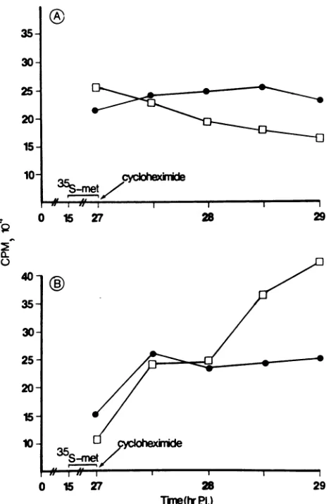
In infected, untreated cells, radiolabeled VP1 accumulated
continually in the nucleus (Fig. 5B), with a concomitant
decrease in the cytoplasmic pool of radiolabeled VP1 (Fig.
5A). In infected cells treated with cycloheximide, VP1
accumulationin the nucleus(Fig.5B)washalted 30to45min
aftertheaddition of cycloheximide.Thesefindingssuggesta
requirement for protein synthesis for continuous transport
and accumulation ofpolyomavirus VP1 in infected-cell
nu-clei.
In order to determine whether the pool of VP1 protein remaining in the cytoplasm during protein synthesis inhibi-tion iscapable of nuclear transport uponremoval of cyclo-heximide, infected MKC were labeled from 15 to 24 h PI with [35S]methionine in methionine-free medium. After the
additionofcycloheximide for 1 h (from 24to 25 h PI), the
cycloheximide was removed and fresh medium was placed
on the cells. At various intervals following the removal of cycloheximide, thecells werefractionated intocytoplasmic and nuclear fractions and the quantity ofradioactive VP1in
each sample was determined. Figure 6 shows that the VP1 remaining in thecytoplasm during cycloheximide treatment
wasfullycapable of nucleartranslocation within30min after removal ofthe protein synthesis inhibitor. As expected, the
rise inthequantity of labeledVP1 inthenuclei ofthesecells wasaccompanied byaconcomitantdecreaseinthequantity
oflabeled VP1 in the cytoplasmic pool.
The rate of newly synthesized VP1 was also assessed
following the removal of the cycloheximide. At 27 h PI,
infected cells were radioactively labeled with [35S]methio-nine following the removal of cycloheximide from the in-fectedcells. As evident in Fig. 7A, newly synthesized VP1 wasdetected in thecytoplasm ofthecellsapproximately 30 min after the removal of the inhibitor. A longer lag period was observed before radioactive VP1 could be detected in
thenucleiof these cells(Fig. 7B). Thislonger lagperiod may be due to the presence of a large pool of presynthesized unlabeled VP1 (awaiting transport to the nucleus) which dilutes the newly synthesized radioactive VP1 in the cyto-plasmof the cell.
In this reportwe describe the construction ofa standard
curveusedfor the specificquantitation ofnanogram
quanti-FIG. 3. Identification of phosphorylated VP1 associated with isolated polyribosomes from infected MKC. Infected MKC were
maintained in phosphate-free Eagle medium for 2 h priorto
radio-activelabeling. At24hP1, cellsgrownonfour 100-mm dishes were
pulsed for 1 h with 500 iLCi of
32Pi
perml in phosphate-free Eagle medium. Polyribosomeswereprepared from the cells by the methodof Schimke (37). Briefly. the cells were incubated with polysome
buffer(25 mM Tris [pH 7.6]. 25 mM NaCI. 5 mM MgCI2. 0.14 M
sucrose. and 500,ug of sodium heparinperml) anddisrupted witha
Dounce homogenizer, and the homogenate was centrifuged for 5
min at 5.000 x g to pellet the nuclei. The nuclear pellet was
disrupted by sonication and immediately frozen to be processed
later. A portion of the nucleus-free cellular lysate was further fractionated by centrifugation at 100.000 x g, and the supernatant
wasusedasthecytoplasmic fraction. The remainder ofthe nucleus-free lysatewas thencentrifuged for 10min at27.000 x g, and the
supernatant was layered over a discontinuous sucrose gradient
containing 2 ml of 2.5 M sucrose and 4 ml of 1 M sucrose (both
prepared in polysome buffer containing 100 xLgofsodium heparin
perml). Thepreparationswerethen centrifugedfor 110 minutesat
180.000xginaBeckman SW41rotor,andthepolysomebandatthe 2.5 M sucrose interface was collected. The nuclear. cytoplasmic.
and polysome preparations were dialyzed against 20 mM MOPS
(morpholinepropanesulfonic acid) buffer. pH 7.0, containing 150 mMNaCI and eitherimmunoprecipitated withrabbit anti-VP1 IgG
and Pansorbin Staphylococcuis aiireius cells (Calbiochem) or
sub-jectedtoSDS-PAGE and blottedontonitrocellulose. The
immuno-precipitates were resuspended in sample buffer and subjected to
SDS-PAGEand autoradiography. The blots wereprobed with the
sameanti-VP1 IgG preparation used for the immunoprecipitation,
and the reactive bandswerevisualizedbyincubation with horserad-ish peroxidase-conjugated protein A (Bio-Rad Laboratories) and
developedwith4-chloro-1-naphthol and H.O.basedon the
recom-mendations of the manufacturer. (A)Immunoprecipitated samples; (B) Westernblots. Lanes 1. nuclear; lanes 2. cytoplasmic; lanes3, polyribosome. Size standards(inkilodaltons)areindicatedbetween
thetwopanels,andthepolyomavirusstructuralproteinsareshown
onboth sides.
tiesofpolyomavirus VP1 protein in infected-cell nuclei and cytoplasm. The standard curve produced by Western
blot-ting wassensitive and linear inthe concentration range of 5 to500 ngof VP1. Figure 1 shows atime courseexperiment of VP1 synthesis in which this standard curvewas used for
VP1 quantitation. We demonstrated the presence of nano-gramquantities ofpolyomavirus VP1 in both thecytoplasm
1 2 3 1 2 3
-68-
vP1-
VP2-
-45-
-36-
VP3-I-VP1
;-VP2
-VP3
-29- -24- -20-VOL.63, 1989
on November 10, 2019 by guest
http://jvi.asm.org/
[image:4.612.53.293.104.263.2]3172 NOTES
._
>i-15 10
9
a
7 6-
5-
3-
1-5
3-
2-1
-
35-
30-25
-20
-
15- 10-imd
24 iS
TrMe(TPO)
26 27 C,0^
U)
40-
35-
30-
25-
20-
15-24 25
Tlme(ITPI)
26 27
FIG. 4. Effectofprotein synthesisinhibitiononVP1cytoplasmic
synthesis and nucleartransport. Infected MKC monolayers were
treated with cycloheximide (100 pg/ml) at 24 h P1. At indicated
intervals following cycloheximide addition, cells were lysed. and
nuclear(B) and cytoplasmic (A) fractions were assayed for VP1
quantitywithastandardVP1curve(inset, panel A)asdescribedin
the text and Fig. 1 legend. Cycloheximide-treated cells (0) were
comparedwithinfected cells nottreatedwithcycloheximide (OI).
and the nucleus of infected cells as early as 15 h PI. VP1 continuedtoaccumulate inthe cytoplasm ofinfected MKC
through 27 h PI, at which point the quantity of VP1 in the
cytoplasm plateaued. This may be due to a heightened
demand for VP1 protein in the nucleus after 27 h for the assemblyprocess. We therefore expectedtoobserve
contin-ualtransport ofnewly synthesized structural proteins (e.g., VP1) into the nucleus of infected cells. VP1 protein
quanti-tationin thenucleus of infected MKC between 15and 36 hPI
demonstrated a continuous pattern of accumulation in the nucleus. Itis also interestingthat VP1proteinwasdetectable
in the nucleus of the infected cells as early as in the
cytoplasm of these cells. This indicates that thetransport of VP1 occurred concurrently with its synthesis and did not
require the build-up of large cytoplasmic pools of synthe-sized VP1prior to transport.
Polyomavirus is one of the finest examples of genetic economyinnature.Withenough genetic informationtocode for only sixproteins (three early tumor antigens and three
3
7S
-, it
0 15 27
2
28 29
0 15 27 28 29
Tie(tr
Pi.)
FIG. 5. Fate ofpresynthesized radiolabeled VP1duringprotein
synthesis inhibition. Infected MKCwere radioactivelylabeledwith 25
pCi
of[35S]methionine
per mlfrom 15 to27 h PI. At 27 hP1,
medium on the cells was removed and replaced with fresh Eagle medium containing cycloheximide (100 p.g/ml) and methionine (4 mg/ml). At indicated intervals following cycloheximide addition, cellswere lysed,andequalamountsofproteinfrom the nuclear(B) and cytoplasmic (A) fractions were subjected to SDS-PAGE. The VP1 bands from stained gels were excised, and the amount of radioactivityineach bandwasdeterminedby scintillationcounting. Cycloheximide-treated cells(@)
were compared withinfected cellsnottreatedwithcycloheximide
(L).
late structural proteins), polyomavirus is ableto expand its functional capabilities by utilizing post-translationally mod-ified versions of its structuralproteins (e.g., VP1) for various required functions (e.g., cellular attachment and hemagglu-tination) (2, 3). It is important to note that the enzymes
responsibleforthesemodifications are aproperty of the host
cell (25). Polyomavirus VP1 has been demonstrated to
separate into six distinct species, A (most basic) through F
(most acidic), based on their isoelectric points (3). Various
posttranslational modifications (e.g., phosphorylation and
sulfation) are thought to be responsible for the different isoelectric points observed with these species. Figure 2
shows that all six species of VP1 were present in the
cytoplasm of infected cells when assayed by isoelectric
focusing. Since VP1 is synthesized in the cytoplasm of the
cell and later transported to the nucleus for assembly, the
presence of modified species of VP1 in the cytoplasm
J. VIROL.
EJ
35S
.. r-..
on0-mmin
I
on November 10, 2019 by guest
http://jvi.asm.org/
[image:5.612.58.302.73.453.2] [image:5.612.324.559.82.440.2]NOTES 3173
25-
23-
21-X_
19-
17-
15-ex& D I
0 24 25 26
Twme(ttPI)
FIG. 6. Fate of presynthesized VP1 upon reversal of pr synthesis inhibition. Infected MKC monolayers were labeled 15 to 24 h PI with 25 ILCi of[39S]methionine per ml. At 24 cycloheximide (100 ,ug/ml) was added to the cells for 1 h and removed, and fresh medium was added to the cells. Cells were lysed at indicated time intervals. Equal amounts of protein each cytoplasmic (0) and nuclear (O) fraction were subject SDS-PAGE, the VP1 band was excised, and the amount ofr
activitywasdeterminedby scintillation counting.
suggested that modification of VP1 occurred in the ( plasm before its translocation into the nucleus of the infe cells.
Essential to understanding the temporal
relationshir
tweensynthesis, posttranslational modification, and nu transport ofpolyomavirus VP1 in infected cells is the d mination of not only the intracellular site but alsothe230
200-130
-100
-20
course of the occurrence ofthese modifications. Quantita-tionof therelative amountsof each VP1 isospecies revealed differences as to their presence at various times during infection. Even thoughat 18h PI all six speciesofVP1 were present in boththe cytoplasmandnucleus ofinfectedcells,
speciesA, the mostbasic species ofVP1, was the
predom-inant species present. VP1 isospeciesAhaspreviously been shownto be associated with the viral minichromosome and
is thought to replace histone Hi early in the assembly
process (3, 45). It is therefore possible that an abundant supply of isospecies A is required early in infection for proper condensation of the viral minichromosome priorto
the assembly ofcapsomere subunits about the DNA. Later in infection, at 27 and 36h PI, the relativequantityof each species resembled the profile observed whenpurified virions
are subjected to the same conditions of isofocusing. This result is expected, since at later times in infection the infected cell has accumulated a large number of mature 27 virions. Pulse-labeling experiments with
3Pi
demonstrated thepresenceofphosphorylated VP1 protein in polyribosomerotein preparations from infected MKC. Thesefindings reflect the
from cotranslational natureof this modification.
h PI. An understanding of the mechanism by which proteins then synthesized in the cytoplasm are specifically sorted to the then different cellularcompartments is essential for an apprecia-from tion of the regulation of
functional
units within eucaryoticred
too-
cells. Nuclearentry oflarger proteins is a selectivemecha-radio- nism and a property of the amino acid sequence of the
protein (10, 18, 32, 38). Recently, the nuclear transport
signal of simian virus 40 (SV40) large tumor antigen was
cyto- identified (19).Itcomprisesahighly basic, lysine-richstretch
scted of amino acids. It was also demonstrated that a synthetic peptide homologous to the SV40 large T antigen transport p be- signal sequence will transport cross-linked proteins to the
clear nucleus (23, 44). In addition, Wychowski et al. have also leter- demonstratedthatthe firsteightamino-terminal amino acids
time of SV40 VP1 and VP2/VP3 amino acids 317 to 323 are
lDO
-
20-0 27 28 29 30
35
S-me*t
0 27 28 29 30
Tine(tr PI) Time(hrPI)
FIG. 7. Recovery of VP1cytoplasmic synthesisandnucleartransportfollowingreversal ofprotein synthesisinhibition.At27 hP1, MKC monolayers were treated with cycloheximide (100 pLg/ml) for 1 h. Upon removal of the inhibitor, fresh medium containing 250 ,Ci of
[35S]methionine per ml was addedtothe cells. At indicated intervals following the removal of the inhibitor, cells werelysedand separated
intocytoplasmic(A) and nuclear(B)fractions. Equal amountsofproteinfrom thesefractionswere thensubjectedtoSDS-PAGE. The VP1 band wasexcised from stainedgels, and the radioactivity was determined by scintillation counting. Cycloheximide-treated (0) cells were compared with infected cellsnottreated withcycloheximide (D).
VOL. 63, 1989
on November 10, 2019 by guest
http://jvi.asm.org/
[image:6.612.64.294.77.275.2] [image:6.612.106.510.470.669.2]3174 NOTES
involved in the migration of theseproteinsinside the nucleus (42, 43). It is interesting that the polyomavirus structural
protein VP1 (first 12 amino-terminal amino acids) and the
last 8 amino acids of VP3 have a similar stretch of basic
amino acids with the potential of transporting cytoplasmic
proteinsto the nucleus.
Inorder toanalyze whether the transport ofpolyomavirus
VP1is an independent process (requiringonly the presence of thesignal sequence), we sought to establish the effect of
protein synthesis inhibition on the accumulation of newly
synthesized VP1in the nucleus of infected MKC. Figures 4
and 5 suggest thatcontinual accumulation of VP1proteinin the nucleus required protein synthesis. If the transport of
VP1 protein was solely dependent on the presence of its
putativenuclear transportsignalsequence, resultsexhibiting
the continued accumulation ofVP1 in the nucleus during
protein synthesis inhibition would have been expected.
Whether the synthesis of viral or cellular products is
re-quired
for the transport process is not known atthispoint.
However,ourresults, togetherwith the work of Stamatoset
al. (40), whichdemonstrates the transport ofvaccinia
virus-expressed VP1 into the nucleus in the absence of other
polyomavirus proteins, extend supportive evidence that
cellular constituents may be involved in this process. Recent
studies by Gharakhanian et al. have demonstrated that the
VP3 protein of SV40, via its carboxyl-terminal 40 amino
acids, is involved in interaction with the VP1 structural
protein (14). This same region of VP3 has been shown by
these authors (13) and others (43) to contain a nuclear
localization signal. Gharakhanian et al. suggest that the
cluster of
positively
charged amino acids (similar to theSV40largeTantigen nuclear translocationsignalsequence)
in this region of VP3 may be involved in electrostatic
interactions between SV40 VP1 and VP3. Whether these
sameinteractionsoccurduring polyomavirusstructural
pro-teinsynthesisis not known. However,given thesimilarities
between the two viruses, it is likely that polyomavirus
structural proteins VP1, VP2, and VP3 undergo similar
protein-protein interactions. It isalsolikelythat
polyomavi-rus VP1 interacts with normal cellular (cytoplasmic or
nu-clear)proteins throughitsfirst 12amino acids for its nuclear
translocation functions. Our results also demonstrated that the pool of VP1 protein remaining in the cytoplasm of the
infected cells during protein synthesis inhibition was fully
capableof nucleartransport upon removalofcycloheximide
(Fig. 6).
With the wealth of accumulated evidence regarding the
involvement of the signal sequence in the translocation of
proteins into the nucleus, it is our goal to focus our future
studies on the examination of VP1 association (interaction) with other viral or cellular proteins in the cytoplasm and nucleus via thesignal sequence. Thesestudies may result in
the identification ofproteins involved in the nuclear
trans-port process ininfected cells.
This investigationwas supported by Public Health Service grant CA-07139 from the National Cancer Institute, NAGW-1197 from NASA,andthe Wesley Foundation of Wichita, Kansas.
We express our appreciation to Viola Hill, Todd Martin, and LaDonna Grenz for excellent technical assistance. We also express
ourappreciation to Melvin Center for critical review of the manu-script.
LITERATURE CITED
1. Anders,D. G., and R. A.Consigli. 1983. Chemical cleavage of polyomavirus major structural protein VP1: identification of cleavageproductsandevidencethat the receptormoiety resides
in thecarboxy-terminal region.J. Virol.48:197-205.
2. Anders,D. G., andR. A. Consigli. 1983. Comparison of non-phosphorylated and phosphorylated species of polyomavirus
major capsidproteinVP1andidentification of themajor phos-phorylationregion. J. Virol.48:206-217.
3. Bolen,J. B.,D.G.Anders,J. Trempy,and R. A.Consigli.1981. Differences in thesubpopulations of the structural proteins of polyomavirionsandcapsids: biologicalfunctions of themultiple VP1species. J. Virol.37:80-91.
4. Bolen, J.B.,andR.A.Consigli.1979.Differentialadsorptionof polyomavirions andcapsids tomouse kidneycells andguinea pigerythrocytes. J.Virol. 32:679-683.
5. Bolen,J. B.,and R.A.Consigli.1980.Separationofneutralizing and hemagglutination-inhibiting antibody activities and speci-ficityofantiseratosodiumdodecylsulfate-derivedpolypeptides ofpolyomavirions. J. Virol. 34:119-129.
6. Brady,J. N., V. D. Winston, and R. A.Consigli. 1977. Dissoci-ation ofpolyomavirusby the chelation of calcium ions found associatedwith purifiedvirions. J. Virol. 23:717-724.
7. Brunck, C.F., and V. Leick. 1969.Rapid equilibriumisopycnic CsCIgradients. Biochim. Biophys. Acta179:136-144.
8. Burglin, T. R., and E. M. De Robertis. 1987. The nuclear migration signal ofXenopus laevis nucleoplasmin. EMBO J. 6:2617-2625.
9. Consigli,R.A., G. R.Griffith,S.J. Marriott, andJ.W.Ludlow. 1986. Biochemicalcharacterizationofpolymavirus-receptor in-teractions, p. 44-53. In R. L. Crowell and K. Lonberg-Holm
(ed.),Virusattachment and entry into cells. American Society forMicrobiology, Washington, D.C.
10. Dingwall, C., S. V. Shannick, and R. A. Laskey. 1982. A polypeptide domain that specifies migration into the nucleus. Cell30:449-458.
11. Eisenman, R. N., C. Y. Tachibana, H. D. Abrams, andS. R. Hann. 1985. v-myc andc-myc-encoded proteins are associated with the nuclear matrix. Mol. Cell. Biol.5:114-126.
12. Garcea, R. L., K. Ballmer-Hofer, and T. L. Benjamin. 1985. Virion assembly defect of polyomavirus hr-t mutants: under-phosphorylationofmajorcapsid protein VP1 before viral DNA encapsidation. J. Virol.54:311-316.
13. Gharakhanian, E., J. Takahashi, J. Clever, and H. Kasamatsu. 1988. In vitro assay for protein-protein interaction: carboxyl-terminal 40 residues of simian virus 40 structural protein VP3 contain a determinant for interaction with VP1. Proc. NatI. Acad. Sci. USA85:6607-6611.
14. Gharakhanian,E.,J. Takahashi, and H. Kasamatsu. 1987. The carboxyl 35 amino acids of SV40 VP3 are essential for its nuclear accumulation. Virology 157:440-448.
15. Griffith, G. R., andR. A.Consigli. 1984. Isolation and charac-terization of monopinocytotic vesicles containing polyomavirus from the cytoplasm ofinfected mouse kidney cells. J. Virol. 50:77-85.
16. Griffith, G. R., and R. A. Consigli. 1986. Cross-linking of a polyomavirus attachment protein to its mouse kidney cell re-ceptor. J. Virol. 58:773-781.
17. Griffith, G. R., S. J.Marriott, D. A. Rintoul, and R. A. Consigli. 1988. Early events in polyomavirus infection: fusion of mono-pinocytotic vesicles containing virions with mouse kidney cell nuclei. Virus. Res. 10:41-52.
18. Hall, M. N., L.Hereford, and I.Herskowitz. 1984. Targeting of E. coli,B-galactosidease to the nucleus in yeast. Cell 36:1057-1065.
19. Kalderon, D., W. D. Richardson, A. F. Markham, and A. E. Smith. 1984. Sequence requirements for nuclear location of simian virus 40 large-T antigen. Nature (London)311:33-38. 20. Klempnauer, K., and A. E. Sippel. 1986. Subnuclear localization
of proteins encoded by the oncogene v-myb and its cellular homologc-mnyb. Mol. Cell. Biol. 6:62-69.
21. Knipe, D. M., and J. L. Smith. 1986. A mutant herpesvirus protein leads to a block in nuclear localization of other viral proteins. Mol. Cell. Biol. 6:2371-2381.
22. Laemmli, U. K. 1970.Cleavageof structural proteins during the assembly of the head of bacteriophage T4. Nature (London) 227:680-685.
J. VIROL.
on November 10, 2019 by guest
http://jvi.asm.org/
NOTES 3175 23. Lanford, R. E., P. Kanda, and R. C. Kennedy. 1986. Induction
of nuclear transport with a synthetic peptide homologous to the SV40 T antigen transport signal. Cell 46:575-582.
24. Lee, B. A., D. W. Maher, M. Hannink, and D. J. Donoghue. 1987. Identification of a signal for nucleartargeting in platelet-derived-growth-factor-related molecules. Mol. Cell. Biol. 7: 3527-3537.
25. Ludlow, J. W., and R. A. Consigli. 1987. Differences in biolog-ical activity and structural protein VP1 phosphorylation of polyomavirus progeny resulting from infection of primary mouse kidney and primary mouse embryo cell cultures. J. Virol. 61:509-515.
26. Ludlow, J. W., and R. A. Consigli. 1987. Polyomaivirus major capsid protein VP1 is modified by tyrosine sulfuration. J. Virol. 61:1708-1711.
27. Mackay, R. L., and R. A. Consigli. 1976. Early events in polyoma virus infection: attachment, penetration. and nuclear entry. J.Virol. 19:620-636.
28. Marriott, S. J., G. R. Griffith, and R. A. Consigli. 1987.
Octyl-p-D-glucopyranoside
extracts polyomavirus receptor moieties from the surface of mouse kidney cells. J. Virol. 61:375-382.29. Marriott, S. J., D. J. Roeder, and R. A. Consigli. 1987. Anti-idiotypic antibodies to a polyomavirus monoclonal antibody recognize cell surface components of mouse kidney cells and prevent polyomavirus infection. J. Virol. 61:2747-2753. 30. McMillen, J., M. S. Center, and R. A.Consigli. 1976. Origin of
the polyoma virus-associated endonuclease. J. Virol. 17:127-131.
31. McMillen, J., and R. A.Consigli.1977. Immunological reactivity ofantisera to sodium dodecyl sulfate-derived polypeptides of polyoma virions. J. Virol. 21:1113-1120.
32. Moreland, R. B., H. G. Nam, L. M. Hereford, and H. M. Fried. 1985. Identification of a nuclear localization signal of a yeast ribosomal protein. Proc. Natl. Acad. Sci. USA 82:6561-6565. 33. O'Farrell,P. H.1975. High-resolutiontwo-dimensional
electro-phoresis of proteins. J. Biol. Chem. 250:4007-4021.
34. O'Farrell, P. Z., and H. M. Goodman. 1976. Resolution of simian virus 40 proteins in whole cell extracts by two-dimen-sional electrophoresis: heterogeneity of the major capsid
pro-tein. Cell 9:289-298.
35. Ponder, B. A. J., A. K. Robbins, and L. V. Crawford. 1977. Phosphorylation of polyoma and SV40 virus proteins. J. (,en. Virol. 37:73-85.
36. Richardson, W. D., B. L. Roberts, and A. E. Smith. 1986. Nuclear translocation signals in polyoma virus large-T. Cell 44:77-85.
37. Schimke, R. T., R. Palacios, D. Sullivan, M. L. Kiley, C. Gonzales, andJ. M. Taylor. 1974. Immunoadsorption of oval-bumin synthesizing polysomes and partial purification of oval-bumin messenger RNA. Methods Enzymol. 30:631-648.
38. Silver, P. A., L. P. Keegan, and M. Ptashne. 1984. Amino terminus of the yeast GAL4 geneproduct is sufficient for nuclear localization. Proc. Natl. Acad. Sci. USA 81:5951-5955. 39. Smith, G. L., and R. A.Consigli. 1972. Transient inhibition of
polyoma virus synthesis by Sendai virus (parainfluenza I). 1. Demonstration andnatureof the inhibitionby inactivatedvirus.
J. Virol. 10:1091-1097.
40. Stamatos,N.M., S.Chakrabati, B. Moss, and J. D. Hare.1987. Expression of polyomavirus virion proteins by a vacciniavirus
vector:association of VP1 and VP2 with the nuclear framework. J. Virol. 61:516-525.
41. Winston, V.D.,J. B. Bolen, and R. A. Consigli. 1980.Isolation and characterization ofpolyoma uncoating intermediates from the nuclei of infected mouse cells. J. Virol. 33:1173-1181. 42. Wychowski, C., D. Benichou, and M. Girard. 1986.Adomain of
SV40 capsid polypeptide VP1 thatspecifies migration into the nucleus. EMBO J. 5:2569-2576.
43. Wychowski, C., D. Benichou, and M. Girard. 1987. The inter-nuiclear location ofsimian virus40 polypeptides VP2 and VP3 depends on a specific amino acid sequence. J. Virol. 61:
3862-3869.
44. Yoneda, Y., T. Arioka, N. Imamoto-Sonobe, H. Sugawa, Y.
Shimonishi,and T. Uchida. 1987.Synthetic peptides containing aregion of SV40largeT-antigen involved in nuclear localization direct the transport ofproteins into the nucleus. Exp. Cell Res. 170:439-452.
45. Yuen, L. K. C., and R. A. Consigli. 1985. Identification and
protein analysis of polyomavirus assembly intermediates from infected primarymouse embryo cells. Virology 144:127-138. VOL. 63,1989
![FIG. PI)fractioncentrifugedclearresuspendedacetone-sample)resuspendedandblottedcytoplasmiccurvetimenuclearofassayedfluoride,tors])mMandtreatedanti-VP1supernatant(31).MKC.[pH infection infected equal 36 7.9], dithiothreitol, fraction)The (inset,courseat a](https://thumb-us.123doks.com/thumbv2/123dok_us/1325404.86358/2.612.60.294.78.309/fractioncentrifugedclearresuspendedacetone-resuspendedandblottedcytoplasmiccurvetimenuclearofassayedfluoride-mmandtreatedanti-supernatant-infection-dithiothreitol-fraction-courseat.webp)