The bioeroding sponge Cliona
orientalis will not tolerate future
projected ocean warming
Blake D. Ramsby
1,2,3, Mia O.
Hoogenboom
1, Hillary A.
Smith
2,3, Steve
Whalan
4&
Nicole S. Webster
2,3,5Coral reefs face many stressors associated with global climate change, including increasing sea surface temperature and ocean acidification. Excavating sponges, such as Cliona spp., are expected to break down reef substrata more quickly as seawater becomes more acidic. However, increased bioerosion
requires that Cliona spp. maintain physiological performance and health under continuing ocean
warming. In this study, we exposed C. orientalis to temperature increments increasing from 23 to 32 °C. At 32 °C, or 3 °C above the maximum monthly mean (MMM) temperature, sponges bleached and the
photosynthetic capacity of Symbiodinium was compromised, consistent with sympatric corals. Cliona orientalis demonstrated little capacity to recover from thermal stress, remaining bleached with reduced
Symbiodinium density and energy reserves after one month at reduced temperature. In comparison,
C. orientalis was not observed to bleach during the 2017 coral bleaching event on the Great Barrier Reef, when temperatures did not reach the 32 °C threshold. While C. orientalis can withstand current
temperature extremes (<3 °C above MMM) under laboratory and natural conditions, this species would not survive ocean temperatures projected for 2100 without acclimatisation or adaptation (≥3 °C above MMM). Hence, as ocean temperatures increase above local thermal thresholds, C. orientalis will have a
negligible impact on reef erosion.
Increasing global temperatures are requiring organisms to acclimate to greater thermal extremes, migrate, or suf-fer reduced fitness and, potentially, local extirpation. The earth’s climate is already estimated to be 0.85 °C warmer than it was in 1880, which is affecting both terrestrial and marine ecosystems1. Much of the thermal energy (~60%) associated with warming has been absorbed by the oceans, resulting in melting sea ice, rising sea levels1, and record temperatures in tropical waters2,3. Ocean warming has already resulted in extensive coral mortality3,4, as evidenced in 2015/2016, when extreme temperatures led to consecutive mass coral bleaching events around the world2,3.
Corals contain photosynthetic dinoflagellates (genus Symbiodinium) that provide them with organic carbon5. However, the symbiosis is thermally sensitive and exposure to elevated temperature disrupts Symbiodinium pho-tosynthesis6 and causes coral bleaching7,8. Some coral species and Symbiodinium ‘types’ are more thermally toler-ant than others9–11, but even tolerant genotypes can be overwhelmed by severe temperature stress3. Nonetheless, in some cases, previous exposure to high temperature or association with tolerant Symbiodinium can lead to greater thermal tolerance of the coral symbiosis9,10,12,13 and accelerate recovery following bleaching9,14,15.
In comparison to reef-building corals, sponges are thought to be relatively tolerant of increasing sea sur-face temperatures16,17. In particular, some bioeroding sponges can tolerate temperatures that induce bleaching in sympatric corals18–22. Bioeroding sponges, principally the genus Cliona, are important members of coral reef communities as they erode the limestone substratum by reducing the pH at the sponge:substratum interface23, dissolving the substratum, and extracting microscopic ‘chips’ of calcium carbonate24,25. Like corals, many bioe-roding sponge species form symbioses with photosynthetic Symbiodinium and photosynthesis enhances their
1College of Science and Engineering and ARC Centre of Excellence for Coral Reef Studies, James Cook University, Townsville, Queensland, Australia. 2Australian Institute of Marine Science, Townsville, Queensland, Australia. 3AIMS@JCU, Australian Institute of Marine Science and James Cook University, Townsville, Queensland, Australia. 4Marine Ecology Research Centre, School of Environment, Science and Engineering, Southern Cross University, Lismore, New South Wales, Australia. 5Australian Centre for Ecogenomics, The University of Queensland, Brisbane, 4072, Queensland, Australia. Correspondence and requests for materials should be addressed to B.D.R. (email: [email protected])
Received: 9 February 2018 Accepted: 15 May 2018 Published: xx xx xxxx
growth and bioerosion26–28. However, while dependence on Symbiodinium may increase the thermal sensitivity of Cliona, little is known about how these sponges will tolerate predicted incremental temperature increases or whether they can recover from extreme thermal stress29,30.
Experimental research combining elevated temperature and reduced pH has shown that sponge bioerosion rates will likely increase under conditions of ocean acidification31–35. However, warming can have negative effects on bioeroding sponges, including bleaching or necrosis; and it is likely that these negative effects will override all other environmental factors29,30,32. For instance, under temperature and pH conditions predicted for 2100, the bioeroding sponge Cliona orientalis bleaches, and the associated reduction in photosynthetic productivity results in a negative energy budget for the sponge despite accelerated rates of erosion29,34. Warming was subse-quently identified as the primary stressor inducing bleaching in a bioeroding sponge30. However, temperature tolerance appears to vary among bioeroding sponge species as bleaching or mortality was not observed in all studies31,36. Therefore, the net effect of climate change on bioeroding sponges, and on their erosion rates, appears to be species-specific.
Identifying thermal thresholds under near-future warming requires measurement of performance across a broad range of incremental temperature changes. This incremental approach enables a holistic understanding of temperature effects by allowing quantification of the optimal temperature for peak physiological performance, along with derivation of sub-lethal and lethal temperature thresholds37,38. A similar approach has been applied to corals to quantify adaptation to local thermal regimes39 and to determine how coral respiration and photosynthe-sis varies with temperature40. Here, we experimentally assessed the ability of C. orientalis to tolerate incrementally increasing sea surface temperatures between 23–32 °C, which represents the annual temperature range for the studied sponges (22–30 °C) and warmer temperatures predicted for 2100 (31, 32 °C)1. In addition, we monitored recovery from temperature stress to evaluate the impact of thermal exposure on sponge survival. Photosynthetic performance of Symbiodinium and energy reserves of the sponge were quantified to identify temperature optima and define thermal thresholds. To contextualize the laboratory experiment, we assessed bleaching severity for C. orientalis during the 2017 mass coral bleaching event.
Results
Response to laboratory thermal exposure.
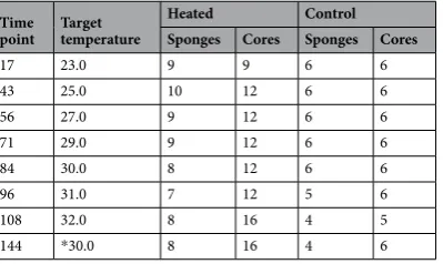
Bleaching and Symbiodinium identity. Cliona orientalis survived temperatures up to 31 °C with no visual signs of bleaching and little evidence of compromised health, such as discolouration or tissue regression (Fig. 1B). However, at 32 °C, more than 50% of the cores became visibly bleached within three to five days and this increased to 70% bleached after 8 days (Fig. 1B). The condition of some cores also visibly deteriorated in the four weeks following bleaching, including the presence of algae at the margin of the cores (Fig. 1C). The narrow bleaching threshold of 32 °C identified for C. orientalis is 2.9 °C above the maxi-mum monthly mean at Orpheus Island. A low level of mortality occurred in both temperature treatments but was constrained to cores from a few specific sponge genotypes. [image:2.595.157.477.48.282.2]Only a single Symbiodinium ITS2 type was associated with C. orientalis, regardless of temperature treatment or bleaching state. ITS2 sequences clustered into 61 OTUs at 97% sequence similarity, but only 21 of these matched the Symbiodinium database, and only 9 were confidently matched with bitscores greater than 100. Of the nine Symbiodinium OTUs, one OTU comprised 96% of the Symbiodinium sequences and was the most abundant OTU in every sponge core. The ITS2 sequence of the dominant OTU was identical to Symbiodinium clade G previously sequenced from C. orientalis (Genbank accession JQ247051) and which was recently described as Symbiodinium endoclionum41. One other OTU occurred in 97% of the cores and was also most similar to Symbiodinium clade G from C. orientalis, differing from the dominant ITS2 sequence by one insertion of eight nucleotide substitutions. The remaining Symbiodinium OTUs were most similar to Symbiodinium clades A, B, or C, however these OTUs comprised <1% of total sequences and occurred in <20% of the samples.
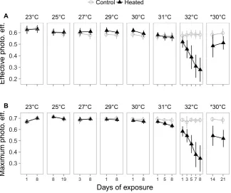
Photosynthesis and respiration. Temperature effects were interpreted as significant temperature*treatment inter-actions which indicate the temperatures at which heated sponge cores responded differently than control cores (Table 3). Photosynthesis was largely unaffected by temperatures up to and including 31 °C, but became inhibited at 32 °C. Sponges at 27 and 29 °C had higher effective (∆F/Fm’), but not maximum (Fv/Fm), photochemical effi-ciency than sponges maintained at 23 °C (Fig. 2; eff. 27 °C: z =−4.5, p< 0.01; eff. 29 °C: z =−3.2, p= 0.01). At 29–31 °C, sponges had significantly lower maximum photochemical efficiencies than control sponges (p< 0.01), but the differences were small, and C. orientalis maintained 93% of maximum photochemical efficiency of control sponges at 31 °C. However, at 32 °C, maximum and effective photochemical efficiencies were 52% and 50% of control sponges, respectively (Table 4). As with photochemical efficiency, the photosynthetic rate (gross oxygen production) did not differ from controls at temperatures up to and including 31 °C (Fig. 3A; p> 0.86) but was 43% lower than controls in sponges exposed to 32 °C (Table 4).
In contrast to photosynthesis, the effect of thermal exposure on sponge respiration was greatest at 29 °C, where heated sponges had 47% higher respiration rates than control sponges (Fig. 3B; z =−3.4, p< 0.01). At tempera-tures close to 29 °C (27, 30, and 31 °C), respiration was 28–35% higher than controls, but these differences were not significant (0.09 <p< 0.29). For sponges at 32 °C, respiration rates were similar to control sponges (Table 4). The ratio of gross photosynthesis to respiration (P/R) was affected at a similar temperature as the respiration rates (Fig. 3C). Sponges at 29, 30, and 31 °C had 79% of the sponge P/R of control sponges, coinciding with faster res-piration rates (Fig. 3B), but the difference was only statistically significant at 30 °C (z = 3.1, p= 0.01). Sponges at 32 °C had 37% of the P/R of control sponges (Table 4), indicating a loss of productivity of the symbiosis.
Time
point Targettemperature Heated(n = 6) (n Control= 3) °C aboveMMM + 1 AccumulatedDHW
17 23.0 23.5 ± 0.1 23.4 ± 0.2 0 0
43 25.0 25.2 ± 0.1 23.1 ± 0.1# 0 0
56 27.0 27.0 ± 0.1 23.3 ± 0.1 0 0
71 29.0 29.0 ± 0.2 23.0 ± 0.2 0 0
84 30.0 30.0 ± 0.1 23.1 ± 0.1 0 0
96 31.0 30.9 ± 0.1# 23.2 ± 0.1 0.8 1.3
108 32.0 32.0 ± 0.1 23.1 ± 0.2 1.9 4.4 144 *30.0 30.1 ± 0.1 23.1 ± 0.3 0 4.4
Table 1. Target and actual temperatures during the laboratory experiment. For each target temperature, tank
temperatures were recorded every 5 minutes over 8 days (mean ± SD°C). #Indicates where the SD was less than 0.05 °C. °C above the maximum monthly mean (MMM) at Orpheus island (29.1 + 1.0 °C) indicates the thermal anomaly above long-term summer temperatures which was used to calculate the accumulated degree heating weeks (DHW) as the product of the thermal exposure and the thermal duration (°C-weeks).
Time
point Targettemperature
Heated Control Sponges Cores Sponges Cores
17 23.0 9 9 6 6
43 25.0 10 12 6 6
56 27.0 9 12 6 6
71 29.0 9 12 6 6
84 30.0 8 12 6 6
96 31.0 7 12 5 6
108 32.0 8 16 4 5
144 *30.0 8 16 4 6
Table 2. The number of cores and original sponges sampled for oxygen flux and tissue contents at each
[image:3.595.160.457.46.160.2] [image:3.595.159.359.241.360.2]Figure 2. Photochemical efficiency of Symbiodinium within C. orientalis. Effective (A) and maximum (B) photochemical efficiencies are shown for control cores measured at 23 °C (grey open circles and lines) and heated cores at elevated temperature (black triangles and lines). Panels separate the temperature of the heated treatment. Points represent means and error bars indicate one SD.
Trans. Out. rem.
Treatment (num. df = 1) Temperature (num. df = 7) Treatment: Temperature (num. df = 7)
SS MS D. df F p SS MS D. df F p SS MS D. df F p
Photosynthesis
Fv/Fm p/(1 − p) N 18.9 18.9 686.1 676.4 <0.01 35.5 5.1 679.0 181.9 <0.01 25.1 3.6 677.9 128.5 <0.01
∆F/Fm’ p/(1 − p) N 0.7 0.7 9.8 106.2 <0.01 23.1 3.3 674.0 106.2 <0.01 11.3 1.6 672.5 52.0 <0.01
Gross P — N <0.1 <0.1 121.4 4.0 <0.01 0.3 <0.1 24.1 128.3 <0.01 <0.1 <0.1 7.6 124.7 <0.01 R — Y <0.1 <0.1 121.0 18.9 <0.01 <0.1 <0.1 128.8 14.9 <0.01 <0.1 <0.1 125.0 3.0 0.01 Gross P:R — Y 14.3 14.3 119.9 41.3 <0.01 25.3 3.6 10.5 126.2 <0.01 12.9 1.8 5.3 122.8 <0.01 Sponge condition
Symb. — N 18944 18944 125.2 8.5 <0.01 173824 24823 129.7 11.1 <0.01 89443 12778 127.4 5.7 <0.01 Chl a — N 25624 25624 123.2 19.6 <0.01 121098 17300 13.2 128.4 <0.01 94888 13556 10.4 13556 <0.01 Chl c — N 990 990 123.1 20.3 <0.01 7072 1010 20.8 128.1 <0.01 4372 624.6 12.8 125.2 <0.01 Chl a:c Log Y <0.1 <0.1 123.3 0.4 0.52 0.8 0.1 5.3 128.6 <0.01 0.1 <0.1 0.8 126.1 0.59 Protein Log N 0.8 0.8 120.5 10.4 <0.01 4.0 0.6 7.9 130.2 <0.01 2.3 0.3 4.6 124.8 <0.01 AFDW Log Y 0.3 0.3 122.1 14.3 <0.01 0.7 0.1 5.8 129.2 <0.01 0.7 0.1 5.5 125.0 <0.01
Table 3. Statistical results of the laboratory experiment. Parameters tested include maximum photochemical
[image:4.595.157.482.47.318.2] [image:4.595.39.555.395.586.2]Figure 3. Oxygen flux rates for net photosynthesis (A), respiration (B), and the ratio of gross photosynthesis
to respiration (C) for C. orientalis between 23 and 32 °C. Control cores (grey open circles and lines) were all measured at 23 °C, while heated cores (black circles and lines) were sampled at the temperature indicated in the legend. *30 indicates samples that were exposed to 32 °C and then returned to 30 °C for four weeks following bleaching. Points represent means and error bars indicate one standard error. Asterisks indicate temperature increments where sponges in control and heated treatments had significantly different responses.
Exposure Recovery
32 °C: Heated vs Ctrl. Heated: 32 °C *30 vs *30 °C: Heated vs Ctrl Min. sig.
temp z p z p z p
Photosynthesis
Fv/Fm 29 C > 32 27.8 <0.01 *30 > 32 11.3 <0.01 C > H 14.2 <0.01
∆F/Fm’ 32 C > 32 15.8 <0.01 *30 > 32 12.2 <0.01 C > H 5.4 <0.01
Gross P 32 C > 32 7.8 <0.01 *30 > 32 −5.6 <0.01 C > H 3.7 <0.01 R — — −0.8 0.99 *30 > 32 −4.8 <0.01 — 1.2 0.89 Gross P:R 30 C > 32 6.1 <0.01 *30 > 32 5.8 <0.01 C > H 3.2 <0.01 Sponge condition
Symb. 32 C > 32 4.3 <0.01 — 1.9 0.42 C > H 5.3 <0.01 Chl a 32 C > 32 3.7 <0.01 32 >*30 12.8 0.33 C > H 8.6 <0.01 Chl c 32 C > 32 3.9 <0.01 32 >*30 3.33 <0.01 C > H 9.5 0.01
Chl a:c — — — — — — — — — —
Protein 32 C > 32 3.0 0.03 32 >*30 3.2 0.01 C > H 4.8 <0.01 AFDW 32 C > 32 2.8 0.05 32 >*30 4.3 <0.01 C > H 6.6 <0.01
Table 4. Results of post-hoc comparisons. To compare the sensitivity of different parameters, the table includes
[image:5.595.155.395.47.318.2] [image:5.595.155.469.426.628.2]Sponge condition. Temperatures less than 32 °C did not significantly affect the condition of C. orientalis and the density of Symbiodinium, chlorophylls a and c2, protein, and organic matter were similar to control sponges (Fig. 4; p> 0.20). At 32 °C, the bleached sponges contained 25% of the Symbiodinium, 35% of chlorophyll a, and 42% of chlorophyll c2 of the control sponges (Table 4). Moreover, sponges at 32 °C contained 83% of the organic matter and 66% of the protein found in control sponges (Table 4), signifying reduced condition of the sponge.
While the a:c2 ratio decreased over the course of the experiment (Table 3, Temperature), it was not strongly affected by temperature, as heated sponges were 91–107% that of control sponges throughout the experiment and differences between treatments were not significant (Table 3, Treatment:Temperature).
Recovery from laboratory thermal exposure.
Bleaching and Symbiodinium identity. The Symbiodinium associated with C. orientalis did not change following bleaching, with all cores dominated by Symbiodinium endoclionum. Four weeks following bleaching, C. orientalis cores appeared white, and both the Symbiodinium density and chlorophyll content revealed that the sponges had not recovered from their bleached state, indicating prolonged holobiont disruption (Fig. 4).Photosynthesis and respiration. C. orientalis recovered some photosynthetic capacity when returned to *30 °C, with photochemical efficiencies, photosynthetic rates, and P/R being higher than when sponges were at 32 °C (Fig. 3, Table 4). However, this response was likely due to other photosynthetic colonisers as Symbiodinium den-sities remained low in sponges at *30 °C (Fig. 3). Regardless, recovery of photosynthesis was incomplete, as pho-tochemical efficiencies, photosynthetic rates, and P/R remained lower than control sponges (Fig. 3, Table 4). The respiration rates of sponges returned to *30 °C were higher than the sponges at 32 °C, but not significantly different from controls (Fig. 3, Table 4).
Sponge condition. Tissue contents indicated that the condition of the 32 °C sponges continued to deteriorate after they were returned to *30 °C (Fig. 4, Table 4). Chlorophyll content, organic matter, and protein content were lower in sponges at *30 °C than at 32 °C, but Symbiodinium density was not significantly different (Table 4). In addition, all measured tissue contents remained lower in sponges returned to *30 °C than control sponges (Fig. 4, Table 4).
Figure 4. Tissue contents of Symbiodinium (A), chlorophyll a (B), ash-free dry weight (C; AFDW), and protein content (D) for C. orientalis between 23 and 32 °C. Controls (grey open circles and lines) were all sampled at 23 °C while heated cores (black circles and lines) were sampled at the temperature indicated in the legend.
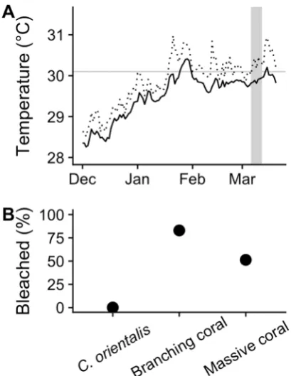
[image:6.595.156.397.48.325.2]Field bleaching surveys.
A total of 133 C. orientalis sponges, 1891 branching corals, and 1068 massive cor-als were counted among the six survey sites. No bleached C. orientalis sponges were observed in any of the video transects. In contrast, 83% (±6.0 SD) of branched coral colonies and 51% (±3.2 SD) of massive coral colonies were bleached (Fig. 5B).Temperatures did not reach 32 °C during the 2017 bleaching event at Orpheus Island (Fig. 5A). During the two weeks preceding the surveys, daily mean temperatures averaged 29.8 °C at 5.8 m depth (Source: Australian Institute of Marine Science; http://data.aims.gov.au) and, during the 12 weeks preceding the surveys, the cumu-lative thermal exposure summed to 1 degree heating week (weeks above 30.1 °C). In comparison, the laboratory experiment indicated that C. orientalis bleached at 32 °C after accumulating 2.5 degree heating weeks (3 days at 32 °C; Table 1).
Discussion
Bioeroding sponges are generally thought to be tolerant of environmental stressors, including elevated tempera-ture, ocean acidification, and eutrophication, raising concerns about increased reef erosion under future projected climate scenarios42,43. Here, we show that incremental increases in ocean temperature up to 30 °C have negligible effects on C. orientalis, but C. orientalis bleaches when exposed to 32 °C, and exhibits little potential for recovery. At the collection site, 32 °C represents an increase of 3 °C above the maximum monthly mean temperature and corresponds to the increase expected under very high greenhouse gas emissions by 2100, but could represent the mean temperature as soon as 2078 (RCP 8.5)1, suggesting that C. orientalis could bleach regularly by the end of this century. The results of this study do not support the hypothesis that bioeroding sponges (particularly those species with photosynthetic symbionts) will play a larger role in structuring future reefs.
Temperature exposure in the laboratory revealed a narrow thermal threshold for C. orientalis, with sponges appearing visibly healthy after 10 days at 31 °C, but bleaching after only 3 days at 32 °C. This narrow threshold is similar to several sympatric coral species that bleached following 1 °C temperature increases between 31 and 33 °C44. In thermally sensitive corals, bleaching coincides with reduced condition and growth7 and C. orientalis exhibited similar negative responses, including a 75% reduction in Symbiodinium density, 17% reduction in organic matter and 44% reduction in protein content of C. orientalis. Few bleached cores exhibited necrosis which had been previously reported in C. orientalis from Orpheus Island after exposure to only 2 °C above MMM. The discrepancy between studies likely results from the faster temperature increases or acute exposures (3–72 h) used in previous research32,45. The thermal threshold identified here is consistent with findings for C. orientalis in the southern GBR, which tolerates exposure to +2.0 °C above MMM (27.3 °C)MMM = 27.3 °C34, but bleaches at +2.7 °C30, and dies at
+3.5 °C34. Taken together, these experimental and field results suggest that C. orientalis can tolerate current ocean temperatures, but will have little capacity to cope with the warmer oceans projected for 2100.
The primary cause of coral bleaching is exposure to extreme ocean temperature, although longer exposure to moderate increases in temperature can also induce bleaching46,47. Cumulative thermal exposure is a product of Figure 5. The temperature (A) and bleaching severity (B) during a natural bleaching event in the Palm Islands,
[image:7.595.155.360.44.309.2]the amount and the duration of stress, which is incorporated into the degree heating weeks (DHW) index, which can be used to accurately predict bleaching3. In the laboratory, C. orientalis bleached after 2.5 DHW, similar to corals that bleach after 2 DHW under natural conditions3. Consistent with our field observations at Orpheus Island, there are few reports of C. orientalis bleaching under natural conditions. In most cases, other Cliona species (C. aprica, C. caribbaea, C. varians, and C. vermifera) have tolerated periods of elevated temperature better than neighbouring corals18–20, including exposures exceeding 31 °C18 and even 33 °C20. In our surveys, C.
orientalis did not bleach although temperatures did not exceed 31 °C, which is below the 32 °C thermal threshold identified during our experiment. A 32 °C threshold is consistent with other Cliona-Symbiodinium symbioses, as C. varians was recently reported to bleach when mean temperatures exceeded 31 °C for 10 days48. The combina-tion of a 32 °C laboratory bleaching threshold with the lack of bleaching during the 2017 coral bleaching event suggests that current summer temperatures could lead to faster local erosion rates in the near future.
Coral bleaching is often preceded by disruption of Symbiodinium photosynthesis6,49 which leads to the pro-duction of toxic oxygen radicals, which must be neutralized to prevent damage to lipids, proteins and DNA50. The mechanisms of bleaching in sponges may be similar, however, if damage to the photosystems was responsible for triggering bleaching in C. orientalis, the response must have been very rapid: when C. orientalis bleached, Symbiodinium still retained ~66% of Fv/Fm which had only declined for 3 days. After eight days of exposure to 32 °C, the photosynthetic capacity of the symbiosis was diminished, coinciding with a loss of Symbiodinium and chlorophyll. Similar effects have previously been observed in bleached C. orientalis30,34, and scleractinian corals, where a loss of Symbiodinium coincides with a loss of lipids, proteins, and organic matter51,52. In addition, bleach-ing can disrupt the bacterial symbioses in C. orientalis53 and scleractinian corals54.
Prior to C. orientalis bleaching, there was some evidence that respiration rates increased (29–31 °C) and that energy reserves were reduced (31 °C), suggesting that the sponges expend resources to maintain their symbiosis at sub-bleaching temperatures. Respiration in C. orientalis was fastest at intermediate temperatures, likely con-tributing to the significant decline in the productivity of the symbiosis. Nevertheless, bleached C. orientalis had similar respiration rates to control sponges despite their reduced condition30. The absence of an effect of bleaching (i.e., absence of Symbiodinium) on respiration rates highlights the need to separate measurement of host and Symbiodinium respiration55. Based on their low biomass relative to the biomass of the sponge tissue, it is likely that
Symbiodinium makes a minor contribution to overall respiration, and other factors such as pumping or feeding rates may dictate energetic demand and respiration in thermally stressed sponges56.
The ability to persist in warming oceans will depend upon recovery of symbionts and energy reserves, before exposure to any subsequent bleaching-inducing temperatures52. After C. orientalis bleached at 32 °C, the sponges did not recover during four weeks at *30 °C, with no recovery of the symbiosis or sponge condition. The only parameter that changed during recovery was photosynthesis, where the rates of oxygen production and photo-chemical efficiency were higher in sponges returned to *30 °C than in sponges at 32 °C. However, based on visual observations and the lack of recovery of Symbiodinium, relatively high photochemical efficiency was likely due to fouling by photosynthetic epibionts rather than a re-establishment of the Symbiodinium population. In cor-als, recovery can take between 1.5 and 10 months and some species do not recover within 12 months9,51,52,57,58. Our experiment indicated that C. orientalis did not recover Symbiodinium under aquarium conditions, but the availability of Symbiodinium may have limited recovery. In other laboratory studies, C. orientalis have recovered Symbiodinium following irradiance-induced bleaching59,60, but further study is necessary to determine whether
C. orientalis can regain symbionts following thermal bleaching under natural conditions. Observations in the Florida Keys, USA indicate that C. varians can recover from thermal bleaching (M. Hill pers. comm.), although some Symbiodinium likely remained within the sponge48.
Association with tolerant Symbiodinium, especially multiple types of Symbiodinium, can aid recovery from coral bleaching14. Here, all C. orientalis cores harboured the same symbiont, S. endoclionum41, and exhibited little flexibility in their symbiotic association before or after bleaching. This may make C. orientalis more vulnerable to warming than reef taxa that can associate with multiple Symbiodinium clades10,12, as C. orientalis harbours S.
endoclionum over a large geographic range41. Little is known about the genetic diversity or physiology of clade G
Symbiodinium, which have only have been found in bioeroding sponges61–63, foraminifera64, and one octocoral species65. A physiological comparison of Clade G to other Symbiodinium has suggested that the Clade G from C.
orientalis are more thermally tolerant than the clade C or D inhabiting scleractinian corals45. However, here we have refined the thermal threshold, showing that while the clade G symbiont in C. orientalis can tolerate current summer temperatures (<32 °C), photosynthesis is impaired at predicted future temperatures (≥32 °C).
Recent mass coral bleaching events are a clear indication that ocean warming is a primary threat to reef corals3 and accelerated bioerosion by Clionaid sponges under ocean acidification would further compound the adverse outcomes of climate change. However, here we show that while the symbiosis between C. orientalis and its asso-ciated Symbiodinium tolerates current maximum sea surface temperatures, the partnership breaks down as sea surface temperatures reach 32 °C. A relatively high tolerance of present day temperature extremes may benefit C. orientalis via coral mortality and increased substratum availability in the short term22,66, however bioeroding sponges with Symbiodinium will be severely affected by ocean temperatures expected by 2100.
Methods
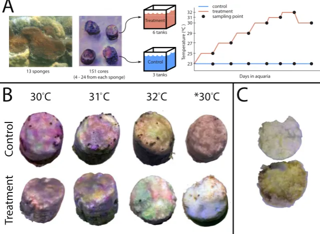
for genotype differences. Sixteen days after drilling, the cores from each sponge were haphazardly divided amongst nine indoor aquaria (50 L), resulting in 15–18 cores per aquarium. Ten sponges were represented in every aquar-ium, but three sponges were not, as they were represented by fewer than nine cores (Table 2).
Each aquarium was continuously supplied with 0.04 µm filtered seawater at 0.8 L/min. Water temperature was regulated by a SeaSim computer-controlled system to reach target temperatures; tanks were additionally buffered against temperature fluctuations using water jackets. LED lighting illuminated aquaria with 300 µmol quanta m−2 s−1 light for 11 h with an additional 1 h ramping period after dawn and before dusk. The irradiance level was less than the saturation irradiance (400 µmol quanta m−2 s−1) that was determined via rapid light curves.The irradiance level (300 µmol quanta m−2 s−1) is comparable to a cloudy day on the reef (302 µmol quanta m−2 s−1), but less than the average irradiance on a clear day (653 µmol quanta m−2 s−1) calculated from67. In terms of the total irra-diance, irradiance in the aquaria was 12 mol d−1 compared to 15 or 31 mol d-1 on a cloudy or sunny day on the reef, respectively calculated from67. Cores were maintained at 23.2 ± 0.3 °C (±SD) for 17 days, after which sponge photosynthesis and respiration were measured and tissue samples were taken (detailed below).
To measure changes in C. orientalis condition and performance as a function of temperature, two tempera-ture treatments were established: three aquaria were maintained under their initial conditions at 23 °C for the duration of the experiment (control) and six aquaria had the temperature increased every two weeks, first by 2 °C increments (23 to 29 °C) then by 1 °C increments (30 to 32 °C). Thus, the temperature targets were 23, 25, 27, 29, 30, 31, and 32 °C. These span the average range of temperatures at the collection site: 22.4 °C in July to 29.1 °C in February, with a maximum monthly mean of 30.5 °C between 2002 and 2010 (Source: Australian Institute of Marine Science; http://weather.aims.gov.au; 1.9 m depth). Each temperature increment was achieved via ramp-ing by 0.5 °C per day up to the target temperature followed by 10 days of exposure to that temperature. The rate of temperature increase is similar to daily changes in mean temperature at the collection site calculated from67. The 25 °C temperature increment was extended from 14 to 24 days due to a logistical issue with the heating and cooling system. With this exception, temperatures were finely controlled throughout the experiment, typically within 0.1 °C of the target temperature and with ~0.1 °C SD among aquaria (Table 1). Exposure of sponges to these temperature increments occurred from July to November and coincided with the natural winter to summer temperature increase during the austral summer.
To evaluate the potential for C. orientalis to recover from bleaching, cores exposed to 32 °C were returned to 30 °C (by 0.25 °C per day) and monitored for a further 28 days. Hereafter, ‘*30 °C’ is used to distinguish the recov-ery period at 30 °C from the incremental increase to 30 °C. To compare the thermal exposure in the laboratory to exposure under natural conditions, degree heating weeks (DHW) of thermal exposure were calculated for each temperature target above the 29.1 + 1.0 °C bleaching threshold for Orpheus Island. DHW was calculated as the product of the °C above 30.1 °C and the duration of temperature ramping (2–4 days) and exposure to the target temperature (10 days). 30 °C was chosen as the recovery temperature as it is below the bleaching threshold and represents a typical summer temperature at the collection site.
For each temperature increment, photosynthetic measurements were taken on two days near the completion of each temperature exposure. Photochemical efficiency of all cores was measured at least twice during exposure to each increment, after one and eight days of exposure, but photochemical efficiency was measured more fre-quently for the 23 °C, 25 °C, 31 °C, and 32 °C temperature increments. For other photosynthetic parameters and tissue contents, 2–3 cores were selected from each tank at each temperature increment, resulting in ~6 and ~12 samples for the control and heated treatments, respectively. Oxygen flux and sponge surface area were measured after nine days at each temperature increment (due to the time required to measure oxygen flux) except for *30 °C, where measurements were taken after 28 days. After 10 days acclimation to each temperature increment (28 days at *30 °C), sponges were frozen in liquid nitrogen for DNA extraction and measurement of tissue contents.
Photosynthesis and respiration.
Photochemical efficiency is an indicator of electron transport during photosynthesis68. For Symbiodinium, decreases in photochemical efficiency can precede bleaching and coin-cide with damage to photosystems69. The photochemical efficiency of photosystem II was measured using a mini-PAM fluorometer (Walz, Effeltrich, Germany) using standard settings (MI = 8; SI = 6; SW = 0.6; G = 2; D = 2). Clear tubing was used to maintain a constant distance between the PAM fibre optic cable and the sponge. Photochemical efficiency was measured at two timepoints: before the lights turned on (Fv/Fm; maximum effi-ciency) and after two hours of constant light exposure (∆F/Fm’; effective efficiency). Changes in these variables over the course of each temperature increment reflect the severity of the stress on the Symbiodinium.Oxygen flux is an integrative measure of respiration by the sponge (and symbionts) and photosynthesis by the Symbiodinium70. For sponges, stress can manifest in altered respiration, decreased photosynthesis, or a reduced ratio of photosynthesis to respiration29,30,71. Oxygen flux was measured using an optical dissolved oxygen meter (Hach HQ30d; Hach, Colorado, USA). Sponge cores were sealed in a darkened chamber (500 mL) for one hour. Water within each chamber was mixed with a magnetic stir bar and the target temperature was maintained using an external water jacket. Respiration rates were calculated by the difference in oxygen concentration between the beginning and end of the incubations. After measurement of respiration, sponges were returned to the aquaria for one hour at 300 µmol quanta m−2 s−1 then sealed in a chamber at 400 µmol quanta m−2 s−1 to measure oxygen production. The photosynthetic rate was calculated using the change in oxygen concentration in the chamber over 40 minutes. Both respiration and photosynthetic rates were adjusted by the amount of oxygen flux in a chamber without a sponge core to account for oxygen flux by microorganisms in the water. The surface area of the selected cores was measured using the aluminium foil method72, sponge tissue was removed (top 1 cm), the cores were frozen in liquid nitrogen and stored at −80 °C.
extracted by incubating sponge tissue in 1 M NaOH at 37 °C for 1 h74. Cells were counted using 4 replicate counts of 0.45 cm2 on a hemocytometer and standardised to the wet weight of sponge tissue. To quantify the photosyn-thetic pigments within C. orientalis tissues, chlorophylls were extracted in two consecutive extractions of 1 mL of ethanol (95%) to ensure complete extraction of pigments. During each extraction, the tissue was homog-enized for 3 min in a bead beater, centrifuged for 5 min (10,000 g), after which the two extracts were pooled. Absorbance of the extract was measured at 630, 647, 664, and 750 nm using a Power Wave Microplate Scanning Spectrophotometer (BIO-TEK Instruments Inc., Vermont, USA). The concentrations of chlorophylls a, b, and c were estimated using the equations of Ritchie75 and standardised to sponge wet weight.
Protein content and tissue organic matter were measured as proxies for sponge condition as these parameters have been shown to decline in bleached corals51. Frozen sponges were weighed (wet weight), lyophilized, and weighed again (dry weight). Proteins were extracted from homogenized dried sponge in 2 mL of 0.125 M NaOH over 24 h at room temperature. Protein concentration was estimated using the Red660 Protein Assay with five concentrations of BSA protein standard and then standardised to the dry weight of the sponge tissue. Organic matter was measured using lyopholized sponge tissue (see Protein content). Dried sponge was combusted at 450 °C for 16 h. Organic matter was estimated as the difference between the dry weight and the ash weight and standardised to the dry weight of the sponge.
Symbiodinium
identity.
To determine whether the sponges switched Symbiodinium types during tempera-ture stress, we identified the Symbiodinium associated with cores from 9 of the original sponge fragments as tem-peratures increased. In total, 35 cores were analysed including cores from the same genotype in each temperature increment. DNA was extracted from frozen sponge tissue (~0.2 g) using the Powerplant Pro DNA Isolation Kit (Mo Bio), including the beadbeating, RNAse, and proteinase K procedures as per the manufacturer’s instructions. DNA extracts were sent to the Australian Centre for Ecogenomics at the University of Queensland, Australia for sequencing. The ITS2 region of ribosomal rDNA was amplified using Symbiodinium-specific ITS2 primers76 and sequenced using Illumina MiSeq250 bp chemistry.Sequences were analysed in Mothur v.1.38.077. Paired reads were combined and screened for quality and chi-meric sequences were identified using Uchime in Mothur. The remaining sequences were clustered into 97% similar operational taxonomic units (OTU). The dataset was reduced to 2000 sequences per sample and the relative abundance and prevalence were calculated for each OTU. Representative sequences were defined using the sequence with the smallest distance to all other sequences within the OTU. Sequences were compared against a curated database of ITS2 sequences including all clades of Symbiodinium using the BLAST algorithm78. Blast results with bit scores less than 100 were discarded.
Bleaching surveys.
Field surveys were conducted to assess the thermal tolerance of C. orientalis during a natural thermal bleaching event, and to contrast bleaching responses between C. orientalis and corals. In March 2017, video transects were filmed at six sites within the Palm Islands Group, three of which were at Orpheus Island where the experimental samples were collected. Survey sites were chosen as replicate exposed and pro-tected locations, as well as to span the depth gradient over which Acropora cover is relatively high at Orpheus Island. At each site, two transects (50 m long and parallel to shore) were filmed at 0–4 m below the lowest astro-nomical tide. Cliona orientalis sponges and scleractinian coral colonies were manually counted along each video transect. Coral colonies were categorized as either branching or massive and we used a simple ‘bleached’ or ‘unbleached’ categorisation due to the absence of a reliable colour reference in the videos. White individuals, as well as individuals with fluorescent discolouration (e.g., blue or pink Acropora spp.) were considered bleached.To compare the thermal exposure during the natural bleaching event to the laboratory experiment, daily mean and maximum temperatures at Orpheus Island (5.8 m depth) were downloaded from the Australian Institute of Marine Science (http://weather.aims.gov.au).
Statistical analysis.
Photosynthesis and sponge condition data were analysed using linear mixed models with treatment, time point, and treatment * time point interaction as well as a random intercept to account for between-sponge differences using the R packages lme479, lmerTest80, and multcomp81. Only the final photochem-ical efficiency was analysed statistphotochem-ically. Symbiodinium density was analysed with an additional random intercept to account for correlations between samples from the same aquarium. Boxplots and residual plots were used to assess whether the data met the assumptions of linear models. Samples with large deviations from fitted values (>1.5*interquartile range) were removed from the analysis. Some response variables were transformed using log (respiration, chl a:c, protein, organic matter) or odds ratios (Fv/Fm, ∆F/Fm’) to meet these assumptions. Planned contrasts were used to test whether heated sponges differed from control sponges at each time point, as well as whether changes occurred after the reduction in temperature following exposure to 32 °C. Results from the planned contrasts are reported with z statistics and P values. P values were corrected using a single-step correc-tion for multiple comparisons.For each benthic category in the bleaching surveys, individuals were pooled between replicate transects at each site. The proportion of bleached individuals was calculated out of the total number of individuals encoun-tered at each site. For each category of taxa, the proportion of bleached individuals was calculated using propor-tion of bleached individuals relative to the total number of colonies and then weighted according to the number of individuals at each site.
References
1. IPCC. IPCC, 2014: Climate Change 2014: Synthesis Report. Contribution of Working Groups I, II and III to the Fifth Assessment Report of the Intergovernmental Panel on Climate Change Core Writing Team, R. K. Pachauri & L. A. Meyer. (IPCC, 2014).
2. Heron, S. F., Maynard, J. A., van Hooidonk, R. & Eakin, C. M. Warming Trends and Bleaching Stress of the World’s Coral Reefs 1985–2012. Sci Rep 6, 38402 (2016).
3. Hughes, T. P. et al. Global warming and recurrent mass bleaching of corals. Nature 543, 373–377 (2017). 4. Doney, S. C. et al. Climate Change Impacts on Marine Ecosystems. Annu. Rev. Marine. Sci. 4, 11–37 (2012).
5. Yellowlees, D., Rees, T. A. V. & Leggat, W. Metabolic interactions between algal symbionts and invertebrate hosts. Plant Cell Environ.
31, 679–694 (2008).
6. Warner, M. E., Fitt, W. K. & Schmidt, G. W. Damage to photosystem II in symbiotic dinoflagellates: a determinant of coral bleaching.
Proc. Natl. Acad. Sci. USA 96, 8007–8012 (1999).
7. McClanahan, T. R., Weil, E., Cortés, J., Baird, A. H. & Ateweberhan, M. In Coral Bleaching 205, 121–138 (Springer Berlin Heidelberg, 2009).
8. Baird, A. H. & Marshall, P. A. Mortality, growth and reproduction in scleractinian corals following bleaching on the Great Barrier Reef. Mar Ecol Prog Ser 237, 133–141 (2002).
9. Grottoli, A. G. et al. The cumulative impact of annual coral bleaching can turn some coral species winners into losers. Global Change Biol 20, 3823–3833 (2014).
10. Abrego, D., Ulstrup, K. E., Willis, B. L. & van Oppen, M. J. H. Species–specific interactions between algal endosymbionts and coral hosts define their bleaching response to heat and light stress. Proc. R. Soc. B 275, 2273–2282 (2008).
11. Díaz-Almeyda, E. M. et al. Intraspecific and interspecific variation in thermotolerance and photoacclimation in Symbiodinium
dinoflagellates. Proc. Biol. Sci. 284, 20171767 (2017).
12. Berkelmans, R. & van Oppen, M. J. H. The role of zooxanthellae in the thermal tolerance of corals: a ‘nugget of hope’ for coral reefs in an era of climate change. Proc. R. Soc. B 273, 2305–2312 (2006).
13. Palumbi, S. R., Barshis, D. J., Traylor-Knowles, N. & Bay, R. A. Mechanisms of reef coral resistance to future climate change. Science
344, 895–898 (2014).
14. Bay, L. K., Doyle, J., Logan, D. M. & Berkelmans, R. Recovery from bleaching is mediated by threshold densities of background thermo-tolerant symbiont types in a reef-building coral. R Soc Open Sci 3, 160322 (2016).
15. Silverstein, R. N., Cunning, R. & Baker, A. C. Change in algal symbiont communities after bleaching, not prior heat exposure, increases heat tolerance of reef corals. Global Change Biol 21, 236–249 (2015).
16. Bell, J. J., Davy, S. K., Jones, T., Taylor, M. W. & Webster, N. S. Could some coral reefs become sponge reefs as our climate changes?
Global Change Biol 19, 2613–2624 (2013).
17. Przeslawski, R., Ahyong, S., Byrne, M., Wörheide, G. & Hutchings, P. A. Beyond corals and fish: the effects of climate change on noncoral benthic invertebrates of tropical reefs. Global Change Biol 14, 2773–2795 (2008).
18. Carballo, J. L., Bautista, E., Nava, H., Cruz-Barraza, J. A. & Chávez, J. A. Boring sponges, an increasing threat for coral reefs affected by bleaching events. Ecol Evol 3, 872–886 (2013).
19. Cortés, J., Murillo, M. M., Guzman, H. M. & Acuña, J. Pérdida de zooxanthelas y muerte de corales y otros organismos arrecifales en el Caribe y Pacifico de Costa Rica. Rev. biol. trop 32, 227–231 (1984).
20. Vicente, V. P. Response of sponges with autotrophic endosymbionts during the coral-bleaching episode in Puerto Rico. Coral Reefs
8, 199–202 (1990).
21. Rützler, K. Impact of Crustose Clionid Sponges on Caribbean Reef Corals. Acta Geol Hisp 37, 61–72 (2002).
22. Schönberg, C. H. L. & Ortiz, J. C. Is sponge bioerosion increasing? Proceedings of 11th International Coral Reef Symp 7–11 (2008). 23. Hatch, W. I. The Implication of Carbonic Anhydrase in the Physiological Mechanism of Penetration of Carbonate Substrata by the
Marine Burrowing Sponge Cliona celata (Demospongiae). Biol Bull 159, 135–147 (1980).
24. Rützler, K. & Rieger, G. Sponge burrowing: Fine structure of Cliona lampa penetrating calcareous substrata. Mar Biol 21, 144–162 (1973).
25. Zundelevich, A., Lazar, B. & Ilan, M. Chemical versus mechanical bioerosion of coral reefs by boring sponges - lessons from Pione
cf. vastifica. J Exp Biol 210, 91–96 (2007).
26. Hill, M. S. Symbiotic zooxanthellae enhance boring and growth rates of the tropical sponge Anthosigmella varians forma varians.
Mar Biol 125, 649–654 (1996).
27. Schönberg, C. H. L. Growth and erosion of the zooxanthellate Australian bioeroding sponge. Proceedings of the 10th International Coral Reef Symposium 168–174 (2006).
28. Weisz, J. B., Massaro, A. J., Ramsby, B. D. & Hill, M. S. Zooxanthellar symbionts shape host sponge trophic status through translocation of carbon. Biol Bull 219, 189–197 (2010).
29. Fang, J. K. H., Schönberg, C. H. L., Mello-Athayde, M. A., Hoegh-Guldberg, O. & Dove, S. Effects of ocean warming and acidification on the energy budget of an excavating sponge. Global Change Biol 20, 1043–1054 (2014).
30. Achlatis, M. et al. Sponge bioerosion on changing reefs: ocean warming poses physiological constraints to the success of a photosymbiotic excavating sponge. Sci Rep 7, 1–13 (2017).
31. Duckworth, A. R. & Peterson, B. J. Effects of seawater temperature and pH on the boring rates of the sponge Cliona celata in scallop shells. Mar Biol 160, 27–35 (2013).
32. Wisshak, M., Schönberg, C. H. L., Form, A. & Freiwald, A. Effects of ocean acidification and global warming on reef bioerosion— lessons from a clionaid sponge. Aquat Biol 19, 111–127 (2013).
33. Wisshak, M., Schönberg, C. H. L., Form, A. & Freiwald, A. Ocean Acidification Accelerates Reef Bioerosion. PLoS ONE 7, e45124 (2012).
34. Fang, J. K. H. et al. Sponge biomass and bioerosion rates increase under ocean warming and acidification. Global Change Biol 19, 3581–3591 (2013).
35. Enochs, I. C. et al. Ocean acidification enhances the bioerosion of a common coral reef sponge: implications for the persistence of the Florida Reef Tract. Bull Mar Sci 91, 271–290 (2015).
36. Stubler, A. D., Furman, B. T. & Peterson, B. J. Sponge erosion under acidification and warming scenarios: differential impacts on living and dead coral. Global Change Biol 21, 4006–4020 (2015).
37. Angilletta, M. J. Thermal adaptation: a theoretical and empirical synthesis: Oxford University Press. (Oxford University Press, 2009). 38. Pörtner, H. O. Climate change and temperature-dependent biogeography: oxygen limitation of thermal tolerance in animals.
Naturwissenschaften 88, 137–146 (2001).
39. Rodolfo-Metalpa, R. et al. Thermally tolerant corals have limited capacity to acclimatize to future warming. Global Change Biol 20, 3036–3049 (2014).
40. Coles, S. L. & Jokiel, P. L. Effects of temperature on photosynthesis and respiration in hermatypic corals. Mar Biol 43, 209–216 (1977).
41. Ramsby, B. D. et al. Sibling species of mutualistic Symbiodinium clade G from bioeroding sponges in the western Pacific and western Atlantic oceans. J Phycol 11, 36–10 (2017).
43. Schönberg, C. H. L., Fang, J. K. H., Carreiro-Silva, M., Tribollet, A. & Wisshak, M. Bioerosion: the other ocean acidification problem.
ICES J. Mar. Sci. 1–31, https://doi.org/10.1093/icesjms/fsw254 (2017).
44. Berkelmans, R. & Willis, B. L. Seasonal and local spatial patterns in the upper thermal limits of corals on the inshore Central Great Barrier Reef. Coral Reefs 18, 219–228 (1999).
45. Schönberg, C. H. L., Suwa, R., Hidaka, M. & Loh, W. K. W. Sponge and coral zooxanthellae in heat and light: preliminary results of photochemical efficiency monitored with pulse amplitude modulated fluorometry. Mar Ecol 29, 247–258 (2008).
46. Berkelmans, R. Time-integrated thermal bleaching thresholds of reefs and their variation on the Great Barrier Reef. Mar Ecol Prog Ser 229, 73–82 (2002).
47. Baker, A. C., Glynn, P. W. & Riegl, B. Climate change and coral reef bleaching: An ecological assessment of long-term impacts, recovery trends and future outlook. Estuar Coast Shelf Sci 80, 435–471 (2008).
48. Hill, M. S., Walter, C. & Bartels, E. A mass bleaching event involving clionaid sponges. Coral Reefs 35, 153–153 (2016).
49. Smith, D. J., Suggett, D. J. & Baker, N. R. Is photoinhibition of zooxanthellae photosynthesis the primary cause of thermal bleaching in corals? Global Change Biol 11, 1–11 (2005).
50. Baird, A. H., Bhagooli, R., Ralph, P. J. & Takahashi, S. Coral bleaching: the role of the host. Trends in Ecology & Evolution 24, 16–20 (2009).
51. Fitt, W. K., Spero, H. J., Halas, J., White, M. W. & Porter, J. W. Recovery of the coral Montastrea annularis in the Florida Keys after the 1987 Caribbean bleaching event. Coral Reefs 12, 57–64 (1993).
52. Rodrigues, L. J. & Grottoli, A. G. Energy reserves and metabolism as indicators of coral recovery from bleaching. Limnology and Oceanography 52, 1874–1882 (2007).
53. Ramsby, B. D., Hoogenboom, M. O., Whalan, S. & Webster N. S. Elevated seawater temperature disrupts the microbiome of an ecologically important bioeroding sponge. Molecular Ecology 27, 2124–2137 (2018).
54. Bourne, D. G., Morrow, K. M. & Webster, N. S. Insights into the Coral Microbiome: Underpinning the Health and Resilience of Reef Ecosystems. Annu. Rev. Microbiol. 70, 317–340 (2016).
55. Hawkins, T. D., Hagemeyer, J. C. G., Hoadley, K. D., Marsh, A. G. & Warner, M. E. Partitioning of Respiration in an Animal-Algal Symbiosis: Implications for Different Aerobic Capacity between Symbiodinium spp. Front. Physiol. 7, (2016).
56. Riisgård, H. U., Thomassen, S., Jakobsen, H., Weeks, J. M. & Larsen, P. S. Suspension feeding in marine sponges Halichondria panicea
and Haliclona urceolus: effects of temperature on filtration rate and energy cost of pumping. Mar Ecol Prog Ser 96, 177–188 (1993). 57. Szmant, A. M. & Gassman, N. J. The effects of prolonged bleaching on the tissue biomass and reproduction of the reef coral
Montastrea annularis. Coral Reefs 8, 217–224 (1990).
58. Grottoli, A. G., Rodrigues, L. J. & Palardy, J. E. Heterotrophic plasticity and resilience in bleached corals. Nature 440, 1186–1189 (2006).
59. Riesgo, A. et al. Transcriptomic analysis of differential host gene expression upon uptake of symbionts: a case study with
Symbiodinium and the major bioeroding sponge Cliona varians. BMC Genomics 15, 376 (2014).
60. Pineda, M.-C. et al. Effects of light attenuation on the sponge holobiont- implications for dredging management. Sci Rep 6, 39038 (2016).
61. Hill, M. S., Allenby, A., Ramsby, B., Schönberg, C. H. L. & Hill, A. L. Symbiodinium diversity among host clionaid sponges from Caribbean and Pacific reefs: Evidence of heteroplasmy and putative host-specific symbiont lineages. Mol Phylogen Evol 59, 81–88 (2011).
62. Schönberg, C. H. L. & Loh, W. K. Molecular identity of the unique symbiotic dinoflagellates found in the bioeroding demosponge
Cliona orientalis. Mar Ecol Prog Ser 299, 157–166 (2005).
63. Granados, C., Camargo, C., Zea, S. & Sánchez, J. A. Phylogenetic relationships among zooxanthellae (Symbiodinium) associated to excavating sponges (Cliona spp.) reveal an unexpected lineage in the Caribbean. Mol Phylogen Evol 49, 554–560 (2008).
64. Pochon, X., Montoya-Burgos, J. I., Stadelmann, B. & Pawlowski, J. Molecular phylogeny, evolutionary rates, and divergence timing of the symbiotic dinoflagellate genus. Symbiodinium. Mol Phylogen Evol 38, 20–30 (2006).
65. Bo, M. et al. First description of algal mutualistic endosymbiosis in a black coral (Anthozoa: Antipatharia). Mar Ecol Prog Ser 435, 1–11 (2011).
66. Chaves-Fonnegra, A. et al. Bleaching events regulate shifts from corals to excavating sponges in algae dominated reefs. Global Change Biol 00, 1–13 (2017).
67. Hoogenboom, M. O., Connolly, S. R. & Anthony, K. Biotic and abiotic correlates of tissue quality for common scleractinian corals.
Mar Ecol Prog Ser 438, 119–128 (2011).
68. Baker, N. R. Chlorophyll fluorescence: a probe of photosynthesis in vivo. Annu Rev Plant Biol 59, 89–113 (2008).
69. Warner, M. E., Lesser, M. P. & Ralph, P. J. In Chlorophyll a fluorescence in aquatic sciences: methods and applications (eds Suggett, D. J., Prášil, O. & Borowitzka, M. A.) 209–222 (2010).
70. Osinga, R., Iglesias-Prieto, R. & Enrquez, S. Measuring Photosynthesis in Symbiotic Invertebrates: A Review of Methodologies, Rates and Processes. In: Applied Photosynthesis (ed Najafpour M), pp. 1–29, https://doi.org/10.5772/29339, InTech (2012). 71. Bennett, H. M. et al. Interactive effects of temperature and pCO2 on sponges: from the cradle to the grave. Global Change Biol 23,
2031–2046 (2016).
72. Marsh, J. A. Primary Productivity of Reef‐Building Calcareous Red Algae. Ecology 51, 255–263 (1970).
73. Fitt, W., Brown, B., Warner, M. & Dunne, R. Coral bleaching: interpretation of thermal tolerance limits and thermal thresholds in tropical corals. Coral Reefs 20, 51–65 (2001).
74. Zamoum, T. & Furla, P. Symbiodinium isolation by NaOH treatment. J Exp Biol 215, 3875–3880 (2012).
75. Ritchie, R. J. Universal chlorophyll equations for estimating chlorophylls a, b, c, and d and total chlorophylls in natural assemblages of photosynthetic organisms using acetone, methanol, or ethanol solvents. Photosynthetica 46, 115–126 (2008).
76. Pochon, X., Pawlowski, J., Zaninetti, L. & Rowan, R. High genetic diversity and relative specificity among Symbiodinium. Mar Biol
139, 1069–1078 (2001).
77. Schloss, P. D. et al. Introducing mothur: open-source, platform-independent, community-supported software for describing and comparing microbial communities. Appl Environ Microbiol 75, 7537–7541 (2009).
78. Arif, C. et al. Assessing Symbiodinium diversity in scleractinian corals via next-generation sequencing-based genotyping of the ITS2 rDNA region. Mol Ecol 23, 4418–4433 (2014).
79. Bates, D., Mächler, M., Bolker, B. & Walker, S. Fitting Linear Mixed-Effects Models using lme4. J Stat Softw 67, 1–48 (2015). 80. Kuznetsova, A., Brockhoff, P. B. & Christensen, R. H. B. lmerTest: Tests in Linear Mixed Effects Models. (R package version 2.0–32,
2016).
81. Hothorn, T., Bretz, F. & Westfall, P. Simultaneous Inference in General Parametric Models. Biom J 50, 346–363 (2008).
Acknowledgements
Author Contributions
B.R., M.H., S.W. and N.W. planned the experiment. B.R. performed the experiment. B.R. and H.S. analysed the data. B.R. wrote the manuscript with significant contributions from M.H., H.S., S.W. and N.W. N.W. was funded by an Australian Research Council Future Fellowship FT120100480. B.R. was sponsored by an AIMS@JCU PhD scholarship.
Additional Information
Competing Interests: The authors declare no competing interests.
Publisher's note: Springer Nature remains neutral with regard to jurisdictional claims in published maps and
institutional affiliations.
Open Access This article is licensed under a Creative Commons Attribution 4.0 International
License, which permits use, sharing, adaptation, distribution and reproduction in any medium or format, as long as you give appropriate credit to the original author(s) and the source, provide a link to the Cre-ative Commons license, and indicate if changes were made. The images or other third party material in this article are included in the article’s Creative Commons license, unless indicated otherwise in a credit line to the material. If material is not included in the article’s Creative Commons license and your intended use is not per-mitted by statutory regulation or exceeds the perper-mitted use, you will need to obtain permission directly from the copyright holder. To view a copy of this license, visit http://creativecommons.org/licenses/by/4.0/.