JOURNAL OFVIROLOGY, Apr. 2006, p. 4168–4173 Vol. 80, No. 8 0022-538X/06/$08.00⫹0 doi:10.1128/JVI.80.8.4168–4173.2006
Copyright © 2006, American Society for Microbiology. All Rights Reserved.
NOTES
Expression of Baculovirus Late and Very Late Genes Depends on
LEF-4, a Component of the Viral RNA Polymerase Whose
Guanyltransferase Function Is Essential
Dagmar Knebel-Mo
¨rsdorf,
3,4Ilja Quadt,
4Yi Li,
2Laura Montier,
2and Linda A. Guarino
1,2*
Departments of Biochemistry and Biophysics1and Entomology,2Texas A&M University, College Station, Texas 77843-2128,
and Department of Neurology3and Max-Planck-Institute for Neurological Research,4
University of Cologne, D-50931 Cologne, Germany
Received 27 December 2005/Accepted 25 January 2006
Baculovirus lef-4 encodes one subunit of the viral RNA polymerase. Here, we demonstrate the essential
nature of LEF-4 by RNA interference and bacmid knockout technology. Silencing of LEF-4 in wild-type virus-infected cells suppressed expression of structural genes, while early expression was unaffected,
demon-strating its essential role in late gene expression. After transfection of insect cells withlef-4mutant bacmid, no
viral progeny was produced, further defining its central role in infection. Cotransfection with wild-typelef-4
plasmid restored normal replication, but plasmid encoding a guanyltransferase-deficient version failed to rescue. These results emphasize the importance of the mRNA capping function of LEF-4.
Baculoviruses are unique among eukaryotic DNA viruses because early genes are transcribed by host RNA polymerase II (5, 13), while late genes are transcribed by a virus-encoded RNA polymerase (6, 8, 10). InAutographa californicamultiple nucleopolyhedrovirus (AcMNPV), the core viral polymerase is composed of four viral proteins, LEF-4, LEF-8, LEF-9, and p47 (10). LEF-8 and LEF-9, which contain motifs common to
and⬘RNA polymerases, are believed to form the catalytic site (19, 26). LEF-4 is an mRNA capping enzyme (7, 9, 15), and the function of p47 is unknown.
The LEF proteins were originally mapped as late expression factors by transient assays that used a late reporter construct and fragments of viral DNA (25). The lef-4 gene was also identified as the site of a temperature-sensitive (ts) mutation with a late expression phenotype (2, 24). To address the es-sential function of LEF-4, we performed RNA silencing exper-iments and gene knockout experexper-iments using bacmid technol-ogy. Previous studies have demonstrated the value of these techniques in the study of essential viral genes inSpodoptera
frugiperdacells (4, 14, 17, 18, 20, 28).
First, a high quality LEF-4 antiserum was produced in rab-bits using LEF-4 protein expressed in bacteria (9).S. frugiperda cells were infected with AcMNPV at 10 PFU/cell, and deter-gent-based nuclear extracts were prepared from infected cells 4, 8, 16, 24, 48, and 72 h postinfection (p.i.). As previously described, the cell pellet was lysed in 0.5% NP-40 and centri-fuged and the supernatant (cytoplasmic fraction) was adjusted to 0.1 N NaOH (21). The pelleted nuclei were resuspended in 1% NP-40 and adjusted to 0.1 N NaOH. Immunoblot
experi-ments showed that LEF-4 antiserum recognized a polypeptide of about 50 kDa from 16 through 72 h p.i., without cross-reactivity (Fig. 1).
To conduct the RNA silencing experiments, specific double-stranded RNA (dsRNA) was prepared using the T7 RiboMax Express RNAi system (Promega). The cloned HindIII-C frag-ment of the AcMNPV genome served as DNA template to generate dsRNA corresponding to 543 bp at the 5⬘terminus of
lef-4 (primer 1a, 5⬘-GGGTAATACGACTCACTATAGGGG
CGATTTTGTGATTGAG-3⬘; primer 1b, 5⬘-GGCAAATTCA TATTCGAGACG-3⬘; primer 2a, 5⬘-GGGTAATACGACTC
[image:1.585.307.532.505.668.2]* Corresponding author. Mailing address: Department of Biochem-istry, Texas A&M University, 2128 TAMU, College Station, TX 77843-2128. Phone: (979) 845-7556. Fax: (979) 845-9274. E-mail: lguarino @tamu.edu.
FIG. 1. Expression of LEF-4 in AcMNPV-infectedS. frugiperda
cells. Detergent-based nuclear extracts were prepared from uninfected
S. frugiperdacells (“un”) or from infected cells at 4, 8, 16, 24, 48, and 72 h p.i. Proteins were resolved on sodium dodecyl sulfate-10% poly-acrylamide gels and stained with the rabbit anti-LEF-4 antiserum. Protein size markers are given on the left.
4168
on November 8, 2019 by guest
http://jvi.asm.org/
ACTATAGGCAAATTCATATTCGAGACG-3⬘; primer 2b, 5⬘-GGGGCGATTTTGTGATTGAG-3⬘). As a control we used dsRNA corresponding to a 600-bp region of the green fluo-rescent protein (GFP) gene.S. frugiperdacells were transfected
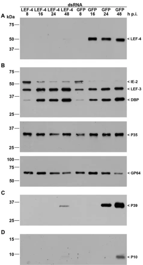
FIG. 2. Inhibition of viral gene expression bylef-4 silencing. S. frugiperdacells were transfected with either LEF-4 or GFP dsRNA. Cells were subsequently infected with AcMNPV (10 PFU/cell) at 20 h posttransfection, and detergent-based nuclear and cytoplasmic extracts were prepared from infected cells 8, 16, 24, and 48 h p.i. Proteins were resolved on sodium dodecyl sulfate-10% polyacrylamide gels and transferred to nitrocellulose membranes (A, B, and C) or resolved on sodium dodecyl sulfate-15% polyacrylamide gels and transferred to polyvinylidene difluoride membranes (D). (A) LEF-4 was stained with rabbit anti-Lef-4 antiserum. (B) Early gene expression was analyzed with rabbit antisera raised against IE2 (16), LEF-3 (3), DBP (22), or P35 (11) or with mouse monoclonal anti-GP64 (AcV5) (12). (C) Late gene expression was analyzed with mouse monoclonal anti-P39 (P10C6) (30). (D) Expression of the very late protein P10 was detect-able with rabbit anti-P10 serum (29). Expression of LEF-4, IE2, LEF-3, DBP, P39, and P10 was analyzed on samples of nuclear protein fractions, and P35 and GP64 expression was detected in cytoplasmic fractions as described previously (21). Protein size markers are shown on the left, and the identities of the viral proteins are indicated on the right.
FIG. 3. Polyhedron formation and polyhedrin expression uponlef-4
inhibition in AcMNPV-infectedS. frugiperdacells. Cells were trans-fected with either LEF-4 dsRNA or GFP dsRNA, intrans-fected with AcMNPV (10 PFU/cells) at 20 h posttransfection, and analyzed at 24 and 48 h p.i. (A) Phase-contrast images are shown, and (B) poly-hedron-containing cells were quantitated at 48 h p.i. (C) Cells transfected with LEF-4 dsRNA, GFP dsRNA, or untransfected cells were infected, and detergent-based nuclear extracts were prepared at 24 and 48 h p.i. Proteins were resolved on sodium dodecyl sulfate-10% polyacrylamide gels and transferred to nitrocellulose, and polyhedrin was viewed by Ponceau staining. Numbers at left are molecular masses in kilodaltons.
on November 8, 2019 by guest
http://jvi.asm.org/
[image:2.585.45.283.85.525.2]with 5g of LEF-4 dsRNA or GFP dsRNA as calcium phos-phate precipitates (BD BaculoGold). Cells were subsequently infected with AcMNPV (10 PFU/cell) at 20 h posttransfection, followed by detergent-based nuclear extraction (21). Immuno-blot analysis showed that transfection of LEF-4 dsRNA si-lenced expression of LEF-4. Transfection of the control GFP dsRNA, however, did not affect LEF-4 expression, thus dem-onstrating specificity (Fig. 2A). The efficiency oflef-4silencing indicates that a high percentage of cells must have taken up the dsRNA.
To study the effect of LEF-4 suppression on viral gene ex-pression, we analyzed proteins expressed early, late, and very late during infection. IE2, an immediate-early protein, is de-tectable from 2 until 12 to 24 h p.i. inS. frugiperdacells (16). The loss of LEF-4 had no effect on IE2 levels, consistent with the assumption that the ie2 promoter is transcribed by host RNA polymerase II (Fig. 2B). We also found no effect on expression patterns of LEF-3 (3), DBP (22), or P35 (11) in infected cells, consistent with the idea that LEF-4 is not in-volved in early gene expression (Fig. 2B). Surprisingly, we observed comparable levels of the membrane protein GP64 in cells treated with LEF-4 dsRNA or GFP dsRNA (Fig. 2B). Since GP64 is known to be regulated by both early and late promoters, we expected that protein levels would be lower in
thelef-4-silenced cells. The similar amounts of protein might
be coincidental, because accumulation of GP64 in normal
in-fection is a function of synthesis combined with the loss of protein due to budding of virus particles. In the case oflef-4 -silenced cells, less budding presumably occurs, due to lower levels of synthesis of viral structural proteins (see below).
To examine the effect of LEF-4 on late genes, protein ex-tracts were probed with antibody against the capsid protein P39 (30). Strong expression of P39 was observed at 24 and 48 h p.i. in cells treated with GFP dsRNA, while only a weak P39 signal was detected in cells with suppressed LEF-4 (Fig. 2C). The very late protein P10 was also shown to be dependent upon LEF-4 synthesis. P10 levels were undetectable in LEF-4 suppressed cells, while P10 was evident in the control cells at 48 h p.i. (Fig. 2D). Furthermore, polyhedrin levels were re-duced. Immunoblot analysis showed a significant reduction in accumulation of polyhedrin protein in cells treated with LEF-4 dsRNA, compared to cells treated with GFP dsRNA or un-treated cells (Fig. 3C). Microscopic analysis revealed the pres-ence of polyhedron-positive cells at 24 and 48 h p.i. in the LEF-4-silenced cells, although the number of cells with poly-hedra was about fivefold lower than that of the control GFP-treated cells (Fig. 3A and B). The few cells that were poly-hedron positive had equivalent numbers of polyhedra per cell, and those polyhedra formed with the same kinetics as in the GFP dsRNA-treated and untreated controls (Fig. 3A and data not shown). This suggests that cells with polyhedra likely
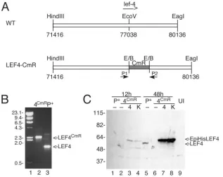
rep-FIG. 4. Construction of BacP⫹/LEF-4CmR. (A) Cloning strategy. A plasmid containing the left 7 kb of the HindIII-C fragment of AcMNPV was
digested with EcoRV, and a 1-kb fragment of pBR325 containing the CAT resistance marker was inserted. The resultant plasmid was transformed into BJ5183 cells containing a modified version of the AcMNPV genome, followed by selection on chloramphenicol, as previously described (1). (B) PCR screening. Correct insertion of CAT intolef-4was verified by PCR using primers that flank thelef-4open reading frame. Lane 2, BacP⫹/LEF-4CmR; lane 3, BacP⫹. The positions of molecular mass markers (lane 1) are indicated on the left in kilodaltons, and the migration of
lef-4andlef-4with the CAT insertion is shown on the right. (C) Immunoblot analysis of LEF-4 expression in transfected cells. Cells were transfected with BacP⫹ (P⫹, lanes 1 and 5) or BacP⫹/LEF-4CmR(4CmR, lanes 2 to 4 and 6 to 8) and cotransfected where indicated with
pHSEpiHisLEF-4 (4, lanes 3 and 7) or pHSEpiHisLEF-4(K255A) (K, lanes 4 and 8). Cells were harvested at 12 h (lanes 1 to 4) or 48 h (lanes 5 to 8) posttransfection. The blot was probed with rabbit LEF-4 antiserum. Untransfected cells were analyzed as a negative control (lane 9). The positions of molecular mass markers are indicated on the left in kilodaltons, and the migration of LEF-4 and EpiHisLEF-4 is indicated on the right.
4170 NOTES J. VIROL.
on November 8, 2019 by guest
http://jvi.asm.org/
[image:3.585.135.452.68.320.2]resent the population of cells that did not take up LEF-4 dsRNA.
Our results strengthen the identification of LEF-4 as an essential component of the viral RNA polymerase that is re-sponsible for late and very late transcription. The role of LEF-4, however, remains open. Previous results showed that
thelef-4ts mutant L104F failed to produce infectious virus at
the nonpermissive temperature (2). Interestingly, a mutant version of LEF-4 with this substitution was not impaired for guanyltransferase activity (15). To address whether LEF-4 is essential for its capping activity, we constructed a virus with an interruptedlef-4gene.
The bacmid BacP⫹is derived from the Bac-to-Bac cloning vector (Invitrogen), which has the AcMNPV genome cloned into a single-copy plasmid (1). BacP⫹ has a reconstructed polyhedrin gene and lacks transposition sites for cloning into the polyhedrin locus. Thelef-4gene was inactivated by insert-ing a 1-kb BstBI fragment of pBR325, containinsert-ing the chloram-phenicol acetyltransferase (CAT) resistance marker under the control of its own promoter, into the EcoRV site of an 8.7-kb genome fragment containing lef-4 (Fig. 4A). The resulting plasmid was digested to excise the viral DNA that was used to transform BacP⫹-containing BJ5183 cells by
electropora-tion. Recombinants were selected by plating on chloram-phenicol, and interruption of thelef-4open reading frame with CAT was confirmed by PCR using plasmids that flank
thelef-4gene (Fig. 4B).
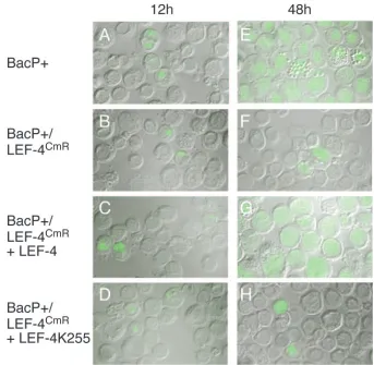
[image:4.585.118.461.66.401.2]Purified bacmid DNA was transfected into Sf9 cells, which were analyzed at 12 h p.i. by immunofluorescence using anti-body against the immediate-early IE1. Approximately 500 total cells were scored for IE1 expression. We found 4 to 12% positive cells, indicating similar transfection efficiencies for the two DNAs (Fig. 5A and B). At 48 h posttransfection, virtually all cells transfected with BacP⫹were positive for IE1 (Fig. 5E), indicating that viral infection had spread to adjacent cells, but
FIG. 5. Infectivity oflef-4 mutant viruses. S. frugiperdacells were transfected with BacP⫹ (A and E), BacP⫹/LEF-4CmR (B and F), or
BacP⫹/LEF-4CmRin the presence of plasmids encoding wild-type LEF-4 (C and G) or a mutant version of LEF-4 that lacks guanyltransferase
activity (D and H). Cells were harvested at 12 or 48 h p.i. and processed for immunofluorescence using a mouse monoclonal antibody raised against IE1. Images were collected using a Zeiss ApoTome/Axioplan 2 microscope. Images shown represent merged images of phase contrast and green fluorescence to visualize percentages of cells expressing IE1.
TABLE 1. Infectious virus produced from transfection of bacmids
Bacmid Plasmid Titer at 48 h
posttransfection
BacP⫹ None 2⫻107
BacP⫹/LEF-4CmR None 0
pHSEpiHisLEF-4 5.3⫻106
pHSEpiHisLEF-4(K255A) 2⫻102a
aAll clones recovered were reversions.
on November 8, 2019 by guest
http://jvi.asm.org/
[image:4.585.301.541.651.717.2]the percentage of IE1-positive cells in BacP⫹/LEF-4CmRhad
not significantly increased (Fig. 5F). The lack of replication was verified by plaque assay of the culture medium at 48 h posttransfection (Table 1).
To determine whether capping enzyme activity was neces-sary for viral infection, we cotransfected BacP⫹/LEF-4CmRand
pHSEpiHisLEF-4 (27), encoding an epitope-tagged version of
lef-4 or a mutant version of the same plasmid encoding an
alanine substitution at lysine 255 (K255A). We have previously shown that this mutant is not active in guanyltransferase assays (15). Immunoblot analysis showed that the two proteins were expressed at equivalent levels by 12 h posttransfection (Fig. 4C). The wild-typelef-4 plasmid rescued the mutant bacmid DNA. The pattern of IE1 immunofluorescence was similar to that seen with the parental bacmid (Fig. 5C and G), and the yield of infectious virus was approximately 25% of that ob-tained with the parental clone (Table 1), indicating that the level of recombination between the bacmid and plasmid was very high. Cotransfection with the K255A mutant, however, did not significantly change the pattern of IE1 immunofluo-rescence (Fig. 5D and H). Some progeny virus was obtained (Table 1), but direct sequencing of four clones revealed that their genomes contained wild-typelef-4. This was possible be-cause the BacP⫹/LEF-4CmR construct was made by inserting
the CAT gene into the LEF-4 open reading frame without deleting anylef-4sequences, including the K255 locus. The fact that only wild-type mutants were obtained with the K255A plasmid indicates that guanyltransferase activity is required for late gene expression and production of progeny virions.
The strategy used in these studies differs somewhat from previous protocols relying on bacmid systems (18, 23, 28). First, we did not construct a “repaired” virus control by inserting the
lef-4gene into the polyhedrin locus. Instead, we relied on the
efficient recombination ability of baculoviruses to produce vi-able virus. The fact that the yield of infectious virus obtained from cotransfection of a wild-typelef-4 plasmid and BacP⫹/ LEF-4CmRwas 25% of that obtained with the parental bacmid
indicates that in vivo recombination is very efficient. This was also evident in the high spread of IE1 fluorescence at 48 h posttransfection. Second, we did not construct bacmids that expressed reporter genes in order to follow infection. Instead we visualized expression of a viral gene.
Taken together, our results further characterize the essential nature of LEF-4 and demonstrate that the guanyltransferase function of LEF-4 is essential for productive infection. In ad-dition, LEF-4 may also be required for its RNA triphosphatase activity and another function that was disrupted by the L105F ts substitution (2). Biochemical assays of a protein with an L105F substitution revealed that it was normal with respect to guanyltranferase and had only a modest decrease in RNA triphosphatase activity, which is probably insignificant (15). Since these are the only two enzymatic activities that the pro-tein is known to possess, it is possible that L105 is important for structural integrity of the polymerase complex, and high temperature inhibits replication because the polymerase dis-sociated although the enzymatic functions of LEF-4 are unaf-fected. Analysis of additional LEF-4 mutants should help to define roles of LEF-4 in viral infection.
We thank Keiju Okano for the antiserum against Bombyx mori
nucleopolyhedrovirus DBP, George F. Rohrmann for LEF-3 anti-serum, Paul D. Friesen for anti-p35 antianti-serum, Gary W. Blissard for monoclonal antibody gp64 (AcV5), Loy Volkman for monoclonal an-tibody p39 (P10C6), and Monique van Oers for anti-p10 antiserum. We are grateful to Ute Schepers for helpful comments on RNA si-lencing experiments.
Research was supported by grant KN536/11-1 from the Deutsche Forschungsgemeinschaft and the Ko¨ln Fortune Program/Faculty of Medicine, University of Cologne, and by grant MCB-0416484 from the National Science Foundation.
REFERENCES
1.Bideshi, D. K., and B. A. Federici.2000. TheTrichoplusia nigranulovirus helicase is unable to support replication ofAutographa californica multicap-sid nucleopolyhedrovirus in cells and larvae ofT. ni. J. Gen. Virol.81:1593– 1599.
2.Carstens, E. B., H. Chan, H. Yu, G. V. Williams, and R. Casselman.1994. Genetic analyses of temperature-sensitive mutations in baculovirus late ex-pression factors. Virology204:323–337.
3.Evans, J. T., and G. F. Rohrmann.1997. The baculovirus single-stranded DNA binding protein, LEF-3, forms a homotrimer in solution. J. Virol. 71:3574–3579.
4.Flores-Jasso, C. F., V. J. Valdes, A. Sampieri, V. Valadez-Graham, F. Recillas-Targa, and L. Vaca.2004. Silencing structural and nonstructural genes in baculovirus by RNA interference. Virus Res.102:75–84.
5.Fuchs, L. Y., M. S. Woods, and R. F. Weaver.1983. Viral transcription during Autographa californica nuclear polyhedrosis virus infection: a novel RNA polymerase induced inSpodoptera frugiperdacells. J. Virol. 48:641–646.
6.Glocker, B., R. R. Hoopes, Jr., L. Hodges, and G. F. Rohrmann.1993. In vitro transcription from baculovirus late gene promoters: accurate mRNA initiation by nuclear extracts prepared from infectedSpodoptera frugiperda
cells. J. Virol.67:3771–3776.
7.Gross, C. H., and S. Shuman.1998. RNA 5⬘-triphosphatase, nucleoside triphosphatase, and guanylyltransferase activities of baculovirus LEF-4 pro-tein. J. Virol.72:10020–10028.
8.Grula, M. A., P. L. Buller, and R. F. Weaver.1981.␣-Amanitin-resistant viral RNA synthesis in nuclei isolated from nuclear polyhedrosis virus-infected
Heliothis zealarvae andSpodoptera frugiperdacells. J. Virol.38:916–921. 9.Guarino, L. A., J. Jin, and W. Dong.1998. Guanylyltransferase activity of
the LEF-4 subunit of baculovirus RNA polymerase. J. Virol.72:10003– 10010.
10.Guarino, L. A., B. Xu, J. Jin, and W. Dong.1998. A virus-encoded RNA polymerase purified from baculovirus-infected cells. J. Virol.72:7985–7991. 11.Hershberger, P. A., D. J. LaCount, and P. D. Friesen.1994. The apoptotic suppressor P35 is required early during baculovirus replication and is tar-geted to the cytosol of infected cells. J. Virol.68:3467–3477.
12.Hohmann, A. W., and P. Faulkner.1983. Monoclonal antibodies to baculo-virus structural proteins: determination of specificities by Western blot anal-ysis. Virology125:432–444.
13.Hoopes, R. R., Jr., and G. F. Rohrmann. 1991. In vitro transcription of baculovirus immediate early genes: accurate mRNA initiation by nuclear extracts from both insect and human cells. Proc. Natl. Acad. Sci. USA 88:4513–4517.
14.Hou, S., X. Chen, H. Wang, M. Tao, and Z. Hu.2002. Efficient method to generate homologous recombinant baculovirus genomes inE. coli. Bio-Techniques32:783–784, 786, 788.
15.Jin, J., W. Dong, and L. A. Guarino.1998. The LEF-4 subunit of baculovirus RNA polymerase has RNA 5⬘-triphosphatase and ATPase activities. J. Virol. 72:10011–10019.
16.Krappa, R., R. Roncarati, and D. Knebel-Morsdorf.1995. Expression of PE38 and IE2, viral members of the C3HC4finger family, during baculovirus
infection: PE38 and IE2 localize to distinct nuclear regions. J. Virol.69: 5287–5293.
17.Li, Y., J. Wang, R. Deng, Q. Zhang, K. Yang, and X. Wang.2005. vlf-1 deletion brought AcMNPV to defect in nucleocapsid formation. Virus Genes31:275–284.
18.Lin, G., and G. W. Blissard.2002. Analysis of anAutographa californica
nucleopolyhedroviruslef-11knockout: LEF-11 is essential for viral DNA replication. J. Virol.76:2770–2779.
19.Lu, A., and L. K. Miller.1994. Identification of three late expression factor genes within the 33.8- to 43.4-map-unit region ofAutographa californica
nuclear polyhedrosis virus. J. Virol.68:6710–6718.
20.Means, J. C., I. Muro, and R. J. Clem.2003. Silencing of the baculovirus Op-iap3gene by RNA interference reveals that it is required for prevention of apoptosis duringOrgyia pseudotsugataM nucleopolyhedrovirus infection of Ld652Y cells. J. Virol.77:4481–4488.
21.Murges, D., I. Quadt, J. Schroer, and D. Knebel-Morsdorf.2001. Dynamic nuclear localization of the baculovirus proteins IE2 and PE38 during the
4172 NOTES J. VIROL.
on November 8, 2019 by guest
http://jvi.asm.org/
infection cycle: the promyelocytic leukemia protein colocalizes with IE2. Exp. Cell Res.264:219–232.
22.Okano, K., V. S. Mikhailov, and S. Maeda.1999. Colocalization of baculo-virus IE-1 and two DNA-binding proteins, DBP and LEF-3, to viral repli-cation factories. J. Virol.73:110–119.
23.Okano, K., A. L. Vanarsdall, and G. F. Rohrmann.2004. Characterization of a baculovirus lacking the alkaline nuclease gene. J. Virol.78:10650–10656. 24.Partington, S., H. Yu, A. Lu, and E. B. Carstens.1990. Isolation of temper-ature sensitive mutants ofAutographa californicanuclear polyhedrosis virus: phenotype characterization of baculovirus mutants defective in very late gene expression. Virology175:91–102.
25.Passarelli, A. L., and L. K. Miller.1993. Identification and characterization oflef-1, a baculovirus gene involved in late and very late gene expression. J. Virol.67:3481–3488.
26.Passarelli, A. L., J. W. Todd, and L. K. Miller.1994. A baculovirus gene involved in late gene expression predicts a large polypeptide with a con-served motif of RNA polymerases. J. Virol.68:4673–4678.
27.Rapp, J. C., J. A. Wilson, and L. K. Miller.1998. Nineteen baculovirus open reading frames, including LEF-12, support late gene expression. J. Virol. 72:10197–10206.
28.Stewart, T. M., I. Huijskens, L. G. Willis, and D. A. Theilmann.2005. The
Autographa californicamultiple nucleopolyhedrovirusie0-ie1gene complex is essential for wild-type virus replication, but either IE0 or IE1 can support virus growth. J. Virol.79:4619–4629.
29.Van Oers, M. M., J. T. Flipsen, C. B. Reusken, and J. M. Vlak.1994. Specificity of baculovirus p10 functions. Virology200:513–523.
30.Whitt, M. A., and J. S. Manning.1988. A phosphorylated 34-kDa protein and a subpopulation of polyhedrin are thiol linked to the carbohydrate layer surrounding a baculovirus occlusion body. Virology163:33–42.