Open Access
Short Report
RNase footprinting demonstrates antigenomic hepatitis delta virus
ribozyme structural rearrangement as a result of self-cleavage
reaction
Larissa Savochkina, Victoria Alekseenkova, Tatyana Belyanko,
Nadezhda Dobrynina and Robert Beabealashvilli*
Address: Russian Cardiology Research and Production Center, 121552 Moscow, Russia
Email: Larissa Savochkina - [email protected]; Victoria Alekseenkova - [email protected]; Tatyana Belyanko - [email protected]; Nadezhda Dobrynina - [email protected]; Robert Beabealashvilli* - [email protected]
* Corresponding author
Abstract
Background: Hepatitis delta virus (HDV) is a satellite virus of hepatitis B. During viral replication the 1700-nucleotide-long genomic RNA and its complement, the antigenomic RNA, undergo self-cleavage catalyzed by internal ribozyme motifs that are essential for propagation of the virus in vivo. These self-cleavage activities are provided by 85-nucleotide-long sequence elements, the genomic and antigenomic forms of HDV ribozyme. Recently four permuted variants of the antigenomic HDV
cis-ribozyme with a self-cleavage site located at the 5' proximity, in the middle, or nearby the 3' end of the molecule were constructed and synthesized. These constructs exhibit equal activity, a bi-phasic kinetics of self-cleavage reaction and reaction products with low and high stability. We have used ribonuclease probing to footprint the structures of uncleaved and post-cleaved forms of the antigenomic HDV ribozymes in solution. Uncleaved ribozymes, associated and individual products of the self-cleavage reaction were analyzed using ribonuclease and Fe(II)-EDTA protection assays to reveal the differences in the structure of pre- and post-cleaved antigenomic HDV ribozyme in solution.
Findings: Our findings demonstrate that a significant conformational change accompanies catalysis in the antigenomic HDV ribozyme in solution, in contrast to minor conformational switch observed in crystals of the genomic form. This study indicates that changes in the structure of stem P1 and stem P4 are minor, those of the region ascribed to stem P2, stem P3 and loop l3 are dramatic, while stem P1.1 results from the self-cleavage reaction.
Conclusion: Our data agree with the structure of post-cleaved and disagree with that of pre-cleaved forms of HDV ribozyme published elsewhere.
Background
According to the double nested pseudoknot model, both genomic and antigenomic ribozymes fold into a similar secondary structure consisting of basepaired stems (P1
-P4), joining sequences (J1/2, J1/4, J4/2) and loops (l3, l4) [1]. Analysis the crystal structure of the 3' product of self-cleaved genomic HDV ribozyme showed, that the ribozyme adopts a tight tertiary structure by forming, in
Published: 16 May 2008
BMC Research Notes 2008, 1:15 doi:10.1186/1756-0500-1-15
Received: 8 May 2008 Accepted: 16 May 2008
This article is available from: http://www.biomedcentral.com/1756-0500/1/15
© 2008 Savochkina et al; licensee BioMed Central Ltd.
addition to the pseudoknot stems P1 - P4, the two-base-pair stem P1.1, thus creating a two-base-pair of nested pseudoknots, which buries the active site in a deep cleft [2,3].
However, when the crystal structure of the pre-cleaved form of cis-acting genomic HDV ribozyme was solved, it turned out that its structure differs from that of the prod-uct form, while the strprod-ucture of stems P1–P4 and P1.1 is the same in both states [4].
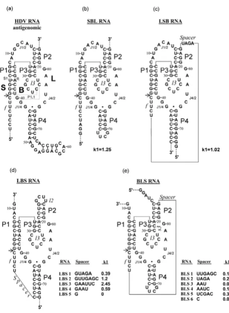
We have prepared a set of permuted variants of HDV ribozyme with different locations of the cleavage site and different spacers connecting three major uninterruptible ribozyme parts (Figure 1). We analyzed the structures of all ribozyme constructs in pre- and post-cleaved states using RNAse and Fe(II)-EDTA footprinting.
Here we present the evidence that the conformations of pre- and post-cleaved antigenomic HDV ribozymes differ dramatically. From analysis of these differences it was concluded that stem P1.1 formed by nucleotides C25 and C24 with G40 and G41 does appear after the self-cleavage reaction.
Structure design of permuted variants of
antigenomic HDV ribozyme
We have prepared four out of six possible permuted vari-ants of the HDV ribozyme. Spacers between L, B and S chains varied in length and nucleotide sequence, while the nucleotide sequences of the chains were invariable and corresponded to those of naturally occurring antige-nomic HDV ribozyme. Figure 1 shows structure and rate constant of the first reaction phase (k1) for all constructs. Briefly, all ribozymes with optimal spacers are almost equally active under individual optimal conditions, while activation energy and optimal temperature vary from ribozyme to ribozyme with the sharpest temperature curve for LSB. In general, the activities of ribozymes belonging to the same family vary with the length and sequence of spacers. A very short spacer makes the ribozyme inactive.
For detailed analysis of the reaction kinetics of all topo-logical variants described here see our previous paper [5] and our laboratory web site [6], where the reader can change the initial conditions and parameters of the reac-tion and observe resulting changes in the kinetic curves. For comparison with the model suggested earlier [5] we have inserted an additional stage that leads to irreversibil-ity of the reaction.
The major difference between SBL (an isomer with "natu-ral" chain connectivity) and the other constructs is the sta-bility of the reaction product. Short and loosely bound 5'proximal fragment of SBL readily dissociates from the
product complex while the products of the other con-structs remain in the complex at temperatures up to 50°C–95°C [5]. This is also the major difference between the variants of HDV cis-ribozyme investigated here and the previously studied products of genomic and antige-nomic ribozymes which lost the 5'proximal fragment after self-cleavage.
Analysis of the secondary structure of
pre-cleaved and post-pre-cleaved antigenomic HDV
ribozymes
The secondary structure of all constructs in pre- and post-cleaved state was probed with T1, U2, A and V1 ribonucle-ases. In addition, for all constructs electrophoretically
[image:2.612.317.545.84.396.2]Nucleotide sequence and proposed secondary structure of designed variants of HDV antigenome ribozyme
Figure 1
Nucleotide sequence and proposed secondary structure
of designed variants of HDV antigenome ribozyme. (a)
purified long fragment of post-cleavage product was probed.
Pre-cleaved ribozymes
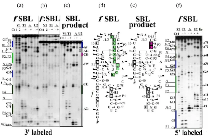
All studied ribozymes which exhibit almost equal activity have similar nuclease digestion profiles for the original molecule (Figures 2a and 2f, 3a and 3f, 4a and 4c). The nuclease cleavage patterns are not the same, but the major features are similar. All joining sequences J1/2 in SBL and LSB, J4/1 in LBS and 3'-part of J2/1 in BLS (not shown) are susceptible to single-strand-specific nucleases but not to the double-strand-specific nuclease V1. Only individual weak V1 cuts are seen in joining sequences (at nucleotides (nt) U10 SBL A9 LSB). The same is true for J4/2 for all pre-cleaved ribozymes of the LBS family (Figure 4a). The bottom of P4 and the ends of R are well accessible to dou-ble-strand-specific nuclease in all studied structures. In all constructs, the middle of R region is probably less accessi-ble than the ends. In pre-cleaved ribozymes, the upstream
regions of P2, P3 and l3 (between nt 15 and 26) are acces-sible to double-strand-specific nuclease. The relative intensities of different bands in this region are different for different ribozymes with slight preference for 15U, 23U and 26U in SBL (Figures 2a and 2f)) and 17U, 23U and 26U in LSB (Figure 3a). In LBS1, LBS3 (Figures 4a and 4c) and in LBS5, BLS1, BLS3 (not shown) the most acces-sible nucleotides are located between 22C and 25C. Digestion of P4 correlates with the activity of LBS. In the most active LBS3 (Figure 4c) the region is the most acces-sible to V1, in less active LBS1 it is only partially accesacces-sible (Figure 4a), while in inactive LBS5 the region is not acces-sible (not shown). This finding suggests that the shorten-ing of joiner J4/1 to a sshorten-ingle nucleotide prevents formation of P4 and makes the ribozyme inactive.
Note that pre-cleaved ribozyme exposed to nucleases at neutral pH in the presence of Mg2+ contains some post-cleavage products. The higher the activity of ribozyme, the
[image:3.612.127.479.326.557.2]RNase probing of SBL RNA Figure 2
RNase probing of SBL RNA. (a)-(c) RNA labeled with 32P at the 3'-end or (f) at the 5'-end. (a) and (f) 32P labeled RNA was treated
with nucleases under the conditions minimizing self-cleavage reaction (20°C, 5 min) or (b) self-cleaved at 50°C for 60 min and self-cleav-age product was analyzed either directly in the mixture for self-cleaving or (c) after re-purifying by electrophoresis in denaturing polyacr-ylamide gel. The lanes marked with a plus sign are reactions carried out at 20°C, pH 7.5, 10 mM MgCl2 with either no added cleavage reagent (Ct) or with ribonuclease V1 (V1), ribonuclease T1 (T1), ribonuclease A (A) and ribonuclease U2 (U2). The minus sign lanes are reactions carried out at 50°C, pH 3.5 with 7 M urea (sequencing markers). Final concentration of RNases, reaction and analysis conditions were as described under Material and Methods (See Additional file 8). Two concentrations of RNase V1 were used: 1 × 10⁻³ units/μl (1)
or 5 × 10⁻³ units/μl (2). V1 products contain a 3'-hydroxyl rather than phosphate, which results in decreased mobility relative to the
greater the proportion of self-cleavage product in a pre-cleaved ribozyme preparation. This accounts for the pres-ence of some minor bands typical of the product in the pattern of a pre-cleaved ribozyme. Vice versa, post-cleaved ribozymes are not fully cleaved and about 20% of uncleaved form is present in each preparation. Therefore, all bands belonging to uncleaved molecule are also present on autoradiograms with a five-fold lower inten-sity. Having this in mind, we considered only the relative intensities of the bands.
Post-cleaved ribozymes
As already mentioned, V1 digestion pattern for pre-cleaved ribozymes shows well-defined bands with differ-ent but comparable intensities for each nucleotide between 15 and 26 nucleotides. This pattern is routine for each uncleaved construct. After cleavage, all these bands are replaced by a strong doublet at nt U17–C18 (SBL, LSB and BLS) or nt C18–C19 (LBS) (Figures 2b, 3b, 4b and 4d).
It is noteworthy that V1 digestion pattern of post-cleaved HDV ribozyme reported by Rosenstein & Been [7] also contains a strong doublet U17–C18 and is practically free from cut sites between nt 15 and 26. Interestingly, point mutations in the 3'product P2 region which disrupt P2
lead to a nuclease-digestion pattern typical of uncleaved unmodified ribozyme (cuts in the upstream strand of P2, P3 and l3 between position 16 and 26), while compensa-tory changes which restore stem P2 of 3'product provide a pattern typical of the 3'-product (cuts at positions 17 and 18) [8]. However, uncleaved LBS exhibits a pattern typical of the uncleaved ribozyme (cuts between position 18 and 26), irrespective of the presence of UUCG which closes nucleotide sequence and forces the formation of stem P2.
Purified large product of ribozyme self-cleavage also exhibits one of the two alternative states for the region between U15 and U26. Note that the product purification procedure includes unwinding the whole structure of RNA by a denaturant and subsequent annealing. After this procedure, the purified products of SBL (Figure 2c) and LBS (LB, Figure 4d) acquire a structure with a doublet at C18 region, while the product of LSB (SB, Figure 3c) regains the structure characteristic of the pre-cleaved form. It should be noted that the product of SBL self-cleavage possesses all elements included in the pseudoknot model. The product of LBS (LB) does not have the S region and therefore cannot form stem P1. The product of LSB (SB) cannot form stem P4 or stem P2 since it lacks L chain. Comparison of Figures 3a and 3c shows that the digestion patterns of the region between G1 and G40 for LSB and SB
RNase probing of LSB RNA Figure 3
RNase probing of LSB RNA. (a)-(c) 3'-32P-labeled and (f)-(h) 5'-32P-labeled LSB RNA was treated with nucleases under different
con-ditions: (a) and (f) 20°C, 5 min (minimizing self-cleavage); (b) and (g) 50°C, 60 min and self-cleavage product was analyzed either directly in the mixture or (c) and (h) after re-purifying by electrophoresis in denaturing polyacrylamide gel. Samples were analyzed as described for Figure 2 except that the concentration of V1 RNase was 1 × 10⁻³ units/μl. Spacer nucleotides C-4 and U-9 were numbered in
are almost identical except for very strong cleavages in R region of SB which are very weak in LSB. Thus, it can be suggested that in uncleaved ribozyme SB fragment is folded as a separate domain without direct interaction with P4 and L fragment. Previously we have shown that SB domain forms autonomously [9]. Obviously, stem P1 is present in SB and there is no candidate for forming a lengthy Watson-Creek double-stranded region with a polypyrimidine stretch from U17 to U26. However, the nucleotides of the region seem to be well stacked as they are well accessible to V1 nuclease and are not susceptible to single-strand-specific nucleases. In contrast to P2 and P3–l3, other parts of post-cleaved SBL and BLS exhibited no changes in nuclease digestion patterns.
It should be noted that the most unexpected changes occurred in the susceptibility of l2 to Fe(II)-EDTA cleavage (Figures 4c and 4d). According to any available 3D model of the ribozyme, this part of the molecule may become fully exposed to the solvate.
We have projected our nuclease probing data on 3D struc-tures of both ribozyme forms. Atomic coordinates depos-ited at the RCSB Protein Data Bank (PDB) [10] under accession codes 1drz and 1cx0 for post-cleaved form [2,3] and 1sj3, 1sj4, 1sjf, 1vbx, 1vby, 1vbz, 1vc0, 1vc5, 1vc6 and
1vc7 for pre-cleaved form [4] were used. Secondary struc-ture homology between antigenomic and genomic HDV ribozymes was used accordingly to Been and Wickham alignments of both ribozyme sequences [11] in order to make the projection of our antigenomic data on the struc-ture of genomic ribozyme (Figures 5a and 5b, see Figure 6).
Figure 5a shows a nuclease cleavage digestion pattern obtained for pre-cleaved SBL ribozyme projected on the model of pre-cleaved genomic ribozyme [4], and Figure 5b shows a nuclease cleavage pattern obtained for cleaved SBL ribozyme projected on the model of post-cleaved genomic form [2]. For comparison, nuclease probing data of the pre-cleaved SBL ribozyme projected on a 3D model of pre-cleaved SBL are given in Figure 5c. The SBL molecule was modeled on the basis of structural data for HDV ribozyme and optimized with Tripos Sybyl V.6 software run on a Silicon Graphics Computer. For this illustration we have selected a model of pre-cleaved SBL possessing stem P2 which formed without L chain as shown in Figure 3c and composed of nt 16–18 and nt 11–13 instead of nt 82–84 (stem P2'). Several models were considered for a possible conformation of the C16 – U32 region of pre-cleaved ribozyme. The model given in Figure 5c was selected because it shows the best fit for the
[image:5.612.148.453.87.291.2]Nuclease V1 and hydroxyl radical (Fe(II)-EDTA) cleavage of 5'-32P-labeled LBS1 and LBS3 RNAs
Figure 4
Nuclease V1 and hydroxyl radical (Fe(II)-EDTA) cleavage of 5'-32P-labeled LBS1 and LBS3 RNAs. (a) LBS1 was treated with
nucleases under the conditions minimizing self-cleavage or (b) incubated at 50°C for 60 min and 32P-labeled product of self-cleavage was
Alignments of the genomic and antigenomic sequences Figure 6
Alignments of the genomic and antigenomic sequences. Red letters correspond to nucleotides C21/24, C22/25 of stem P1.1 and
catalytic C75/76.
RNase probing data for SBL ribozyme were projected on 3D model of HDV ribozyme Figure 5
RNase probing data for SBL ribozyme were projected on 3D model of HDV ribozyme. (a) RNase probing data for
X-ray data, does not contradict RNAse cleavage pattern, and maintains reasonable orientation of catalytic C76 rel-ative to the cut site phosphate and does not use the 3'end part of L chain for the formation of stem P2. The projec-tion of the RNAse cleavage data on a number of 3D mod-els of antigenomic HDV ribozyme can be found at our laboratory site [6].
As seen from Figure 5a, the major cut sites for both single-strand-specific (red balls) and double-single-strand-specific (orange balls) nucleases located in the vicinity of the self-cleavage site (green ball) are in conflict with the ribozyme model. The area within black circle in the proximity to C24, C25 (C21, C22 in the genomic ribozyme) contains nuclease cut sites identified by RNAse mapping which are inaccessible to nucleases due to their large size. All other cut sites in this figure and all cut sites in Figure 5b are located at the periphery of the molecule and are accessible to nucleases from the outside. From these data it can be concluded that at least in pre-cleaved antigenomic ribozyme nucleotides C24 and C25 (C21, C22 in the genomic ribozyme) are located on the molecule surface and, consequently, cannot form stem P1.1.
For details and discussion see Additional files 1, 2, 3, 4, 5, 6, 7, 8.
Conclusion
Dramatic, yet similar changes in the nuclease digestion pattern after the self-cleavage reaction together with the ability of all constructed ribozymes to efficient self-cleav-age led us to suggestion that pre- and post-reactive confor-mations of the ribozyme variants differing in topology and location of cut site have common features, such as the absence of stems P2 and P3 in the form existing in the product and absence of stem P1.1. These stems result from conformational changes during cleavage.
Competing interests
The authors declare that they have no competing interests.
Authors' contributions
All experimental procedures were carried out by VA, TB and ND. LS contributed to the design and coordination of the study and helped to draft the manuscript. RB con-ceived of the study, and participated in the design of the study and performed the data analysis. All authors read and approved the final manuscript.
Additional material
Additional file 1
Projection of nuclease probing data on 3D structure of ribozymes. Locali-zation of the RNAse cleavage sites on the HDV ribozyme structure 3D models.
Click here for file
[http://www.biomedcentral.com/content/supplementary/1756-0500-1-15-S1.doc]
Additional file 2
Projection of nuclease probing data on 3D structure of pre-cleaved genomic HDV ribozyme. Atomic coordinates and RasMol script directing RasMol program to show nuclease cleavage pattern for pre-cleaved ribozyme projected on its 3D model. All details how to use the rsm files are given in Additional File 1.
Click here for file
[http://www.biomedcentral.com/content/supplementary/1756-0500-1-15-S2.rsm]
Additional file 3
Projection of nuclease probing data on 3D structure of post-cleaved genomic HDV ribozyme. Atomic coordinates and RasMol script directing RasMol program to show nuclease cleavage pattern for post-cleaved ribozyme projected on its 3D model. All details how to use the rsm files are given in Additional File 1.
Click here for file
[http://www.biomedcentral.com/content/supplementary/1756-0500-1-15-S3.rsm]
Additional file 4
Projection of nuclease probing data on 3D structure of pre-cleaved SBL ribozyme. Atomic coordinates and RasMol script directing RasMol gram to show nuclease cleavage pattern for pre-cleaved SBL ribozyme pro-jected on its 3D model. All details how to use the rsm files are given in Additional File 1.
Click here for file
[http://www.biomedcentral.com/content/supplementary/1756-0500-1-15-S4.rsm]
Additional file 5
The model of the interaction of single-strand-specific nucleases with the pre-cleaved HDV ribozyme. Atomic coordinates and RasMol script of sin-gle-strand-specific nucleases and pre-cleaved HDV ribozyme. All details how to use the rsm files are given in Additional File 1.
Click here for file
[http://www.biomedcentral.com/content/supplementary/1756-0500-1-15-S5.rsm]
Additional file 6
The model of the interaction of single-strand-specific nucleases with the pre-cleaved HDV ribozyme. 2D projection of an overlaid structure of sin-gle-strand-specific nucleases and pre-cleaved HDV ribozyme.
Click here for file
Publish with BioMed Central and every scientist can read your work free of charge "BioMed Central will be the most significant development for disseminating the results of biomedical researc h in our lifetime."
Sir Paul Nurse, Cancer Research UK
Your research papers will be:
available free of charge to the entire biomedical community
peer reviewed and published immediately upon acceptance
cited in PubMed and archived on PubMed Central
yours — you keep the copyright
Submit your manuscript here:
http://www.biomedcentral.com/info/publishing_adv.asp
BioMedcentral
Acknowledgements
We are grateful to Natalya Scripina for her help in the study and for useful discussion of the manuscript. We thank Andrey Fesenko for assistance in preparing of the paper. Mark Lukin is thanked for synthesizing some oligos for the DNA-template synthesis.
References
1. Perrotta AT, Been MD: A pseudoknot-like structure required for efficient self-cleavage of hepatitis delta virus RNA. Nature
1991, 350:434-436.
2. Ferre-D'Amare AR, Zho K, Doudna JA: Crystal structure of a hepatitis delta virus ribozyme. Nature 1998, 395:567-574. 3. Ferre-D'Amare AR, Doudna JA: Crystallization and structure
determination of a hepatitis delta virus ribozyme: use of the RNA-binding protein U1A as a crystallization module. J Mol Biol 2000, 295:541-556.
4. Ke A, Zhou K, Ding F, Cate JH, Doudna JA: A conformational switch controls hepatitis delta virus ribozyme catalysis.
Nature 2004, 429:201-205.
5. Alekseenkova VA, Belyanko TI, Timofeeva AV, Savochkina LP, Heu-mann H, Beabealashvilli RSh: Kinetic self-cleavage models for permuted variants of the antigenomic HDV ribozyme. Mole-kulyarnaya Biologiya (Moscow) 2001, 35:750-768. (English Translation,
SpringerLink.com)
6. Properties of antigenomic HDV ribozyme [http://labgen.car dio.ru/en/HDV_ribozyme]
7. Rosenstein SP, Been MD: Evidence that genomic and antige-nomic RNA self-cleaving elements from hepatitis delta virus have similar secondary structures. Nucleic Acids Res 1991, 19:5409-5416.
8. Been MD, Perrotta AT, Rosenstein SP: Secondary structure of the self-cleaving RNA of hepatitis delta virus: applications to cat-alytic RNA design. Biochemistry 1992, 31:11843-11852.
9. Belyanko TI, Alekseenkova VA, Savochkina LP, Lukin MA, Beabealash-villi RSh: Properties of antigenomic hepatitis delta virus ribozyme that consists of three RNA oligomer strands.
Biokhimiia (Moscow) 2003, 68:1231-1238. (English Translation, Spring-erLink.com)
10. Berman HM, Westbrook J, Feng Z, Gilliland G, Bhat TN, Weissig H, Shindyalov IN, Bourne PE: The Protein Data Bank. Nucleic Acids Res 2000, 28:235-242.
11. Been MD, Wickham GS: Self-cleaving ribozymes of hepatitis delta virus RNA. Eur J Biochem 1997, 247:741-753.
Additional file 7
A comparative analysis of data obtained by RNAse footprinting and X-ray diffraction. The major discrepancy between the ribozyme structures in solution and that obtained upon crystallization.
Click here for file
[http://www.biomedcentral.com/content/supplementary/1756-0500-1-15-S7.doc]
Additional file 8
Material and methods. Enzymes, reagents, preparation of DNA-templates and RNA-product, ribonuclease and Fe(II)-EDTA probing.
Click here for file