STUDY OF MORPHOLOGY OF
ABDOMINAL AORTA IN 50 SPECIMENS
by
Karpagajothi J 20107101 M.D. Anatomy
WORD COUNT 11560
CHARACTER COUNT 59148
TIME SUBMITTED 19-DEC-2012 03:03PM
18
%
SIMILARITY INDEX4
%
INTERNET SOURCES
18
%
PUBLICATIONS2
%
STUDENT PAPERS
1
2
3
4
5
6
7
8
9
10
11
12
13
14
STUDY OF MORPHOLOGY OF ABDOMINAL AORTA IN 50
SPECIMENS
ORIGINALITY REPORT
PRIMARY SOURCES
Panagouli, E.. "A morphometric study concerning the branching points of the m…
Publication
Neil Pennington. "The anterior visceral branches of the abdominal aorta and the…
Publication
Gurses, Ilke Ali. "Bilateral variations of renal and testicular arteries", Internationa…
Publication
Q. Liu. "Endovascular graft exclusion for abdominal aortic aneurysms: 3D contr…
Publication
"Vascular Anatomy and Exposures", Vascular Reconstructions, 2000
Publication
George R. Harrison. "The Anatomy and Physiology of the Diaphragm", Springe…
Publication
www.rjme.ro
Internet Source
John H. Anderson. "Abnormalities of the duodenum", British Journal of Surgery…
Publication
Joseph H. Needles. "The caudal level of termination of the spinal cord in americ…
Publication
J.-P. Beregi. "Anatomic variation in the origin of the main renal arteries: spiral CT…
Publication
Joseph C. Presti. "Urology", Surgery, 2008
Publication
HILEL NATHAN. "An Unusual Case of Right and Left Testicular Arteries Arch…
Publication
smj.sma.org.sg
Internet Source
Joshua Yahel. "The topographic relationships of the unpaired visceral branche…
15
16
17
18
19
20
21
22
23
24
25
26
27
28
29
30
31
32
Rao, T. Ramesh. "Aberrant renal arteries and its clinical significance: a case re…
Publication
"Chapter I. Angionephrogram of the Normal Kidney", Acta Radiologica [Old Se…
Publication
Rollo E. McCotter. "Regarding the length and extent of the human medulla spi…
Publication
Kazuya Yoshinaga. "Morphological study of a horseshoe kidney with special r…
Publication
Panyanetinad, Ornkes. "Rare combined variations of renal, testicular and supr…
Publication
Salve, Vishal Manoharrao. "Variant origin of right testicular artery - a rare case…
Publication
Gülnur Özgüner. "Development of the abdominal aorta and iliac arteries during…
Publication
www.ispub.com
Internet Source
Topaz, O.. "Origin of a common trunk for the inferior phrenic arteries from the r…
Publication
Chaeles F. Sonntag. "On the Anatomy, Physiology, and Pathology of the Chim…
Publication
Sarita Sylvia. "Bilateral variant testicular arteries with double renal arteries", Ca…
Publication
WILLIAM THORBURN. "ON INJURIES OF THE CAUDA EQUINA", Brain, 18…
Publication
Olave, E.. "Niveles de Origen de las Arterias Renales y Mesentérica Superior…
Publication
Earl W. Cauldwell. "The visceral branches of the abdominal aorta: Topographi…
Publication
pre-pg.blogspot.com
Internet Source
ROLF WELLER. "Year-round chemical aerosol records in continental Antarcti…
Publication
Patasi, Beata. "A case report: accessory right renal artery", International Journ…
Publication
Marios Loukas. "Clinical anatomy of the inferior phrenic artery", Clinical Anatom…
33
34
35
36
37
38
39
40
41
42
43
44
45
46
47
48
49
50
"Part I: Anatomy", Acta Radiologica, 1971
Publication
K. S. Satyapal. "Additional renal arteries incidence and morphometry", Surgica…
Publication
KOCABIYIK, Necdet, YALÇIN, Bülent, KILIÇ, Cenk, KIRICI, Yalçın and OZA…
Publication
Marios Loukas. "Rare case of right accessory renal artery originating as a com…
Publication
Seiichiro Kitamura. "Rare case of the inferior mesenteric artery arising from the…
Publication
Deep A. Patel. "Clinical manifestations of noncoronary atherosclerotic vascula…
Publication
C. J. HODSON. "PART I RADIOLOGY", British Journal of Urology, 05/1969
Publication
P. Bordei. "Morphological aspects of the inferior suprarenal artery", Surgical a…
Publication
Xue, H.-G.. "Duplicate testicular veins accompanied by anomalies of the testic…
Publication
plumbot.com
Internet Source
Munekazu Naito. "Left testicular artery arching over the ipsilateral renal vein", A…
Publication
www.innerbody.com
Internet Source
Yamaki, K.i.. "A rare case of absence of the celiac trunk: the left gastric, the sp…
Publication
Zvia Paz. "Anatomical basis for celiac trunk and superior mesenteric artery en…
Publication
Henry Haimovici. "Transperitoneal Exposure of the Abdominal Aorta and Iliac …
Publication
Owings W. Kincaid. "Abdominal Aortography", New England Journal of Medici…
Publication
Hiroshi Okamoto. "Alternate venous drainage and return of warmed blood com…
Publication
R. Deepthinath. "Multiple variations in the paired arteries of the abdominal aort…
51
52
53
54
55
56
57
58
EXCLUDE QUOTES OFF
EXCLUDE BIBLIOGRAPHY OFF
EXCLUDE MATCHES < 12 WORDS G. A. G. Mitchell. "The spread of acute intraperitoneal effusions", British Journ…
Publication
Submitted to EDMC
Student Paper
Kaplan, A.. "Myelographic defects of herniated intervertebral discs simulating c…
Publication
M. E. Lucarotti. "Distribution of aortic diameter in a screened male population"…
Publication
"Part I: Anatomy", Acta Radiologica, 1965
Publication
C. G. Tribble. "Celiac Artery Compression Syndrome: Report of a Case and R…
Publication
D. A. Packham. "THE SYMPTOMATOLOGY AND DIAGNOSIS OF RETROP…
Publication
N. Muthukumar, J. Arunthathi, V. Sunda. "Split cord malformation and neurente…
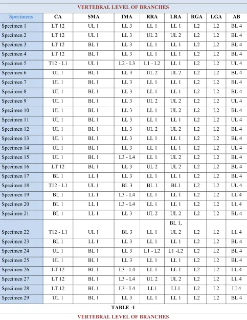
TABLE -1
VERTEBRAL LEVEL OF BRANCHES
Specimens CA SMA IMA RRA LRA RGA LGA AB
Specimen 1 LT 12 UL 1 LL 3 LL 1 LL 1 L2 L2 BL 4 Specimen 2 LT 12 UL 1 LL 3 UL 2 UL 2 L2 L2 BL 4 Specimen 3 LT 12 BL 1 LL 3 LL 1 LL 1 L2 L2 BL 4 Specimen 4 LT 12 BL 1 LL 3 LL 1 LL 1 L2 L2 BL 4 Specimen 5 T12 - L1 UL 1 L2 - L3 L1 - L2 LL 1 L2 L2 UL 4 Specimen 6 UL 1 BL 1 LL 3 UL 2 UL 2 L2 L2 BL 4 Specimen 7 UL 1 BL 1 LL 3 LL 1 LL 1 L2 L2 BL 4 Specimen 8 UL 1 BL 1 LL 3 LL 1 LL 1 L2 L2 BL 4 Specimen 9 UL 1 BL 1 LL 3 UL 2 UL 2 L2 L2 UL 4 Specimen 10 UL 1 BL 1 LL 3 LL 1 UL 2 L2 L2 BL 4 Specimen 11 UL 1 BL 1 LL 3 LL 1 LL 1 L2 L2 UL 4 Specimen 12 UL 1 BL 1 LL 3 UL 2 UL 2 L2 L2 BL 4 Specimen 13 UL 1 BL 1 LL 3 LL 1 LL 1 L2 L2 BL 4 Specimen 14 UL 1 BL 1 LL 3 LL 1 LL 1 L2 L2 UL 4 Specimen 15 UL 1 BL 1 L3 - L4 LL 1 UL 2 L2 L2 BL 4 Specimen 16 LT 12 BL 1 LL 3 UL 2 UL 2 L2 L2 BL 4 Specimen 17 BL 1 LL 1 LL 3 LL 1 LL 1 L2 L2 BL 4 Specimen 18 T12 - L1 UL 1 BL 3 BL 1 BL1 L2 L2 UL 4 Specimen 19 BL 1 LL 1 L3 - L4 LL 1 LL 1 L2 L2 LL 4 Specimen 20 BL 1 LL 1 L3 - L4 LL 1 LL 1 L2 L2 LL 4 Specimen 21 BL 1 LL 1 LL 3 UL 2 UL 2 L2 L2 BL 4 Specimen 22 T12 - L1 UL 1 BL 3 LL 1
BL 1,
UL 2 L2 L2 LL 4 Specimen 23 BL 1 LL 1 LL 3 LL 1 LL 1 L2 L2 BL 4 Specimen 24 UL 1 BL 1 LL 3 L1 - L2 L1 -L2 L2 L2 BL 4 Specimen 25 UL 1 BL 1 LL 3 LL 1 LL 1 L2 L2 BL 4 Specimen 26 LT 12 BL 1 L3 - L4 LL 1 LL 1 L2 L2 LL 4 Specimen 27 LT 12 BL 1 L3 - L4 UL 2 UL 2 L2 L2 LL 4 Specimen 28 LT 12 BL 1 L3 - L4 LL1 LL1 L2 L2 LL4 Specimen 29 UL 1 BL 1 LL 3 LL 1 LL 1 L2 L2 BL 4
TABLE -1
Specimens CA SMA IMA RRA LRA RGA LGA AB
Specimen 30 UL 1 BL 1 LL 3 LL 1 LL 1 L2 L2 BL 4 Specimen 31 UL 1 BL 1 LL 3 LL 1 LL 1 L2 L2 BL 4 Specimen 32 LT 12 LT 12 L3 - L4 UL 1 UL 1 L2 L2 L4 - 5 Specimen 33 LT 12 T12 - L1 BL 2 UL 1 UL 1 RL LL1 LL 3 Specimen 34 LT 12 UL 1 L3 - L4 LL 1 LL 1 L2 L2 BL 4 Specimen 35 LT 12 UL 1 L3 - L4 LL 1 LL 1 L2 L2 BL 4 Specimen 36 T12 - L1 UL 1 LL 3 LL 1 LL 1 L2 L2 BL 4 Specimen 37 T12 - L1 UL 1 LL 3 UL 2 UL 2 L2 L2 BL 4 Specimen 38 T12 - L1 UL 1 LL 3 LL 1 LL 1 L2 L2 BL 4 Specimen 39 T12 - L1 UL 1 LL 3 LL 1 LL 1 L2 L2 BL 4 Specimen 40 T12 - L1 UL 1 LL 3 LL 1 LL 1 L2 L2 BL 4 Specimen 41 LT 12 UL 1 L3 - L4 LL 1 LL 1 L2 L2 BL 4 Specimen 42 T12 - L1 UL 1 LL 3 LL 1 LL 1 L2 L2 BL 4 Specimen 43 T12 - L1 UL 1 BL 3 BL 1 LL 1 L2 L1 -2 UL 4 Specimen 44 LT 12 BL 1 LL 2
UL 1, LL 1
BL 1,
LL 1 VO VO UL 4 Specimen 45 T12 - L1 UL 1 LL 3 BL 1 LL 1
L2 -
L3 LL1 LL 4 Specimen 46 T12 - L1 UL 1 BL 3 LL 1 LL 1 L2 L2 LL 4 Specimen 47 T12 - L1 UL 1 L3 - L4 LL 1 LL 1 L2 L2 BL 4 Specimen 48 T12 - L1 UL 1 BL 3 LL 1 LL 1 L2 L2 UL 4 Specimen 49 T12 - L1 UL 1 LL 3 LL 1 LL 1 L2 L2 BL 4 Specimen 50 T12 - L1 UL 1 BL 3 BL 1 LL 1 L2 L2 UL 4
TABLE-2
SPECIMEN 25 11.5
SPECIMEN 26 12.7
SPECIMEN 27 11.9
SPECIMEN 28 11.5
SPECIMEN 29 11
SPECIMEN 30 10.4
SPECIMEN 31 10.8
SPECIMEN 32 13.1
SPECIMEN 33 11.4
SPECIMEN 34 11.5
SPECIMEN 35 12
SPECIMEN 36 12.4
SPECIMEN 37 11.9
SPECIMEN 38 12.1
SPECIMEN 39 11.2
SPECIMEN 40 11.7
SPECIMEN 41 11.2
SPECIMEN 42 11.8
SPECIMEN 43 11.4
SPECIMEN 44 12
SPECIMEN 45 12.6
SPECIMEN 46 12.3
SPECIMEN 47 11.2
SPECIMEN 48 10.8
SPECIMEN 49 10.9
SPECIMEN 50 11.1
SPECIMENS
TOTAL
LENGTH OF
AA
SPECIMEN 1 10.9
SPECIMEN 2 11.5
SPECIMEN 3 12
SPECIMEN 4 11.7
SPECIMEN 5 11.4
SPECIMEN 6 12.4
SPECIMEN 7 12.5
SPECIMEN 8 12.3
SPECIMEN 9 12.7
SPECIMEN 10 12.1
SPECIMEN 11 12.4
SPECIMEN 12 10.8
SPECIMEN 13 11.7
SPECIMEN 14 11.6
SPECIMEN 15 11.6
SPECIMEN 16 11.9
SPECIMEN 17 11.4
SPECIMEN 18 11
SPECIMEN 19 10.4
SPECIMEN 20 11
SPECIMEN 21 10.5
SPECIMEN 22 12.5
SPECIMEN 23 11.2
TABLE-3
SPECIMEN SUPRA REANAL AORTIC DIAMETER cm MID AORTIC DIAMETER cm ABOVE BIFUR DIAMETER cmSpecimen 1 1.94 1.82 1.44
Specimen 2 1.92 1.78 1.42
Specimen 3 1.94 1.88 1.32
Specimen 4 1.91 1.8 1.52
Specimen 5 1.92 1.72 1.49
Specimen 6 1.94 1.74 1.43
Specimen 7 1.92 1.81 1.41
Specimen 8 1.93 1.66 1.42
Specimen 9 1.79 1.67 1.45
Specimen 10 1.76 1.62 1.43
Specimen 11 1.74 1.71 1.44
Specimen 12 1.82 1.72 1.32
Specimen 13 1.85 1.82 1.35
Specimen 14 1.93 1.77 1.38
Specimen 15 1.92 1.81 1.32
Specimen 16 1.98 1.69 1.34
Specimen 17 1.75 1.62 1.37
Specimen 18 1.74 1.59 1.39
Specimen 19 1.72 1.57 1.32
Specimen 20 1.68 1.5 1.49
Specimen 21 1.66 1.52 1.42
Specimen 22 1.68 1.53 1.44
Specimen 23 1.69 1.51 1.4
Specimen 24 1.69 1.51 1.41
SPECIMEN SUPRA REANAL AORTIC DIAMETER cm MID AORTIC DIAMETER cm ABOVE BIFUR DIAMETER cm
Specimen 26 1.79 1.55 1.44
Specimen 27 1.82 1.61 1.41
Specimen 28 2.2 1.92 1.73
Specimen 29 2.12 1.9 1.7
Specimen 30 1.97 1.84 1.45
Specimen 31 1.92 1.82 1.46
Specimen 32 2.31 1.99 1.83
Specimen 33 1.96 1.84 1.39
Specimen 34 1.75 1.64 1.33
Specimen 35 1.79 1.69 1.42
Specimen 36 1.7 1.59 1.39
Specimen 37 1.68 1.55 1.42
Specimen 38 1.72 1.59 1.38
Specimen 39 1.74 1.57 1.36
Specimen 40 1.83 1.69 1.3
Specimen 41 1.94 1.82 1.41
Specimen 42 1.92 1.79 1.44
Specimen 43 1.98 1.84 1.45
Specimen 44 2.31 1.97 1.83
Specimen 45 1.72 1.68 1.43
Specimen 46 1.52 1.38 1.3
Specimen 47 1.68 1.54 1.46
Specimen 48 1.94 1.62 1.48
Specimen 49 1.74 1.61 1.36
TABLE-4
SPECIMENS Distance between AB-CA (cm) Distance between CA-SMA (cm) Distance between SMA-IMA (cm) Distance between IMA-AB (cm)Specimen 1 10.4 0.6 5.7 4.1
Specimen 2 11 1.2 5.9 3.9
Specimen 3 11.5 1.1 6.1 4.3
Specimen 4 11.2 1.2 5.9 4.1
Specimen 5 10.9 0.7 5.7 4.5
Specimen 6 11.9 1 6.6 4.3
Specimen 7 12 1.2 6.9 3.9
Specimen 8 11.8 1.3 6.5 4.1
Specimen 9 11.6 1.3 6.4 3.9
Specimen 10 11.6 1.5 5.8 4.3
Specimen 11 11.9 1.3 7 3.6
Specimen 12 10.6 1.3 5.2 3.8
Specimen 13 11.2 1.7 5.7 3.8
Specimen 14 11.1 1.5 5.5 4.1
Specimen 15 11.1 1.2 5.2 4.7
Specimen 16 11.4 1.5 5.7 4.2
Specimen 17 10.9 0.7 6.3 3.9
Specimen 18 10.5 0.8 6.1 3.6
Specimen 19 9.9 0.7 6 3.2
Specimen 20 10.5 0.9 6.1 3.5
Specimen 21 10 0.7 6 3.3
Specimen 22 12 1.5 6.4 4.1
Specimen 23 10.7 0.8 6.1 3.8
Specimen 24 10.4 0.6 6.3 3.5
SPECIMENS Distance between AB-CA (cm) Distance between CA-SMA (cm) Distance between SMA-IMA (cm) Distance between IMA-AB (cm)
Specimen 26 12.2 1.2 6.8 4.2
Specimen 27 11.4 1.1 5.9 4.4
Specimen 28 11 1.3 5.5 4.2
Specimen 29 10.6 1.5 5.2 3.9
Specimen 30 9.9 0.8 5.4 3.7
Specimen 31 10.3 1.1 5.6 3.6
Specimen 32 13.1 0 8.4 4.7
Specimen 33 10.9 1.2 5.9 3.8
Specimen 34 11 1.2 6.2 3.6
Specimen 35 11.5 1.2 6.9 3.4
Specimen 36 11.9 1.3 6.5 4.1
Specimen 37 11.4 1.5 6.2 3.7
Specimen 38 11.7 1.7 6.2 3.8
Specimen 39 10.7 0.9 6.3 3.5
Specimen 40 11.2 0.7 6.8 3.7
Specimen 41 10.7 0.8 6.5 3.5
Specimen 42 11.3 1 6.5 3.8
Specimen 43 10.9 1.1 6.4 3.4
Specimen 44 11.5 1.2 6.2 4.1
Specimen 45 12.1 1.2 7.1 3.8
Specimen 46 11.8 1.5 6.8 3.5
Specimen 47 10.7 1.3 5.9 3.4
Specimen 48 10.3 1.2 5.6 3.5
Specimen 49 10.4 1.1 5.5 3.8
ROW LABELS
COUNT OF
CELIAC AXIS
Body of first lumbar vertebra 5
Lower border of twelfth thoracic
vertebra 14
Inter vertebral disc between twelfth
thoracic & First lumbar vertebra 16
Upper border of first lumbar vertebra 15
CHART-1
BL 1 10%
LT 12 28%
T12 - L1 32% UL 1
30%
VERTEBRAL LEVEL- CA
ROW LABELS
COUNT OF
SUPERIOR MESENTRIC ARTERY
Body of first lumbar vertebra 22
Lower border of first lumbar vertebra 5
Lower border of twelfth thoracic vertebra 1
Inter vertebral disc between twelfth thoracic & First lumbar
vertebra 1
Upper border of first lumbar vertebra 21
CHART- 2 BL 1 44% LL 1 10% LT 12 2% T12 - L1
2% UL 1 42%
VERTEBRAL LEVEL- SMA
ROW LABELS
COUNT OF INFERIOR
MESENTERICARTERY
Body of second lumbar vertebra 1
Body of third lumbar vertebra 6
Inter vertebral disc between second & third
lumbar vertebra 1
Inter vertebral disc between third & fourth
lumbar vertebra 11
Lower border of second lumbar vertebra 1
Lower border of third lumbar vertebra 30
CHART- 3
BL 2
2% 12%BL 3
L2 - L3 2%
L3 - L4 22%
LL 2 2% LL 3
60%
VERTEBRAL LEVEL IMA
ROW LABELS
COUNT OF RIGHT
RENAL ARTERY
Body of first lumbar vertebra 4
Inter vertebral disc between first & second lumbar
vertebra 2
Lower border of second lumbar vertebra 33
Upper border of first lumbar vertebra 2
Upper Border Of First Lumbar Vertebra &
Lower border of first lumbar vertebra 1
Upper border of second lumbar vertebra 8
CHART- 4
VERTEBRAL LEVEL- RRA
ROW LABELS
COUNT OF
LEFT RENAL ARTERY
Body of first lumbar vertebra &
Lower border of first lumbar vertebra 1
Body of first lumbar vertebra &
Upper border of second lumbar vertebra 1
Body of first lumbar vertebra 1
Inter vertebral disc between first & second lumbar
vertebra 1
Lower border of first lumbar vertebra 34
Upper border of first lumbar vertebra 2
Upper border of second lumbar vertebra 10
CHART- 5
BL 1, LL 1
1 2%
BL 1, UL 2
1 2%
BL1 1
2% L1 -L2 1 2% LL 1 34 68% UL 1 2 4% UL 2 10 20%
VERTEBRAL LEVEL LRA
ROW LABELS
COUNT OF AORTIC
BIFUR
Body of fourth lumbar vertebra 31
Inter vertebral disc between fourth & fifth
lumbar vertebra 1
Lower border of third lumbar vertebra 1
Lower border of fourth lumbar vertebra 8
Upper border of fourth lumbar vertebra 9
CHART - 6
BL 4 31 62%
L4 - L5 1 2% LL 3 1 2% LL 4 8 16% UL 4 9 18%
VERTEBRAL LEVEL AB
RANGE COUNT
10.4-10.9 9
11-11.9 25
12-13.1 16
CHART - 7
0 5 10 15 20 25
10.4-10.9 11-11.9 12-13.1 9
25
16
TOTAL LENGTH OF AA
RANGE COUNT
1.52 - 1.69 9
1.7 - 1.79 15
1.82 - 1.98 22
2.12 - 2.31 4
CHART - 8
0 5 10 15 20 25
1.52 - 1.69 1.7 - 1.79 1.82 - 1.98 2.12 - 2.31 9
15
22
4
SUPRA REANAL AORTIC DIAMETER cm
RANGE COUNT
1.38 - 1.49 1
1.5 - 1.59 14
1.6 -1.69 13
1.7 - 1.79 7
1.8 - 1.89 11
1.9 - 1.99 4
50
CHART - 9
0 2 4 6 8 10 12 14 1.38 -1.49 1.5 -1.59
1.6 -1.69 1.7 -1.79 1.8 -1.89 1.9 -1.99 1 14 13 7 11 4
MID AORTIC DIAMETER cm
RANGE COUNT
1.3 - 1.39 18
1.4 - 1.49 27
1.5 - 1.59 1
1.6 - 1.69 0
1.7 - 1.79 2
1.8 - 1.83 2
CHART - 10
0 5 10 15 20 25 30
1.3 - 1.39 1.4 - 1.49 1.5 - 1.59 1.6 - 1.69 1.7 - 1.79 1.8 - 1.83 18
27
1 0 2 2
ABOVE BIFURCATION DIAMETER cm
ROW LABELS SUM OF COUNT
0.6 - 0.9 13
1 - 1.7 36
Common Celiac Mesenteric
Trunk 1
CHART -11 0 5 10 15 20 25 30 35 40
0.6 - 0.9 1 - 1.7 CCMT
Total 13 36 1
13
36
1 DISTANCE BETWEEN CA - SMA
RANGE COUNT
5.2 - 5.9 21
6 - 6.9 26
7 - 8.4 3
CHART - 12
0 5 10 15 20 25 30
5.2 - 5.9 6 - 6.9 7 - 8.4 21
26
3 DISTANCE BETWEEN SMA-IMA
RANGE COUNT
3.2 - 3.9 32
4.1 - 4.7 18
CHART – 13
0 5 10 15 20 25 30 35
3.2 - 3.9 4.1 - 4.7
32
18
DISTANCE BETWEEN IMA - AB
Figure: 1
Figure: 2
Figure: 3
Figure: 4
Figure: 5
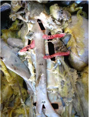
Bilateral accessory renal arteries were seen.
Renal orgin of gonadal arteries were seen.
Abdominal aorta shows dilatation just below the Inferior
Figure: 6
Figure: 7
Measurement of Diameter of AA
Figure: 8
[image:123.612.54.272.376.538.2]Measurement of length of AA
Figure: 9
[image:123.612.294.506.379.536.2]Figure: 10
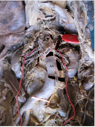
Aorta shows kinking at inferior mesenteric artery level. Aorta bifurcation at higher level (lower border of 3rd lumbar vertebra).
Figure: 11
[image:124.612.205.403.341.609.2]Figure: 12 & 13
Figure: 14
Figure: 15, 16& 17
Figure: 18
Figure: 19
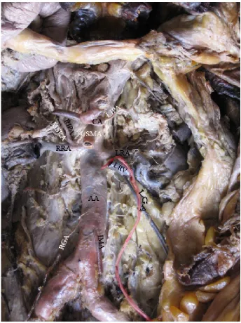
Figure - Shows Right Inferior Phrenic Artery, Middle Supra Renal Artery arise from renal artery from common trunk. Left Inferior Phrenic Artery from Celiac artery and left middle supra
1
BIBILIOGRAPHY
1. Adachi B., Das Arteriensystem der Japaner, Band II, Verlag der
Kaliserlich- Japanischen universitat zu Kyoto, Maruzen
publishing Co., 1928, 28,38,54
2. Adachi, B. (1928) Anatamie der Japaner.I.Das.Arterian system der
Japaner. II.Aorta. Thoracalis. Arcus.Plantarie Profundus
Maruzen.Kyoto quoted by cauld well and Anson in Americian Jr.
of Anat.
3. Anson BJ Mc Vay CB The topographical positions and the
mutual relations of the visceral branches of the abdominal aorta: a
study of 100 consecutive cadavers. Anat Rec. 1936:67:7-15.
4. Bandopadhyay, M. & Saha, A. Three rare variations in the
course of the gonadal artery Int.J. Morphol., 27 (3): 655-658,
2009.
5. Banowsky LHW, Surgical anatomy. In: NOVICK AC, STREEM
SB, PONTES JE (eds) Stewart’s operative urology, Williams &
Wilkins, Baltimore, 1989
6. Basmajian, J.V. (1980) Grants method of Anatomy by regions
2
7. Bordei P, Sapte E, Iliescu D double renal arteries originating
from the aorta Surg Radiol Anat 2004, 26: 474-9
8. Bordei P, Sapte E, Iliescu D, Dina C, the morphology and the
surgical importance of the gonadal arteries originating from the
renal artery, Surg Radiol Anat, 2007, 29 (5): 367-371
9. Cauldwel EW, Anson BJ (1943) Visceral branches of the
abdominal aorta topographicsl relationships. AM J Anat 73:27-57
10.Chithriki M, Jaibaji M, Steele RD The anatomical relationship
of the aortic bifurcation to the lumbar vertebrae: a MRI study.
Surg Radiol Anat 2002-24: 308-12
11.Cicekcibasi A.E, Salbaca A, Salbacak A, Sekar M. Ziylan T,
Buyukmumcu.M, Uysal II: The origin of gonadal arteries in
human fetuses: anatomical variations Ann Anat 2002, 184 (3):
275-279 perbmed Abstract.
12.Cicekcibasi AE, Ziylan T, Salbacak A, Et al An investigation of
the origin, location and variations of the renal arteries in human
fetus: Ann Anat 2005; 187:421.7
13.Coray,F., and Aubert (1913) Arteries de, I, intestingrele et des
colons, Bibiliog, Anat.vol.23 PP.221-254 Quoted by cauldwell &
3
14.Deepthinath R, Satheesha Nayak B, Mehtra RB et al, Multiple
variations in the paired arteries of the abdominal aorta, Clin Anat
2006 ; 19: 556-8
15.Dutta A.K. Esse0ntials of Human Anatomy- Part I 8th edition (2008) Kolkata.
16.Dunbar, J. D., Molnar, W., Beman, F. F. and Marable, S. A.:
compression of the Celiac Trunk and Abdominal Angina.
Preliminary Report of 15 Cases. Amer. J. Roentgen., 95:731, 1965.
17.Feller I, Woodburne Rt. Surgical anatomy of the abdominal
aorta. Ann.Surg.1961 Dec; 154(6) Suppl:239-252
18.George Ruggles Topography of the unpaired visceral Branches of
the abodominal Aorta. J.Anat 1935 Jan:69:176
19.Gokan.T, Hashimoto T, Matsui.S, Kushihashi.T, Nobusawa .H,
Munechika.H, Helical CTdemonstration of dialated inferior
phrenic arteries as entrahepatic collateral arteries of hepato cellular
carcinomas.J omput Assit Tomagr. 2001:25:68- 73
20.Gray (2008)Text book of Gray’s Anatomy 40th edi.Williams and Warwick, Churchill. Livingstone.
21.GWON DI, KO GY, YOON HK, SUNG KB, LEE JM, RYU
4
artery anatomy, variations, pathologic conditions and
interventional management, Radiographics 2007, 27 (3): 687 -705.
22.Hasan A. Al Zahrani, FRCS (Glas): Mohammed Rawas;
FRCP (C): Abduraul Maimani, FRCP (C); Maher Gasab, MD,
BAHA; Aba Al Khail,MD The Normal measurements of
abdominal Aortic Diameters in the Saudi Population, Saudi
Medical Journal Volume 16 No. 3 May 1995.
23.Heidsielk, E.(1928) Zur skeletopic de grossen, Aote.der
Baucharota Anat. Ans.vol.66 PP.6-24. Quoted by cauldwell and
Anson in Amer.Jr.of Anatomy.
24.Hollinshead WH Anatomy for Surgeons 1971:2 New York
Harper and Row .579-80
25.James E.Crouch (1970) Text book of functional Human Anatomy
chapter -12, Lea & Febiger. Philadelphia
26.Jamieson, C.W. (1986)Cocliac axis compression syndrome
Brit.Med.J.(Clin Res.) July19, 293 (6540) 159-60.
27.Koichi Adachi, Takamasa Iwawawa and Tsuyosi Ono,
screening for Abodominal Aortic Aneurysms During a basic
medical checkup in Residents of a Japanese rural community Surg.
5
28.Lakchayapakorn K, Siriprkarn Y (2008) Anatomical variations
of the position of the aortic bifurcation, iliocava junction and iliac
veins in relation to the lumbar vertebra. J med Assoc Thai,
91:1564-1570.
29.Last R.J. (1984) Anatomy Regional and applied 7th Ed., Section 5 30.Lelli, F: Maurelli, V.; Maranillo, E. & Valderrama – Canales
F.J. Arched and retrocaval testicular arteries: a case report. Eur.
J.Anat.11 (2) : 119-22, 2007
31.Lipshutz, B (1917) composite study of the colliac axis artery.
Ann. Surgery 65:159.
32.Loukas M, Hullett J, Wagner T Clinical anatomy of the inferior
phrenic artery. Clin Anat 2005; 18:357-65
33.Lucarotti ME, Shaw E, Heather BP (1992) Distribution of aortic
diameter in a screened male population . Br J. Surg 79:641 -642
34.Manguidi, C.(1893) Topographic del principals raoni viscerali
deil aorta abdominal Vellarchi Milano. Quoted by Heidsieck.
Quoted by cauld well and Anson in Amer Jr.of.Anat.1954.
35.Michels, N. A.: Blood Supply and Anatomy of Upper Abdominal
6
36.Mohammed A. Bakheit, MBBS, Ph.D, Mohammed A.
Mtabagani, B.Sc, Ph.D¸Anomalies of the renal, phrenic
suprarenal arteries. Saudi Med J 2004: Vol. 25(3): 376-38
37.Moore KL, Dalley AF, Agur AMR (2010) Clinically oriented
anatomy 6th Ed. Lippincott Williams & Wilkins, New Delhi, pp.314
38.Naito, M.; Terayama, H.; Nakamura, Y.;Hayashi, S.;Miyaki,
T.& Itoh,M. Left testicular artery arching over the ipsilateral renal
vein. Asian J.Androl., 8 (1): 107-10, 2006.
39.Neil Pennington Roger W.Soames The anterior visceral
branches of the abdominal aorta and their relationship to the renal
arteries. Surg Radiol Anat (2005) 27: 395-403.
40.Notkovich H, variations of the testicular and overian arteries in
relation to the renal pedicle, Surg Gynecol Obstet, 1956, 103 (4)
487-495,
41.Panyanetinad O (2011) Rare combined variations of renal,
testicular and suprarenal arteries, Internet J Anat Variations, 4
17-19.
42.Piao DX, Ohtsuka A, Murakami T. Typology of abdominal
arteries with special reference to inferior phrenic arteries and their
7
43.PICK JM, ANSON BJ, The inferior phrenic artery origin and
suprarenal branches, Anat Rec, 1940, 78:413-427
44.Prakash, Varsha Mokhasi, T. Rajini, M. Shashirekha
Department of Anatomy, Vydehi Institute of Medical Sciences and
Research Centre, Whitefield, Bengaluru, Karnataka, India. The
abdominal aorta and its branches anatomical variations and clinical
implications Folia Morphol. Vol.70. No:4, pp-282-286
45.Raikos.R1), G.K.PRASKEVAS1) K.NATSIS1) a.TZIKAS2),
S.N. NJAU2) 1) Department of Anatomy 2) Department of
Forensic Medicine and Toxicology Medical School, Aristotle
University of Tessaloniki, Greece Romanian Jouranal of
Morphology and Embryology 2010 , 51 (3): 585-587
46.Riddell AM, Khalili K, Sequential adrenal infarction without
MRI 0 detectable hemorrhage in primary antiphospholipid –
antibody syndrome. AJR Am J Roentgenol 2004; 183:220-222
47.Romanes, G.J. (2009) 16th edi.cunning text book of Anatomy.P 1297-1302.
48.S.Nayak Associate Professor of Anatomy Melaka Manipal
Medical College (Manipal Campus): Abnormal course of right
renal artery and ovarian vessels: A case Report. The Internet
8
49.Satchidhanandam. S.R.(1987) Madurai Medical College,
Madurai. Dissertation on ventral branches of Abdominal Aorta.
50.Sarita Sylvia 1, Sridhar Varma kakarlapudi1, Venkata
Ramana Vollala2, Bhagath Kumar Potu3, Raghu Jetti2,
Srinivasa Rao Balla4, Mohandas Rao5 and Narendra Pamidi2
Bilateral variant testicular arteries with double renal arteries Cases
Jouranl 2009, 2:114
51.Satyapal KS, Haffejee AA, Singh B, et al Additional renal
arteries: incidence and morphometry. Surg.Radiol Anat
2001;23:33-8.
52.Schaeffer, J.P. (1953)Morris human Anatomy 11th edi.P.690-708. 53.Schellhammer F, Von den Driesch P, Gaitzch A.
Pseudocoarctation of the abdominal aorta, Vasa. 1997:26:308-10
54.Schwal be, G., and Pfitzner, W. (1897) Varietaten. Statistik and
Anthropologic: Morphogische. Arbeit.Vol.3, PP.459
55.SINGH G, NG YK, BAY BH, Bilateral accessory renal arteries
associated with some anomalies of the ovarian arteries : a case
study, Clin Anat, 1998, 11(6): 417-420.
56.Siniluoto TM, Hellstrom PA, Paivansalo MJ, Leionen AS.
Testicular infarction following ethanol embolization of a renal
9
57.Spark.j.i Epidemiology of abdominal aortic aneurysms in the
Asian community, British journal of Surgery, 2002, Vol: 88 (3)
P:382-384.9
58.Skoog, S.J.; Roberts, K.P.; Goldstein, M.& Pryor, J.L. The
adolescent varicocele: what’s new with an old problem in young
patients? Pediatrics, 100 (1): 112-21, 1997.
59.Songur A, Toktas M, Alkoc O, Acar T, Uzun I, Bas O, Ozen
OA (2010) Abdominal aorta and its branches; morphometry –
variations in autopsy cases. Eur.J Gen Med, 7-321-325.
60.Soni S.Wadha A (2010) Multiple variations in the paired arteries
of abdominal aorta clinical implications J.Clin Diagn
Res.4:2622-2625
61.Ssoson, Jaroschelvitsch.A.J. (1926) Zur Chirurgicohen Anatomil
der aorta bifurcation. Zeitscher.F.Anat. Ent. Wick
Lungagesch.vol79.49-57.Quoted b cauldwell & Anson in
Amer.Jr.of Anatomy.
62.Sridhar Varmal .K : Narendra pamidill: Venkata R. Vollalall
Common celiacomesenteric trunk: a rare anatomic variation J.vasc
10
63.Taniguchi,T.(1931) Beitrag Zur Topographic der groseen Aote
der Bauchatta aorta. Folia Anat. Japan Vol.9 PP.201-214.Quoted
by Cauldwell Anson in Amer.Jr.of. Anatomy.
64.Thane, G.D.(1892) In Quains Elements of Anatomy vol.II, Part II,
Longmen’s Green & Co., London.
65.Thoreak, PC (1954)Anatomy in Surgery, 3rd Edn.Chap.23
66.Tsukamoto, N. (1929)Uber dil Arterien in dei bauchhonte Bei den
Japanera Kerboga Ku Zassulu Bd, 2 (Japanisch). Quoted by
cauldwell and Anson in Amer.J. Anatomy,.