HISTOPATHOLOGICAL SPECTRUM OF OVARIAN TUMORS: A DESCRIPTIVE STUDY
Dissertation submitted in
Partial fulfillment of the regulations required for the award of
M.D. DEGREE IN
PATHOLOGY
THE TAMILNADU
DR.M.G.R. MEDICAL UNIVERSITY,
CHENNAI
CERTIFICATE
This is to certify that the dissertation entitled “Histopathological
spectrum of ovarian tumors – A descriptive study” is a record of
bonafide work done by Dr. Darshana S. Patil in the Department of
Pathology, Coimbatore Medical College, Coimbatore and submitted in
partial fulfillment of the requirements for the award of M.D. Degree in
Pathology by the Tamilnadu Dr. MGR University, Chennai. This work
has not previously formed the basis for the award of a degree or diploma.
Dr. M. Murthy, M.D. Dr. R.Vimala, M.D.
Professor and Head, Dean,
Department of Pathology, Coimbatore Medical College,
Coimbatore Medical College, Coimbatore - 14.
ACKNOWLEDGEMENT
I express my deep gratitude to Dr. R.Vimala M.D. Dean,
Coimbatore Medical College, for granting me permission to undertake
this study.
With an immense sense of gratitude, I humbly thank
Dr. M.Murthy, M.D. Professor and Head, Department of Pathology,
Coimbatore Medical College, for his scholarly guidance, advice and for
having rendered his valuable time and encouragement, without which this
study would not have been feasible.
I express my heartfelt thanks to all Additional Professors and all
Assistant Professors of Department of Pathology, Coimbatore Medical
College, for their constant support and encouragement throughout the
work. I thank all the technical staff of Department of Pathology,
Coimbatore Medical College, for their sincere and timely technical
assistance.
I thank my family members for their blessings and moral support.
CONTENTS
SR.NO. PARTICULARS PAGE NO.
1. INTRODUCTION 1
2. AIMS AND OBJECTIVES 4
3. NEED FOR THE STUDY 5
4. REVIEW OF LITERATURE 6
5. MATERIAL AND METHODS 36
6. OBSERVATION AND RESULTS 39
7. DISCUSSION 55
8. SUMMARY AND CONCLUSION 67
9. BIBLIOGRAPHY
10. ANNEXURES
Annexure I - Proforma
Annexure II - H & E Stain procedure
Annexure III - PAS Stain procedure
Annexure IV - Reticulin Stain Procedure
Annexure V - Immunohistochemistry Procedure
INTRODUCTION
Ovarian cancer is the fifth most common malignancy among
women and second most common gynecologic malignancy. It is the most
common cause of death due to malignancy of female genital tract.1
Ovarian malignancies constitute about 4% of the total cancers in females
and 25% of malignant tumors of the female genital tract.2 In India, the
ovary is next in importance to cervix as the seat of cancer of female
genital tract. Surface epithelial ovarian tumors constitute two thirds of all
ovarian tumors and malignant forms account for 90% of ovarian cancers.3
They exist in different histological patterns and exhibit varying degree of
aggressiveness.4
Ovary is subjected to monthly endocrine and traumatic insults
during normal ovulatory cycles and becomes susceptible to
tumorogenesis. Repeated ovulatory rupture and repair theoretically
creates opportunities for malignant gene mutations. This may explain the
apparent protective effects of oral contraceptives, late menarche, early
menopause, multiparity, and breast feeding. Each of these factors
from the tumors, different tumors tend to involve different age groups
preferentially. The complex anatomy of the ovary and its peculiar
physiology with the constant cyclical changes from puberty to
menopause give rise to number of cell types, each of which is capable of
giving rise to tumors. Both primary and secondary tumors of the ovaries
are relatively frequent showing a variety of histopathological patterns.6
Since germ cells are totipotential, it is not surprising that a very wide
variety of neoplasms may develop from them. Germ cell tumors are the
commonest ovarian neoplasms in young age group and constitute two
thirds of ovarian tumors in this age group, out of which one third are
malignant. These tumors account for 25-30% of all ovarian tumors, with
great majority of them in the category of mature cystic teratoma, the most
common variety of ovarian neoplasm in humans.3 Ovarian tumors in
children and adolescent girls constitute an important part of
gynecological oncology. Detection of these tumors at such a young age
creates much anxiety to parents and throws up a great challenge to
doctors in charge as the question of operative safety, chance of
malignancy and prospects of future child bearing associated with
radiotherapy by chemotherapy and arrival of newer chemotherapeutic
drugs has made the prognosis much better today. The main aim lies in
distinguishing ovarian neoplasms from the wide spectrum of
non-neoplastic lesions. Despite the new techniques in imaging and clinical
diagnosis, the diagnosis of ovarian tumors is primarily dependent upon
histopathological examination.
Determination of various histologic patterns of ovarian tumors is
very important in diagnosis as well as prognosis of ovarian tumors.
Prognosis of the tumors can also be predicted from the degree of
differentiation of the tumors. The stage and laterality of the tumors also
indicates their nature for example, tumors in the sex cord stromal
category are almost always confined to a single ovary. On the other hand,
approximately 65% of the metastatic tumors are bilateral. In this study, it
was aimed to find out the histopathological patterns which are more
prevalent in our population and whether they are significantly different
from other populations.7 Their characteristics with regard to age, size,
laterality, gross features were evaluated. Incidence of benign versus
AIMS AND OBJECTIVES
1. To evaluate the incidence of different histopathological subtypes of
ovarian tumors in and around Coimbatore district.
2. To study the incidence of malignancy, both primary and secondary
in ovarian tumors
3. To study the distribution of ovarian tumors with respect to various
parameters like age, clinical presentation, parity, menstural
status, bilaterality, size, gross features and constistency of the tumors.
4. To study the gross morphological patterns of the various
histopathological tumor types.
5. To evaluate the association between various parameters of ovarian
NEED FOR THE STUDY
Ovarian tumors pose a major threat to public health worldwide. In
global terms, ovarian cancer is the fifth most common cancer in females.
Two thirds of ovarian tumors occur in reproductive age group. It has
been estimated that one woman in 70 (1.4%) will develop ovarian cancer
at some point during her lifetime. Its lower cure rates (less than 40%)
result in 6% of total deaths in females.
Thus studying the histopathological spectrum of the ovarian
tumors will help to understand the relative incidence of malignancy over
benign, distribution of different tumors according to age. Studying the
correlation of tumors with various parameters like age at menarche,
menstrual status and parity can help to understand the risk factors and
pathogenesis of ovarian tumors. This study can also contribute to
understand the correlation of gross morphological features with
histopathological diagnosis which can certainly help us to arrive at a
close differential diagnosis even only with gross examination alone in
REVIEW OF LITERATURE
Ovarian neoplasms have become increasingly important not only
because of the large variety of neoplastic entities but more because they
have gradually increased the mortality rate in female genital tract
cancers.8 As proposed by Kurman et al, efforts at early detection and new
therapeutic approaches to reduce mortality have been largely
unsuccessful, because the origin and pathogenesis of ovarian cancer is
poorly understood.9
Embryological development of the ovary:
As emphasized by Harold N et al in his study, in order to
understand the broad range of ovarian tumors and the clinical
significance of each tumor, we must review embryological origins of
various histological subtypes.10 The developing ovary contains three
types of cell, the oocytes, the follicular cells and the supporting stromal
cells.
Oocytes are derivatives of primordial germ cells that can result in
germ cell tumors and represent the most common benign or malignant
important in regulation of estrogen production and maturation of
follicles. These cells give rise to epithelial tumors which are the
commonest adult ovarian tumors. The stromal granulosa and theca cells
are mesenchymal in origin. They surround follicular cells, support their
development and are the origin of ovarian stromal tumors.
Anatomy of the ovary:
Ovaries are situated one on each side of uterus close to lateral
pelvic wall.11 Each adult ovary measures 3-4 cm in length, 1.5- 3cm in
width; 0.6-1.5cm in thickness .The average weight is 5-8 gms.
Histology:
The surface of the ovary is covered by a single layer of cuboidal
cells, which forms germinal epithelium that is continuous with the
mesothelial covering. Beneath the epithelium a tough connective tissue of
tunica albuginea is present under which lies the thicker outer cortex and
the inner medulla. Cortex shows follicles at varying stages of maturation.
Classification:
The primary purpose of classification of ovarian tumors is to
affected by ovarian carcinoma. As proposed by Langley F.A et al, the
classification of ovarian tumors is primarily morphologic but is intended
to reflect current concepts of embryogenesis and histogenesis of this
complex organ.11,12 In 1973 World health organization (WHO) gave
classification based primarily on gross features. In late 1980, the
International Society of Gynecological Pathologists gave a new
Classification, which is adopted, as
WHO CLASSIFICATION:
I. SURFACE EPITHELIAL – STROMAL TUMOURS
A. SEROUS TUMOURS
1. Benign
a. Cystadenoma and papillary cystadenoma
b. Surface papilloma
c. Adenofibroma and cystadenofibroma.
2. Of borderline malignancy
a. Cystadenoma and papillary cystadenoma
b. Surface papilloma
c. Adenofibroma and cystadenofibroma
3. Malignant
b. Surface papillary carcinoma
c. Malignant adenofibroma and cystadenofibroma.
B. MUCINOUS TUMOURS
Same as for serous tumors except that surface forms are exceptional.
C. ENDOMETRIOD TUMOURS
1. Same as for serous tumors except that surface forms are rare.
2. Stromal and mixed epithelial stromal tumors
a. Adenosarcoma
b. Stromal sarcoma
a. Mesodermal (Mullerian) mixed tumours
D. CLEAR CELL TUMOURS
Same as for serous tumors except that surface forms are not reported.
E. TRANSITIONAL CELL TUMORS
1. Brenner tumor
2. Of borderline malignancy (proliferating)
3. Malignant
4. Transitional cell carcinoma (non Brenner type)
G. MIXED EPITHELIAL TUMOURS
2. Of borderline malignancy
3. Malignant
H. UNDIFFERENTIATED CARCINOMA I. UNCLASSIFIED
II. SEX CORD-STROMAL TUMOURS
A. GRANULOSA-STROMAL CELL TUMOURS
1. Granulosa cell tumour
a. Adult
b. Juvenile
2. Tumors in the thecoma-fibroma group
a. Thecoma
i) Typical
ii) Luteinized
b. Fibroma
c. Fibrosarcoma
d. Stromal tumor
e. Sclerosing stromal tumor
g. Unclassified
B. SERTOLI-STROMAL CELL TUMOURS:
1. Well differentiated
a. Sertoli cell tumour; tubular androblastoma
b. Sertoli-Leydig cell tumour
c. Leydig cell tumour / hilus cell tumour
d. Stromal Leydig cell tumour
2. Of intermediate differentiation
3. Poorly differentiated [sarcomatioid]
4. Retiform
5. With heterologous elements
6. Mixed
C. SEX CORD TUMOURS WITH ANNULAR TUBULES D. GYNANDROBLASTOMA
E. UNCLASSIFIED
F. STEROID [LIPID] CELL TUMUORS
1. Stromal luteoma
2. Leydig cell tumor
b. Leydig cell tumor non hilar type
3. Adrenal cortical type
4. Not otherwise specified
III. GERM CELL TUMOURS
A. DYSGERMINOMA B. YOLK SAC TUMOURS
C. EMBRYONAL CARCINOMA D. POLYEMBRYOMA
E. CHORIOCARCINOMA F. TERATOMAS
1. Immature
2. Mature
a. Solid
b. Cystic (Dermoid cyst)
c. Secondary tumors transformation of a component
d. Fetiform (homunculus)
2. Monodermal and highly specialized
b. Carcinoid
c. Strumal carcinoid d. Mucinous carcinoid
e. Neuroectodermal tumors
f. Others
G. MIXED GERM CELL TUMORS
IV. GONADOBLASTOMA
1. Pure
2. Mixed with dysgerminoma or other form of germ cell tumor
V. GERM CELL- SEX CORD- STROMAL TUMORS
VI. TUMOURS OF RETE OVARII
1. Adenoma and cystadenoma
2. Carcinoma
VII.MESOTHELIAL TUMORS
VIII. TUMOURS OF UNCERTAIN ORIGIN
1. Small cell carcinoma hypercalcemic type
2. Tumor of wolffian origin
3. Hepatoid carcinoma
5. Wilms tumor
IX. SOFT TISSUE TUMOURS NOT SPECIFIC TO OVARY
X. UNCLASSIFIED TUMOURS
XI. SECONDARY [ METASTATIC ] TUMOURS
I.SURFACE EPITHELIAL – STROMAL TUMORS:
As seen in study by Ramesh Dawar et al (2004) this category
constitutes almost two thirds of all ovarian tumors. 90% of all ovarian
cancers are surface epithelial carcinomas.13
They are subclassified according to following patterns:
1) Cell type: serous / mucinous / endometroid / clear / transitional
2) Exophytic / Intracystic
3) Pattern of growth: cystic /solid
4) Amount of epithelium / stroma (adenoma / adenofibroma)
5) Cell proliferation, nuclear atypia and stromal invasion:
Benign / borderline / malignant.
A. SEROUS TUMORS:
These tumors comprise 30-40 % of all ovarian tumors and are
M. Leitao et al, 70% are benign, 5-10% are borderline and 20-25% are
malignant.14 Bilaterality is present in 12-20% cases.
1) Benign serous tumors:
Grossly these are usually endophytic cystadenomas but may be
exophytic surface papillomas. Grossly, they are unilocular thin walled
cysts filled with serous fluid with smooth inner surface. Multiloculation,
mucinous contents and polypoid excrescence may also be seen.
Microscopic examination reveals fallopian tube like epithelial lining.
Stroma may be collagenous or markedly edematous. In
cystadenofibromas, glands and cysts are scattered within predominantly
fibrous stroma.
2) Serous borderline ovarian tumors:
They constitute 25-30% of malignant serous tumors with 30%
bilaterality. The decision whether an ovarian serous tumor is malignant
or borderline is made purely on the basis of morphologic features of
primary ovarian tumor regardless of presence or absence of peritoneal,
lymph node or even lung metastasis.15,16 These tumors are separated from
carcinoma by the absence of stromal invasion. These tumors are
excrescences. Microscopy shows stratified cuboidal to columnar pithelial
cells and ciliated cells. As revealed by Seidman JD et al, arborizing
papillae / hierarchical branching, varying nuclear atypia and absence of
frank stromal invasion characterize them.17
Micropapillary Variant:
Histologically it is characterized by an exuberant cellular proliferation
that emanates from the surfaces of fibrous papillae without hierarchical
branching.18,19
Peritoneal implants:
The serous borderline tumors are associated with implants in 30%
of cases. These are divided into non-invasive and invasive types. Non
invasive which constitute 88% of all, are further subdivided into
epithelial and desmoplastic types.
3) Malignant serous tumors:
Serous carcinomas account for about 30 % of all ovarian cancers. These
range in size from microscopically undetectable to over 20 cm in
diameter and are bilateral in one third of cases as seen in study done by
CM Shahbaz Sarwar et al.20 Grossly they show predominant solid
examination reveals obvious stromal invasion with high grade nuclear
features and irregularly branching highly cellular papillae with little or no
stromal support. Psammoma bodies are commonly seen. As revealed in
study by Naseer A Shaikh et al, the majority of patients present at
advanced stage when the prognosis is poor.21
B) MUCINOUS TUMORS:
These tumors account for 10-15% of all ovarian tumors.22 They generally
occur in older women. They may be associated with dermoid cysts,
Brenner tumors or mucinous tumors of other organs.
1) Benign mucinous cystadenoma:
These tend to be largest of all ovarian tumors. Most of these
tumors are usually unilateral multilocular cystic masses containing
viscous mucoid or watery material. Microscopically they show cysts or
glands lined by mucinous columnar epithelium with minimal cellular
stratification and basally located bland nuclei.
2) Borderline mucinous cystadenoma:
The lining epithelium shows stratification not more than 3 layers and
may form filiform intracystic papillae with absent frank stromal invasion.
Intestinal type (85%) and Endocervical /mullerian type (15%) As in study
by Shappell et al, borderline tumors are qualified as intraepithelial
carcinoma based on the presence of marked cytologic atypia or a
complex cribriform growth pattern involving the epithelium covering the
surface of papillae or lining cystic spaces. Tumors with stromal invasion
≤5 mm are classified as microinvasive carcinoma.23
3) Malignant mucinous ovarian tumor (mucinous adenocarcinoma):
These are relatively uncommon tumors accounting for 10% of
ovarian cancers. They are multicystic with cut section showing both solid
and papillary cystic areas with areas of hemorrhage and necrosis. As seen
in study by Stephen S Raab et al, microscopically the lining epithelium
shows stratification of more than 4 layers and atypical changes. Areas of
necrosis and stromal invasion also seen. 24
C) ENDOMETROID TUMORS:
These tumors account for approximately 2-4% of all ovarian
tumors and 10-20% of all ovarian cancers25. These occur in women in
perimenopausal age. As seen in study by Karen A.Bell et al most
Endometrioid carcinoma:
These tumors present as solid, friable or cystic mass with hemorrhagic
content. Histopathological examination shows that these resemble the
ordinary type of endometrial adenocarcinoma hence the name. Study
done by Falkenberry SS et al shows that 15-20% of endometroid
carcinoma show synchronus endometrial adenocarcinoma.27-31
D) CLEAR CELL TUMORS:
Clear cell carcinomas constitute 6% of surface epithelial stromal
Cancers.25,32 They most commonly occur in the 5th to 6th decade of life
as seen in study by by Crozeir MA et al 33,34. These are predominantly
solid with cystic areas containing one or more white or yellow polypoid
masses protruding in the lumen. As seen in study by Longacre TA et al,
clear cell carcinoma shows a variety of patterns and cell types which are
admixed. The common patterns are tubulocystic, papillary, reticular and
solid sheets.35Cores of papillae are lined by one or two layers of hobnail
cells with clear cells forming solid sheets. The stroma in ovarian clear
E) BRENNER TUMOR:
These tumors constitute 1 to 2 % of all ovarian neoplasms25.
Average age at presentation was between 30-50 years in study done by
Ehrlich CE et al 37,38 . These are hormonally active accompanied by signs
of hyperesternism. Most authors currently favor an origin from surface
ovarian epithelium or cyst derived from them through the process of
metaplasia. As seen in study Cuatrecasas et al(2009),Brenner tumor is
seen in association with mucinous cystadenoma and transitional cell
tumor of the bladder.39 Most Brenner tumors are benign. These are solid,
sharply circumscribed and lobulated fibrous masses.37,38 These tumors
consist of nests and trabeculae of transitional cells surrounded by
abundant stroma. Nuclei of tumor cells are oval with longitudinal groove
(coffee bean).
F) TRANSITIONAL CELL CARCINOMA:
These tumors in ovary are broadly similar to transitional cell
carcinoma of urinary tract in architectural arrangement and they lack a
benign or borderline Brenner component. As seen in study by Eichhorn,
these are usually cystic with few solid areas. Microscopically solid or
papillary growth pattern is seen and the papillae are multilayered.
II. SEX CORD STROMAL TUMORS:
These comprise of a heterogeneous group of neoplasms accounting for
8% of all ovarian neoplasms25. They are the most common functioning
ovarian tumors.
A.1) GRANULOSA STROMAL CELL TUMOR:
These comprise of two groups:
1) The granulosa cell tumor
2) The thecoma - fibroma group
GRANULOSA CELL TUMOR:
These are the ovarian neoplasms showing differentiation towards
follicular granulosa cells. They are mainly of two types – Juvenile and
adult granulosa cell tumor. Adult granulosa cell tumors account for 1-2 %
of all ovarian tumors and 95% of all granulosa cell tumors. These occur
in menopausal and postmenopausal age group. Three fourths are
associated with hyperestrenism which leads to endometrial hyperplasia
and carcinoma in approximately 5% of cases. These are unilateral with
color. The most common histologic patterns include diffuse, trabecular,
insular, and microfollicular. An additional morphologic variant, a
pseudopapillary pattern, may also be seen in granulosa cell tumors of
both adult and juvenile type, and can give rise to problems in their
distinction from transitional cell neoplasms was studied by Irving, Julie
et al. 40 These tumors are potentially malignant. Juvenile granulose cell tumor usually presents in the first decade of life, almost all presenting
within third decade. Young, Robert H. M.B et al found 82% of the
prepubertal patients presenting with isosexual pseudo- precocity. 41
Juvenile granulosa cell tumor differs from adult granulose cell tumor
such that former shows nuclear hyperchromasia, increased mitotis, rare
nuclear grooves and call exner bodies.
A.2) THECOMA- FIBROMA GROUP: a) Thecoma:
These constitute 4% of all ovarian tumors and are subclassified in
typical and leutinized subtypes. They affect postmenopausal females.
They show estrogenic manifestations. These are unilateral, yellow, solid
and have a well-defined capsule, and are firm in consistency. They are
round to spindle and have finely dispersed chromatin with pale,
vacuolated lipid rich cytoplasm.42 Silver stains demonstrate reticulin
fibrils investing around each tumor cells.
b) FIBROMA:
The most common tumor in the sex cord-stromal category, which
accounts for 4% of human ovarian tumors, is the fibroma. Fibroma
occurs invariably after puberty, which is often seen between 50-60 years.
They may be associated with meigs syndrome or basal cell nevus
syndrome. These are smooth, lobulated and firm in consistency with
average 6 cm diameter. On cut section, these are white in color. These
shows spindle shaped fibroblastic cells that grow in interlacing fascicles
and whorls. The tumor cells have small ovoid nuclei with pointed ends.
No cytologic atypia or mitotic activity is present. 43
c) FIBROSARCOMA:
These are the most common ovarian sarcomas. They affect older
women. Grossly these are large, unilateral, solid tumors with
hemorrhagic and necrotic areas. These are densely cellular, with spindle
to severe cytological atypia and high mitotic rate (average 4 - 25
mitosis/10HPF).44
e) Sclerosing stromal tumor of ovary:
It is a rare ovarian neoplasm occurring predominantly in young
women and is histologically characterized by cellular heterogeneity,
prominent vasculature, and a pseudolobular appearance composed of
cellular and hypocellular areas. As seen in study by Kawauchi S et al,
immunohistochemistry demonstrates an expression of vascular
permeability factor/vascular endothelial growth factor(VPF/VEGF) in the
luteinized theca like cells.45 Grossly they are solid tumors with white and
yellow areas, edema and cyst formation.
B) SERTOLI - LEYDIG CELL TUMOR:
These tumors account for less than 0.5% of all ovarian tumors.
They may occur at any age but common around 25 years. These often
produce androgens. They vary in gross appearance from solid yellow
lobulated masses to uni or multilocular cysts. Sertoli-Leydig cell tumors
are subdivided into well-differentiated forms, tumors of intermediate
type. As in study by Oliva, Esther et al EMA, inhibin, and
chromogranin represent the most helpful triad of immunomarkers serving
to exclude two common mimics of Sertoli cell tumors (endometrioid
carcinoma [inhibin−; EMA+; chromogranin−] and carcinoid tumor
[inhibin−; EMA+; chromogranin+]).46
C) SEX CORD TUMOR WITH ANNULAR TUBULES:
It is a distinctive ovarian tumor, which present with
hyperestrenism. Its morphologic hallmark is the presence of simple and
complex annular tubules containing eosinophilic hyaline bodies, which
are often calcified. Tumors associated with Peutz –Jegher syndrome are
typically multifocal, bilateral, calcified and usually benign in nature.47
D) GYNANDROBLASTOMA:
It is an extremely rare tumor. It is the term used for sex cord –
stromal tumor composed of a mixture in similar amounts of clearly
identifiable granulosa-theca cells and sertoli leydig cell elements. The
inor component should account at least 10% of the tumor.2
III. LIPID CELL (LIPOID, STEROID CELL) TUMOR:
This group consists of ovarian tumors composed of cells resembling
lipid-rich.
They are subdivided into four subtypes:
1. stromal luteoma
2. leydig cell tumor – hilus / nonhilus
3. adrenal cortical type
4. not other wise specified
LEYDIG CELL TUMOR:
These are usually unilateral and grossly present as small dark
reddish brown to yellow nodules. Microscopically these are composed
large round to polygonal cells with abundant eosinophilic cytoplasm that
shows positivity for fat stains.48 Endocrine atypia is common. About 25%
of tumors have tendency to develop malignancy. Malignant tumors tend
to be larger with foci of necrosis and hemorrhage.
IV. GERM CELL TUMORS:
These tumors constitute approximately 30% of all ovarian
neoplasms. Most of them are seen in children and young adults.
Approximately 95% of tumors are benign cystic teratomas. The germ cell
theory suggests origin from a meiotic germ cell. It could be due to failure
of meiosis 1 or meiosis 2.
A) DYSGERMINOMA:
These are the most common malignant germ cell tumors of the
ovary which account for 1 % of all ovarian cancers. As seen in study
done by Archana Amatya et al, it is the most common malignant tumor
occurring children and young women and in patients with gonadal
dysgenesis.49 Grossly they have convoluted external surface and solid
gray white cut surface. Presence of calcification suggests underlying
gonadoblastoma. Microscopically they show monotonous population of
cells arranged in nests separated by fibrous septa with lymphocytic
infiltration. The cells have abundant clear cytoplasm, prominent
cytoplasmic membranes, round vesicular nucleus and central prominent
nucleoli. The PAS stain helps in demonstrating the presence of
intracytoplasmic glycogen. Liang Cheng et al revealed importance of
OCT 4 marker in dysgerminoma.50
B) YOLK SAC TUMOR (ENDODERMAL SINUS TUMOR):
These neoplasm constitute 20 % of malignant germ cell tumors.
cystic areas. Areas of hemorrhage and necrosis also seen. These tumors
have a more than one growth pattern recapitulating yolk sac
development, which can be reticular/microcystic, solid, papillary,
polyvesicular vitelline or hepatoid. Schiller Duval bodies are the
characteristic feature of this tumor. Eosinophilic PAS +ve diastase
resistant hyaline globules are a frequent finding in yolk sac tumors.51
C) EMBRYONAL CARCINOMA:
These tumors occur in young age group with a median age of 15
years. They present with precocious puberty, vaginal bleeding, hirsutism
and amenorrhea. They are large, unilateral exhibiting solid and variegated
cut surface. Foci of hemorrhage and necrosis are present. Microscopy
shows large primitive cells distributed in solid sheets. Occasionally
papillae and abortive glandular structures are formed. Nuclei are round
and vesicular. Numerous mitotic figures are present.52
D) POLYEMBRYOMAS:
These are very rare neoplasms containing large number of
embryoid bodies at various stages of development embedded in primitive
E) CHORIOCARCINOMA:
Primary ovarian choriocarcinoma are rare, highly malignant
tumors usually developing before puberty. Primary ovarian
choriocarcinoma are usually a component of mixed germ cell tumor.
Grossly they are hemorrhagic and friable. They show the typical
bilamellar pattern with admixture of syncitial and cytotrophoblastic
elements in a necrotic and hemorrhagic background.
F) TERATOMA:
These tumors are characterized by presence of tissues derived from
two or all three embryonic layers.These are divided into three types.
1) Immature teratoma
2) Mature teratoma
3) Monodermal teratoma
1)IMMMATURE TERATOMA:
It is a malignant teratoma constituting 3% of all ovarian teratomas
and 20% of all malignant germ cell neoplasms. It occurs in children and
partly solid and cystic areas. Microscopy shows the tissues derived from
all the three germ layers are present growing as disorganized mixture of
mature and immature elements Prognosis of these tumors depends mainly
on relative amount of primitive neuroepithelial tissue. The most widely
used grading system (Norris system) stratifies immature teratoma into
three grades:
Grade 1 - Rare, primitive neuroepithelium not greater than 1 hpf/ slide
Grade 2 - Primitive neuroepithelium not greater than 3 hpf/ slide
Grade 3 - Primitive neuroepithelium greater than or equal to 4 hpf/ slide
2) MATURE CYSTIC TERATOMA:
These are most common ovarian neoplasm accounting for 27–44%
of all ovarian neoplasms. Two thirds of the patients are between 20 and
40 years. These are almost always cystic. Cyst content is greasy, largely
composed of keratin, sebum and hairs. Teeth are often present.
Microscopically variable admixture of mature ectodermal,
mesodermal and endodermal elements is seen. In almost 2% of the cases
and particularly in older women, dermoid cysts undergo a secondary
occasionally an adenocarcinoma, sarcoma, or other type of malignant
neoplasm can occur. As observed by Ronnett,Brigitte M et al mature
cystic teratoma can coexist with mucinous cystadenoma giving rise to
pseudomyxoma peritonei or with Brenner tumor and fibrothecoma.53
3) MONODERMAL TERATOMA:
These are the teratoma in which one element predominates over all
others. Struma ovarii is the most common which consists of thyroid
follicles with eosinophilic colloid and lined by columnar or cuboidal cells
with uniform round nucleus. Carcinoid tumors are the second common
monodermal teratomas. Microscopic study shows round to cuboidal
tumor cells with a round nucleus with coarse salt and pepper chromatin
and small nucleoli. Four types are - mucinous, insular, trabacular and
strumal carcinoids.
G) MALIGNANT MIXED GERM CELL TUMOR:
These comprise 5 to 10 % of all malignant germ cell tumors.54
These show combination of two or more neoplastic germ cell elements.
common. Proper identification of various components is necessary which
requires careful gross examination and extensive sampling.
V. MIXED GERM CELL - SEXCORD STROMAL TUMORS: GONADOBLASTOMA:
These are tumors composed of a combination of germ cells and sex
cord stromal cells. It is a rare tumor which occurs almost exclusively in
dysgenetic gonads. They vary in gross appearance from soft and fleshy to
firm and completely calcified. They show admixture of primitive germ
cells resembling those of dysgerminoma with sex cord stromal cells
resembling that of immature sertoli and granulosa cells arranged in small
islands. Hyalinization and calcification are common.
VI. TUMORS OF THE RETE OVARII:
These are relatively uncommon tumors, which includes adenoma,
cystadenoma and carcinoma.
VII. MESOTHELIAL TUMORS: A) ADENOMATOID TUMOR:
These are benign mesothelial tumors. Histologically they resemble
B) MESOTHELIOMA: These are very rare.
VIII. TUMORS OF UNCERTAIN ORIGIN: A) SMALL CELL CARCINOMA:
There are two types of small cell carcinoma designated as hypercalcemic
and pulmonary type.
Hypercalcemic type:
Extrapulmonary small cell carcinoma is a clinicopathological
entity distinct from pulmonary small cell carcinoma. As revealed by
Reckova M et al, it is a high-grade ovarian malignant tumor, which
occurs in young females.55 It is usually unilateral with two thirds of
patients showing hypercalcemia. The tumor is large and solid grey white
resembling dysgerminoma or lymphoma. Large tumors may show
hemorrhagic and necrotic areas. Two histological patterns are identified.
A diffuse population of small, closely packed cells with scant cytoplasm
and small nuclei constitute the common pattern. The nuclei are
hyperchromatic, with fine chromatin and prominent nucleoli. Mitotic
figures are numerous. Cytoplasmic hyaline globules are seen. In second
eosinophilic cytoplasm and prominent nucleoli are seen.
Pulmonary type:
These highly malignant tumors resemble lung small cell
carcinoma. Metastasis from lung primary should always excluded.
B) FEMALE ADENEXAL TUMOR OF WOLFFIAN ORIGIN:
These are benign unilateral tumors. These arise from mesonephric/
wollfian remnants.56 The tumor cells show diffuse, trabecular and
microcystic growth pattern. Small to medium sized epithelial cells are
embedded in fibrous stroma as seen in study by Karminjed MH et al.57
Almost always inhibin positivity is also recorded.
IX. SOFT TISSUE TUMORS NOT SPECIFIC TO OVARY:
These include various mesenchymal neoplasms arising from those
tissue elements for example smooth musle, nerves or blood vessels which
are not involved in specific gonadal function. These may be leiomyomas
as seen in study by Murakami M et al or leiomyosarcomas,
rhabdomyomas, rhabdomyosarcomas, chondromas, chondrasarcomas or
X.METASTATIC TUMORS (SECONDARY TUMORS):
The ovary is a common site of metastasis in female genital tract as
observed by Mazur MT et al.59 Colon , stomach, breast, uterus and lung
constitute the common primary sites. As seen in study by Zhao et al most
of the meatastatic tumors occurred in premenopausal patients and had
bilateral presentation.60 Metastatic tumors in the ovary vary in
appearance. In some cases the ovary is completely replaced by solid
white tumor or cystic tumor with solid areas. The microscopic
appearance varies with the nature of primary
tumor. They resemble the primary to a variable degree.
Immunohistochemical stains like CK7 and CK20 are helpful in
establishing the diagnosis.
Krukenberg tumor:
These are adenocarcinomas with distinctive histological
appearance. They show typical mucin filled signet ring cells associated
with striking proliferation of ovarian stroma. Stomach is the usual
primary site. Grossly, these are solid bosselated masses with grey white
cut surface. Special stains for mucin and immunostains for epithelial
MATERIAL AND METHODS
STUDY DESGIN: Descriptive study.
PLACE OF THE STUDY AND STUDY PERIOD:
This study was conducted in the Department of Pathology,
Coimbatore Medical College, Coimbatore. The study period extended
from March 2009 to June 2010.
SAMPLE SIZE:
This study included all the specimens of ovarian tumors sent for
histopathological examination to the Department of Pathology at
Coimbatore Medical College, Coimbatore between March 2007 &
February 2010.
INCLUSION CRITERIA:
1. All the ovarian tumors, irrespective of their clinical features, stage of
the disease or type of surgical procedure implemented were included.
2. Hysterectomy specimens with incidental ovarian tumors were also
EXCLUSION CRITERIA:
Non neoplastic ovarian lesions like simple ovarian cyst,
tubo-ovarian mass and polycystic ovaries were excluded.
METHODOLOGY:
Relevant data like parity, clinical presentation, age of the patient
was collected in a proforma. For specimens of ovarian tumors from May
2009 to February 2010, gross observation of the specimens received was
done. For proper fixation, tumors were cut serially at 1cm thickness. The
specimens were fixed in 10% formalin for 24-48 hours. After fixation,
sections were given from representative areas. Sections were cut at 4-5
micrometer thickness & stained with H&E. Slides and blocks were
retrieved for cases from March 2007 to April 2009. All stained
histopathology slides were studied in detail. Histopathology reports for
each tumor were retrieved from department of pathology, Coimbatore
Medical College. Special stains of PAS, Reticulin were done whenever
necessary. All details of the specimen consisting of gross features,
microscopic features and final diagnosis were studied. World Health
of the data was done to find out the incidence of various types of tumors,
age of presentation of various tumor types, gross features and
histopathological patterns of individual tumor types. Incidence of benign
versus malignant tumors was studied. This was a descriptive study and
analysis of data was done using chi square test and logistic regression
analysis.
OBSERVATIONS AND RESULTS
The incidence and clinical presentation of the different ovarian
tumors is extremely variable. The preoperative diagnosis of the tumors is
often difficult with only clinical examination and even on exploration,
though some investigations like peritoneal fluid cytology or serum LDH
may help in predicting the nature of pathology. Hence one has to depend
on the microscopic appearance of the tumor for further management.8
The present study of ovarian tumors was undertaken for a period of three
years from March 2007 to February 2010. Totally 301 cases were studied
[image:43.612.106.524.484.621.2]in this period.
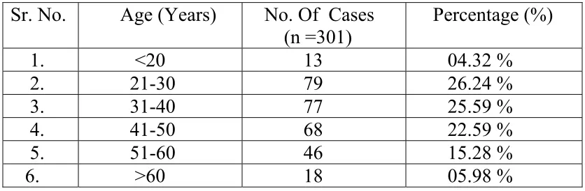
TABLE 1: AGE DISTRIBUTION
Sr. No. Age (Years) No. Of Cases (n =301)
Percentage (%)
1. <20 13 04.32 % 2. 21-30 79 26.24 % 3. 31-40 77 25.59 % 4. 41-50 68 22.59 % 5. 51-60 46 15.28 % 6. >60 18 05.98 %
Table 1 shows that the majority of tumors ie. 270 out of 301 (89.7
years only one tenth tumors were seen. Approximately 50 % tumors were
present in reproductive age group.
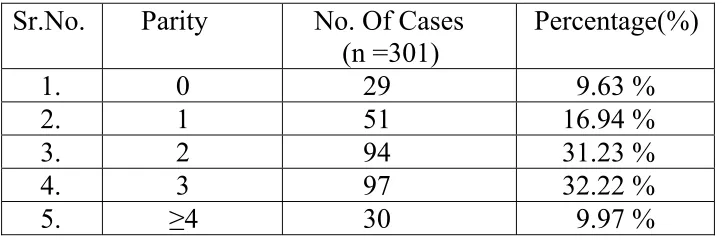
TABLE 2 : PARITY STATUS
[image:44.612.136.497.183.303.2]
Table 2 shows the parity status observed in the present study.
There were 29(9.63%) nulliparous patients and 145(48.17%) were of
parity one or two. Patients with parity three or more were 127(42.19%).
TABLE 3: AGE AT MENARCHE
Sr.No. Age At Menarche (Yr) No. Of Cases (n =301) Percentage (%)
1. <11 10 3.32 %
2. 11-12 46 15.28 %
3. 12-13 94 31.23 %
4. 13-14 47 15.61 %
5. 14-15 65 21.59 %
6. >15 39 12.96 %
Table 3 shows the age at menarche recorded for the patients in the Sr.No. Parity No. Of Cases
(n =301)
Percentage(%)
[image:44.612.130.533.473.612.2]present study. Majority of the patients, 245 (81.39%) attained menarche
after the age of 12. The other 56 patients attained menarche before age of
12.
TABLE 4 : MENSTRUAL STATUS
Sr.No. Category No. Of Cases (n =301) Percentage (%)
1. Premenopausal 211 70.10 %
[image:45.612.108.519.499.617.2]2. Postmenopausal 90 29.90 %
Table 4 shows menstrual status among the patients of the present
study. Premenopausal age group was more common than post
menopausal, contributing to 70.10% of the tumors.
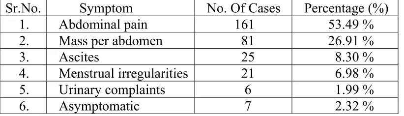
TABLE 5: MODE OF PRESENTATION
Table 5 shows that abdominal pain was the commonest
presentation followed by mass per abdomen. Asymptomatic ovarian
tumors were least common.
TABLE 6: LATERALITY OF THE TUMORS
[image:46.612.116.485.251.320.2]Sr. No. Laterality No. Of Cases (n =301) Percentage (%) 1. Unilateral 268 89.04 % 2. Bilateral 33 10.96 %
Table 6 shows the incidence of unilateral and bilateral tumors.
Most of the tumors were unilateral. 33 out of the 301 tumors (11%) had
bilateral presentation.
TABLE 7: INCIDENCE OF BILATERALITY IN BENIGN
VERSUS MALIGNANT TUMORS
[image:46.612.128.446.544.612.2]Sr.No. Type Unilateral Bilateral Percentage (%) 1. Benign 213 12 5.63% 2. Malignant 55 21 38.18%
Table 7 shows that 12 out of 213 (5.63%) benign tumors and 21
bilateral tumors 21 (64%) were malignant as against 12(5.63%) benign
tumors. Thus malignant tumors were common to present bilaterally.
[image:47.612.106.506.261.442.2]
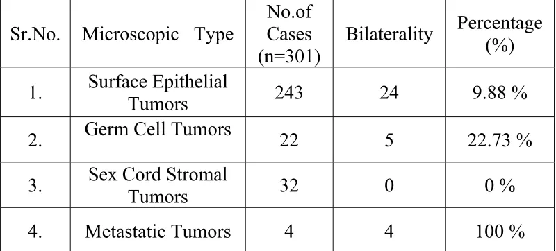
TABLE 8: INCIDENCE OF BILATERALITY IN THE MAJOR HISTOPATHOLOGICAL SUBTYPES
Table 8 shows that all the metastatic tumors had bilateral presentation
whereas none of the sex cord stromal tumors were bilateral. Out of 12
cases of endometrioid carcinoma in present study 6 (50%) had bilateral
presentation.
Sr.No. Microscopic Type
No.of Cases (n=301)
Bilaterality Percentage (%)
1. Surface Epithelial Tumors 243 24 9.88 %
2. Germ Cell Tumors 22 5 22.73 %
3. Sex Cord Stromal Tumors 32 0 0 %
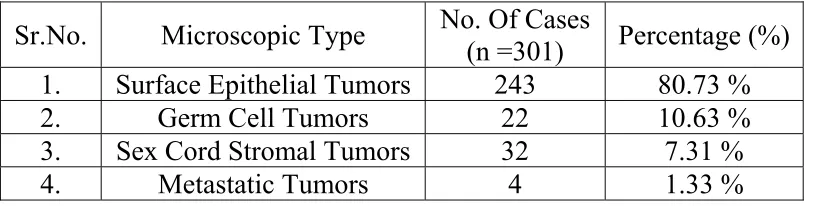
TABLE 9: MAJOR HISTOPATHOLOGICAL SUBTYPES
Table 9 shows the incidence of the major histopathological subtypes of
ovarian tumors. The most common histopathological pattern encountered
in the present study was surface epithelial tumors followed by germ cell
tumors. Metastatic tumors were the least common variety.
[image:48.612.106.514.147.250.2]
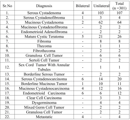
TABLE 10: LATERALITY IN INDIVIDUAL
HISTOPATHOLOGICAL SUBTYPE
Sr.No Diagnosis Bilateral Unilateral (n =301) Total
1. Serous Cystadenoma 4 103 107 2. Serous Cystadenofibroma 1 3 4
3. Mucinous Cystadenoma 2 62 64
4. Mucinous Cystadenofibroma - 2 2 5. Endometrioid Adenofibroma - 2 2 6. Mature Cystic Teratoma 5 21 26
7. Fibroma - 8 8
8. Thecoma - 1 1
9. Fibrothecoma - 2 2
10. Granulosa Cell Tumor - 6 6 11. Sertoli Cell Tumor - 2 2
12. Sex Cord Tumor With Annular Tubules - 1 1 13. Borderline Serous Tumor - 2 2
14. Serous Cystadenocarcinoma 6 14 20 15. Borderline Mucinous Tumor 1 10 11
16. Mucinous Cystadenocarcinoma 4 12 16 17. Endometrioid Carcinoma 6 6 12
18. Clear Cell Carcinoma - 3 3
19. Dysgerminoma - 4 4
20. Mixed Germ Cell Tumor - 2 2
21. Granulosa Cell Tumor - 2 2
[image:49.612.102.557.167.588.2]22. Metastatic 4 - 4
Table 10 shows the laterality observed in individual
teratoma was common to present bilaterally. In primary ovarian
carcinomas, endometrioid carcinoma and serous cystadenocarcinoma
showed higher incidence of bilaterality.
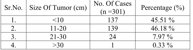
TABLE 11: SIZE OF THE TUMORS
Sr.No. Size Of Tumor (cm) No. Of Cases (n =301) Percentage (%)
1. <10 137 45.51 %
2. 11-20 139 46.18 %
3. 21-30 24 7.97 %
4. >30 1 0.33 %
[image:50.612.128.510.253.352.2]
Table 11 shows the distribution of tumors according to their size.
Majority of the tumors i.e. 276 (91.69%) were less than 20 cms in the
largest dimension. Only 24 tumors had their largest dimension between
21 to 30 cms. The largest tumor was measuring 32x25x12 cms which
was unilateral benign serous cystadenoma affecting a 27 year old lady.
Bilateral endometrioid adenocarcinoma was the smallest tumor in this
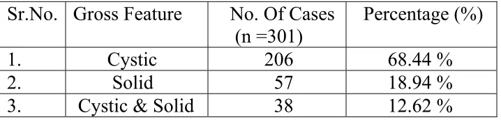
TABLE 12: GROSS FEATURES
Table 12 shows the details of the gross morphological features of
the ovarian tumors included in the present study. The commonest gross
morphology was the cystic nature of the tumors contributing to 68.44%.
The tumors presenting with gross cystic morphology were benign tumors
especially of surface epithelial category. Among tumors with solid or
complex morphology, majority were malignant.
[image:51.612.122.554.500.585.2]TABLE 13: NATURE OF THE TUMORS
Table 13 shows that out of 301 cases studied, majority were benign
tumors 225 (74.75%), followed by malignant 63(20.93 %) and 13
(4.32 %) cases of borderline tumors.
Sr.No. Gross Feature No. Of Cases (n =301)
Percentage (%)
1. Cystic 206 68.44 % 2. Solid 57 18.94 % 3. Cystic & Solid 38 12.62 %
Sr. NO Nature Of Tumor No. Of Cases
(n =301) Percentage (%)
1. Benign 225 74.75 %
2. Borderline 13 4.32 %
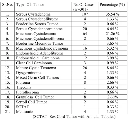
TABLE 14: HISTOMORPHOLOGICAL TYPES OF THE OVARIAN TUMORS
Sr.No. Type Of Tumor No.Of Cases (n =301)
Percentage (%)
1. Serous Cystadenoma 107 35.54 % 2. Serous Cystadenofibroma 4 1.33 % 3. Borderline Serous Tumor 2 0.66 % 4. Serous Cystadenocarcinoma 20 6.64 % 5. Mucinous Cystadenoma 64 21.26 % 6. Mucinous Cystadenofibroma 2 0.66 % 7. Borderline Mucinous Tumor 11 3.65 % 8. Mucinous Cystadenocarcinoma 16 5.32 % 9. Endometrioid Adenofibroma 2 0.66 % 10. Endometrioid Carcinoma 12 3.99 % 11. Clear Cell Carcinoma 3 0.99 %
12. Mature Cystic Teratoma 26 8.64 %
13. Dysgerminoma 4 1.33 %
14. Mixed Germ Cell Tumors 2 0.66 %
15. Fibroma 8 2.66 %
16. Thecoma 1 0.33 %
17. Fibrothecoma 2 0.66 %
18. Granulosa Cell Tumor 8 2.66% 19. Sertoli Cell Tumor 2 0.66 %
20. SCTAT 1 0.33 %
21. Metastatic 4 1.33 %
(SCTAT- Sex Cord Tumor with Annular Tubules)
Table 14 shows the incidence of each histomorphological subtype.
Benign serous cystadenoma was the commonest histopathological pattern
encountered in present study contributing to 35.54% followed by benign
tumor with annular tubules was seen. No transitional cell tumors were
seen in this study.
TABLE 15: AGE DISTRIBUTION OF THE
BENIGN / BORDERLINE / MALIGNANT OVARIAN TUMORS
AGE(Yr) BENIGN BORDERLINE MALIGNANT TOTAL
<20 9 - 4 13
21-30 67 5 7 79
31-40 67 1 9 77
41-50 45 4 19 68
51-60 28 3 15 46
[image:53.612.129.542.250.370.2]>60 9 - 9 18
Table 15 shows age distribution of benign and malignant ovarian tumors.
Malignant tumors commonly affected older age group (>40 years) with
only malignant germ cell tumors involving younger age group. On the
contrary, benign tumors predominantly involved patients < 40 year age.
The increasing incidence of malignancy with increasing age was found to
be statistically significant.
TABLE 16: AGE DISTRIBUTION OF THE MAJOR TUMOR CATEGORIES
AGE (Yr)
Surface Epithelial Tumors
Germ Cell Tumors
Sex cord Stromal Tumors
Metastatic Tumors
Total
<20 6 4 3 - 13 21-30 61 13 3 2 79 31-40 60 11 6 - 77 41-50 60 2 5 1 68 51-60 41 2 3 - 46 >60 15 - 2 1 18 Total 243 32 22 4 301
Table 16 shows the distribution of major histopathological types
according to age. Surface epithelial tumors were uncommon to present in
adolescent age group whereas germ cell tumors had predilection for this
age group. After age of 40 years, surface epithelial was the
predominating category with rare germ cell tumors.
TABLE 17: AGE DISTRIBUTION OF THE VARIOUS HISTOMORPHOLOGICAL TYPES OF THE TUMORS
NO Type Of Tumor <20 Yr. 21-30 31-40 41-50 51-60 >60 Yr. 1. Serous Cystadenoma 2 35 29 23 13 5 2. Serous Cystadenofibroma 1 1 1 - 1 - 3. Borderline Serous Tumor - 1 1 - - - 4. Serous Cystadenocarcinoma - 1 2 9 6 2 5. Mucinous Cystadenoma 3 17 20 15 8 1 6. Mucinous Cystadenofibroma - - - 1 1 7. Borderline Mucinous Tumor - 4 - 4 3 - 8. Mucinous Cystadenocarcinoma - 2 5 3 4 2 9. Endometrioid Adenofibroma - - - - 1 1 10. Endometrioid Carcinoma - - 2 6 2 2 11. Clear Cell Carcinoma - - - - 2 1 12. Mature Cystic Teratoma - 11 11 2 2 -
13. Dysgerminoma 2 2 - - - -
14. Mixed Germ Cell Tumors 2 - - -
15. Fibroma 2 - 4 2 - -
16. Thecoma - - - - 1 -
17. Fibrothecoma - 1 - - - 1
18. Granulosa Cell Tumor - 1 1 3 2 1 19. Sertoli Cell Tumor 1 1 - - - -
20. SCTAT - - 1 - - -
21. Metastatic - 2 - 1 - 1
[image:55.612.111.550.151.538.2]
Table 17 shows the age distribution of each histopathological
subtype of ovarian tumors included in present study. In patients less than
20 years of age, most of the tumors were benign except malignant germ
TABLE 18: VARIABLES RELATED TO THE RISK OF MALIGNANCY
Sr.
No. VARIABLE TOTAL MALIGNANT % p VALUE
1. AGE:
≤50 Years 237 49 20.67% p <0.01 *
>50 Years 64 27 42.19%
2. GRAVIDITY:
≤1 80 32 40.00% p < 0.01 *
>1 221 44 19.91%
3. MENOPAUSAL STATUS
PRE MENOPAUSAL 211 40 13.29% p < 0.01 * POST MENOPAUSAL 90 36 40.00%
4. MORPHOLOGY: TUMOR
CYSTIC 206 12 3.97%
p < 0.01 *
SOLID 57 42 73.68%
COMPLEX 38 22 57.89%
5. TUMOR DIAMETER:
≤10 cm 137 29 21.17%
p > 0.05
>10 cm 164 47 28.66%
6. LATERALITY
UNILATERAL 268 55 20.52% p < 0.01 * BILATERAL 33 21 63.64%
7. AGE AT MENARCHE
≤12 YEARS 56 17 30.36%
p > 0.05 >12 YEARS 245 59 24.08%
TABLE 19: LOGISTIC REGRESSION ANALYSIS
Variable Odds(B)Log S.E. Wald Df Prob. Sig. Exp(B)
Age 1.166 0.622 4.072 1 0.01 * 1.181 Parity -1.562 0.468 11.160 1 0.00 * 0.210 Age at Menarche -0.077 0.486 0.025 1 0.87 Ns 0.926 Menstrual status 1.497 0.603 6.170 1 0.01 * 4.469 Laterality 1.439 0.525 7.509 1 0.01 * 4.217 Size -0.051 0.384 0.018 1 0.89 Ns 0.950 Tumor Morphology 3.145 0.385 66.669 1 0.00 * 23.213
* = Significant NS = Not significant
Table 18 and 19 show the risk of malignancy associated with various
variables related to ovarian tumors. Logistic regression analysis is
applied to predict the probability of a particular outcome in relation to
several other independent variables. Here, Logistic regression analysis
was used to find out the probability (Odds) of the patients developing
malignant tumor. The model was constructed based on the set of
independent variables listed above. Increasing age, nulliparity or low
parity, postmenopausal status, bilaterality and complex or solid tumor
morphology conferred higher risk of malignancy. Early menarche and
TABLE 20: PREDICTIBILITY OF THE MODEL
Predicted by the equation
Observed data
Malignant Benign Total Percent Correct Malignant 208 17 225 92.44% Benign 20 56 76 73.68%
Using the above seven variables together, a model was created to predict
the risk of malignancy in a tumor. The following statistics was used to
assess the overall effectiveness of the model.
-2 Log Likelihood 190.312
Cox & Snell - R2 0.392
Nagelkerke - R2 0.579
Chi-Square Df Significance 149.851 7 *
* - Significant at 1 % level (P<0.01)
Overall 87.71% of the cases were correctly classified by the model. The –
2 Log Likelihood value is 190.312 which was used to test the
effectiveness of the model using chi-square test. The chi-square value
was 149.851, which was significant at 1% level indicating that the model
DISCUSSION
Because of the anatomical location, ovarian tumors may remain
unnoticed for a long period of time. These tumors can cause abdominal
pain and abdominal distension. Based on histological patterns, these
tumors are divided into benign, borderline and malignant. The common
variants are epithelial cell tumors, germ cell tumors, and sex cord stromal
Tumors. The ovary is one of the common sites to get metastatic deposits
from other abdominal malignancies.62
DISTRIBUTION OF OVARIAN TUMORS ACCORDING TO AGE:
Ovarian tumors are common in all age groups and no age is
exempted. The age range in the present study was 14 to 76 years. The
youngest patient in this study was a 14 year old girl with left sided
dysgerminoma measuring 17x14x10 cms which is the most common
germ cell tumor of adolescent age group. She presented with mass per
cystadenocarcinoma with omental deposits. She presented with ascites
and the tumor was measuring 5x4x3 cms. The maximum number of cases
in present study were between 21 and 60 years. This age group included
89.7% of tumors. This was in concordance with following studies:
The comparative analysis with above studies revealed similar results as
present study. 2nd to 5th decade was the most common age group
affected. Adolescent age group constituted 4.32 % in this study. This is
also comparable to study done by Deshpande et al where the incidence
of ovarian tumors in this age group was 4.2 %.67
PARITY STATUS:
Increasing parity is associated with a reduction in the risk of ovarian
cancer, but it is not clear whether this association applies to all the
histopathological types and to borderline tumours.68 Nulliparity and low Sr.No. Age
(Yr)
Present study
Pilli et al 63
R Jha et al 64
Kayastha et al 65
Kar et al 66
1. <20 4.32 % 7 % 6.8 % 6.3 % 7 % 2. 21-30 26.24 % 58 % 20.5 % 28.5 % 42 % 3. 31-40 25.59 % 26.7 % 27.4 %
4. 41-50 22.59 % 30 % 21.1 % 26.3 % 46 % 5. 51-60 15.28 % 14.3 % 11.6 %
parity were associated with increased relative risk of ovarian tumors in
the present study. In this study, tumors in nullipara and women with low
parity (two children) contributed to 57.80 % of all tumors which was
consistent with findings of study done by Kayastha et al.65 In their study
tumors in nullipara and women with low parity contributed to 58.93 %.
Similar results were obtained in study by Adami et al.68
AGE OF MENARCHE:
Increased risk of ovarian tumors associated with early menarche is put
forward by various studies like study by Adami et al and Hildreth et
al.68,69 In the present study only 18.60 % patients attained menarche
before 12 years of age. This is in contrast to the above mentioned studies.
As in present study, Kayastha et al recorded similar finding of 91.7 %
tumors in patients with menarche after age of 12 years.65 This difference
may be explained by the fact that both studies by Adami et al and
Hildreth et al were conducted in western population where as the present
study and study by Kayastha et al were conducted in Indian subcontinent.
The onset of menarche is influenced by female biology, as well as
from western countries have been shown to have younger age at
menarche with possible role played by nutritional status.
MENSTRUAL STATUS:
Similar to other studies, present study revealed higher incidence of
ovarian tumors in reproductive age group. Higher incidence of ovarian
tumors was observed in 21-40 years and premenopausal age group. In
postmenopausal patients, comparatively higher incidence of malignant
tumors was observed. Similar findings were recorded in study by
R Jha et al and also in study by Pilli et al and by Merino et al.63,64,70
MODE OF PRESENTATION :
Some of the ovarian tumors may be incidentally diagnosed on ultrasound
whereas others may be symptomatic. Present study reveals that the
presentation of the ovarian tumors is variable. If patient presented with
more than one complaint then the predominant symptom was considered
as the presenting symptom. In the present study the commonest
presenting symptom was pain in the abdomen 209 (69.43%) followed by
whereas menstrual irregularities including post menopausal bleeding in
19 (6.31 %). Asymptomatic patients were only 7 (2.32 %) with these
tumors being incidentally diagnosed on ultrasound done for other cause
or as a routine work up. The results comply well with a study carried out
by Rashid et al, in which abdominal pain was the commonest presenting
complaint (59%) followed by abdominal mass/distension (37%).71 Study
done by Sumaria Yasmin et al showed similar findings.7 In contrast to
this, in another retrospective analysis by Jamal et al the commonest
mode of presentation was bleeding per vaginum, followed by pain
abdomen, pelvic mass and gastric intestinal symptoms.72
UNILATERAL / BILATERAL PRESENTATION:
In the present study most of the tumors were unilateral. Only 33 out 301
tumors (constituting almost 11%) had bilateral presentation. Among
bilateral tumors 21 (64%) were malignant. Thus among bilateral tumors,
malignant tumors are more common . 21 out of 76 (27.63 %) malignant
tumors presented bilaterally as against 12 out of 225 (5.33 %) benign
tumors. This finding was similar to study done by R Jha et al who
encountered only 6.7 % benign tumors and 42.3 % malignant tumors
of bilaterality was present in the study by Kar et al which showed 26.8%
bilaterality.66 Out of 12 cases of endometrioid carcinoma present in this
study 6 (50%) had bilateral presentation. Serous carcinoma showed 42.85
% and mucinous carcinoma 33.33 % bilaterality. The most common
benign tumor to present bilaterally was mature cystic teratoma, which
showed 23.8 % bilaterality. Tumours in the sex cord stromal category are
almost always confined to a single ovary, similar observation is made in
this study.7 From 32 sex-cord stromal tumors, none had bilateral
presentation. As seen in study by Zhao et al most of the meatastatic
tumors occur in premenopausal patients and have bilateral presentation.60
In the present study all the metastatic tumors had bilateral presentation.
Ovarian dermoids can be bilateral in 15% cases.73
SIZE OF THE TUMOR:
Largest dimension of the tumor was utilized to categorize the tumors
according to the size. In present study tumors ranged in size from 3cms to
32 cms. Almost all the tumors i.e. 276 (91.69%) were less than 20 cms in
the largest dimension. Only 24 tumors had their largest dimension
which was unilateral benign serous cystadenoma affecting a 27 year old
lady. Bilateral endometrioid adenocarcinoma was the smallest tumor in
this study measuring 3x2x1.5 cms present in a 55 year lady.This finding
correlated with study by Pilli et al, In their study the largest tumor
measured 33x23x22 cms and smallest one measured 3x2x1 cms.63
GROSS FEATURES :
In the present study, 215 out of 301 (71.43 %) tumors had purely cystic
architecture. Solid tumors were 57 out of 301 and thus comprised 18.94
%. Combined solid and cystic presentation was present in 29 tumors
(9.63 %). Study done by Kar et al had 58.21 % cystic tumors, 13.43 %
solid and 28.36 % combined solid and cystic tumors.66 The relatively
high proportion of tumors with complex architecture and less percentage
of cystic tumors in study by Kar et al can be explained by the fact that
this study had more number of malignant tumors. Patients with solid or
complex ovarian tumors are at high risk of ovarian malignancy was
HISTOPATHOLOGICAL PATTERNS:
In this study WHO classification was implemented to classify the tumors.
Surface epithelial tumors constituted the most common category in the
present study contributing to 80.73 % of all tumors followed by germ cell
tumors, sex cord stromal tumors and metastatic tumors in decreasing
order of frequency as seen in other studies from India and neighboring
countries like Pakistan and Nepal.
Benign serous cystadenoma was the commonest tumor type with 35.54%
of tumors. This was followed by benign mucinous cystadenoma having
incidence of 21.26%. Cases of Serous, mucinous and endometrioid
cystadenofibromas were also reported. Among malignant tumors, serous Histopatho-
logical type
Present study
Shahbaz et al 75
Naseer et al 21
Kar et al 66
Pilli et al 63
Kayasth et al 65
Surface Epithelial Tumors
80.73 % 83.3% 81.0% 79% 70.9% 72.6%
Germ Cell Tumors
10.63 % 14.0% 10.95% 16% 21.2% 25.3%
Sex Cord Stromal Tumors
7.31 % 2.7% 5.03% 1.5% 6.7% 2.10%
Metastatic Tumors
cystadenocarcinoma was the most common category followed by
mucinous cystadenocarcinomas. Cases of endometrioid and clear cell
carcinomas were also present. In borderline surface epithelial tumor
category borderline mucinous tumor dominated the category along with
only 2 cases of serous borderline tumors. No transitional cell tumors were
seen in this study. Benign mature cystic teratoma was the most common
germ cell tumor whereas fibroma and granulosa cell tumor were the most
common sex cord stromal tumors. Metastatic Krukenberg tumors were
also present. Single case of sex cord tumor with annular tubules was
seen. Some differences in incidence of certain ovarian tumors in the
study population were noticed when compared to other studies. The most
marked one was the higher incidence of endometrioid adenocarcinoma in
the present study, with 12 cases being reported. Another important
feature regarding this tumor category was high incidence of bilateral
presentation which was observed in 50% cases. Another important
difference was in the incidence of the borderline surface epithelial tumors
with higher incidence recorded in present study. Both differences in
tumor morphology, the higher incidence of endometrioid carcinoma type
Sr No.
Type Of Tumor Present Study
Naseer et al 21
R Jha et al 64
1. Serous Cystadenoma 35.54 % 42.07 27.33% 2. Serous Cystadenofibroma 1.33 % 0.62% 3. Borderline Serous Tumor 0.66 % - - 4. Serous Cystadenocarcinoma 6.64 % 8.78% 7.45% 5. Mucinous Cystadenoma 21.26 % 16.85% 13.04% 6. Mucinous Cystadenofibroma 0.66 % -
7. Borderline Mucinous Tumor 3.65 % - - 8. Mucinous Cystadenocarcinoma 5.32 % 12.11% 3.73% 9. Endometrioid Adenofibroma 0.66 % - - 10. Endometrioid Carcinoma 3.99 % - - 11. Clear Cell Carcinoma 0.99 % - -
12. Mature Cystic Teratoma 8.64 % 7.2% 40.37% 13. Dysgerminoma 1.33 % 2.73% -
14. Mixed Germ Cell Tumors 0.66 % - - 15. Fibroma 2.66 % 1.29% 0.62 16. Thecoma 0.33 % 0.41% 1.86 17. Fibrothecoma 0.66 % - - 18. Granulosa Cell Tumor 2.66% 3.6% 0.62 19. Sertoli Cell Tumor 0.66 % - - 20. SCTAT 0.33 % - - 21. Metastatic 1.33 % 1.58% 1.24 (SCTAT- Sex Cord Stromal Tumor with Annular Tubules)
BENIGN / BORDERLINE / MALIGNANT:
Out of 301 cases studied, majority were b