International Journal of COPD 2018:13 2387–2398
International Journal of COPD
Dove
press
submit your manuscript | www.dovepress.com 2387
O r I g I n a l r e s e a r C h
open access to scientific and medical research
Open access Full Text article
Systematic analysis of transcriptomic profiles of
COPD airway epithelium using next-generation
sequencing and bioinformatics
Wei-an Chang1,2
Ming-Ju Tsai1–4
shu-Fang Jian1
Chau-Chyun sheu1–4
Po-lin Kuo1,5
1graduate Institute of Clinical
Medicine, College of Medicine, Kaohsiung Medical University,
2Division of Pulmonary and Critical
Care Medicine, Department of Internal Medicine, Kaohsiung Medical University hospital, Kaohsiung Medical University, 3Department of
Internal Medicine, school of Medicine, College of Medicine, Kaohsiung Medical University, 4Department
of respiratory Therapy, school of Medicine, College of Medicine, Kaohsiung Medical University,
5Center for Infectious Disease and
Cancer research, Kaohsiung Medical University, Kaohsiung, Taiwan
Introduction: COPD is a chronic inflammatory disease of lung. The inflammatory response in COPD is associated with neutrophils, macrophages, T lymphocytes, and bronchial epithelial cells, and occurs mainly in the small airway, leading to irreversible airflow limitation.
Methods: In order to investigate the microRNA–mRNA interaction in the microenvironment of the COPD airway, we used next-generation sequencing and bioinformatics in this study.
Results: We identified four genes with microRNA–mRNA interactions involved in COPD small-airway bronchial epithelial cells: NT5E, SDK1, TNS1, and PCDH7. Furthermore, miR6511a-5p–NT5E interaction was found to be involved in small-airway bronchial epithelial cells, large-airway bronchial epithelial cells, and alveolar macrophages.
Conclusion: Our results showed that miR6511a-5p–NT5E interaction plays an important role in COPD, which might be associated with cell–cell contact, activation of leukocytes, activa-tion of T lymphocytes, and cellular homeostasis. These findings provide new informaactiva-tion for further investigations of the COPD microenvironment, and may help to develop new diagnostic or therapeutic strategies targeting the bronchial epithelium for COPD.
Keywords: bioinformatics, COPD, epithelium, miR6511a-5p, next-generation sequencing, NT5E
Introduction
COPD affected 174.5 million people and caused 3.2 million deaths worldwide in 2015.1 It is progressive and usually causes major disability.2 COPD is a chronic inflammatory disease of the lung. The inflammatory response of COPD is associated with inhaled noxious substances, such as smoke and air pollution.3 Neutrophils, macrophages, and T lymphocytes are increased in the lung with COPD.3 As a defensive barrier of airways, bronchial epithelial cells are also affected in the inflammatory process of COPD.4 After being stimulated by inhaled insults, bronchial epithelial cells initiate immune and inflammatory responses, including recruiting macrophages, neutrophils, and dendritic cells.4 The inflammatory response occurs mainly in the small airway and results in significant airflow limitation, which is not fully reversible. With the progression of COPD, the inflamed small airways undergo remodeling processes involving airway-wall thickening, reduced airway diameter, and increasing resistance to respiratory flow.5 Recently, epithelial–mesenchymal transition caused by repeated stimulation was identified as a potential source of fibroblasts and myofibroblasts that could contribute to the chronic remodeling of airways.6
MicroRNAs have been recognized as important regulators in the gene regula-tion of COPD.7 Downregulated miR128 in smokers, downregulated miR181d and miR30a-3p in COPD lung tissue, and increased miR223 in lungs of smokers have Correspondence: Chau-Chyun sheu
Division of Pulmonary and Critical Care Medicine, Department of Internal Medicine, Kaohsiung Medical University hospital, 100 Tzyou First road, Kaohsiung 807, Taiwan Tel +88 67 312 1101 ext 5901 Fax +88 67 316 1210 email [email protected]
Journal name: International Journal of COPD Article Designation: Original Research Year: 2018
Volume: 13
Running head verso: Chang et al
Running head recto: Transcriptomic profiles of COPD airway epithelium DOI: 173206
International Journal of Chronic Obstructive Pulmonary Disease downloaded from https://www.dovepress.com/ by 118.70.13.36 on 22-Aug-2020
For personal use only.
Dovepress
Chang et al
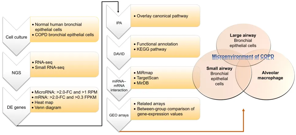
been demonstrated.7 In a study investigating muscle wast-ing of COPD patients, the results showed that increased miR424-5p was associated with muscle wasting.8 These studies showed that microRNA–mRNA interactions may play important roles in COPD, as we have demonstrated in asthma.9 In order to investigate microRNA–mRNA interac-tions in the microenvironment of COPD airways, we used COPD bronchial epithelial (DHBE) cells and normal bron-chial epithelial cells to perform next-generation sequencing (NGS) and bioinformatic analyses (Figure 1).
Methods
Primary cells
Primary normal human bronchial epithelial (NHBE) and DHBE cells were purchased from Lonza (Basel, Switzerland). Cell cultures were performed according to the manufacturer’s protocol. Cells were grown in bronchial epithelial cell basal media (BEGM™ Bronchial Epithelial Cell Growth Medium BulletKit™; Lonza), supplemented with 2 mL bovine pituitary extract, 0.5 mL hydrocortisone, 0.5 mL human epidermal growth factor (hEGF), 0.5 mL epinephrine, 0.5 mL transferrin, 0.5 mL insulin, 0.5 mL retinoic acid, 0.5 mL triiodothyronine, and 0.5 mL GA1000. Cells were maintained at 37°C in a 5% CO2 incubator and passaged with Reagent-Pack (Lonza) containing trypsin–EDTA (EDTA, trypsin neutralizing solution, and HEPES
[4-(2-hydroxyethyl)-1-piperazineethanesulfonic acid]-buffered solution). Cells were harvested for NGS analysis after cultivation from primary cells for one generation.
next-generation sequencing
Expression profiles of microRNAs and mRNAs were exam-ined using NGS. Total RNA of both normal and DHBE cells was extracted using Trizol reagent (Thermo Fisher Scientific, Waltham, MA, USA) according to the manufacturer’s pro-tocol. Purified RNA was then quantified at OD260 using an ND-1000 spectrophotometer (NanoDrop; Thermo Fisher Scientific) and quality assessed with a 2,100 bioanalyzer with RNA 6000 LabChip kit (Agilent Technologies, Santa Clara, CA, USA) at Welgene Biotech (Taipei, Taiwan).
For small-RNA library construction and deep sequencing, samples were prepared using an Illumina sample-preparation kit according to the TruSeq small-RNA library sample-preparation guide. In summary, total RNA was ligated with 3′ and 5′ adaptors and reverse-transcribed into cDNA followed by polymerase chain-reaction amplification. The harvested cDNA constructs were fractionated by size and purified with 6% polyacrylamide-gel electrophoresis, and bands contain-ing 18–40-nucleotide RNA fragments (140–155 nucleotides in length with both adapters) were selected. Libraries were then sequenced on an Illumina instrument (75SE cycle, single-end) and sequencing results processed with Illumina
&HOOFXOWXUH
• 1RUPDOKXPDQEURQFKLDO HSLWKHOLDOFHOOV
• &23'EURQFKLDOHSLWKHOLDOFHOOV
• 51$VHT
• 6PDOO51$VHT
• 0LFUR51$!)&DQG!530
• P51$!)&DQG!)3.0
• +HDWPDS
• 9HQQGLDJUDP
,3$
•2YHUOD\FDQRQLFDOSDWKZD\
•0L5PDS
•7DUJHW6FDQ
•0LU'%
•5HODWHGDUUD\V
•%HWZHHQJURXSFRPSDULVRQRI JHQHH[SUHVVLRQYDOXHV
•)XQFWLRQDODQQRWDWLRQ
•.(**SDWKZD\
/DUJHDLUZD\
%URQFKLDO HSLWKHOLDOFHOOV
6PDOODLUZD\
%URQFKLDO HSLWKHOLDO FHOOV
$OYHRODU PDFURSKDJH
'$9,'
PL51$± P51$ LQWHUDFWLRQ
*(2DUUD\V
1*6
'(JHQHV
Figure 1 Flowchart of study design.
Notes: In order to investigate the roles of microrna–mrna interactions in the microenvironment of COPD, we used normal human bronchial epithelial cells and COPD bronchial epithelial cells for next-generation sequencing (ngs). Then, we analyzed the ngs data of with several bioinformatic tools, including Mirmap, Ingenuity Pathway analysis (IPa), the Database for annotation, Visualization, and Integrated Discovery (DaVID), MirDB, Targetscan, and the gene expression Omnibus (geO) database. Abbreviations: De, differentially expressed; FC, fold change; rPM, reads per million; FPKM, fragments per kilobase of transcript per million mapped reads; Kegg, Kyoto encyclopedia of genes and genomes.
International Journal of Chronic Obstructive Pulmonary Disease downloaded from https://www.dovepress.com/ by 118.70.13.36 on 22-Aug-2020
Dovepress Transcriptomic profiles of COPD airway epithelium
software. For small-RNA-sequence analysis, sequencing data were applied to go through a filtering process to obtain qualified reads. Trimmomatic was used to trim or remove reads according to quality scores.10 Qualifying reads were then analyzed using miRDeep2 to clip the 3′ adapter sequence and shorter reads (,18 nucleotides) removed, before aligning reads with the human genome from the University of California, Santa Cruz.11 Because microRNAs are usually mapped to several genomic locations, only reads mapped perfectly to the genome five or more times were used for microRNA detection. MiRDeep2 was used to estimate expression levels of microRNAs. The criteria for microRNA selection were fold change .2, and reads per million .1.
For transcriptome sequencing, a library was con-structed with Agilent’s SureSelect Strand specific RNA-library-preparation kit for 75SE (single-end or paired-end) and sequencing performed on the Solexa platform. Sequences were determined directly using sequencing-by-synthesis tech-nology via the TruSeq SBS kit. Raw sequences were obtained from Illumina Pipeline software bcl2fastq version 2.0 and expected to generate 30 million reads per sample. The sequences generated went through a filtering process to obtain qualifying reads. Trimmomatic was implemented to trim or remove the reads according to the quality score.10 Qualifying reads were analyzed using TopHat/Cufflinks to estimate gene-expression level, calculated as fragments per kilobase of transcript per million mapped reads.12 For differential-expression analysis, the Cummerbund statistical package was employed to perform statistical analyses of gene-expression profiles. The reference genome and gene annotations were retrieved from the Ensembl database.
Mirmap database analysis
MiRmap is an open-source software library providing com-prehensive microRNA-target prediction (http://mirmap. ezlab.org).13 Putative target genes can be identified by cal-culating the complementary ability of microRNA–mRNA interactions. The predictor also estimates mRNA-repression strength for ranking potential candidate targets by a combi-nation of thermodynamic, evolutionary, probabilistic, and sequence-based features. The prediction results provide a list of putative target genes with MiRmap score, which is a predic-tive reference value. In this study, the criterion for selection of putative microRNA targets was MiRmap score $99.
MirDB database analysis
MirDB is an online database for predicting microRNA-target and functional annotations. In MirDB, MirTarget was used for
predicting all the targets. MirTarget was designed by analyzing microRNA-target interactions from high-throughput sequenc-ing experiments. MirDB can predict microRNA targets in five species, including human, mouse, rat, dog, and chicken.14,15
Targetscan database analysis
TargetScan is an online database for predicting biologi-cal targets of microRNAs. It searches for the presence of conserved 8mer, 7mer and 6mer sites, matching the seed region of each microRNA. Predictions are ranked based on the predicted efficacy of targeting or by their probability of conserved targeting.16
DaVID database analysis
The Database for Annotation, Visualization, and Integrated Discovery (DAVID), which integrates multiple functional annotation databases, including Gene Ontology, biological processes, or Kyoto Encyclopedia of Genes and Genomes pathways, is a powerful tool for gene-function classification (https://david.ncifcrf.gov).17 A list of interesting genes can be classified into clusters of related biological functions, sig-naling pathways, or diseases by calculating the similarity of global annotation profiles with an agglomeration algorithm. It also provides an Ease score, which is a modified Fisher’s exact P-value. The reference score represents how specifi-cally the user genes are involved in the category (eg, signaling pathways). In this study, we selected an Ease score of 0.1 as default and 1 to extend clustering range.
gene expression Omnibus database
analysis
The Gene Expression Omnibus (GEO) is a web database that collects submitted high-throughput gene-expression data of microarrays, chips, or NGS (https://www.ncbi.nlm. nih.gov/geo).18 Microarrays of accession numbers GSE4498 (small-airway bronchial epithelial cells), GSE5056 (large-airway bronchial epithelial cells), and GSE2125 (alveolar macrophages) were used in this study. GSE4498 assessed gene expression (HG133 Plus 2.0 array) in ten phenotypi-cally normal smokers compared to 12 matched nonsmokers.19 GSE5056 assessed gene expression in 13 phenotypically normal smokers and nine normal nonsmokers.20 GSE2125 assessed gene expression in 15 cigarette smokers and 15 nonsmokers.21 Raw data extracted from GEO were replot-ted and statistically analyzed with Student’s t-test using GraphPad Prism 7 software (GraphPad Software, La Jolla, CA, USA).
International Journal of Chronic Obstructive Pulmonary Disease downloaded from https://www.dovepress.com/ by 118.70.13.36 on 22-Aug-2020
Dovepress
Chang et al
Ingenuity Pathway analysis
Ingenuity Pathway Analysis (IPA) software (Ingenuity Systems, Redwood City, CA, USA) contains a large data-base with detailed and structured findings reviewed by experts. This IPA database was derived from thousands of biological, chemical, and medical studies, and provides researchers with quick searching. IPA also enables analysis, integration, and recognition of data from gene and single-nucleotide-polymorphism arrays, RNA and small-RNA sequencing, proteomics, and many other biological experi-ments. In addition, deeper understanding and identification of related signaling pathways, upstream regulators, molecular interactions, disease processes, and candidate biomarkers are also available in IPA.22
Results
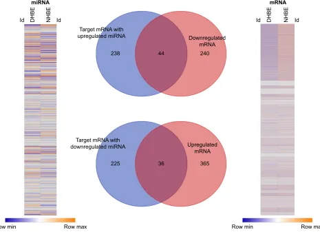
In order to identify the potential microRNA–mRNA inter-actions involved in the homeostasis of COPD epithelium,
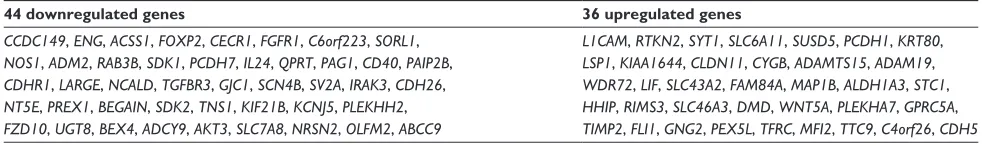
we analyzed the expressions of genes and microRNAs using NGS data from DHBE and NHBE cells (Figure 2). The gene-expression heat map of differentially expressed genes revealed 685 genes with fold change .2, including 284 downregulated genes and 401 upregulated genes. The heat map of microRNA expression revealed 144 microRNAs with fold change .2. Based on the MiRmap web-based database, we predicted 543 mRNA as targets of these 144 microRNAs, including 282 targets of upregulated microRNAs and 261 targets of downregulated microRNAs. Venn diagrams of microRNA–mRNA interactions showed that 44 genes were downregulated and 36 genes upregulated in DHBE cells compared to NHBE cells. These 80 dysregu-lated genes with potential microRNA–mRNA interactions are listed in Table 1.
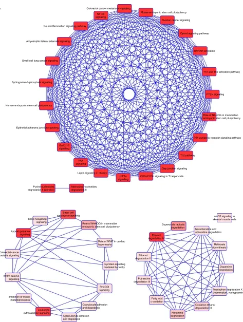
We then used IPA to analyze the function of these 80 dys-regulated genes. Possible pathways related to these 80 genes are demonstrated in Figure 3. Figure 3A shows pathways
7DUJHWP51$ZLWK XSUHJXODWHGPL51$
7DUJHWP51$ZLWK GRZQUHJXODWHGPL51$
'RZQUHJXODWHG P51$
8SUHJXODWHG P51$ '+%( 1+%(
5RZPLQ 5RZPD[
PL51$
,G ,G
5RZPLQ 5RZPD[
'+%( 1+%(
P51$
,G ,G
Figure 2 Identification of genes with potential microRNA–mRNA interactions in COPD bronchial epithelial cells.
Notes: gene-expression heat map (right) of differentially expressed genes revealed 685 genes with fold change .2. The heat map (left) of differentially expressed micrornas revealed 144 micrornas with fold change .2 and reads per million (rPM) .1. according to the Mirmap web-based database, we predicted 543 mrnas as targets of these 144 micrornas. Venn diagrams of microrna–mrna interactions shows that 44 genes were downregulated and 36 genes upregulated in COPD bronchial epithelial (DhBe) cells compared to normal human bronchial epithelial (nhBe) cells. The selection threshold for microrna-target prediction was Mirmap score $99.
International Journal of Chronic Obstructive Pulmonary Disease downloaded from https://www.dovepress.com/ by 118.70.13.36 on 22-Aug-2020
Dovepress Transcriptomic profiles of COPD airway epithelium
related to 44 downregulated genes, and Figure 3B shows pathways related to 36 upregulated genes. In Figure 3A, epithelial adherens junction signaling, T-helper (TH)-1- and TH2-activation pathways, and the TH2 pathway were involved in downregulation of the 44 genes. This implies that some of these genes may be associated with the func-tion of bronchial epithelium adherence and immunity of the COPD lung. Because most microRNAs could have down-regulated gene expression, we focused on the interaction between upregulated microRNA and downregulated mRNA. To analyze the possible mechanisms of these 44 down-regulated genes in DHBE cells, we used DAVID (Figure 4). The results showed that these 44 genes were involved in the functions of membrane, transmembrane helix, transmem-brane, nucleotide bonding, cell adhesion, calcium, disulfide bonds, and signals. We further analyzed the disease and function of 44 downregulated genes with IPA and then performed meta-analyses with MirDB and TargetScan. The meta-analyses showed 17 upregulated microRNA with inter-actions with 18 downregulated mRNAs (Table 2). To vali-date the identified 18 downregulated mRNAs in clinical COPD samples, we used the GEO database and selected a representative microarray (accession number GSE4498) that contains bronchial epithelial cells in small airways from 12 nonsmokers and ten smokers. Four genes with significant differences in mRNA expression between nonsmokers and smokers were found in GSE4498: NT5E, SDK1, TNS1, and PCDH7 (Figure 5).
To investigate further the role of these four genes that were downregulated in the small airway of COPD, in the microenvironment of other parts of the COPD airways, we used the GSE5056 (large-airway bronchial epithelial cells) and GSE2125 (alveolar macrophages) databases for meta-analyses. The results showed NT5E was the only downregulated gene in bronchial epithelial cells of the small airway, large airway, and alveolar macrophages (Figure 6). Expression data for TNS11, PCDH, and SDK1 were not available in the GSE5056 data set. Our data showed that hsa-miR6511a-5p was the possible upstream regulator for NT5E (Table 2). NT5E may be involved in cell–cell contact,
activation of leukocytes, activation of T lymphocytes, and cellular homeostasis. We also analyzed the 36 upregulated mRNAs and found only two interactions: miR20b-5p– SLC46A3 and miR145-5p–FLI1. However, of SLC16A3 and FLI1 overexpression was not validated in the data sets of GSE4498, GSE5056, or GSE2125 (data not shown).
Discussion
COPD is a disease with abnormal inflammatory airway processes and can lead to structural changes in lung parenchyma, airways, and vessels.23 Macrophages in the lung are important immunoeffector cells. Lung macrophages provide innate and adaptive immunoresponses to inhaled foreign matters in the lung.24 Bronchial epithelial cells of large and small airways and alveolar macrophages could be considered important components of the COPD microenvi-ronment. Further investigation of gene regulations in this COPD microenvironment may provide potential therapeu-tic implications and knowledge of COPD pathogenesis. In this study, we analyzed the gene-expression profiles of microRNAs and mRNAs in NHBE and DHBE cells by NGS. We used bioinformatics to investigate the potential molecular mechanisms of gene regulation. Using MiRmap for predict-ing targets and Venn diagrams for intersection analysis, we focused on 44 potential microRNA–mRNA interactions (upregulated microRNA–downregulated mRNA). After meta-analysis with MirDB and TargetScan website, we focused on 17 upregulated microRNAs with interactions with 18 downregulated mRNAs. We further analyzed these 18 genes in the GEO database. The database we chose was GSE4498 (microarray data from small-airway bronchial epithelial cells), and four genes with significant and identical changes were found. In order to analyze the role of these four genes in the microenvironment of COPD, we further analyzed these four genes in the GSE5056 (large-airway bronchial epithelial cells) and GSE2125 (alveolar mac-rophages) databases. The results showed that NT5E was the only significantly downregulated gene. According to our NGS data, miR6511a-5p was the upstream regulator of NT5E. In the IPA analysis, NT5E was associated with Table 1 Dysregulated genes with potential microrna–mrna interactions in COPD bronchial epithelial cells
44 downregulated genes 36 upregulated genes
CCDC149, ENG, ACSS1, FOXP2, CECR1, FGFR1, C6orf223, SORL1, NOS1, ADM2, RAB3B, SDK1, PCDH7, IL24, QPRT, PAG1, CD40, PAIP2B, CDHR1, LARGE, NCALD, TGFBR3, GJC1, SCN4B, SV2A, IRAK3, CDH26, NT5E, PREX1, BEGAIN, SDK2, TNS1, KIF21B, KCNJ5, PLEKHH2, FZD10, UGT8, BEX4, ADCY9, AKT3, SLC7A8, NRSN2, OLFM2, ABCC9
L1CAM, RTKN2, SYT1, SLC6A11, SUSD5, PCDH1, KRT80, LSP1, KIAA1644, CLDN11, CYGB, ADAMTS15, ADAM19, WDR72, LIF, SLC43A2, FAM84A, MAP1B, ALDH1A3, STC1, HHIP, RIMS3, SLC46A3, DMD, WNT5A, PLEKHA7, GPRC5A, TIMP2, FLI1, GNG2, PEX5L, TFRC, MFI2, TTC9, C4orf26, CDH5
International Journal of Chronic Obstructive Pulmonary Disease downloaded from https://www.dovepress.com/ by 118.70.13.36 on 22-Aug-2020
Dovepress
Chang et al
Figure 3 Functional analysis of dysregulated genes identified in COPD epithelial cells by Ingenuity Pathway Analysis (IPA).
Notes: The 80 identified dysregulated genes with potential microRNA–mRNA interactions were analyzed by IPA. (A) Pathways related to 44 downregulated genes; (B) pathways related to 36 upregulated genes.
Abbreviations: Tr, thyroid hormone receptor; rXr, retinoid X receptor; PTen, phosphatase and tensin homolog; FaK, focal adhesion kinase; nFaT, nuclear factor of activated T-cell.
&RORUHFWDOFDQFHUPHWDVWDVLVVLJQDOLQJ 1)κ%
VLJQDOLQJ
1HXURLQIODPPDWLRQVLJQDOLQJSDWKZD\
$P\RWURSKLFODWHUDOVFOHURVLVVLJQDOLQJ
6PDOOFHOOOXQJFDQFHUVLJQDOLQJ
6SKLQJRVLQHSKRVSKDWHVLJQDOLQJ
+XPDQHPEU\RQLFVWHPFHOOSOXULSRWHQF\
(SLWKHOLDODGKHUHQVMXQFWLRQVLJQDOLQJ
*α VLJQDOLQJ
)$. VLJQDOLQJ
/HSWLQVLJQDOLQJLQREHVLW\
+,)α
VLJQDOLQJ L&26L&26/VLJQDOLQJLQ7KHOSHUFHOOV *DSMXQFWLRQVLJQDOLQJ
7KSDWKZD\
3<SXULJHQLFUHFHSWRUVLJQDOLQJSDWKZD\ 37(1VLJQDOLQJ 7KDQG7KDFWLYDWLRQSDWKZD\ 755;5DFWLYDWLRQ
2SLRLGVLJQDOLQJSDWKZD\ 2YDULDQFDQFHUVLJQDOLQJ
0RXVHHPEU\RQLFVWHPFHOOSOXULSRWHQF\
5ROHRI1$12*LQPDPPDOLDQ HPEU\RQLFVWHPFHOOSOXULSRWHQF\
3XULQHQXFOHRWLGHV
GHJUDGDWLRQ,,DHURELF $GHQRVLQHQXFOHRWLGHVGHJUDGDWLRQ,,
6XSHUR[LGHUDGLFDOV
GHJUDGDWLRQ 1RUDGUHQDOLQHDQG DGUHQDOLQHGHJUDGDWLRQ
5HWLQRDWH ELRV\QWKHVLV,
'RSDPLQH GHJUDGDWLRQ
7U\SWRSKDQGHJUDGDWLRQ; PDPPDOLDQYLDWU\SWDPLQH
2[LGDWLYHHWKDQRO GHJUDGDWLRQ,,, +LVWDPLQH
GHJUDGDWLRQ )DWW\DFLG
αR[LGDWLRQ 3XWUHVFLQH GHJUDGDWLRQ,,, (WKDQRO GHJUDGDWLRQ,,
(WKDQRO GHJUDGDWLRQ,9
Q126VLJQDOLQJLQ VNHOHWDOPXVFOHFHOOV %DVDOFHOO
FDUFLQRPDVLJQDOLQJ 6RQLFKHRJHKRJ
VLJQDOLQJ
$[RQDOJXLGDQFH VLJQDOLQJ
&RORUHFWDOFDQFHU PHWDVWDVLVVLJQDOLQJ
:QWβFDWHQLQ VLJQDOLQJ
,QKLELWLRQRIPDWUL[ PHWDOORSURWHDVHV
/HXNRF\WH H[WUDYDVDWLRQVLJQDOLQJ
$JUDQXORF\WHDGKHVLRQ DQGGLDSHGHVLV
*UDQXORF\WHDGKHVLRQ DQGGLDSHGHVLV
5KR*', VLJQDOLQJ
*SURWHLQVLJQDOLQJ PHGLDWHGE\WXEE\ 5ROHRI1)$7LQFDUGLDF K\SHUWURSK\ 5ROHRI1$12*LQPDPPDOLDQ HPEU\RQLFVWHPFHOOSOXULSRWHQF\
%
$
International Journal of Chronic Obstructive Pulmonary Disease downloaded from https://www.dovepress.com/ by 118.70.13.36 on 22-Aug-2020
Dovepress Transcriptomic profiles of COPD airway epithelium
0HPEUDQH
7UDQVPHPEUDQH KHOL[
7UDQVPHPEUDQH 1XFOHRWLGH
ERQGLQJ &HOODGKHVLRQ
&DOFLXP 'LVXOILGHERQG
6LJQDO
Figure 4 Possible mechanisms of dysregulated genes identified in COPD epithelial cells analyzed by Database for Annotation, Visualization, and Integrated Discovery (DaVID).
Notes: Functional annotation of the 44 downregulated genes was determined by gene ontology using DaVID. These 44 genes are involved in the functioning of membrane, transmembrane helix, transmembrane, nucleotide bonding, cell adhesion, calcium, disulfide bonds, and signals.
cell–cell contact, activation of leukocytes, activation of T lymphocytes, and cellular homeostasis.
The NT5E gene is associated with cellular functions dependent on G-protein-coupled receptors specific for adenosine. These functions include proliferation, apoptosis, and activation.25 In a study of glioma cell lines, inhibition of ecto-5′-nucleotidase, the enzyme encoded by the NT5E gene, led to significant reduction in glioma-cell proliferation.26 In a study of human airways, ecto-5′-nucleotidase was found to be responsible for the production of adenosine on the mucosal surface of human airway epithelial cells and was an important factor in the regulation of adenosine-mediated epithelial functions.27 In a radiation-induced pulmonary fibrosis study, inhibition of ecto-5′-nucleotidase significantly reduced radiation-induced lung fibrosis.28 In our study, the downregulation of NT5E discovered from NGS analysis was validated in the GEO databases of COPD small airway, large airway, and alveolar macrophages. Accompanying the results of IPA analysis, NT5E, regulated by miR6511a-5p, could be an important factor in the microenvironment of the COPD airway.
In our study, downregulation of SDK1, TNS1, and PCDH1 discovered from NGS analysis was also validated in the GEO database of COPD small-airway epithelial cells. The SDK1 gene encodes SDK1, which is an adhesion
molecule. SDK1 is activated by cellular stress, such as with reactive oxygen species.29 SDK1 may be also associ-ated with asbestos-exposure-relassoci-ated lung malignancies.29 In a study investigating cross-species cancer genes, SDK1 was both significantly amplified and significantly deleted, which may indicate that the SDK1 gene resides in unstable regions of the genome.30 Therefore, the down-regulated SDK1 shown in our study might have been related to tumorigenesis and associated with lung cancer in COPD patients.
The TNS1 gene encodes tensin 1, which is involved in fibrillar adhesion formation. It might also be related to cell migration and cartilage development.31 A genome-wide association study of COPD also showed that patient forced expiratory volume in 1 second (FEV1) or FEV1:forced vital capacity (FVC) ratio was associated with common variants at locus 2q35 in TNS1.32 A study on TNS1 function showed significant reduction in proliferation and migration in endothelial cells isolated from TNS1-knockout mice or those silenced with TNS1 siRNA.33 In our study, we found an opposite direction for TNS1 expression in small-airway bronchial epithelial cells and alveolar macrophages of COPD lungs. These findings suggest that proliferation and migration might be decreased in DHBE cells and increased in COPD alveolar macrophages.
International Journal of Chronic Obstructive Pulmonary Disease downloaded from https://www.dovepress.com/ by 118.70.13.36 on 22-Aug-2020
Dovepress
Chang et al
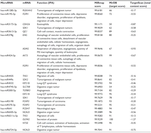
Table 2 Downregulated genes with microrna–mrna interactions and their functions
MicroRNA mRNA Function (IPA) MiRmap
score
MirDB (target score)
TargetScan (total context score)
hsa-mir1285-3p PLEKHH2 Tumorigenesis of malignant tumor 99.6181 66 -0.17
hsa-mir195-5p FGFR1 Proliferation of connective tissue cells, depressive disorder, angiogenesis, proliferation of lipoblasts, migration of cells, major depression
99.8560 72 -0.55
hsa-mir3173-3p CDH26 eosinophilia 99.1171 50 -0.87
SDK1 Tumorigenesis of malignant tumors 99.9039 62 -0.58
hsa-mir3613-3p GJC1 Cell–cell contact, muscle contraction 99.8337 89 -0.63
hsa-mir378g ENG autophagy of vascular endothelial cells, proliferation of connective tissue cells, detachment of vascular endothelial cells, cellular homeostasis, angiogenesis, autophagy of cells, migration of cells, organism death
99.8158 80 -0.25
ADM2 respiration of adipocytes, angiogenesis, quantity of M2 macrophages, quantity of leukocytes
99.9646 67 -0.93
hsa-mir424-5p AKT3 autophagy of vascular endothelial cells, proliferation of connective tissue cells, autophagy of cells, migration of cells, cellular homeostasis
99.0670 99 -0.67
FGFR1 Proliferation of connective tissue cells, depressive disorder, angiogenesis, proliferation of lipoblasts, migration of cells, major depression
99.8506 72 -0.55
hsa-mir4435 TNS1 Migration of cells 99.8208 74 -0.16
hsa-mir449a SDK2 Tumorigenesis of malignant tumor 99.8641 83 -0.41
hsa-mir4532 KIF21B long-QT syndrome 99.5702 94 -0.46
hsa-mir4745-5p SLC7A8 Digestive organ tumor 99.6903 54 -0.25
hsa-mir50015p TGFBR3 angiogenesis 99.7104 82 -0.29
KIF21B long-QT syndrome 99.9772 95 -0.34
SDK1 Tumorigenesis of malignant tumor 99.7629 78 -0.17
hsa-mir5190 FOXP2 Tumorigenesis of carcinoma 99.1872 93 -0.20
hsa-mir570-3p FOXP2 Tumorigenesis of carcinoma 99.5231 95 -0.21
hsa-mir641 PCDH7 neurological signs 99.4883 94 -0.47
hsa-mir6505-5p NCALD Digestive organ tumor 99.2089 96 -0.56
hsa-mir6511a-5p TNS1 Migration of cells 99.9283 91 -0.13
OLFM2 secretion of protein 99.9239 100 -1.27
NT5E Cell–cell contact, activation of leukocytes, activation of T lymphocytes, cellular homeostasis
99.5498 95 -0.35
hsa-mir6724-5p NCALD Digestive organ tumor 99.7041 91 -0.75
Note: Predicted by Ingenuity Pathway analysis (IPa) and meta-analysis with Mirmap, MirDB, and Targetscan.
The PCDH7 gene encodes Pcdh7, which is a member of the cadherin superfamily.34 The PCDH7 gene product is an inte-gral membrane protein involves in cell–cell recognition and adhesion.35 A study of gastric cancer demonstrated that Pcdh7 inhibited cell migration and invasion.35 Therefore, downregu-lation of PCDH7 could promote cell migration. In the nervous system, protocadherins are widely expressed transmem-brane proteins, and overexpression of Pcdh7 would cause intrinsic apoptotic pathways in primary cortical neurons.36 Our IPA analysis also showed that PCDH7 was associated with neurological signs. However, in the pathogenesis of COPD, Pcdh7 might be more related to migration and adhesion of bronchial epithelial cells of the small airway.
To date, there has been neither cure against COPD nor effective biomarkers for diagnosing COPD. In this study,
we identified potential microRNA–mRNA interactions that regulate the homeostasis of DHBE cells. Our results showed that the miR6511a-5p–NT5E interaction plays an important role in COPD and might be associated with cell–cell contact, activation of leukocytes, activation of T lymphocytes, and cellular homeostasis. We also found that miR3173-3p–SDK1, miR4435–TNS1, and miR641–PCDH7 interactions might be associated with COPD pathogenesis in small-airway bron-chial epithelial cells (Figure 7). However, there are several limitations in this study. Firstly, it is not easy to obtain bronchial epithelial cells from either healthy subjects or COPD patients. Potentially dysregulated microRNAs, mRNAs, and their interactions in COPD were generated from only one nor-mal subject and one COPD patient. Although we used GEO to validate the differentially expressed genes and TargetScan
International Journal of Chronic Obstructive Pulmonary Disease downloaded from https://www.dovepress.com/ by 118.70.13.36 on 22-Aug-2020
Dovepress Transcriptomic profiles of COPD airway epithelium
and MirDB to validate the interaction between microRNA and gene expression predicted by MiRmap, these findings need to be validated in more clinical samples. Secondly, the DHBE cells, cultivated in the same normal epithelial culture medium, had already gone out of the actual microenvironment in COPD airways. As this was a screening and bioinformatic study, our findings need further validation.
Conclusion
In conclusion, this study showed that miR6511a-5p–NT5E, miR3173-3p–SDK1, miR4435–TNS1, and miR641–PCDH7 interactions might play important roles in the bronchial epi-thelium of COPD. These findings provide new information
for further investigations of the COPD microenvironment, and may help in development of new diagnostic or therapeutic strategies targeting bronchial epithelium for COPD.
Acknowledgments
The authors gratefully acknowledge the support of research grants from the Ministry of Science and Technology (MOST 106-2314-B-037-016-MY2, MOST 104-2320-B-037-014-MY3), Kaohsiung Medical University Hospital Research Founda-tion (KMUHS10601, KMUH106-6R15), Kaohsiung Medi-cal University Research Foundation (105KMUOR05), and the KMU-KMUH Co-Project of Key Research (KMU-DK 107009 from Kaohsiung Medical University).
100 80 60 40 20 0 Nonsmokers FGFR 1 expression (arbitrary units) Smokers FGFR1 NS 600 400 200 0 Nonsmokers FOXP2 expression (arbitrary units) Smokers FOXP2 NS 30 20 10 0 Nonsmokers GJC1 expression (arbitrary units) Smokers GJC1 NS 80 60 40 20 0 Nonsmokers KIF21B expression (arbitrary units) Smokers KIF21B NS 80 60 40 20 0 Nonsmokers SDK2 expression (arbitrary units) Smokers SDK2 NS 150 100 50 0 Nonsmokers PLEKHH2 expression (arbitrary units) Smokers PLEKHH2 NS 35 30 25 20 15 Nonsmokers OLFM 2 expression (arbitrary units) Smokers OLFM2 NS 800 600 400 200 0 Nonsmokers NCAL D expression (arbitrary units) Smokers NCALD NS 500 400 300 200 100 0 Nonsmokers SLC7A8 expression (arbitrary units) Smokers SLC7A8 NS 600 400 200 0 Nonsmokers TGFBR3 expression (arbitrary units) Smokers TGFBR3 NS 250 200 150 100 50 0 Nonsmokers ADM 2 expression (arbitrary units) Smokers ADM2 NS 60 40 20 0 Nonsmokers AKT 3 expression (arbitrary units) Smokers AKT3 NS 500 400 300 200 100 0 Nonsmokers CDH26 expression (arbitrary units) Smokers CDH26 NS 250 200 150 100 50 0 Nonsmokers EN G expression (arbitrary units) Smokers ENG NS 600 400 200 0 Nonsmokers NT5E expression (arbitrary units) Smokers NT5E *** 250 200 150 100 50 0 Nonsmokers SDK1 expression (arbitrary units) Smokers SDK1 *** 800 600 400 200 0 Nonsmokers TNS1 expression (arbitrary units) Smokers TNS1 ** 200 150 100 50 0 Nonsmokers PCDH7 expression (arbitrary units) Smokers PCDH7 *
Figure 5 gene expression Omnibus (geO) database analysis of 18 downregulated genes with potential microrna–mrna interactions in COPD small airway.
Notes: gene expression of the 18 downregulated genes with potential microrna–mrna interactions was analyzed using gse4498 microarray data from the geO database. The results showed that expression of NT5E, SDK1, TNS1, and PCDH7 was significantly downregulated in patients with COPD compared to normal controls. *P,0.05; **P,0.01; ***P,0.001.
Abbreviation: NS, not significant.
International Journal of Chronic Obstructive Pulmonary Disease downloaded from https://www.dovepress.com/ by 118.70.13.36 on 22-Aug-2020
Dovepress
Chang et al
/DUJHDLUZD\
17(
1RQVPRNHUV
17(
H[SUHVVLRQ
DUELWUDU\XQLWV 6PRNHUV
0DFURSKDJHV
17(
1RQVPRNHUV
17(
H[SUHVVLRQ
DUELWUDU\XQLWV 6PRNHUV
716
1RQVPRNHUV
716
H[SUHVVLRQ
DUELWUDU\XQLWV 6PRNHUV
3&'+
1RQVPRNHUV
3&'+
H[SUHVVLRQ
DUELWUDU\XQLWV 6PRNHUV
16
6'.
1RQVPRNHUV
6'.
H[SUHVVLRQ
DUELWUDU\XQLWV 6PRNHUV
16
Figure 6 gene expression Omnibus database analysis of the four genes downregulated in COPD small airway, large airway, and alveolar macrophages.
Notes: The four downregulated gene with potential microrna–mrna interactions validated in the gse4498 database (COPD small-airway bronchial epithelial cells) were further analyzed in the gse5056 database (large airway) and gse2125 database (alveolar macrophages). NT5E was the only significantly downregulated mRNA in all databases. *P,0.05; **P,0.01; ***P,0.001.
Abbreviation: NS, not significant.
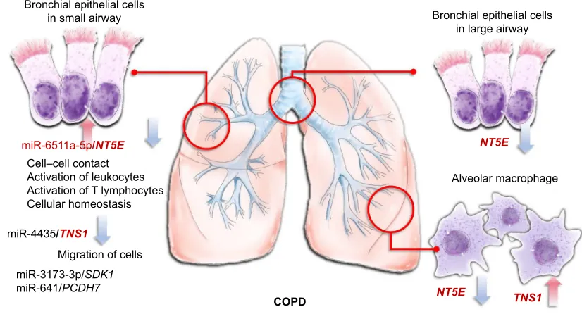
Figure 7 Microrna–mrna interactions in the microenvironment of COPD.
Notes: The mir6511a-5p–NT5E interaction plays an important role in COPD, and may be associated with cell–cell contact, activation of leukocytes, activation of T lymphocytes, and cellular homeostasis. mir3173-3p–SDK1, mir4435–TNS1, and mir641–PCDH7 interactions might also be associated with COPD pathogenesis in small-airway bronchial epithelial cells.
%URQFKLDOHSLWKHOLDOFHOOV LQVPDOODLUZD\
&23'
&HOO±FHOOFRQWDFW $FWLYDWLRQRIOHXNRF\WHV $FWLYDWLRQRI7O\PSKRF\WHV &HOOXODUKRPHRVWDVLV
PL5S6'.
PL53&'+
0LJUDWLRQRIFHOOV
%URQFKLDOHSLWKHOLDOFHOOV LQODUJHDLUZD\
17(
17( 716
PL5DS17(
$OYHRODUPDFURSKDJH
PL5716
Disclosure
The authors report no conflicts of interest in this work.
References
1. GBD 2015 Chronic Respiratory Disease Collaborators. Global, regional, and national deaths, prevalence, disability-adjusted life years, and years lived with disability for chronic obstructive pulmonary disease and asthma, 1990–2015: a systematic analysis for the Global Burden of Disease Study 2015. Lancet Respir Med. 2017;5(9):691–706. 2. Ecenarro PS, Iguiñiz MI, Tejada SP, et al. Management of COPD in
end-of-life care by Spanish pulmonologists. COPD. Epub 2018 Mar 20.
3. Sinden NJ, Stockley RA. Systemic inflammation and comorbidity in COPD: a result of ‘overspill’ of inflammatory mediators from the lungs? Review of the evidence. Thorax. 2010;65(10):930–936.
4. Gao W, Li L, Wang Y, et al. Bronchial epithelial cells: the key effector cells in the pathogenesis of chronic obstructive pulmonary disease? Respirology. 2015;20(5):722–729.
5. Cosio MG, Saetta M, Agusti A. Immunologic aspects of chronic obstructive pulmonary disease. N Engl J Med. 2009;360(23): 2445–2454.
6. Pain M, Bermudez O, Lacoste P, et al. Tissue remodelling in chronic bronchial diseases: from the epithelial to mesenchymal phenotype. Eur Respir Rev. 2014;23(131):118–130.
International Journal of Chronic Obstructive Pulmonary Disease downloaded from https://www.dovepress.com/ by 118.70.13.36 on 22-Aug-2020
Dovepress Transcriptomic profiles of COPD airway epithelium
7. Stolzenburg LR, Harris A. The role of microRNAs in chronic respira-tory disease: recent insights. Biol Chem. 2018;399(3):219–234. 8. Connolly M, Paul R, Farre-Garros R, et al. miR-424-5p reduces
ribosomal RNA and protein synthesis in muscle wasting. J Cachexia Sarcopenia Muscle. 2018;9(2):400–416.
9. Sheu CC, Tsai MJ, Chen FW, et al. Identification of novel genetic regulations associated with airway epithelial homeostasis using next-generation sequencing data and bioinformatics approaches. Oncotarget. 2017;8(47):82674–82688.
10. Bolger AM, Lohse M, Usadel B. Trimmomatic: a flexible trimmer for Illumina sequence data. Bioinformatics. 2014;30(15):2114–2120. 11. Friedländer MR, Mackowiak SD, Li N, Chen W, Rajewsky N.
miRD-eep2 accurately identifies known and hundreds of novel microRNA genes in seven animal clades. Nucleic Acids Res. 2012;40(1):37–52. 12. Trapnell C, Roberts A, Goff L, et al. Differential gene and transcript
expression analysis of RNA-seq experiments with TopHat and Cuf-flinks. Nat Protoc. 2012;7(3):562–578.
13. Vejnar CE, Zdobnov EM. MiRmap: comprehensive prediction of microRNA target repression strength. Nucleic Acids Res. 2012;40(22): 11673–11683.
14. Wong N, Wang X. miRDB: an online resource for microRNA target prediction and functional annotations. Nucleic Acids Res. 2015;43: D146–D152.
15. Wang X. Improving microRNA target prediction by modeling with unambiguously identified microRNA-target pairs from CLIP-ligation studies. Bioinformatics. 2016;32(9):1316–1322.
16. Agarwal V, Bell GW, Nam JW, Bartel DP. Predicting effective microRNA target sites in mammalian mRNAs. Elife. 2015;4:e05005. 17. Huang DW, Sherman BT, Tan Q, et al. The DAVID gene functional
classification tool: a novel biological module-centric algorithm to functionally analyze large gene lists. Genome Biol. 2007;8(9):R183. 18. Clough E, Barrett T. The Gene Expression Omnibus Database. Methods
Mol Biol. 2016;1418:93–110.
19. Tilley AE, Harvey BG, Heguy A, et al. Down-regulation of the Notch pathway in human airway epithelium in association with smoking and chronic obstructive pulmonary disease. Am J Respir Crit Care Med. 2009; 179(6):457–466.
20. Carolan BJ, Heguy A, Harvey BG, Leopold PL, Ferris B, Crystal RG. Up-regulation of expression of the ubiquitin carboxyl-terminal hydro-lase L1 gene in human airway epithelium of cigarette smokers. Cancer Res. 2006;66(22):10729–10740.
21. Woodruff PG, Koth LL, Yang YH, et al. A distinctive alveolar mac-rophage activation state induced by cigarette smoking. Am J Respir Crit Care Med. 2005;172(11):1383–1392.
22. Thomas S, Bonchev D. A survey of current software for network analysis in molecular biology. Hum Genomics. 2010;4(5):353–360.
23. Sohal SS. Epithelial and endothelial cell plasticity in chronic obstructive pulmonary disease (COPD). Respir Investig. 2017;55(2):104–113. 24. Yamasaki K, Eeden SF. Lung macrophage phenotypes and functional
responses: role in the pathogenesis of COPD. Int J Mol Sci. 2018; 19(2):E582.
25. Fausther M, Lavoie EG, Goree JR, Baldini G, Dranoff JA. NT5E mutations that cause human disease are associated with intracellular mistrafficking of NT5E protein. PLoS One. 2014;9(6):e98568. 26. Braganhol E, Tamajusuku AS, Bernardi A, Wink MR, Battastini AM.
Ecto-5′-nucleotidase/CD73 inhibition by quercetin in the human U138MG
glioma cell line. Biochim Biophys Acta. 2007;1770(9):1352–1359. 27. Picher M, Burch LH, Hirsh AJ, Spychala J, Boucher RC. Ecto
5′-nucleotidase and nonspecific alkaline phosphatase: two
AMP-hydrolyzing ectoenzymes with distinct roles in human airways. J Biol Chem. 2003;278(15):13468–13479.
28. Wirsdörfer F, de Leve S, Cappuccini F, et al. Extracellular adenosine
production by ecto-5′-nucleotidase (CD73) enhances radiation-induced
lung fibrosis. Cancer Res. 2016;76(10):3045–3056.
29. Mäki-Nevala S, Sarhadi VK, Knuuttila A, et al. Driver gene and novel mutations in asbestos-exposed lung adenocarcinoma and malignant meso-thelioma detected by exome sequencing. Lung. 2016;194(1):125–135. 30. Mattison J, Kool J, Uren AG, et al. Novel candidate cancer genes
identified by a large-scale cross-species comparative oncogenomics approach. Cancer Res. 2010;70(3):883–895.
31. Brunner M, Millon-Frémillon A, Chevalier G, et al. Osteoblast
miner-alization requires β1 integrin/ICAP-1-dependent fibronectin deposition.
J Cell Biol. 2011;194(2):307–322.
32. Repapi E, Sayers I, Wain LV, et al. Genome-wide association study identifies five loci associated with lung function. Nat Genet. 2010;42(1): 36–44.
33. Shih YP, Sun P, Wang A, et al. Tensin1 positively regulates RhoA activity through its interaction with DLC1. Biochim Biophys Acta. 1853; 2015(12):3258–3265.
34. Yoshida K, Yoshitomo-Nakagawa K, Seki N, Sasaki M, Sugano S. Cloning, expression analysis, and chromosomal localization of BH-protocadherin (PCDH7), a novel member of the cadherin superfamily. Genomics. 1998;49(3):458–461.
35. Chen HF, Ma RR, He JY, et al. Protocadherin 7 inhibits cell migra-tion and invasion through E-cadherin in gastric cancer. Tumour Biol. 2017;39(4):1010428317697551.
36. Xiao H, Sun Z, Wan J, Hou S, Xiong Y. Overexpression of protocad-herin 7 inhibits neuronal survival by downregulating BIRC5 in vitro. Exp Cell Res. 2018;366(1):71–80.
International Journal of Chronic Obstructive Pulmonary Disease downloaded from https://www.dovepress.com/ by 118.70.13.36 on 22-Aug-2020
International Journal of COPD
Publish your work in this journal
Submit your manuscript here: http://www.dovepress.com/international-journal-of-chronic-obstructive-pulmonary-disease-journal The International Journal of COPD is an international, peer-reviewed
journal of therapeutics and pharmacology focusing on concise rapid reporting of clinical studies and reviews in COPD. Special focus is given to the pathophysiological processes underlying the disease, intervention programs, patient focused education, and self management protocols.
This journal is indexed on PubMed Central, MedLine and CAS. The manuscript management system is completely online and includes a very quick and fair peer-review system, which is all easy to use. Visit http://www.dovepress.com/testimonials.php to read real quotes from published authors.
Dovepress
Dove
press
Chang et al
International Journal of Chronic Obstructive Pulmonary Disease downloaded from https://www.dovepress.com/ by 118.70.13.36 on 22-Aug-2020