DOI: 10.1534/genetics.107.077081
Evolution of Gene Sequence in Response to Chromosomal Location
Carlos Dı´az-Castillo
1and Kent G. Golic
Department of Biology, University of Utah, Salt Lake City, Utah 84112
Manuscript received April 10, 2007 Accepted for publication June 6, 2007
ABSTRACT
Evolutionary forces acting on the repetitive DNA of heterochromatin are not constrained by the same considerations that apply to protein-coding genes. Consequently, such sequences are subject to rapid evolutionary change. By examining the Troponin C gene family of Drosophila melanogaster, which has euchromatic and heterochromatic members, we find that protein-coding genes also evolve in response to their chromosomal location. The heterochromatic members of the family show a reduced CG content and increased variation in DNA sequence. We show that the CG reduction applies broadly to the protein-coding sequences of genes located at the heterochromatin:euchromatin interface, with a very strong correlation between CG content and the distance from centric heterochromatin. We also observe a similar trend in the transition from telomeric heterochromatin to euchromatin. We propose that the methylation of DNA is one of the forces driving this sequence evolution.
D
ETAILED examination of the heterochromatic regions around eukaryotic centromeres has distin-guished two subregions with differences in the structure of their chromatin and in their sequence composition (Heitz1934; Gattiand Pimpinelli1992). The central region, referred to as a-heterochromatin, hosts the centromere and is the most compacted chromosome region. In the polytene chromosomes of Drosophila, it shows the lowest degree of replication and consists mainly of highly repetitive elements. The region referred to asb-heterochromatin is generally thought to be lo-cated between thea-heterochromatin and the euchro-matin of each chromosome arm. Its intermediate location also reflects the intermediate nature of its molecular and cytological characteristics. b-Heterochromatin is moderately compacted, moderately replicated in poly-tene chromosomes, and is formed by moderately re-petitive elements interspersed with genes at a lower density than in euchromatic locations (Ashburneret al. 2004).Theb-heterochromatic genes present some unusual structural and regulatory characteristics. They span larger regions than is typical for euchromatic genes, mainly due to the possession of extremely large introns containing many insertions of transposable elements (Devlinet al.1990; Biggset al.1994; Tulinet al.2002; Dimitri et al. 2003). Although there is nothing obvi-ously distinctive about the proteins that these genes encode, their regulation is in many ways contrary to that of euchromatic genes. Their expression is reduced
when they are relocated away from centric heterochro-matin (Khvostova 1939; Hessler 1958; Wakimoto and Hearn1990; Eberlet al.1993), and suppressors or enhancers of euchromatic gene variegation often have the opposite effect on the variegation of heterochro-matic genes (Schultz 1936; Baker and Rein 1962; Wakimotoand Hearn1990; Hearnet al.1991; Luet al. 2000; Weilerand Wakimoto2002). Thus, both gene structure and expression appear to be influenced by a heterochromatic location.
Gene families are especially valuable for the study of molecular evolution since they afford the possibility of making several kinds of comparisons. One type of com-parative analysis uniquely available with multi-gene families is the comparison of paralogs, those family members found within a single genome. Ideally, these analyses would make use of DNA sequence variation, both in coding and noncoding elements, gene exon structures and gene expression patterns, and known mutant phenotypes. These paralogous comparisons will help to detect conservation or divergence of function and/or structure and to deduce roles for each family member. Ultimately, this should allow us to interpret how such DNA and protein sequence changes are related to functional specializations, chromosome loca-tions, or any other specific characteristic of the studied paralogs.
To specifically explore whether genes located near heterochromatin experience unique selective forces because of their location, we chose to examine the
Troponin C(TNC) family ofDrosophila melanogaster, which has members in euchromatin and inb-heterochromatin (Figure 1). TNC is the component of the sarcomeric thin filament that senses increases in cytosolic calcium 1Corresponding author:Department of Biology, University of Utah, 257
South 1400 East, Salt Lake City, UT 84112. E-mail: [email protected]
and mediates myofibril contraction (Filatov et al. 1999). Three of the fiveTNCgenes of D. melanogaster,
TNC25D,TNC47D, andTNC73F, are located in euchro-matin and are expressed throughout development (Herranz et al. 2004). The genes that complete the family, TNC41C and TNC41F6, are located in the b-heterochromatin of2Rand are expressed almost ex-clusively in late pupae and adults (Herranzet al.2004). These b-heterochromatic members span larger chro-mosome regions than their euchromatic relatives due to the possession of larger introns (Figure 1). Since this is a common characteristic shared with otherb -heterochro-matic genes, it is probable that these genes have been present in ab-heterochromatin environment for some time and their characteristics may reveal the influence of such a chromosomal position.
It is true that it is somewhat unusual to use compar-ative analyses of paralogous genes because their origin and time of divergence may be very different, influenc-ing their consideration as totally independent genes in the associated statistical analyses. We think this is not a big obstacle in this case. Herranzet al.(2005) estimated that the last duplication event affecting theTNCgene family in the D. melanogasterevolutionary line has oc-curred .60 MYA. It is known that much younger genes have accumulated a considerable amount of divergence (Zhang et al. 2004), suggesting that the time passed since the lastTNCduplication might have been more than enough for those genes to have accu-mulated changes independently. In fact,TNC47Dand
TNC73F, the two genes resulting from the last TNC
duplication event that occurred in theD. melanogaster
evolutionary line, diverged distinctively since the dupli-cation, both with respect to their expression patterns
and at the level of intron structure (Herranz et al. 2005).
The sequence characteristics of theTNCgenes led us to look for similar tendencies in genes ofD. melanogaster
at all heterochromatin:euchromatin boundaries and within the predominantly or entirely heterochromatic chromosomes4andY. Our findings are reported here.
MATERIALS AND METHODS
Sequences: The nucleotide sequences of the mem-bers of the TNC family of D. melanogaster were retrieved from FlyBase (FlyBase 1999): TNC25D (Fbgn0031692), TNC41C(FBgn0013348),TNC41F6(FBgn0033027),TNC47D
(FBgn0010423), and TNC73F (FBgn0010424). The data of genes located in the heterochromatin–euchromatin transi-tion regions were retrieved from FlyBase (FlyBase 1999) (Figures 4 and 5, and supplemental Data 2 at http://www. genetics.org/supplemental/).
To study the base composition of comparable tracts of the coding units located in theYchromosome and their putative autosomal orthologs, we retrieved their nucleotide protein-coding sequences from public databases and used the anneal-ing tools of Gene Jockey II Sequence Processor (Biosoft) to better define the regions that show the highest identity of sequence. The sequences of genes located in theY chromo-some were retrieved from the nucleotide databases of the National Center for Biotechnology Information (NCBI, http:// www.ncbi.nlm.nih.gov):kl-2(AF313479),kl-3(AF313480),kl-5
(AF136243), ORY (AF427496), Pp1-Y1 (AF427493), Pp1-Y2
(AF427494), and Ppr-Y(AF427495). The sequence of their putative autosomal paralogs were retrieved from FlyBase (FlyBase1999):CG9068(FBtr0087138),CG9492(FBtr0082100), Dhc 93AB (FBtr0084046), CG6059 (FBtr0085130), PpN58A
(FBtr0071734),Pp1-87B(FBtr0082595),CG13125-PA (FBtr0079898), andCG13125-PB (FBtr0079899).
Variation measures: The sequences of theTNC genes of
Sequence Processor (Biosoft) and curated manually to mini-mize gaps and maximini-mize the sequence identity (supplemental Data 1 at http://www.genetics.org/supplemental/). Since we were interested in the detection of evolutionary trends de-pendent on the relative location of theTNCgenes, we based our measures on codon changes. Codon changes can reflect evolutionary trends at the protein level and at the genomic level. In these comparisons, codon changes were divided into synonymous (S; codon changes that resulted in no change in the amino acid encoded) and nonsynonymous (N; codon changes that did produce a difference in the translated amino acid sequence) and were reported as the fraction of total codons. Changes that resulted in the deletion or insertion of codons were considered nonsynonymous. Multiple substi-tutions at a codon position were considered synonymous or nonsynonymous depending on the encoded amino acid re-gardless of the chain of substitutions that led to the current sequence.
Base composition and codon analyses:The base composi-tion data of all protein-coding or noncoding nucleotide sequences were obtained using Gene Jockey II Sequence Processor (Biosoft) and ApE (http://www.biology.utah.edu/ jorgensen/wayned/ape/).
The effective number of codons (Nc) (Wright1990) of the TNC genes ofD. melanogaster studied were calculated using CodonW 1.4.2 (http://bioweb.pasteur.fr/seqanal/interfaces/ codonw.html). TheNcvalue is a measure of overall codon bias and ranges between 20 (when only 1 codon is used for each amino acid) and 61 (when codons are used randomly). GeneQuest 5.01 (DNASTAR) was used to obtain the codon
frequency values used to calculate the data represented in Table 3 and Figure 3.
Statistical analyses:The statistical significance of the differ-ences observed between the codon variation measures of two independent sets of TNCgenes ofD. melanogaster(Table 1) were analyzed using the nonparametric Mann-Whitney test provided by GraphPad InStat for Macintosh (GraphPad Software).
The statistical significance of the correlation of the changes in the codon frequencies partitioned according to whether they had been caused by transitions or transversions com-patible with a decrease of the CG content in the third codon position of heterochromaticTNCgenes (Table 3 and Figure 3) was analyzed using the nonparametric Spearman test provided by GraphPad InStat for Macintosh (GraphPad Software).
The statistical significance of the correlation between genes’ protein-coding CG composition and distance from centro-mere or telocentro-mere (Table 5, Figures 4 and 5, and supplemental Data 2 at http://www.genetics.org/supplemental/) was ana-lyzed using the nonparametric Spearman test provided by GraphPad InStat for Macintosh (GraphPad Software).
Other software used: Power Macintosh MegAlign 5.01 (DNASTAR) was used to draw a phylogenetic tree of the
TNCgenes ofD. melanogasterbased in the alignments of the nucleotide protein-coding sequences used in the codon variation analyses (Figure 1 and supplemental Data 1 at http://www.genetics.org/supplemental/). Microsoft Excel X for Mac was used to manipulate and represent data. Microsoft PowerPoint X for Mac, Microsoft Word X for Mac, and Adobe TABLE 1
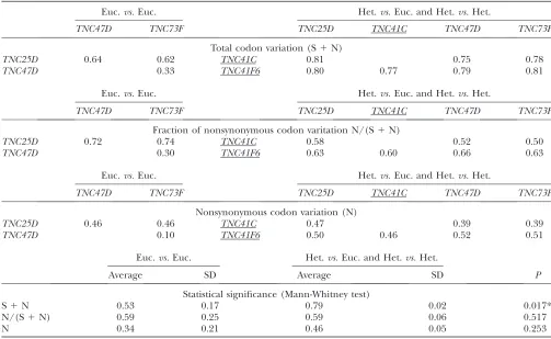
Variation measures obtained from the comparison of the nucleotide protein-coding sequences of theTNCgenes ofD. melanogaster
Euc.vs.Euc. Het.vs.Euc. and Het.vs.Het.
TNC47D TNC73F TNC25D TNC41C TNC47D TNC73F
Total codon variation (S1N)
TNC25D 0.64 0.62 TNC41C 0.81 0.75 0.78
TNC47D 0.33 TNC41F6 0.80 0.77 0.79 0.81
Euc.vs.Euc. Het.vs.Euc. and Het.vs.Het.
TNC47D TNC73F TNC25D TNC41C TNC47D TNC73F
Fraction of nonsynonymous codon varitation N/(S1N)
TNC25D 0.72 0.74 TNC41C 0.58 0.52 0.50
TNC47D 0.30 TNC41F6 0.63 0.60 0.66 0.63
Euc.vs.Euc. Het.vs.Euc. and Het.vs.Het.
TNC47D TNC73F TNC25D TNC41C TNC47D TNC73F
Nonsynonymous codon variation (N)
TNC25D 0.46 0.46 TNC41C 0.47 0.39 0.39
TNC47D 0.10 TNC41F6 0.50 0.46 0.52 0.51
Euc.vs.Euc. Het.vs.Euc. and Het.vs.Het.
Average SD Average SD P
Statistical significance (Mann-Whitney test)
S1N 0.53 0.17 0.79 0.02 0.017*
N/(S1N) 0.59 0.25 0.59 0.06 0.517
N 0.34 0.21 0.46 0.05 0.253
Illustrator 10 were used in the preparation of the manuscript, tables, and figures.
RESULTS
Comparison of euchromatic and heterochromatic
TNCgenes:The examination of the genomes of related species has revealed that heterochromatic DNA can change rapidly during evolution (Powell1997). More specifically, the centromeric DNA of eukaryotic spe-cies shows rapid evolution (Henikoff et al.2001). Al-though this high rate of change in heterochromatin pertains to the noncoding sequences that are located in a-heterochromatin, we wondered whether a similar trend might be visible in the genes located in the adjacent b-heterochromatin. A comparative study of a paralo-gous group of genes with euchromatic and heterochro-matic locations, like those that encode for TNC in
D. melanogaster, might be a good way to answer this question (Figure 1).
If the heterochromaticTNCgenes are under a rapid evolution trend, similar to the one reported for the centromere repeats or heterochromatic DNA in gen-eral, we should be able to detect it at the nucleotide level and possibly even at the protein level. An examination of the codons of these genes allows detection of both nucleotide and the protein variation. We compared the protein-coding sequences of the TNC genes two at a time, quantified the divergent codons in each of those comparisons, and presented the results as a fraction of the total number of codons compared.
Synonymous changes (S) are those in which the di-vergence detected between two codons does not result in an amino acid change; nonsynonymous changes (N) are those that do produce an amino acid change. Changes that resulted in the partial or total deletion or insertion of codons were considered nonsynonymous. The addi-tion of synonymous and nonsynonymous changes (S1N) will inform us of the degree of variation accumulated at the nucleotide level, while nonsynonymous changes either in absolute (N) or relative½N/(S1N)terms will tell us more specifically about the variability that has occurred at the protein level.
A special problem when dealing with the analysis of variability at the codon level is posed by multiple substi-tutions in a single codon. In such cases, there is no easy way of knowing if those were caused by a succession of only one kind of substitution or by the alternation of synonymous and nonsynonymous substitutions. Some approaches have been devised to deal with this in-convenience (for general reference, use Graurand Li 2000). These approaches makea priori considerations about the probability of the different base substitutions: either the probability of the individual base changes is the same in all cases or they aren’t and we need to introduce corrections to somehow reflect the biases (for instance, usually synonymous substitutions are more
fre-quent than nonsynonymous). At this point in our study, we chose not to make assumptions about what type of sequence substitutions might occur in heterochromatic environments, because the eventual detection of biases at this level is the final aim of our work. Therefore, we limited our consideration to synonymous or nonsynon-ymous changes, regardless of the chain of events that drove to these changes. This approach ensures that we are not introducing biased assumptions at the outset.
Once we obtained the values for synonymous and nonsynonymous variation in the TNC genes, we parti-tioned them into two groups. One group is formed by the parameters obtained from comparisons in which at least one of the genes is heterochromatic. These data will in-form us of the degree of variation heterochromaticTNC
genes accumulate on average. The other group is formed by the parameters obtained from comparisons of two euchromatic genes. This second group will act as a base-line to allow us to determine if the heterochromaticTNC
genes differ significantly in their accumulated changes. We found that the heterochromaticTNCgenes have accumulated higher codon divergence (S1N) than the euchromatic genes (Table 1;P¼0.0167). However, this has not led to an increased variation in the proteins that the heterochromatic genes encode. When comparing the average divergence of euchromatic genes with the average divergence of heterochromatic genes, the frac-tion of codon changes that produce changes in the amino acid sequence½N/(S1N)does not differ signif-icantly (Table 1;P¼0.5167). Even when considered as the absolute number of amino acid changes (N), the heterochromatic genes do not show a significantly in-creased degree of variation (Table 1;P¼0.2526).
Thus, heterochromaticTNCgenes seem to accumu-late elevated nucleotide variation, although this varia-tion is not translated into an elevated variavaria-tion of the proteins they encode. This result suggests that the proteins encoded by the heterochromatic genes are subject to the same functional constraints as the euchro-matic genes, consistent with their role as the primary sources of TNC in the adult muscles (Herranz et al. 2004). The elevated nucleotide variation is reminiscent of the rapid evolution detected for noncoding elements of heterochromatic centromeric elements (Henikoff et al.2001).
Specific biases detected in heterochromatic TNC
genes:To try and gain some understanding of the pos-sible causes of the elevated nucleotide variation of the heterochromatic genes, we analyzed the sequence with respect to other parameters, such as base composition and codon bias. The initial analysis of theD. melanogaster
like those found in the adjacenta-heterochromatin, or whether the protein-coding segments also had a re-duced CG content. To test whether a general reduction in CG content might account for the increased variation
of the heterochromaticTNCgenes, we determined the base composition of the fiveTNCgenes and found that the b-heterochromatic genes are CG-depleted relative to their euchromatic counterparts (Table 2). This re-duction affects both protein-coding and non-protein-coding sequences (Figure 2). This finding that the b-heterochromatic TNC genes exhibit a reduced CG content in their coding regions clearly shows that this is not solely due to the accumulation of repetitive sequence elements in the noncoding parts of the gene and further suggests that it is not merely a regulatory adaptation specific for the region.
Finally, we also detected a difference between the heterochromatic and the euchromaticTNCgenes ofD. melanogasterforNc(Wright1990).Ncvalues are higher in the case of heterochromatic TNC genes (Table 2), which is an indication that those genes have lower codon bias than their euchromatic relatives.
TABLE 2
Base composition, CG content, andNcvalues of the
nucleotide protein-coding sequences of theTNC genes ofD. melanogaster
TNC25D TNC41C TNC41F6 TNC47D TNC73F
A% 27 30 32 25 24
C% 26 17 17 26 25
G% 29 25 25 29 31
T% 19 27 26 20 19
CG% 55 42 42 55 56
Nc 42.52 49.03 61.00 35.88 31.46
Heterochromatic genes are underlined.
In summary, the heterochromatic TNC genes of D. melanogasterhave elevated nucleotide variation relative to the euchromatic paralogs; even though the encoded proteins do not vary significantly from those encoded by the euchromatic genes, they exhibit a depletion of the CG content of coding and noncoding elements and they have reduced codon biases.
Biased mutation as a mechanism for heterochro-matic CG depletion:The fact that both protein-coding and noncoding sequences of the heterochromaticTNC
genes ofD. melanogasterexhibit CG depletion without a significant alteration in the proteins they encode sug-gests that this cannot be explained by a selection bias. Instead, we considered whether some force acting di-rectly at the level of DNA could be responsible for the reduced CG content of the heterochromatic genes. In other words, we wondered whether a biased mutational process could account for the differences between the heterochromatic and euchromatic genes. For example, as we will later discuss, it has been proposed that the occurrence of meiotic recombination may exert a muta-tional bias toward CG in the euchromatic regions where recombination occurs (Marais 2003). We wondered whether there might be evidence for a biased mutational process in heterochromatic regions. By comparing the different extents of common trends shown by the het-erochromaticTNCgenes ofD. melanogaster, we might be able to identify traces of such a mechanism. Therefore, we undertook an analysis of some of the sequence param-eters obtained from theTNCgenes ofD. melanogaster.
We studied the base composition of the three codon positions by analyzing the nucleotide protein-coding sequences of theTNCgenes ofD. melanogaster. The aver-age CG percentaver-ages found in the heterochromaticTNC
genes of D. melanogaster are: 1st ¼ 58.8, 2nd ¼ 30.6, and 3rd ¼ 37.8. For the euchromatic TNC genes of
D. melanogaster, the values are: 1st¼60.5, 2nd¼27, and 3rd ¼ 78.2. The differences in CG content for the heterochromatic genes at each position are: 1st¼ 1.7, 2nd¼3.6, and 3rd¼ 40.4, indicating that the drop in CG content detected in the heterochromaticTNCgenes of D. melanogaster is based almost entirely on CG de-pletion of the third codon position, whereas the com-position of the first and second com-positions shows very little change. This is not a surprise considering the degeneracy of the genetic code and is consistent with our finding that the proteins encoded by these genes have not significantly diverged.
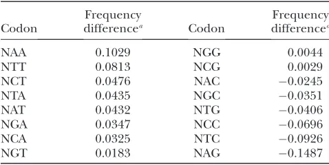
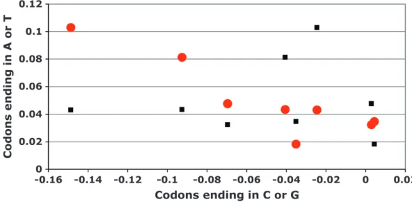
Next, we examined the frequencies of codons grouped according to the composition of the second and the third positions. The practical immutability of the bases in the second codon position allowed us to study possible trends in the changes that occurred at the third codon position, both at the single nucleotide and the di-nucleotide level. If a mutagenic process were to work mainly to reduce C and G bases in the third codon positions, we expect the frequency of codons with C or G
in the third position in heterochromatic genes to de-crease, while the frequency of codons with T or A in the third position should increase. Based on the results we presented in the Table 3, we can say that this is generally correct for the heterochromatic TNC genes of D. melanogaster. With the exception of NGG and NCG that show a slight increase of frequency in heterochromatic genes, the frequencies of codons ending in T or A are increased, whereas the frequencies of the other codons ending in C or G are decreased in heterochromatic genes. Mutations that reduce CG content can be of two kinds: transitional (C to T and G to A) or transversional (C to A and G to T). We wondered to what degree the third position CG depletion could be attributed to each of these two types of mutation. To determine that, we partitioned the changes of the codon frequencies found in Table 3 according to whether they had been caused by transitions or transversions in the third position that re-sulted in a decrease of the CG content. For instance, the decrease of CG content of a protein-coding sequence based in transitions occurred in the third base of NAC codons will result in NAT, whereas the transversion will result in NAA. This arrangement of the data is plotted in Figure 3, showing that inD. melanogaster, there is a sta-tistically significant negative correlation when arranged according to transitions (r¼ 0.8333;P¼0.0154) but not according to transversions (r¼ 0.2381;P¼0.9768). This means that the codons of a pair were enriched or depleted in comparable magnitudes only when the third position changes were transitions. Thus, the reduc-tion in CG content of the heterochromaticTNCgenes of
D. melanogastermight have been caused by the action of a mutagenic mechanism that mainly generates transitions. If the main mechanism responsible for CG depletion of the heterochromaticTNCgenes ofD. melanogasteris indeed based in the production of transitions in the third codon position, we should most easily detect this
TABLE 3
Changes of the frequencies of codons group according to the composition of their second and third bases in the
heterochromaticTNCgenes ofD. melanogaster
Codon
Frequency
differencea Codon
Frequency differencea
NAA 0.1029 NGG 0.0044
NTT 0.0813 NCG 0.0029
NCT 0.0476 NAC 0.0245
NTA 0.0435 NGC 0.0351
NAT 0.0432 NTG 0.0406
NGA 0.0347 NCC 0.0696
NCA 0.0325 NTC 0.0926
NGT 0.0183 NAG 0.1487
a
trend when analyzing the codons that specify amino acids based on only on the first two positions: serine (UCN), leucine (CUN), proline (CCN), arginine (CGN), threonine (ACN), valine (GUN), alanine (GCN), and glycine (GGN). This time, we compared the frequency of changes that occurred in the third base of this subset of codons in two groups of protein-coding sequence alignments of theTNCgenes ofD. melanogaster, which already permitted us to obtain the codon variation pa-rameters (supplemental Data 1 at http://www.genetics. org/supplemental/). The first set of alignments was formed by comparing one heterochromaticTNCgene and one euchromatic TNC gene, whereas the second set of alignments was formed by comparing two genes that were euchromatic. When we subtract the average frequencies of third base codon changes obtained in the euchromaticTNCgenevs.euchromaticTNCgene com-parisons from the average values obtained in the het-erochromatic TNC gene vs. euchromatic TNC gene comparisons, we will have an indication of the type of changes that are preferred in the heterochromaticTNC
genes. As expected, in Table 4, we can see that changes that decrease CG content are elevated in the hetero-chromaticTNCgenes, whereas those that would increase CG content in the third codon position are reduced. In the case of changes that result in loss of C alone, the transition (NNC to NNT) is more frequent than the transversion (NNC to NNA). However, the opposite is true for changes that result in the depletion of G; transversions (NNG to NNT) are more common than transitions (NNG to NNA).
DNA methylation as a mechanism for CG reduction:
The methylation of cytosine increases its tendency to spontaneously deaminate (Shenet al.1994). While the unmodified base is deaminated into uracil, methylated cytosines deaminate into thymines. Methylated sequen-ces of very different organisms are known to be hotspots for sequence variation and are characteristically en-riched in C to T and G to A transitions (Coulondreet al. 1978; Cooper and Yousouffian 1988; Selker 1990; Joneset al.1991; Greenblattet al.1994; Singeret al.
1995; Yang et al. 1996; Colot Et Rossignol 1999; Watterset al.1999). Moreover, the methylation of DNA has been proposed to contribute to regional differences in base composition found in other genomes (Fryxell and Zuckerkandl2000; Eyre-Walkerand Hurst2001). If DNA methylation was responsible for the trends we found in the heterochromatic TNC genes of D. mela-nogaster, we should find noticeable traces of its activity in the analyses we just presented. One of those expected traces would be the depletion of CT dinucleotides, since CT is reported to be the preferentially methylated di-nucleotide inD. melanogaster(Lykoet al.2000). The two codons produced from DNA with a CT dinucleotide in the last position are either NCT or NAG (with CT on the complementary strand). Of these, NAG is more amena-ble to change, since deamination, and a C to T tran-sition on the opposite strand, will produce no change in the encoded amino acid (NAG to NAA). As shown in Table 3, NAG codons show the greatest decrease and NAA the greatest increase in the heterochromaticTNC
genes ofD. melanogaster.
TABLE 4
Changes of the frequencies of third base mutations in codons that specify amino acids based in their two first bases in
the heterochromaticTNCgenes ofD. melanogaster
Mutation
Frequency
differencea Mutation
Frequency differencea
NNC to NNT 0.1482 NNA to NNG 0.0105 NNC to NNA 0.0837 NNT to NNC 0.0523 NNT to NNA 0.0744 NNC to NNG 0.0649 NNG to NNT 0.0730 NNA to NNC 0.0780 NNG to NNA 0.0389 NNT to NNG 0.0853 NNA to NNT 0.0195 NNG to NNC 0.1469
a
Value calculated by subtracting the average frequency of each mutation obtained from the alignments of the nucleo-tide protein-coding sequences of two euchromaticTNCgenes from the average frequency of each mutation obtained from the alignments of the nucleotide protein-coding sequences of one heterochromatic and one euchromaticTNCgene.
Our results also show that the frequency of codons ending with CT increases, a finding that does not follow directly from the hypothesis that methylation of C in CT dinucleotides is responsible for CG depletion of het-erochromatic genes. However, the decrease of CG con-tent in the heterochromaticTNCgenes ofD. melanogaster
is almost entirely the result of changes in the third codon base, whereas the second position barely changes. As discussed previously, this is likely a result of selection for conservation of protein function. Therefore, it might only be possible to increase the frequency of codons ending with CT as a result of transitions that occur in the third position of codons ending with CC. Thus, the se-quence differences between heterochromatic and euchro-maticTNCgenes are compatible with changes provoked by cytosine methylation.
If a DNA methylation-based mechanism is responsi-ble for the CG depletion at the third codon position, we should also find a higher frequency of CG-reducing transitions in those codons that specify amino acids based on the first two positions. Consistent with this expectation, NNC to NNT substitutions are by far the most frequent changes in the heterochromatic TNC
genes ofD. melanogaster(Table 4). Quite surprising is the result that NNG to NNA changes, though increased, do not show a higher frequency in the same set of genes. Furthermore, contrary to our expectations, NNG to NNT transversions seem to be clearly preferred to NNG to NNA transitions in the heterochromaticTNCgenes of D. melanogaster. Though the elevated frequency of NNC to NNT substitutions speaks in favor of the activity of a DNA methylation-based mechanism, the somehow lower-than-expected frequency of NNG to NNA tran-sitions might indicate that this is not the only mecha-nism responsible for the CG depletion found in the heterochromaticTNCgenes ofD. melanogaster.
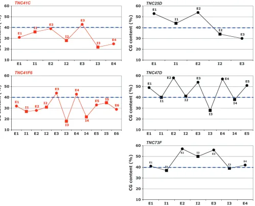
CG content of protein-coding segments in hetero-chromatic-euchromatic transition regions:The data pre-sented above support the possible existence of a DNA methylation-based mechanism that, at least in part, has caused the accumulation of AT-biased sequence varia-tion in the heterochromatic TNC genes of D. mela-nogaster. If the CG depletion is caused by methylation of heterochromatic DNA, then it seems probable that the AT composition bias should not be restricted to the heterochromaticTNCgenes but should be seen with all heterochromatic genes. To determine whether the re-duced CG content ofTNC41CandTNC41F6reflects a regional influence, we examined the protein-coding regions of genes found in the first 3 Mbp of DNA sequence at the base of2Rin the vicinity of both genes. The available sequence at the base of each chromosome arm begins where the repetitive content is reduced sufficiently to allow the assembly of shotgun-sequenced fragments—essentially, in b-heterochromatin. We ex-tended our analyses far enough from the centromere to be sure that some of the genes studied were clearly
located in euchromatin. The result, presented in Figure 4, shows that the CG depletion of gene protein-coding sequences inb-heterochromatin of2Ris characteristic of that region as a whole, and the strength of the effect depends on the proximity toa-heterochromatin.
Because the reduction in CG content was not specific to the TNC genes, we became interested in whether CG reduction was a common characteristic of the protein-coding sequences of genes in the vicinity of heterochromatin. We surveyed genes located in all heterochromatin–euchromatin transition regions of the genome. Our survey encompassed genes found near centromeric and telomeric heterochromatin. Most of these regions on the major chromosomes (X, 2, 3) appear to show trends of a general increase in CG con-tent of protein-coding segments as the genes become more distant from the nearest heterochromatic region (Figures 4 and 5). Around centromeres, the CG content is higher for genes that are more distant from the centromere (Figure 4). At the chromosome tips, CG content is higher for genes with a more internal lo-cation, placing them further from the telomere (Figure 5). Those general trends are manifest to differing extents in the surveyed regions, and in 8 of the 10 surveyed regions onX,2, and3, the trend was statistically significant (Table 5).
The strength of the correlations between CG content and distance to the centromere or telomere are not homogeneous, as thervalues compiled in Table 5 show. Some of theser values are rather small, meaning that many genes in these regions do not strictly conform to the CG trend. This suggests the rather unsurprising conclusion that other forces may also act to influence the DNA sequence of genes in these regions.
Within the genome of D. melanogaster, the chromo-somes with the highest proportion of heterochromatin by far are theYand4(Gattiand Pimpinelli1992). If the reduction in CG content is truly a result of a het-erochromatic environment, we should notice it very clearly in these two chromosomes. The tiny chromo-some4is known to have characteristics of heterochro-matin throughout its length (Sunet al.2000; Sunet al. 2004). Accordingly, we find that the CG contents of the coding regions of almost all the genes along this chromosome are very low (Figure 4). Despite that, no statistical significance was found when studying the correlation of the CG contents and the distance to the centromere of the chromosome4(Table 5). Wanget al. (2002), who surveyed nucleotide variation in a world-wide sampling of chromosomes 4 of D. melanogaster, identified a region, limited by the genesCG11091and
toy, with levels of variation typical of other putatively euchromatic autosomal regions. Visual inspection of the data presented in Figure 4 does seem to indicate a region of somewhat higher CG content coinciding with the
euchromatin-like region, the relevance of the data of the chromosome4is even higher. Replicating the analyses of the other heterochromatin–euchromatin transition regions, we studied the statistical significance of the correlation of the CG contents of the protein-coding sequence of the genes located in chromosome 4 and their distance to its centromere going as far as to include genes presumably located in euchromatin, those within the region CG1109-toy. We considered the region lo-cated between the centromere and the genetoyas the centromeric transition region of chromosome4and the region between the geneCG11152and the telomere as the telomeric transition region. With this assumption, we found that the correlation between CG content of protein-coding sequences and their distance from the
centromere or telomere was also statistically significant (Table 5).
the proximity to telomeres or centromeres affects the degree of the CG depletion, since many of these coding units hadn’t been finely mapped. It is true though that the CG content of kl-2, kl-3, and kl-5 progressively in-creases as their distance from the centromere inin-creases (Table 6) (Gattiand Pimpinelli1992).
DISCUSSION
It has been previously noted that a gradient of increasing CG content is apparent in the chromosomal DNA ofD. melanogasteras it passes from centric hetero-chromatin to euhetero-chromatin (Adamset al.2000). By spe-cifically examining the protein-coding regions of genes in these parts of the chromosomes, we found that this
result cannot be solely explained by the accumulation of transposable elements of low CG content or as an adap-tation of noncoding DNA to a heterochromatic location. Our analysis of genes at the boundaries of heterochro-matin and euchroheterochro-matin reveals that the composition of the protein-coding sequences of genes in such locations reflects their position. Genes closer to centric or telo-meric heterochromatin tend to have a lower CG content than genes that are more removed, and this tendency is proportional to their proximity to heterochromatin.
showing a much lower CG content. This example strongly suggests that the DNA sequences of the genes located in heterochromatin, while still constrained by the necessity of encoding functional proteins, are evolving in response to the influence of their chromosomal location.
The evolution of heterochromatin, and of genes in the vicinity of heterochromatin, has interested geneticists since the discoveries that heterochromatin exhibited many unique properties, including housing the sites for chromosome segregation (Anderson1925), having novel modes of gene regulation (Mullerand Painter1932), and possessing very distinct sequence content (Kliman and Hey1993; Heyand Kliman2002; Ashburneret al. 2004). Regional differences of CG content have been detected within the genomes of several organisms (Bernardi1989; Sharpet al.1989; Carulliet al.1993; Sharpand Lloyd1993; Dujonet al.1994; Feldmann
et al.1994; Bernardi1995; Deschavanneand Filipski 1995; Bradnam et al.1999; Eyre-Walker and Hurst 2001; Daubinand Perrie` re 2003; Zhangand Zhang 2004), and a positive correlation between CG content and recombination rates has been identified (Ikemura and Wada1991; Gerton et al.2000; Fullertonet al. 2001; Marais et al. 2001, 2003; Birdsell2002; Kong et al.2002; Maraisand Piganeau2002; Meunierand Duret2004). It has long been known that heterochro-matic regions have greatly reduced levels of recombina-tion (Kliman and Hey 1993; Heyand Kliman 2002; Ashburner et al.2004). The basis for the correlation between reduced recombination and CG content has been the subject of much interest.
One class of model supposes that the reduced CG content of heterochromatic regions is an indirect con-sequence of the reduction in recombination in these TABLE 5
Statistical analyses of the CG content patterns in the main heterochromatin–euchromatin transition regions and the chromosome4of the genome ofD. melanogasterrepresented in Figure 4
Chromosome compartment
No. of protein-coding genes studied
Total no. of protein-coding genes
Coverage (%)
Spearman test (95% confidence
interval)
r P
Xc 212 258 82 0.4458 ,0.0001*
Xt 79 94 84 0.3617 0.0011*
2Lc 285 331 86 0.2162 0.0002*
2Lt 105 127 83 0.4437 ,0.0001*
2Rc 156 192 81 0.5766 ,0.0001*
2Rt 115 139 83 0.3728 ,0.0001*
3Lc 144 167 86 0.3775 ,0.0001*
3Lt 80 106 75 0.3704 0.0007*
3Rc 297 374 79 0.0966 0.0966
3Rt 75 92 82 0.1597 0.1710
4 51 90 57 0.1511 0.2900
4c 39 70 56 0.5179 0.0007*
4t 19 35 54 0.4771 0.0389*
* Statistically significant; c, centromere; t, telomere.
TABLE 6
CG content of comparable regions of some of the coding units found in the chromosomeYofD. melanogaster and their putative orthologs located out of this chromosome
Yparalogs Autosomal paralogs
Genes
Sequence fragment
(bp) CG % Genes
Sequences fragment
(bp) CG (%)
kl-2 2-2695 35 CG9068 973-3675 47
kl-3 1-8370 38 CG9492 4795-13242 54
kl-5 1-1737 44 Dhc 93AB 4893-6639 54
ORY 1-1989 34 CG6059 662-2650 54
Pp1-Y1 102-840 45 PpN58A 245-982 52
Pp1-Y2 1-897 43 Pp1-87B 230-1126 57
Ppr-Y 73-1686 34 CG13125-PA 97-1686 50
parts of the chromosomes. When a natural DNA se-quence variant arises that substitutes C for Tor G for A in the third position of a codon, any selective advantage provided by that variant is likely to be extremely small because the majority of such changes encode the same or similar amino acids. Since most mutations are ex-pected to be deleterious, infrequent mutations providing only a slight selective advantage are likely to be lost be-cause of the stronger selection against a number of linked mutations that are disadvantageous (Charlesworth et al.1993). Alternatively, if a single highly advantageous mutation were to arise, it could be rapidly swept to fixation and carry with it all tightly linked variants, regardless of whether they were beneficial or not (Maynard Smith and Haigh 1974). Both scenarios illustrate the inability of slightly advantageous mutations to increase in frequency unless they can be separated from the effects of neighboring mutations by recombi-nation. Considering that T-to-C or A-to-G changes in the third position of a codon have, in general, very low or no adaptive value at the protein level, the existence of codon preferences is thought to be an adaptation for more efficient translation. In support of this, highly expressed genes of Drosophila tend to contain codons ending in C or G (Shieldset al.1988). However, the very slight advantage conferred by more efficient translation of a single codon can only be selected if it occurs in a region of high recombination. Thus, it is thought, the higher CG content of euchromatin reflects selection for more efficient translation in these regions of high recombination.
An alternative but not exclusive hypothesis asserts that the CG enrichment of euchromatin is a more direct consequence of recombination. DNA double-strand breaks are initiators of meiotic recombination. The mech-anisms that repair the double-strand breaks, generate recombinants, and repair heteroduplex mismatches have preferences that could result in CG enrichment of regions with higher levels of recombination (Brown and Jiricny1988; Holmeset al.1990; Varletet al.1990, 1996; Bill et al. 1998; Smith and Nicolas 1998; Nickoloffet al.1999; Petranovicet al.2000; Birdsell 2002).
The distribution of recombination might then have a dual influence over DNA base composition: the lack of recombination in heterochromatin impedes selection for preferred codons, leading to a lower CG content when compared with regions having normal rates of recombination. Additionally, characteristics of the re-combination mechanism might itself favor an increase in CG content (Marais2003).
The heterochromatic TNC genes of both D. mela-nogastershow reduced codon bias, consistent with the hypothesis discussed above. However, if selection for favored codons did not operate in regions with low recombination, we might expect to see the coding sequences of genes in these regions equilibrate in
the range of 50% CG content. Such is not the case for the heterochromatic TNC genes of D. melanogaster, which have a CG content of,50%. Furthermore, for several of the heterochromatic regions, we surveyed the CG content of the most heterochromatin-proximal genes tends to be ,50%. This raises the question of whether other mechanisms are responsible for CG depletion of genes in or near heterochromatin in
D. melanogaster.
We suggest that the reduction in CG content in and near heterochromatin inD. melanogastermay be, at least partly, a consequence of the methylation of this DNA. The existence of DNA methylation in Drosophila was not known until recently, subsequent to the identifica-tion of a putative DNA methylase gene (Hunget al.1999; Tweedieet al. 1999; Lyko, 2001; Kunert et al. 2003). Chemical analysis then revealed the existence of a small amount of cytosine methylation for a short period during early development (Gowheret al.2000; Lykoet al.2000). The finding of methylated DNA and genes coding for putative DNA methyltransferases and methyl-DNA-binding proteins in other invertebrate species, including within the same Drosophila genus, supports the possibility of the existence of a methylated component of the genome of D. melanogaster (Garcia et al. 2007; Schaefer and Lyko2007). Methylated cytosine residues are known to be especially prone to mutation, undergoing spontane-ous deamination to T (Shenet al.1994). The elevated susceptibility of methylated cytosine to mutation can ex-plain the CG depletion in heterochromatin if this region of the chromosome is methylated at a higher rate than the rest of the genome.
genome would correspond to, and possibly account for, the CG depletion gradient that we observed for genes in this region. In the future, the availability of more sensitive methods for detecting 5-methylcytosine should allow direct testing of this hypothesis.
Several investigators have examined trends in se-quence substitutions by analyzing DNA sese-quences of different species of the Drosophila genus (Akashi1996; Rodriguez-Trelleset al.1999, 2000; Takano-Shimizu 1999, 2001; Bachtrog2003; Powellet al.2003; Kern and Begun2004; Koet al.2006). The results of Takano -Shimizu(2001) and Koet al.(2006) seem very interesting to us, because they show there exists a general bias in AT-increasing substitutions in the tip of theXchromosome of species belonging to theD. melanogasterspecies sub-group. This bias is consistent with the reduction in CG content in heterochromatic regions in general and the telomeric region of theXchromosome in particular. On the other hand, those studies also showed that within theXtelomeric regions, there are restricted intervals of DNA that have strong biases of CG-increasing substitu-tions in the D. teissieri–D. yakuba andD.erecta–D. orena
lineages. Those CG-increasing biases coincide with re-gional increments of recombination rates, and we already discussed the dual contribution recombination might have to elevate CG content. If our hypothesis that DNA methylation is responsible for CG reduction around heterochromatin inD. melanogasteris correct, it seems then that more than one mechanism might contribute to the base composition of genomes. Regional biases in the action of such mechanisms as recombination or DNA methylation may define discrete regions with different compositions.
A somewhat controversial aspect of our hypothesis is related to the role of Dnmt2. Enzymes that belong to the Dnmt2 subfamily exhibit a high degree of conservation and are the eukaryotic DNA methyltransferases with the broadest phylogenetic distribution (Golland Bestor 2005; Ponger and Li 2005). This may be taken as evidence of the importance of these enzymes, yet elimi-nation of Dnmt2 function does not have a large effect on viability or fertility (Pinarbasiet al.1996; Okanoet al. 1998; Kunertet al.2003; Fisheret al.2004; Gutierrez and Sommer2004; Kuhlmannet al.2005; Linet al.2005; Gollet al.2006). However, the biggest question about Dnmt2 is related to its enzymatic function. In recent years, a number of reports have emerged purporting to demonstrate that Dnmt2 methylates DNA (Pinarbasi et al.1996; Hermannet al.2003; Kunertet al.2003; Liu et al.2003; NarsaReddyet al. 2003; Tanget al.2003; Fisher et al.2004; Mundet al. 2004; Kuhlmannet al. 2005; Ferres-Marcoet al.2006; Katohet al.2006) or that it does not methylate DNA (Okanoet al.1998; Van Den Wyngaert et al. 1998; Yoder and Bestor1998; Donget al.2001, Gollet al.2006). Yet, more recently it has been shown that Dnmt2 methylates RNA, not DNA,
in vitro (Goll et al. 2006, Rai et al. 2007) and has an
in vivorole in zebrafish that is more consistent with RNA methylation than DNA methylation (Rai et al. 2007). Interestingly, Jefferyand Nakielny(2004) showed that mammal Dnmt3, an established DNA methyltransfer-ase, was able to bind a small interfering RNA molecule
in vitro, while mammalian Dnmt2 did not. At the moment, it is not clear what role these various interactions play in mediating the biological role of the enzymes. We consider it possible that Dnmt2 has DNA methylation activity but that it requires a cofactor that was absent from thein vitropreparations. Alternatively, our hypoth-esis that DNA methylation has played a role in the evolution of heterochromatic DNA sequence in Dro-sophila does not depend specifically on Dnmt2 having cytosine methylation activity. It is sufficient that there is a cytosine DNA methylation activity in D. melanogaster, even if the gene encoding that function has not been positively identified.
The function of DNA methylation in Drosophila:
One of the functions that has been attributed to DNA methylation is the control of transposable elements (Yoderet al. 1997; Bird 2002; Galagan and Selker 2004; Golland Bestor2005; Pongerand Li2005). In D. melanogaster, we propose that DNA methylation may be responsible for inducing sequence alterations in het-erochromatin, where transposable elements are found in high quantity (Ashburneret al.2004). Actually, several of the sequences found to be methylated in Drosophila adults are transposable elements or heterochromatic repeats (Salzberget al.2004). InDictyostelium discoideum, it has been reported that Dnmt2 methylates the DNA sequence of several transposable elements (Kuhlmann et al.2005; Katohet al.2006).
A similar role for DNA methylation may be found in
Neurospora crassa, where repeated sequences are subject to mutation and silencing, a process termed RIP, for repeat-induced point mutation (for review, see Galagan and Selker2004). A putative DNA methylase, encoded by theridgene, is required for the mutagenic process and may function to generate methylated cytosines, which are then subject to a high rate of deamination leading to sequence alteration. This process has been extremely efficient; no intact transposons have been identified in theN. crassagenome. The sequence alterations mediated by RIP are especially prominent in the region of cen-tromeric heterochromatin in Neurospora. This is very similar to our finding of strong CG reduction for coding sequences located in proximity to centric heterochroma-tin. The CG reduction exhibited by genes in this region might then be viewed as an accident of their proximity to large numbers of transposon sequences.
combat transposons, including transcriptional and post-transcriptional silencing (Bird2002; Golland Bestor 2005; Cerutti and Casas-Molano 2006). To under-stand the degree to which these mechanisms may act to control transposons and the extent of their cooperation in this process is likely to be an interesting area of future investigation.
In this article, we showed that paralogous compar-isons are a powerful tool for understanding the way genes and genomes evolve. The study of a single gene family formed by just five genes inD. melanogaster per-mitted us to detect possible signatures of the activity of a particular sequence-altering mechanism acting in spe-cific chromosome regions. We realize that our conclu-sions remain speculative and need confirmation. In the coming years, many more species of the Drosophila ge-nus will have their genomes sequenced. We have some data about theTNCgene family in 11 more Drosophila species, includingD. pseudoobscura, whose genome was already published (Richards et al. 2005). Unfortu-nately, at this time, the quality of the assembly of the genome sequences in these species does not allow us to be certain of the locations of all theTNCgenes in these species, which is why analyses of these genes are not included in this article.
Another kind of analysis that will help us to validate our hypothesis will be to identify and examine other gene families with members located in heterochromatin and euchromatin or even single genes that have changed location from euchromatin to heterochromatin. For these purposes, having accurately assembled genome sequen-ces of additional Drosophila species will be extremely valuable.
We thank Roberto Marco and Alfredo Villasante for their help in the first stages of the study here presented. This work was supported by NIH grant GM065604.
LITERATURE CITED
Adams, M. D., S. E. Celniker, R. A. Holt, C. A. Evans, J. D. Gocayne
et al., 2000 The genome sequence ofDrosophila melanogaster. Sci-ence287:2185–2195.
Akashi, H., 1996 Molecular evolution betweenDrosophila
melano-gasterandD. simulans: reduced codon bias, faster rates of amino acid substitution, and larger proteins inD. melanogaster.Genetics
144:1297–1307.
Anderson, E. G., 1925 Crossing over in a case of attachedX
chro-mosomes inDrosophila melanogaster.Genetics10:403–417. Ashburner, M., K. G. Golicand R. S. Hawley, 2004 Drosophila: A
Laboratory Handbook.Cold Spring Harbor Laboratory Press, Cold Spring Harbor, NY.
Bachtrog, D., 2003 Protein evolution and codon usage bias on the
neo-sex chromosomes ofDrosophila Miranda.Genetics165:1221– 1232.
Baker, W. K., and A. Rein, 1962 The dichotomous action ofY
chro-mosomes on the expression of position-effect variegation. Genet-ics47:1399–1407.
Bernardi, G., 1989 The isochore organization of the human
ge-nome. Annu. Rev. Genet.23:637–661.
Bernardi, G., 1995 The human genome: organization and
evolu-tionary history. Annu. Rev. Genet.29:445–476.
Biggs, W. H. III, K. H. Zavitz, B. Dickson, A.van derStraten,
D. Brunneret al., 1994 The Drosophilarolledlocus encodes
a MAP kinase required in the sevenless signal transduction path-way. EMBO J.13:1628–1635.
Bill, C. A., W. A. Duran, N. R. Miselis and J. A. Nickoloff,
1998 Efficient repair of all types of single-base mismatches in recombination intermediates in Chinese hamster ovary cells. Competition between long-patch and G-T glycosylase-mediated repair of G-T mismatches. Genetics149:1935–1943.
Bird, A., 2002 DNA methylation patterns and epigenetic memory.
Genes Dev.16:6–21.
Birdsell, J. A., 2002 Integrating genomics, bioinformatics, and
clas-sical genetics to study the effects of recombination on genome evolution. Mol. Biol. Evol.19:1181–1197.
Bradnam, K. R., C. Seoighe, P. M. Sharp and K. H. Wolfe,
1999 G1C content variation along and amongSaccharomyces cer-evisiaechromosomes. Mol. Biol. Evol.16:666–675.
Brown, T. C., and J. Jiricny, 1988 Different base/base mispairs are
correlated with different efficiencies and specificities in monkey kidney cells. Cell54:705–711.
Carulli, J. P., D. E. Krane, D. L. Hartl and H. Ochman,
1993 Compositional heterogeneity and patterns of molecular evolution in the Drosophila genome. Genetics134:837–845. Carvalho, A. B., B. A. Dobo, M. D. Vibranovskiand A. G. Clark,
2001 Identification of five new genes on the Y chromosome of
Drosophila melanogaster.Proc. Natl. Acad. Sci. USA98:13225–13230. Cerutti, H., and J. A. Casas-Mollano, 2006 On the origin of
RNA-mediated silencing: from protist to man. Curr. Genet.50:81–99. Charlesworth, B., M. T. Morgan and D. Charlesworth,
1993 The effect of deleterious mutations on neutral molecular variation. Genetics134:1289–1303.
Colot, V., and J. L. Rossignol, 1999 Eukaryotic DNA methylation
as an evolutionary device. BioEssays21:402–411.
Cooper, D. N., and H. Yousouffian, 1988 The CpG dinucleotide
and human genetic disease. Hum. Genet.78:151–155. Coulondre, C., J. H. Miller, P. J. Farabaugh and W. Gilbert,
1978 Molecular basis of base substitution hotspots inEscherichia coli.Nature274:775–780.
Daubin, V., and G. Perrie` re, 2003 G1C3 structuring along the genome:
a common feature in prokaryotes. Mol. Biol. Evol.20:471–483. Deschavanne, P., and J. Filipski, 1995 Correlation of GC content
with replication timing and repair mechanisms in weakly ex-pressedE.coligenes. Nucleic Acids Res.23:1350–1353. Devlin, R. H., B. Binghamand B. T. Wakimoto, 1990 The
organi-zation and expression of thelightgene, a heterochromatic gene ofDrosophila melanogaster.Genetics125:129–140.
Dimitri, P., N. Corradini, F. Rossi, F. Verni, G. Cenci et al.,
2003 Vital genes in the heterochromatin of chromosomes 2
and3ofDrosophila melanogaster.Genetica117:209–215. Dong, A., J. A. Yoder, X. Zhang, L. Zhou, T. H. Bestor et al.,
2001 Structure of human DNMT2, and enigmatic DNA methyl-transferase homolog that displays denaturant-resistant binding to DNA. Nucleic Acids Res.29:439–448.
Dujon, B., D. Alexandraki, B. Andre, W. Ansorge, V. Baladron
et al., 1994 Complete DNA sequence of yeast chromosome
XI.Nature369:371–378.
Eberl, D. F., B. J. Duyffand A. J. Hilliker, 1993 The role of
het-erochromatin in the expression of a heterochromatic gene, the
rolledlocus ofDrosophila melanogaster.Genetics134:277–292. Eyre-Walker, A., and L. D. Hurst, 2001 The evolution of
iso-chores. Nat Rev Genet2:549–555.
Feldmann, H., M. Aigle, G. Aljinovic, B. Andre, M. C. Bacletet al.,
1994 Complete DNA sequence of yeast chromosomeII.EMBO J.13:5795–5809.
Ferres-Marco, D., I. Gutierrez-Garcia, D. M. Vallejo, J. Bolivar,
F. J. Gutierrez-Avin˜ o et al., 2006 Epigenetic silencers and
Notch collaborate to promote malignant tumours byRb silenc-ing. Nature439:430–436.
Filatov, V. L., A. G. Katrukha, T. V. Bularginaand N. B. Gusev,
1999 Troponin: structure, properties, and mechanism of func-tioning. Biochemistry64:969–985.
Fisher, O., R. Siman-Tovand S. Ankri, 2004 Characterization of
FlyBase, 1999 The FlyBase database of the Drosophila genome
projects and community literature. Nucleic Acids Res.27:85–88. Fryxell, K. J., and E. Zuckerkandl, 2000 Cytosine deamination
plays a primary role in the evolution of mammalian isochores. Mol. Biol. Evol.17:1371–1383.
Fullerton, S. M., A. Bernardo Carvalho and A. G. Clark,
2001 Local rates of recombination are positively correlated with GC content in the human genome. Mol. Biol. Evol.18:1139–1142. Galagan, J. E., and E. U. Selker, 2004 RIP: the evolutionary cost of
genome defense. Trends Genet.20:417–423.
Garcia, R. N., M. F. D’a´ vila, L. J. Robe, E. L. Loreto, Y. Panzera
et al., 2007 First evidence of methylation in the genome of Dro-sophila willistoni. Genetica131:91–105.
Gatti, M., and S. Pimpinelli, 1992 Functional elements in
Drosoph-ila melanogasterheterochromatin. Annu. Rev. Genet.26:239–275. Gerton, J. L., J. DeRisi, R. Shroff, M. Lichten, P. O. Brownet al.,
2000 Inaugural article: global mapping of meiotic recombination hotspots and coldspots in the yeastSaccharomyces cerevisiae.Proc. Natl. Acad. Sci. USA97:11383–11390.
Goll, M. G., and T. H. Bestor, 2005 Eukaryotic cytosine DNA
methyltransferases. Annu. Rev. Biochem.74:481–514. Goll, M. G., F. Kirpekar, K. A. Maggert, J. A. Yoder, C. -L. Hsieh
et al., 2006 Methylation of tRNAAspby the DNA methyltransfer-ase homolog Dnmt2. Science311:395–398.
Gowher, H., O. Leismannand A. Jeltsch, 2000 DNA ofDrosophila
melanogastercontains 5-methylcytosine. EMBO J.19:6918–6923. Graur, D., and W. -H. Li, 2000 Fundamentals of Molecular Evolution,
Ed. 2. Sinauer Associates, Sunderland, MA.
Greenblatt, M. S., W. P. Bennett, M. Hollsteinand C. C. Harris,
1994 Mutations in thep53tumor suppressor gene: clues to can-cer etiology and molecular pathogenesis. Cancan-cer Res.54:4855– 4878.
Gutierrez, A., and R. J. Sommer, 2004 Evolution ofdnmt-2 andmbd
-2-like genes in the free-living nematodesPristionchus pacificus,
Caenorhabditis elegansand Caenorhabditis briggsae.Nucleic Acids Res.32:6388–6396.
Hearn, M. G., A. Hedrick, T. A. Grigliattiand B. T. Wakimoto,
1991 The effect of modifiers of position-effect variegation on the variegation of heterochromatic genes ofDrosophila melanogaster.
Genetics128:785–797.
Heitz, E., 1934 Uber- und b-Heterochromatin sowie Konstanz
und Bau der Chromomeren bei Drosophila. Biol. Zentbl.54:
588–609.
Henikoff, S., K. Ahmadand H. S. Malik, 2001 The centromere
par-adox: stable inheritance with rapidly evolving DNA. Science293:
1098–1102.
Hermann, A., S. Schmittand A. Jeltsch, 2003 The human Dnmt2
has residual DNA-(cytosine-C5) methyltransferase activity. J. Biol. Chem.278:31717–31721.
Herranz, R., C. Dı´az-Castillo, T. P. Nguyen, T. L. Lovato, R. M.
Crippset al., 2004 Expression patterns of the wholetroponin
Cgene repertoire during Drosophila development. Gene Expr. Patterns4:183–190.
Herranz, R., J. Mateosand R. Marco, 2005 Diversification and
in-dependent evolution of troponin C genes in insects. J. Mol. Evol.
60:31–44.
Hessler, A. Y., 1958 V-type position effects at thelightlocus in
Dro-sophila melanogaster.Genetics43:395–403.
Hey, J., and R. M. Kliman, 2002 Interactions between natural
selec-tion, recombination and gene density in the genes of Drosophila. Genetics160:595–608.
Holmes, J. Jr., S. Clark and P. Modrich, 1990 Strand-specific
mismatch correction in nuclear extracts of human and Drosoph-ila melanogastercell lines. Proc. Natl. Acad. Sci. USA87:5837– 5841.
Hung, M. S., N. Karthikeyan, B. Huang, H. C. Koo, J. Kigeret al.,
1999 Drosophila proteins related to vertebrate DNA (5-cytosine) methyltransferases. Proc. Natl. Acad. Sci. USA96:11940–11945. Ikemura, T., and K. Wada, 1991 Evident diversity of codon usage
patterns of human genes with respect to chromosome banding patterns and chromosome numbers; relation between nucleotide sequence data and cytogenetic data. Nucleic Acids Res.19:4333– 4339.
Jeffery, L., and S. Nakielny, 2004 Components of the DNA
meth-ylation system of chromatin control are RNA-binding proteins. J. Biol. Chem.279:49479–49487.
Jones, P.A., J. D. Buckley, B. E. Henderson, R. K. Rossand M. C.
Pike, 1991 From gene to carcinogen: a rapidly evolving field
in molecular epidemiology. Cancer Res.51:3617–3620. Katoh, M., T. Curk, Q. Xu, B. Zupan, A. Kuspa et al.,
2006 Developmentally regulated DNA methylation in Dictyoste-lium discoideum.Eukaryotic Cell5:18–25.
Kern, A. D., and D. J. Begun, 2004 Patterns of polymorphism and
divergence from noncoding sequences ofDrosophila melanogaster
and D. simulans: evidence for nonequilibrium processes. Mol. Biol. Evol.22:51–62.
Khvostova, V. V., 1939 The role played by the inert chromosome
regions in the position effect of thecubitus interruptusgene in Dro-sophila melanogaster.Izv. Akad. Nauk. SSSR1939:541–574. Kidwell, M. G., and D. Lisch, 1997 Transposable elements as
sour-ces of variation in animals and plants. Proc. Natl. Acad. Sci. USA
94:7704–7711.
Kliman, R. M., and J. Hey, 1993 Reduced natural selection
associ-ated with low recombination inDrosophila melanogaster.Mol. Biol. Evol.10:1239–1258.
Ko, W. -Y., S. Piaoand H. Akashi, 2006 Strong region-specific
het-erogeneity in base composition evolution on the DrosophilaX
chromosome. Genetics174:349–362.
Kong, A., D. F. Gudbjartsson, J. Sainz, G. M. Jonsdottir, S. A.
Gudjonssonet al., 2002 A high-resolution recombination map
of the human genome. Nat. Genet.31:241–247.
Kuhlmann, M., B. E. Borisova, M. Kaller, P. Larsson, D. Stach
et al., 2005 Silencing of retrotransposons in Dictyostelium by DNA methylation and RNAi. Nucleic Acids Res.33:6405–6417. Kunert, N., J. Marhold, J. Stanke, D. Stachand F. Lyko, 2003 A
Dnmt2-like protein mediates DNA methylation in Drosophila. Development130:5083–5090.
Lin, M. -J., L. -Y. Tang, M. NarsaReddyand C. -K. Shen, 2005 DNA
methyltransferase gene dDnmt2 and longevity of Drosophila. J. Biol. Chem.280:861–864.
Liu, K., Y. F. Wang, C. Cantemirand M. T. Muller, 2003
En-dogenous assays of DNA methyltransferases: evidence for differ-ential activities of DNMT1, DNMT2, and DNMT3 in mammalian cells in vitro. Mol. Cell. Biol.23:2709–2719.
Lu, B. Y., P. C. Emtage, B. J. Duyf, A. J. Hillikerand J. C. Eissenberg,
2000 Heterochromatin protein 1 is required for the normal ex-pression of two heterochromatin genes in Drosophila. Genetics
155:699–708.
Lyko, F., 2001 DNA methylation learns to fly. Trends Genet.17:169–172.
Lyko, F., B. H. Ramsahoyeand R. Jaenisch, 2000 DNA methylation
inDrosophila melanogaster.Nature408:538–540.
Maggert, K. A., and K. G. Golic, 2002 TheYchromosome of
Dro-sophila melanogaster exhibits chromosome-wide imprinting. Ge-netics162:1245–1258.
Marais, G., 2003 Biased gene conversion: implications for genome
and sex evolution. Trends Genet.19:330–338.
Marais, G., and G. Piganeau, 2002 Hill-Robertson interference is a
minor determinant of variations in codon bias acrossDrosophila melanogasterandCaenorhabditis elegansgenomes. Mol. Biol. Evol.
19:1399–1406.
Marais, G., D. Mouchiroudand L. Duret, 2001 Does recombination
improve selection on codon usage? Lessons from nematode and fly complete genomes. Proc. Natl. Acad. Sci. USA98:5688–5692. Marais, G., D. Mouchiroudand L. Duret, 2003 Neutral effect of
recombination on base composition in Drosophila. Genet. Res.
81:79–87.
Mathieu, O., G. Picardand S. Tourmente, 2002 Methylation of a
heterochromatin-euchromatin transition region in Arabidopsis thalianachromosome5left arm. Chromosome Res.10:455–466. MaynardSmith, J., and J. Haigh, 1974 The hitch-hiking effect of a
favorable gene. Genet. Res.231:1114–1116.
Meunier, J., and L. Duret, 2004 Recombination drives the
evolu-tion of GC-content in the human genome. Mol. Biol. Evol.21:
984–990.
Muller, H. J., and T. S. Painter, 1932 The differentiation of the sex