Cite as: Gherri E. & Forster B. 2015. Independent effects of eye gaze and spatial attention on the processing of tactile events: Evidence from event-related potentials. Biological Psychology. doi: 10.1016/j.biopsycho.2015.05.008.
Independent effects of eye gaze and spatial attention
on the processing of tactile events:
Evidence from event-related potentials
Elena Gherri1 and Bettina Forster
Department of Psychology, Cognitive Neuroscience Research Unit
City University London, UK
Corresponding author:
Dr. Elena Gherri
1 Present Address:
Department of Psychology The University of Edinburgh 7 George Square
Abstract
Directing one’s gaze at a body part reduces detection speed and enhances the processing of tactile stimuli presented at the gazed location. Given the close links between spatial attention and the oculomotor system it is possible that these gaze-dependent modulations of touch are mediated by attentional mechanisms. To investigate this possibility, gaze direction and sustained tactile attention were orthogonally manipulated in the present study. Participants covertly attended to one hand to perform a tactile target-nontarget discrimination while they gazed at the same or opposite hand. Spatial attention resulted in enhancements of the somatosensory P100 and Nd components. In contrast, gaze resulted in modulations of the N140 component with more positive ERPs for gazed than non gazed stimuli. This dissociation in the pattern and timing of the effects of gaze and attention on somatosensory processing reveals that gaze and attention have independent effects on touch.
Keywords:
Highlights
Is the effect of gaze on touch mediated by attentional mechanisms?
Previous studies manipulated gaze direction but not spatial attention
Here, both gaze and spatial attention are simultaneously manipulated
SEPs modulations by gaze and attention have different patterns and time courses
Introduction
When we feel a touch at a certain location on our body, we tend to direct our eyes to that location to look at the source of stimulation. While this orienting behavior does not alter directly the tactile input, that is the operations of the mechanoreceptors on the stimulated skin, a number of recent studies have now demonstrated that tactile processing is modulated not only by the availability of visual information about the stimulated body part but also by the direction of the eyes.
Viewing the touched body part during a tactile task improved the discrimination of stimuli and lowered the tactile threshold (e.g. Kennett et al., 2001; Press et al., 2004; Tipper et al., 1998; 2001). For instance, responses to tactile targets were faster when presented to the visible hand (displayed on a monitor thorough a video camera) than when they were presented to the non visible hand, suggesting that vision of the hand facilitated the discrimination of tactile stimuli (Tipper et al., 1998). In addition, psychophysical studies showed improved performance in a two-point discrimination threshold task (2ptD) when participants viewed their stimulated arm, as compared to when their arm was not visible or when a neutral object was presented in the same location (Kennett et al., 2001; Press et al., 2004). Crucially, in these experiments, visual enhancement of touch was observed despite the fact that vision of the tactually stimulated body site was completely non-informative (i.e. viewing the body did not provide any information about the tactile stimulation). Neuroimaging studies have started to unravel the neural mechanisms underlying this facilitatory effect of vision on touch. For instance, TMS and ERP evidence showed that non-informative vision can modulate early somatosensory processing, already within the primary somatosensory cortex, and that the multisensory integration of visual and tactile information is likely to be responsible for the visual enhancement of touch (e.g. Cardini et al., 2011; 2012; Forster and Eimer, 2005; Fiorio and Haggard, 2005; Longo et al., 2011; Taylor-Clarke et al., 2002).
mediated by changes in the position of the eyes (gaze direction). Intriguingly, it has been observed that also gaze direction can modulate tactile perception even when no visual information relative to the stimulated body site is available (Honoré et al., 1989; Pierson et al., 1991; Tipper et al., 1998). While the effect of eye gaze on touch has been less investigated, initial evidence suggests that gazing towards a specific body location facilitates the processing of tactile stimuli presented at that location. Behavioural studies in which participants were asked to detect or discriminate tactile stimuli presented to either hands while their gaze was directed to one of the hands, showed faster responses to stimuli presented to the hand gaze was directed, under conditions where the hands were hidden from view (Honoré et al., 1989; Pierson et al., 1991; Tipper et al., 1998).
The studies described above suggest that gazing to the tactually stimulated body part can improve tactile processing and that this effect is independent from the presence of visual information. While the available evidence indicates that vision and gaze have independent effects on touch (e.g. Forster and Eimer, 2005; Tipper et al., 1998), the functional and neural mechanisms that mediate the effect of gaze on touch remain almost entirely unknown. The proprioceptive orienting of the eyes towards the tactually stimulated body site has been suggested as one of the possible mechanisms responsible for the observed changes in tactile processing due to the manipulation of gaze direction (Tipper et al., 2001; 1998; Honoré et al., 1898). In addition, it has been proposed that the effect of gaze on touch might be mediated by spatial attention (Forster and Eimer, 2005; Pierson et al., 1991). Given that the mechanisms responsible for the allocation of attention in space are closely linked to the oculomotor system, spatial attention might be automatically directed to the gazed body location. While these two hypotheses are not mutually exclusive, none of them has been directly investigated.
enhanced N140 components followed by enhanced sustained negativities. Importantly, the pattern and time course of this effect of gaze (Forster and Eimer, 2005) are remarkably similar to those reported in previous ERP studies of covert tactile spatial attention (e.g. Eimer and Forster, 2003; Forster and Eimer, 2004; García-Larrea et al., 1995; Michie et al., 1987). When participants are explicitly instructed to covertly attend to one of their hands (and to maintain their gaze onto a ‘neutral’ central location equidistant to both hands), enhanced mid-latency somatosensory ERP components (P100 and/or N140), followed by enhanced Nd components (Eimer and Forster, 2003; Michie, 1984; Josiassen et al., 1982) are typically elicited by attended stimuli.
The similarities between the effects of gaze (Forster and Eimer, 2005) and of
spatial attention (e.g. Eimer and Forster, 2003; Forster and Eimer, 2004; García-Larrea et al., 1995; Michie et al., 1987) on somatosensory processing might suggest that manipulating gaze direction activates the same mechanisms that are responsible for the covert orienting of spatial attention in touch. However, existing data cannot provide unequivocal support for the hypothesis of a functional link between gaze and spatial attention. In all previous studies on the effect of gaze on touch (Honoré et al., 1989; Tipper et al., 1998; Forster and Eimer, 2005; Hesse et al., 2004; Pierson et al., 1991) tactile stimuli were equally likely to be presented to either hands, and participants had no incentive to focus their attention on a specific location. In other words, spatial attention was not directly manipulated. Under these experimental conditions it is possible that spatial attention was directed to the gazed location simply because it was not engaged in any other specific spatial task. Thus, current evidence does not allow to disentangle between the effects of gaze and of spatial attention on touch.
Material and methods
Participants
Nine paid volunteers (2 males) aged 21-35 (mean age of 26.9 years) participated in the experiment. Two were left handed and they all had normal or corrected-to-normal vision by self-report. All participants gave written informed consent. The study was performed in accordance with the ethical standards laid down in the 1964 Declaration of Helsinki and was approved by the Ethics committee, Department of Psychology, City University London.
Stimuli and apparatus
Participants sat in a dimly lit experimental chamber. Tactile stimuli were presented using a 12V solenoids, driving a metal rod with a blunt conical tip to the top segment of the index fingers, making contact with the fingers whenever a current was passed through the solenoid. Two tactile stimulators were used, each attached with adhesive medical tape to the left and right index finger, placed so that the metal rod made contact with the outer side of the top phalanx.
Tactile stimuli were either continuous (non-target stimuli), consisting of one rod contacting one finger for 200 ms, or contained a 6-ms gap where this contact was interrupted after a duration of 97 ms (gap stimuli). Throughout the experimental blocks, white noise (62 dB SPL) was continuously delivered from a loudspeaker centrally located in front of the participants, to mask any sounds made by tactile stimulators.
gaze direction throughout all experimental blocks, participants had to hold a small stick between their middle and the index finger with each hand. These sticks passed through two small holes practiced on the horizontal panel so that their top segment was visible. The white circles used as fixation points were glued to the top of these sticks. Participants’ body posture was unchanged through the experiment, with the exception of their gaze that was maintained to the left or right fixation point (corresponding with the left and right hand, respectively) in different blocks.
Procedure
Each trial started with a 200 ms stimulus presentation (either target or non-target) followed by a 1000 ms interval used to collect vocal responses. Intertrial interval was varied randomly between 200 and 300 ms.
The experiment consisted of 12 blocks, with 64 trials per block. In each block, a non-target stimulus was presented on 48 trials with equal probability to the task-relevant or to the task-irrelevant hand. A target stimulus was presented in the remaining 16 trials. Of these, twelve were trials where a target stimulus was presented to the task-relevant hand, while four were trials where a target stimulus was presented to the task-irrelevant hand. Participants were instructed to respond vocally (by saying ‘yes’) whenever a target stimulus was presented to the task-relevant hand, they had to ignore target stimuli to the task-irrelevant hand as well as all non-target stimuli. At the beginning of the session, a block of trials was run to familiarize participants with the task and the stimuli.
for each combination of task-relevant hand (attend to the left hand vs. attend to the right hand) and gaze-direction (left hand vs. right hand). Prior to the beginning of each block, participants were instructed/reminded about the task-relevant hand (left or right hand) and about the gaze direction (left or right hand) they had to maintain throughout the block.
Participants’ gaze direction was monitored via a video camera throughout the experiment.
G recording and data analyses
EEG was recorded from 28 Ag–AgCl electrodes (Fp1, Fp2, F7, F8, F3, F4, Fz, Fc5, Fc6, Fc1, Fc2, Fcz, T7, T8, C3, C4, Cz, Cp5, Cp6, Cp1, Cp2, P7, P8, P3, P4, Pz, O1, O2) relative to a right earlobe reference. Horizontal EOG was recorded unipolarly from the outer canthi of both eyes. Electrode impedance was kept below 5 kΩ, and efforts were made to equalize the impedance of the earlobe electrodes. Amplifier bandpass was 0.1– 100 Hz, and digitization rate was 500 Hz. EEG was digitally re-referenced to the average of the left and right earlobes and HEOG was averaged for the left and right eye. Trials with eye blinks (Fp1 or Fp2 exceeding ±60 µV relative to baseline), horizontal eye movements (HEOG exceeding ±30 µV relative to baseline) or other artefacts (a voltage exceeding ±60 µV at any other electrode location relative to baseline) were excluded. On average, these artefact rejection criteria led to the exclusion of 6.5% of trials.
CP5/6) sites, contralateral and ipsilateral to the stimulated hand, and for midline sites (Fcz, Cz, and Pz).
To investigate the effects of gaze and attention on somatosensory processing, separate repeated measures analyses of variance were carried out for each time window (40-60ms; 70-90ms; 90-120ms;130-150ms; 160-240ms) and each electrode site (lateral anterior, central and posterior sites, contralateral and ipsilateral to the stimulated hand, and midline sites) with gaze (stimulus presented to the gazed hand versus the non gazed hand: G+ vs. G-), attention (stimulus presented to the attended versus unattended hand: A+ vs. A-), stimulus location (left versus right), and electrode site (F7/8, F3/4, and FC5/6 for lateral anterior electrodes; FC1/2, C3/4, and CP1/2, for central electrodes; P7/8, P3/4, and CP5/6 for lateral posterior electrodes and Fcz, Cz, and Pz for midline electrodes) as within-subjects factors.
When appropriate, Greenhouse-Geisser adjustments to the degrees of freedom were performed. For all ERP analyses, only significant main effects or interactions are reported.
Participants were instructed to vocally respond to attended targets only. Thus, they had to discriminate between targets and nontargets presented to the attended hand, while ignoring all stimuli to the unattended hand. Error rates were computed and reported separately for the different types of trials (false alarms to unattended targets, false alarms to nontargets and failure to respond attended targets). In addition, accuracy rates were calculated across all types of trials as a function of gaze (stimulus presented to the gazed, G+, vs. non gazed, G-, hand) and compared with paired t-tests. The latency of vocal responses was measured with a voice key relative to the gap onset of the target stimuli (97 ms after stimulus onset), as target/non-target discriminations were only possible after this interval. Only correct vocal responses between 200 and 900 ms post stimulus were used to compute the mean RTs for the response analysis. To investigate the effect of gaze on participants’ response speed, mean RTs were calculated separately for gazed (G+) and non gazed (G-) attended targets and were compared with paired t-tests.
Behavioural results
False alarms to nontargets occurred on less than 0.2% of these trials (0.06% on G+ and 0.51% on G- trials). No false alarm occurred when targets were presented to the unattended hand. Participants failed to respond to attended targets on 2.62% of these trials. When overall accuracy rates (calculated across all trials) were compared as a function of gaze, higher accuracy in the G+ as compared to the G- condition was observed, 99.5% and 99.1%, respectively, t(8)=2.7, p=.026. The analysis of correct vocal RTs to attended targets presented to the gazed and non gazed hand did not show any statistically reliable difference between G + and G – conditions, 475 and 483 ms, respectively; t(8)=-1.5, p=.16).
EEG results
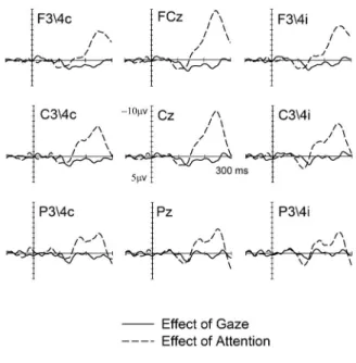
Figures 1 and 2 show SEPs elicited by non-target stimuli in the 300 ms interval after stimulus onset. SEPs are displayed separately for midline sites Fcz, Cz and Pz (centre) and for lateral sites F3/4, C3/4 and P3/4 ipsilateral (right) and contralateral (left) to the stimulated hand. Figure 1 shows the effect of Gaze on tactile processing, obtained by comparing ERP waveforms elicited by non-target stimuli delivered to the hand to which gaze was directed (G+, solid line) and to the other non gazed hand (G-, dashed line), collapsed across currently attended and unattended stimuli (A+ and A-, respectively). The effect of sustained spatial attention on touch is represented in Figure 2 where ERP waveforms are shown separately for tactile non-target stimuli to the attended hand (A+, solid line) and to the unattended hand (A-, dashed line), collapsed across the current direction of gaze (G+ and G- trials). The corresponding difference waveforms for the effects of gaze and attention can be observed in Figure 3 (solid and dashed lines, respectively), while Figure 4 shows separately the four different experimental conditions (A+G+; A-G-; A+G- and A-G+) as observed at central electrodes C3/4 contralateral to the stimulated hand.
in which distinct somatosensory ERP components were elicited. The manipulation of gaze resulted in selective modulations of ERPs elicited in the N140 time window. As shown in Figure 1, reduced N140 component were observed for gazed stimuli (G+ trials) as compared to non gazed stimuli (G-), that is, ERPs elicited by stimuli to the gazed hand (G+) were more positive than those to the non gazed hand (G-). In contrast, sustained spatial attention resulted in enhanced P100 components for attended stimuli (A+) as compared to unattended ones (A-) (Figure 2). While no effect of spatial attention was visible in the N140 time range, enhanced negativities for tactile stimuli to the attended as compared to the unattended hand were visible in the time range of the processing negativity (Nd, Figure 2).
(G-, dashed line) in the 300 ms following stimulus onset (relative to a 100 ms pre-stimulus baseline) at fronto-central (FC3/4), central (C3/4) and parietal (P3/4) electrodes contralateral (c) and ipsilateral (i) to the stimulated hand as well as at midline electrodes (Fcz, Cz, Pz).
No significant effect of gaze emerged in the P45 (40-60ms post stimulus), N80 (70-90 ms), or P100 ((70-90-120 ms) time ranges. In contrast, an enhanced positivity for G+ as compared to G- trials was reliably present in the N140 time window between 130 and 150 ms post-stimulus. In this time window, significant main effects of gaze were found at anterior and central electrodes ipsilateral and contralateral to the stimulated hand as well as at contralateral posterior and midline electrodes (all F(1, 8)>6.04; all p<.04). Gaze x Electrode Site interactions were present at anterior electrodes both ipsilateral and contralateral to the stimulated hand (both F(2, 16)>5.67; both p<.022), and follow-up analyses showed a significant effect of gaze on N140 amplitudes at all sites (all F(1, 8)>5.3; all p<.05) with the exception of contralateral F7/8 where this effect approached significance (F(1, 8)=4.3; p=.72). In the subsequent time window (160-240 ms post stimulus) no main effects of gaze were observed at any of the electrode sites. Table 1 summarizes the mean amplitude values and mean standard errors of tactile ERPs elicited by gazed (G+) and non gazed (G-) stimuli. These values are reported for the specific time windows and electrodes sites where statistically reliable main effects of gaze were observed.
Gaze
Contralateral electrodes Ipsilateral electrodes
Anterior Central Posterior Anterior Central Posterior Midline
N140 (130-150ms)
G+ -1.35 (0.42) -0.059 (0.5) 0.1 (0.5) 0.18 (0.53) 0.7 (0.59) - 0.56 (0.63)
G- -1.9 (0.46) -0.9 (0.48) -0.67 (0.43) -0.49 (0.53) -0.02 (0.55) - -0.24 (0.56)
Table 1. Mean amplitude values (µV) and mean standard errors of ERPs elicited by gazed (G+) and non gazed (G-) tactile stimuli for the time windows and electrode sites in which significant main effects of gaze emerged.
Reliable effects of attention were first observed in the P100 time range (90-120 ms post-stimulus), with enhanced P100 amplitudes for A+ relative to A- stimuli, as indicated by main effects of attention at central electrodes contralateral and ipsilateral to the
all p<.036). In contrast, no significant modulation of the N140 component was observed between 130 and 150 ms post stimulus. Finally, in the 160-240 ms time window,
enhanced negativities for A+ as compared to A- trials were observed at central and posterior electrodes contralateral and ipsilateral to the stimulated hand as well as midline sites (all F(1,8)>7.08; all p<.03). Attention x Electrode Site interactions were observed in the Nd time range for posterior electrodes both contralateral and ipsilateral to the
stimulated hand (both F(2, 16)>4.6; both p<.026) as well as for midline sites (F(2, 16)=11.6; p=.005), and follow-up analyses revealed significant main effects of attention at all sites (all F(1, 8)>6.7; all p<.032), except for ipsilateral P7/8 were this effect did not reach statistical significance (F(1, 8)=3; p=.12). Table 2 summarizes the mean amplitude values and mean standard errors of tactile ERPs elicited by attended (A+) and unattended (A-) stimuli. These values are reported for the specific time windows and electrodes sites where statistically reliable main effects of attention were observed.
Attention
Contralateral electrodes Ipsilateral electrodes
Anterior Central Posterior Anterior Central Posterior Midline
P100 (90-120ms)
A+ 0.59 (0.38) 1.6 (0.47) - - 1.9 (0.38) - 1.9 (0.41)
A- 0.1 (0.45) 1 (0.54) - - 1.1 (0.4) - 1.1 (0.45)
Nd
(160-240ms)
A+ - 0.38 (0.68) 0.1 (0.62) - 0.3 (0.5) 0.17 (0.38) 0.5 (0.6)
A- - 2.35 (0.58) 1.4 (0.51) - 2.23 (0.44) 1.05 (0.33) 2.6 (0.56)
Table 2. Mean amplitude values (µV) and mean standard errors of ERPs elicited by attended (A+) and unattended (A-) tactile stimuli for the time windows and electrode sites where significant main effects of attention were found.
interactions were present at any of the electrode sites. Figure 4 shows separately ERPs elicited by the four different experimental conditions. The main difference between ERP waveforms is primarily due to attention in the P100 and Nd time windows (A+ and A-, solid vs. dashed lines, respectively), while it is mainly driven by gaze in the N140 interval (G+ and G-, black vs. grey lines).
General discussion
Directing gaze to a body site improves the discrimination of tactile stimuli presented to that location, as demonstrated by initial behavioural and electrophysiological evidence (Forster and Eimer, 2005; Honoré et al., 1989; Pierson et al., 1991; Tipper et al., 1998). However, the neural mechanisms underpinning the effect of gaze on touch remain almost completely unexplored. Here, we directly investigated the relationship between the effects of gaze and spatial attention on tactile processing, to uncover whether these effects are mediated by shared neural mechanisms or whether, in contrast, they represent the outcome of distinct processes. To this aim we orthogonally manipulated the directions of gaze and spatial attention, by instructing participants to covertly attend one of their hands, while directing their gaze to the same hand or to the opposite unattended hand (in different blocks of trials). Because participants’ hands were covered during the experiment, attention and gaze were directed towards the location occupied by the hands. Thus, the effects of gaze and attention on touch were investigated in the absence of visual information about the tactually stimulated body part.
window (130-150 ms post-stimulus onset), later attentional ERP modulations were present between 160 and 240 ms post- stimulus overlapping with the descending flank of the N140 component and with the subsequent processing negativity. These enhanced negativities for stimuli presented to the attended as compared to the unattended hand were present over central and posterior electrodes and maximal over midline sites (Figure 4, right column, bottom panel). While a number of different factors contribute to determine the specific time course of the attentional modulations of tactile ERPs (such as the specific type of attention task, e.g. Forster and Eimer, 2003; the availability of visual information relative to the stimulated body site, e.g. Sambo et al., 2009; the difficulty of the discrimination task (e.g. Michie et al.,1987) and the stimulated body site (e.g. Gillmeister et al., 2010), the observation that tactile spatial attention resulted in modulations of both perceptual (here reflected by P100 enhancements) and post-perceptual (as reflected by Nd modulations) stages of somatosensory processing is in line with previous ERP studies (e.g. Sambo and Forster, 2011) and demonstrates that participants engaged with the tactile attention task and covertly attended the task-relevant hand1.
Crucially, ERPs elicited by tactile stimuli presented to the hands were systematically modulated not only by the direction of covert attention but also by the direction of gaze. A reliable difference between ERPs elicited by gazed (G+) and non gazed (G-) tactile stimuli was observed between 130 and 150 ms post-stimulus, overlapping with the N140 somatosensory ERP component. More specifically, reduced
1 Recent evidence suggests that the position of the eyes in the orbit (central vs.
Figure 5. Topographical voltage maps for the effects of Gaze (left panels) and Attention (right panels) on somatosensory ERPs are shown separately for the P100 (90-120 ms interval post-stimulus; top panels), N140 (130-150 ms interval; central panels) and Nd (160-240 ms; bottom panels) components. Left maps (Effect of Gaze) display the voltage distributions of the difference amplitudes obtained by subtracting ERPs to tactile non-target stimuli presented to the non gazed hand (G-) from those elicited by stimuli to the gazed hand (G+). Right maps (Effect of Attention) display the voltage distributions of the difference amplitudes obtained by subtracting ERPs to tactile non-target stimuli presented to the unattended hand (A-) from those elicited by stimuli to the attended hand (A+). Amplitude scales range between -2.0 to +2.0 µV for the P100 and for the N140 components, while in the Nd time range amplitude values range between -6.0 to +6.0 µV. Positive voltage values are plotted in red while negative values are plotted in green.
The aim of the present study was to disentangle the effect of gaze direction on touch from that of spatial attention. First, we asked whether an effect of gaze on touch can be observed when both spatial attention and gaze direction are independently manipulated. Results were clear-cut. Under these experimental conditions, gaze direction still resulted in systematic modulations of somatosensory processing. This finding confirms the presence of an effect of gaze on touch, expanding results of earlier investigations (Forster and Eimer, 2005; Honoré et al., 1989; Pierson et al., 1991; Tipper et al., 1998). Second, we asked whether shared mechanisms are responsible for both the effects of gaze and spatial attention on touch. To address this question we directly compared gaze-dependent and attention-dependent modulations of tactile processing. Both the pattern and the specific time course of these SEPs modulations indicate that the mechanisms underlying the effect of gaze are independent of those responsible for spatial attention.
components for gazed stimuli (that is ERPs were more positive for G+ than for G-between 130 and 150 ms post stimulus). This finding provides the first direct indication that gazing to the tactually stimulated body site activates neural mechanisms which are different from those of spatial attention.
This conclusion is further supported by the specific time course of the effects of gaze and attention. While both gaze and spatial attention had reliable effects on touch, they influenced different stages of somatosensory processing. Results demonstrated a clear dissociation between the timing and therefore the associated locus of the effects of gaze and attention on touch. Sustained spatial attention resulted in enhanced positivities starting around 90 ms after stimulus onset. In contrast, the earliest effects of gaze were only observed after 130 ms post-stimulus. Thus, the effects of attention and gaze overlapped with different somatosensory ERP components, with attention affecting the P100 and gaze modulating the following N140 component. Both the P100 and the N140 somatosensory ERP components are considered mid-latency somatosensory ERP components representing modality-specific stages of tactile processing, but they are characterized by distinct neural generators. While the P100 component originates bilaterally from SII (Hari et al., 1984; Forster and Eimer, 2003; Frot and Maguiere, 1999), multiple neural generators are likely to be responsible for the N140 component including SII and bilateral frontal areas (Allison et al. 1992; Hari et al., 1984; 1993; Kakigi et al., 2000; Mima et al., 1998). The dissociation between gaze-dependent and attention-dependent modulations of somatosensory processing is not only related to the onset of these effects, but also to their time course. Results revealed that the effects of gaze on touch were short-lived and exclusively present in the N140 time-range, while the effects of attention were also observed during later stages of processing (between 160 and 240 ms post-stimulus). Taken together, these results suggest that the changes provoked by gaze and attention occur at different stages of somatosensory processing providing additional evidence that the effects of gaze and of spatial attention are mediated by different mechanisms.
for a direct relationship between visual N1 component amplitude and response speed). The results of the present experiment appear to confirm a relationship between the ERP modulations in the N140 time window and the behavioural performance. While it is reasonable to expect a strong relationship between the Nd attentional modulations and the behavioural performance, the present task, which was devised to provide a strong incentive for participants to fully focus attention on the task-relevant hand and to maximize the number of trials for the ERP analysis, does not allow such comparison (due to the fact that only attended targets required a response). For this reason, one question which remains open is whether the dissociation observed in the ERP data in the present study would be reflected by independent effects of gaze and attention on performance. Future behavioural studies should directly assess the behavioural effects of gaze and attention on touch when these factors are orthogonally manipulated. If gaze and attention impact different stages of processing, as suggested by the present ERP data, they should have additive effects on the behavioural data (i.e., main effects for both variables and no interaction), following the additive-factors logic (e.g. Sternberg, 1969).
gaze direction but not spatial attention were manipulated, there is the possibility that spatial attention was at least in part responsible for the observed effects of gaze. Overall, the present study provides the first direct ERP evidence that the effects of gaze and spatial attention on touch are mediated by distinct neural mechanisms.
This conclusion has relevant implications for the understanding of the mechanisms of spatial attention and its links with the oculomotor system. Previous studies have demonstrated that the attentional and oculomotor processes are closely, or even mandatorily, linked during the dynamic programming of a saccadic eye movement. For instance, planning a saccadic eye movement towards a spatial location elicits supramodal shifts of attention able to enhance the processing of stimuli presented close to the target location not only in the visual modality (e.g., Deubel and Schneider 1996; Eimer et al., 2006; 2007; Hoffman and Subramaniam 1995; Kowler et al., 1995), but also in the tactile modality (e.g. Gherri and Eimer, 2008; Gherri and Forster, 2012a; 2012b; Juravle and Deuble, 2009; Rorden et al., 2002). In contrast, when the oculomotor system is preset to maintain fixation as opposed to plan a saccade, the attentional and oculomotor processes can be dissociated, as suggested by recent studies on fixational eye movements (Tse et al., 2002; 2004; Horowitz et al., 2007). In line with these observations our results suggest that maintaining gaze on a specific body location does not necessarily results in attentional modulations of tactile stimuli presented at the gazed location. Different operations of the oculomotor system are likely to be coupled in a different way to spatial attention mechanisms. While directing the eyes (or planning to direct the eyes) toward a relevant location elicit a shift of attention toward that location, maintaining the eyes on the same location (fixation) can be de-coupled from attentional processing when gaze and spatial attention are independently manipulated, as observed in the present study.
eccentric position, the perceived location of a tactile stimulus is systematically shifted in the direction of gaze as demonstrated by systematic errors related to gaze-direction when participants are asked to localize tactile stimuli (e.g. Harrar and Harris, 2009; Harrar et al., 2013; Pritchett and Harris, 2011). Thus, coding of tactile space requires the integration of tactile and eye position information. Importantly, such integration may only occur within higher level brain areas. Tactile stimuli are initially encoded according to a somatotopic representation of the body which is independent of body posture and of eye position. Only during later stages of processing, tactile information is recoded from somatotopic onto an external representation of space which takes into account the position of the body and of the eyes and is based on the integration of tactile with proprioceptive and visual information about the body (e.g. Azañón and Soto-Faraco, 2008; Longo et al., 2010; Röder et al., 2004). It has been suggested that the posterior parietal cortex might play a pivotal role in the remapping of touch from somatotopic into external coordinates (e.g. Azañón et al., 2010). In line with this observation, and consistent with electrophysiological studies of postural remapping of touch in external space (Heed and Röder, 2010; Rigato et al., 2013), we observed a reliable effects of gaze on touch only after 130 ms post-stimulus onset, suggesting a neural activation of areas in and beyond SII. Given that gaze direction can be used as one of the reference points against which external space is coded (e.g. Harrar and Harris, 2009; Harrar et al., , 2013; Pritchett and Harris, 2011), proprioceptive signals of the position of the eyes might influence tactile processing via back projections from multimodal brain areas starting from 130 ms after stimulus presentation. While this hypothesis is speculative at present and should be further investigated in future studies, postural cues of eye position might be responsible for the effect of gaze on touch observed in the present study.
Acknowledgements
References
Allison, T., McCarthy, G., Wood, C.C., 1992. The relationship between human long latency somatosensory evoked-potentials recorded from the cortical surface and from the scalp. Electroencephalography and Clinical Neurophysiology 84, 301-314.
Azañón, E., Soto-Faraco, S., 2008. Changing reference frames during the encoding of tactile events. Current Biology 18, 1044-1049.
Azañón, E., Longo, M. R., Soto-Faraco, S., Haggard, P., 2010. The posterior parietal cortex remaps touch into external space. Current Biology 20, 1304-1309.
Cardini, F., Longo, M. R., Haggard, P., 2011. Vision of the body modulates somatosensory intracortical inhibition. Cerebral Cortex 21, 2014-2022.
Cardini, F., Longo, M. R., Driver, J., Haggard, P., 2012. Rapid enhancement of touch from non-informative vision of the hand. Neuropsychologia 50, 1954-1960.
Desmedt, J. E., Robertson, D., 1977. Differential enhancement of early and late components of the cerebral somatosensory evoked potentials during forced-paced cognitive tasks in man. The Journal of physiology 271, 761-782.
Deubel, H., Schneider, W.X., 1996. Saccade Target Selection and Object Recognition: Evidence for a Common Attentional Mechanism. Vision Research 36, 1827-1837.
Eimer, M., Forster, B., 2003. Modulations of early somatosensory ERP components by transient and sustained spatial attention. Experimental Brain Research 151, 24-31.
Eimer, M., Velzen, J. V., Gherri, E., Press, C., 2007. ERP correlates of shared control mechanisms involved in saccade preparation and in covert attention. Brain research 1135, 154-166.
Fiorio, M., Haggard, P., 2005. Viewing the body prepares the brain for touch: effects of TMS over somatosensory cortex. European Journal of Neuroscience 22, 773–777.
Forster, B., Eimer, M., 2004. The attentional selection of spatial and non-spatial attributes in touch: ERP evidence for parallel and independent processes. Biological Psychology 66, 1–20.
Forster, B., Eimer, M., 2005. Vision and gaze direction modulate tactile processing in somatosensory cortex: evidence from event-related brain potentials. Experimental Brain Research 165, 8–18.
Frot, M., Maguiere, F., 1999. Timing and spatial distribution of somatosensory responses recorded in the upper bank of the sylvian fissure (SII area) in humans. Cerebral Cortex 9, 854-863.
García-Larrea, L., Lukaszewicz, A.C., Mauguire, F., 1995. Somatosensory responses during selective spatial attention: The N120- to-N140 transition. Psychophysiology 32, 526–537.
Gherri, E., Eimer, M., 2008. Links between eye movement preparation and the attentional processing of tactile events: an event-related brain potential study. Clinical neurophysiology 119, 2587-2597.
Gherri, E., Forster, B., 2012a. Crossing the hands disrupts tactile spatial attention but not motor attention: Evidence from event-related potentials. Neuropsychologia 50, 2303-16.
Gherri, E., Forster, B., 2012b. The orienting of attention during eye and hand movements: ERP evidence for similar frame of reference but different spatially-specific modulations of tactile processing. Biological Psychology 91, 172-84.
Gillmeister, H., Sambo, C.F., Forster, B., 2010. Which finger? Early effects of attentional selection within the hand are absent when the hand is viewed. European Journal of Neuroscience 31, 1874-1881.
Hari, R., Karhu, J., Hämäläinen, M., Knuutila, J., Salonen, O., Sams, M., Vilkman, V., 1993. Functional organization of the human first and second somatosensory cortices: a neuromagnetic study. European Journal of Neuroscience 5, 724–734.
Hari, R., Reinikainen, K., Kaukoranta, E., Hämäläinen, M., Ilmoniemi, R., Penttinen, A., Salminen, J., Teszner, D., 1984. Somatosensory evoked magnetic fields from SI and SII in man. Electroencephalography and Clinical Neurophysiology 57, 254–263.
Harrar, V., Harris, L. R., 2009. Eye position affects the perceived location of touch. Experimental Brain Research 198, 403–410.
Harrar, V., Pritchett, L. M., Harris, L. R., 2012. Segmented space: measuring tactile localisation in body coordinates. Multisensory research 26, 3-18.
Heed, T., Röder, B., 2010. Common Anatomical and External Coding for Hands and Feet in Tactile Attention: Evidence from Event-related Potentials. Journal of Cognitive Neuroscience 22, 184-202.
Hesse, C.W., Seiss, E., Bracewell, R.M., Praamstra, P., 2004. Absence of gaze direction effects on EEG measures of sensorimotor function. Clinical Neurophysiology 115, 29–38.
Hoffman, J. E., Subramaniam, B., 1995. The role of visual attention in saccadic eye movements. Perception and Psychophysics 57, 787-795.
Honore, J., Bourdeaud’Hui, M., Sparrow, L., 1989. Reduction of cutaneous reaction time by directing the eyes towards the source of stimulation. Neuropsychologia 27, 367–371.
Jones, A., Forster, B., 2014. Neural correlates of endogenous attention, exogenous attention and inhibition of return in touch. European Journal of Neuroscience 40, 2389-2398.
Josiassen, R. C., Shagrass, C., Roemer, R. A., Ercegovac, D. E., Straumanis, J. J., 1982. Somatosensory evoked potentials change with a selective attention task. Psychophysiology 19, 146-159.
Juravle, G., Deubel, H., 2009. Action preparation enhances the processing of tactile targets. Experimental brain research 198(2-3), 301-311.
Kakigi, R., Hoshiyama, M., Shimojo, M., Naka, D., Yamasaki, H., Watanabe, S., Nakamura, A., 2000. The somatosensory evoked magnetic fields. Progress in neurobiology 61, 495-523.
Kennett, S., Taylor-Clarke, M., Haggard, P., 2001. Noninformative vision improves the spatial resolution of touch in humans. Current Biology 11, 1188–1191. Kowler, E., Anderson, E., Dosher, B., Blaser, E., 1995. The role of attention in the
programming of saccades. Vision Research 35, 1897-1916.
Longo, M. R., Azañón, E., Haggard, P., 2010. More than skin deep: body representation beyond primary somatosensory cortex. Neuropsychologia 48, 655-668.
Longo, M.R., Pernigo, S., Haggard, P., 2011. Vision of the body modulates processing primary somatosensory cortex. Neuroscience Letters 489, 159– 163.
Michie, P. T., 1984. Selective attention effects on somatosensory event-related potentials. Annals of the New York Academy of Science 425, 250–255. Michie, P. T., Bearpark, H. M., Crawford, J. M., Glue, L. C. T., 1987. The effects
of spatial selective attention on the somatosensory event-related potential. Psychophysiology 24, 449–463.
Pierson, J. M., Bradshaw, J. L., Meyer, T. F. Howard, M. J, Bradshaw, A., 1991. Direction of gaze during a vibrotactile choice reaction time task. Neuropsychologia 29, 925-928.
Press, C., Taylor-Clarke, M., Kennett, S., Haggard, P., 2004. Visual enhancement of touch in spatial body representation. Experimental Brain Research 154, 238-245.
Pritchett, L., Harris, L. R., 2011. Perceived touch location is affected by both eye and head position. Experimental Brain Research 213, 229-234.
Rigato, S., Bremner, A.J., Mason, L., Pickering, A., Davis, R., van Velzen, J., 2013. The electrophysiological time course of somatosensory spatial remapping: vision of the hands modulates effects of posture on somatosensory evoked potentials. European Journal of Neuroscience 38, 2884-92.
Röder, B., Rösler, F., Spence, C., 2004. Early vision impairs tactile perception in the blind. Current Biology 14, 121-124.
Rorden, C., Greene, K., Sasine, G. M., Baylis, G. C., 2002. Enhanced Tactile Performance at the Destination of an Upcoming Saccade. Current Biology 12, 1429–1434.
Sambo, C., Forster, B., 2011. Sustained spatial attention in touch: modality-specific and multimodal mechanisms. The Scientific World Journal 11, 199-213. Sambo, C.F., Gillmeister, H., Forster, B., 2009. Viewing the body modulates neural
mechanisms underlying sustained spatial attention in touch. European Journal of Neuroscience 30, 143-150.
Sternberg, S., 1969. The discovery of processing stages: Extensions of Donders' method. In: Koster W. G. (Ed.), Attention and performance II, Acta Psychologica 30, pp. 276-315.
Talsma, D., Mulckhuyse, M., Theeuwes, J., 2007. Faster, more intense! The Relation between Electrophysiological Reflections of Attentional Orienting, Sensory Gain Control, and Speed of Responding. Brain research 1178, 92-105.
Tipper, S. P., Phillips, N., Dancer, C., Lloyd, D., Howard, L. A., McGlone, F. (2001). Vision influences tactile perception at body sites that cannot be viewed directly. Experimental Brain Research 139, 160-167.
Tipper, S.P., Lloyd, D., Shorland, B., Dancer, C., Howard, L.A., McGlone, F., 1998. Vision influences tactile perception without proprioceptive orienting, NeuroReport 9, 1741–1744.
Tse, P. U., Sheinberg, D. L., Logothetis, N. K., 2002. Fixational eye movements are not affected by abrupt onsets that capture attention. Vision research 42, 1663-1669.
Tse, P. U., Sheinberg, D. S., Logothetis, N. K., 2004. The distribution of microsaccade directions need not reveal the location of attention: reply to Rolfs, Engbert, and Kliegl. Psychological Science, 708-710.