ISSN (Print) : 2320 – 3765 ISSN (Online): 2278 – 8875
I
nternational
J
ournal of
A
dvanced
R
esearch in
E
lectrical,
E
lectronics and
I
nstrumentation
E
ngineering
(An ISO 3297: 2007 Certified Organization)
Vol. 3, Issue 9, September 2014
Frequency Dependent Attenuation of EM
Radiation on Biological Tissues
Ch. Rajani Chandra1, P.S.N. Bharani1, Y. Ratna Kumar2
PG Student [BME], Department of ECE, Andhra University Collegeof Engineering (A), Andhra Pradesh, India1
PG Student [BME], Department of ECE, Andhra University Collegeof Engineering (A), Andhra Pradesh, India1
Assistant Professor (c), Department of ECE, Andhra University Collegeof Engineering (A), Andhra Pradesh, India2
ABSTRACT: The human body is a non-homogeneous medium and it is characterized by different fundamental
parameters. All of them vary from location to location. Human body acts as a transmission medium for electrical signals. This offers novel data communication in biomedical monitoring systems. The human body is characterized as a transmission medium for electrical current by means of numerical simulations and measurements. Several attempts have been made to model the human body as electrical channel. Here the variations of penetration characteristics of the Electromagnetic radiation of the biological tissues had been observed.
KEYWORDS: Non-homogeneous medium, transmission medium, Electromagnetic radiation, penetration
characteristics, biological tissues, electrical current
I. INTRODUCTION
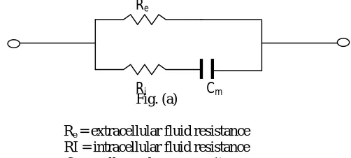
It is common practice to study the behavior of electromagnetic waves in homogenous medium. In order to apply electromagnetic energy for the treatment of different tumors, the characteristics of the biological tissues are required to be established. It is well known that the homogenous media is characterized by constant values of permittivity, permeability and conductivity throughout the medium. On the other hand, non-homogeneous medium is a medium for which the above fundamental parameters are not constant and are different from point to point in the media. The human body is a non-homogeneous medium and it is characterized by different fundamental parameters. Using the novel data communication in biomedical monitoring systems [6], The hyperthermia treatments are categorized as a whole body, regional or local. The rational for the whole body hyperthermia is that the cancer is a systemic disease and the cancer cells have metastasized throughout the body in most of the cases.It has been possible to find out relative electromagnetic energy absorption characteristics of the data on fundamental constants of the tissues. In order to apply electromagnetic energy as a function of frequency, The equivalent and simplified circuit(Fig. (a)) for a cell or a tissue [7].
Fig. (a)
Re = extracellular fluid resistance
RI = intracellular fluid resistance Cm = cell membrane capacitance
In the presence of an electromagnetic field, the biological tissue is considered as a medium with losses [8], [9], [10], [11], [12].At frequency ω of complex electric field Eˆ→and complex magnetic excitation Hˆ-> the Ampere law [13] is given by:
Re
ISSN (Print) : 2320 – 3765 ISSN (Online): 2278 – 8875
I
nternational
J
ournal of
A
dvanced
R
esearch in
E
lectrical,
E
lectronics and
I
nstrumentation
E
ngineering
(An ISO 3297: 2007 Certified Organization)
Vol. 3, Issue 9, September 2014
⃑ ⃑= ̂ ⃑ (1)
The complex permittivity εˆ characterizes the biological tissue with conduction losses, and can be written in the following form [9], [11], [12]:
εˆ = ε (ε′ − jε′′) (2) Where =8. 8×10 -12 F/m is the absolute permittivity
ε′= And ε"= are the relative permittivities with the conductivity σ and the dielectric permittivity ε of the biological tissue. ε’ and ε’’ are often indirectly measured by using RF impedance bridges and are provided into tables as a function of the frequency [9], [11]. A new approach is adopted here to evaluate the power attenuation caused by the presence of a biological tissue between the transmitter and receiver coupled coils. The magnetic field propagation in the biological tissue which is defined by its parameters of permittivity ε, conductivity σ and permeability of vacuum μ0, is according to the magnetic wave equation, [13], using the Laplacian operator ∇ :
1/ ∇ ^⃑= ^⃑+ ^⃑ (3)
For a sinusoïdal variation of magnetic field ^⃑=⃑ Equation (3) becomes:
∇ ^⃑=j ( +j ) ^⃑= ^⃑ (4)
where γˆ (m−1 ) = α + jβ is the propagation constant and α, β are attenuation and phase constants, respectively.
= є 1 + −1 (5)
= є 1 + + 1 (6)
The losses of the medium are represented by the tangent of loss [7].
=є"
є = (7)
Tissue Characteristics of Human Body
The attenuation constant in the biological tissue is a function of frequency, conductivity, permittivity and permeability of the bio-tissue. When an EM wave is allowed to incident on the tissue in such a way that electric field is parallel to the interference between the tissues, two adjacent tissues absorb power. Under these conditions, the electric fields are tangent to the interference between the pair of tissues and they are equal. When the electric fields are perpendicular to the interface, the conduction and displacement current becomes continuous across the interference. The following cases are considered for finding at the relative absorption of EM energy.The electric field vector parallel to the interference between the tissues.The electric field vector perpendicular to the interference between tissues.
II. RESULTS
The attenuation constant of a non-homogenous medium [human body] is evaluated by the expression (8)
1 1
2 2 2
2
(8)
ISSN (Print) : 2320 – 3765 ISSN (Online): 2278 – 8875
I
nternational
J
ournal of
A
dvanced
R
esearch in
E
lectrical,
E
lectronics and
I
nstrumentation
E
ngineering
(An ISO 3297: 2007 Certified Organization)
Vol. 3, Issue 9, September 2014
Fig. 1 (a) Fig. 1 (b) Fig. 1 (c)
Fig. 1 (a) A plot of conductivities of dry skin tissues at corresponding tissues Fig. 1 (b) A plot of permittivities of dry skin tissues at corresponding tissues Fig. 1 (c) A plot of Attenuation of dry skin tissues at corresponding tissues
In Fig. 1(a), it is observed that the conductivity is very low at lower frequencies and while going towards the high frequencies the conductivity increases with increase in frequency.
In Fig. 1(b), the permittivity of dry skin tissues is observed to be high at low frequencies and gradually decreases with increase in the frequency.
In Fig. 1(c), the minimum attenuation of electromagnetic radiation by the dry skin tissues is observed at the frequencies around 10 KHz and maximum attenuation at around 10 MHz.
Fig. 2 (a) Fig. 2 (b) Fig. 2 (c)
Fig. 2 (a) A plot of conductivities of wet skin tissues at corresponding tissues Fig. 2 (b) A plot of permittivities of wet skin tissues at corresponding tissues Fig. 2 (c) A plot of Attenuation of wet skin tissues at corresponding tissues
In Fig.2(a), it is observed that the conductivity is linearly increasing with the increase of frequency of radiation. In Fig.2(b), the permittivity of wet skin tissues is observed to be high at low frequencies and gradually decreases with increase in the frequency.
In Fig.2(c), the maximum attenuation of electromagnetic radiation by the wet skin tissues is observed at the frequencies around 1MHz .
-0.1 0 0.1 0.2 0.3 0.4 0.5 0.6 0.7 0.8 0.9 1
10000 100000 1000000 10000000 100000000 1000000000
FREQUENCY (Hz) C O N D U C T IV IT Y (s /m ) 0 100 200 300 400 500 600 700 800
0 2 4 6 8 10
R EL A TI V E P ER M IT TI V IT Y( є r ) FREQUENCY(MHz) 0 0.2 0.4 0.6 0.8 1 1.2 1.4
10000 100000 1000000 10000000 100000000 1000000000
FREQUENCY(Hz) A T T E N U A T IO N 0 0.1 0.2 0.3 0.4 0.5 0.6 0.7 0.8 0.9 1
10000 100000 1000000 10000000 100000000 1000000000
FREQUENCY(Hz) C O N D U C T IV IT Y (s /m ) 0 100 200 300 400 500 600 700 800
0 5 10
R EL A TI V E P ER M IT TI V IT Y( є r) FREQUENCY(MHz) 0 0.5 1 1.5 2 2.5 3 3.5
10000 100000 1000000 10000000 100000000 1000000000
ISSN (Print) : 2320 – 3765 ISSN (Online): 2278 – 8875
I
nternational
J
ournal of
A
dvanced
R
esearch in
E
lectrical,
E
lectronics and
I
nstrumentation
E
ngineering
(An ISO 3297: 2007 Certified Organization)
Vol. 3, Issue 9, September 2014
Fig. 3 (a) Fig. 3 (b) Fig. 3 (c)
Fig. 3 (a) A plot of conductivities of fat tissues at corresponding tissues Fig. 3 (b) A plot of permittivities of fat tissues at corresponding tissues Fig. 3 (c) A plot of Attenuation of fat tissues at corresponding tissues
In Fig. 3(a), it is observed that the conductivity takes place even at the low frequencies and also increases with frequency
In Fig. 3(b), the permittivity of fat tissues is observed to be high at low frequencies and gradually decreases with increase in the frequency.
In Fig. 3(c), the maximum attenuation of electromagnetic radiation by the fat tissues is observed at the frequencies around 10 KHz .
Fig. 4 (a) Fig. 4 (b) Fig. 4 (c)
Fig. 4 (a) A plot of conductivities of muscle tissues at corresponding tissues Fig. 4 (b) A plot of permittivities of muscle tissues at corresponding tissues Fig. 4 (c) A plot of Attenuation of muscle tissues at corresponding tissues
In Fig. 4(a), it is observed that the conductivity takes place even at the low frequencies and also increases with frequency
In Fig. 4(b), the permittivity of muscle tissues is observed to be high at low frequencies and gradually decreases with increase in the frequency.
In Fig. 4(c), the maximum attenuation of electromagnetic radiation by the muscle tissues is observed at the frequencies around 10 KHz .
0 0.01 0.02 0.03 0.04 0.05 0.06
10000 100000 1000000 10000000 100000000 1000000000
FREQUENCY(Hz) C O N D U C T IV IT Y ( s /m ) 0 100 200 300 400 500 600 700 800
0 5 10
R EL A TI V E P ER M IT TI V IT Y( є r) FREQUENCY(MHz) 0 5 10 15 20 25 30 35 40 45
10000 100000 1000000 10000000 100000000 1000000000
FREQUENCY(Hz) A T T E N U A T IO N 0 0.2 0.4 0.6 0.8 1 1.2
10000 100000 1000000 10000000 100000000 1000000000
FREQUENCY(Hz) C O N D U C T IV IT Y (s /m ) 0 100 200 300 400 500 600 700 800
0 5 10
R EL A TI V E P ER M IT TI V IT Y( є r) FREQUENCY(MHz) 0 5 10 15 20 25 30 35 40 45
10000 100000 1000000 10000000 100000000 1000000000
ISSN (Print) : 2320 – 3765 ISSN (Online): 2278 – 8875
I
nternational
J
ournal of
A
dvanced
R
esearch in
E
lectrical,
E
lectronics and
I
nstrumentation
E
ngineering
(An ISO 3297: 2007 Certified Organization)
Vol. 3, Issue 9, September 2014
Fig. 5 (a) Fig. 5 (b) Fig. 5 (c)
Fig. 5 (a) A plot of conductivities of corticle bone tissues at corresponding tissues Fig. 5 (b) A plot of permittivities of corticle bone tissues at corresponding tissues Fig. 5 (c) A plot of Attenuation of corticle bone tissues at corresponding tissues
In Fig. 5(a), it is observed that the conductivity takes place only at high frequencies
In Fig. 5(b), the permittivity of bone tissues is observed to be high at low frequencies and gradually decreases with increase in the frequency.
In Fig. 5(c), the attenuation of bone tissues is observed to be high at low frequencies and gradually decreases with increase in the frequency.
Fig. 6 (a) Fig. 6 (b) Fig. 6 (c)
Fig. 6 (a) A plot of conductivities of bone marrow tissues at corresponding tissues Fig. 6 (b) A plot of permittivities of bone marrow tissues at corresponding tissues Fig. 6 (c) A plot of Attenuation of bone marrow tissues at corresponding tissues
In Fig. 6(a), it is observed that the conductivity takes place only at high frequencies
In Fig. 6(b), the permittivity of bone marrow tissues is observed to be high at low frequencies and gradually decreases with increase in the frequency.
In Fig. 6(c), the attenuation of bone marrow tissues is observed only at high frequencies around 100 MHz
III. CONCLUSION
Thus variations of the penetration characteristics such as conductivity and relative permittivity are plotted at various frequencies. From these we can obtain the attenuation of Electromagnetic radiation on different biological tissues such as skin, fat, muscle, corticle bone and bone marrow at the corresponding frequency range of 10KHz to 1GHz.
-0.1 0 0.1 0.2 0.3 0.4 0.5 0.6 0.7
1 10 100 1000 10000 100000 1000000 1000000
0 1E+08 1E+09 FREQUENCY(Hz) C O N D U C T IV IT Y (s /m ) 0 100 200 300 400 500 600 700 800
0 5 10
R EL A TI V E P ER M IT TI V IT Y( є r ) FREQUENCY(MHz) 0 10 20 30 40 50 60 70 80
10000 100000 1000000 10000000 100000000 1000000000
FREQUENCY(Hz) A T T E N U A T IO N ( d B ) -0.05 0 0.05 0.1 0.15 0.2 0.25 0.3 0.35
10000 100000 1000000 10000000 100000000 1000000000
FREQUENCY(Hz) C O N D U C T IV IT Y (s /m ) 0 100 200 300 400 500 600 700 800
0 5 10
R EL A TI V E P ER M IT TI V IT Y( є r) FREQUENCY(MHz) -0.05 0 0.05 0.1 0.15 0.2 0.25 0.3 0.35
10000 100000 1000000 10000000 100000000 1000000000
ISSN (Print) : 2320 – 3765 ISSN (Online): 2278 – 8875
I
nternational
J
ournal of
A
dvanced
R
esearch in
E
lectrical,
E
lectronics and
I
nstrumentation
E
ngineering
(An ISO 3297: 2007 Certified Organization)
Vol. 3, Issue 9, September 2014
REFERENCES
1. T. Handa, S. Shoji, S. Ike, S. Takeda and T. Sekiguchi, “A very low power consumption wireless ECG monitoring system using the body as a
signal transmission medium.” In proc. Int. Conf. Transducers, Solid State Sensors, Actuators, 1997, pp-1003-1007.
2. T. G. Zimmeraman. “Personal area network (PAN).’ M..S. Thesis. Media Lab, Massachusetts Inst. Technol., Cambridge. M.A .1995.
3. K..Partridge. B.Dahlquist. A.Veiseh.A.Cain. A.Foreman. J.Goldberg and G.Borriello, ‘Emperical measurements of intra-body communication
performance under varied physical configurations’ in proc. A.C.M. Symp. User interface software technol – 2001 pp 183 to 190.
4. M. Fukumoto and Y.Tonomure, “body coupled fingering, Wireless wearable key board.” In proc .Conf.Human Factors Comp.Syst.(CHI) 1997.
PP. 147-154.
5. K.Fujii. M.Takahashi. K. Ito, K.Hachisuka. Y.Terauchi, Y.Kishi and K.Sasaki, “ A study on the transmission mechanism for wearable
devices using the human body as a transmission channel” I E I C E trans. Communication vol. E88-B.no 6 pp 2401 to 2410. 2005.
6. Marc Simon Wegmueller et al., “An attempt to model the human body as a communication channel”, IEEE Transactions on biomedical
engineering, Vol.54.No.10,pp-1851-1857,October 2007.
7. Hiroshi Kanai, Makoto Haeno, and Katsuyuki Sakamoto, “Electrical measurement of fluid distribution in legs and arms”, Medical Progress
through Technology 12, 159-170, 1987.
8. M. Mehenni, "Optimization of the elements of a telemetering chain", Thesis of Doctorate, National School Polytechnic of Algiers, 1992.
9. M. A. Stuchly, and S. S. Stuchly, "Dielectric properties of biological substances― tabulated", Newspaper of Microwave Power, 1980, vol. 15, no. 1, pp. 19-26.
10. H. P. Schwan, "Electrical and acoustic properties of biological materials and biomedical applications ", IEEE Trans Biomedical Eng. 1984, vol.
31, no. 12, pp. 872-878.
11. H. P. Schwan, and K. R. Foster, "RF-field interactions with biological systems: electrical properties and biophysical mechanisms ", Proceedings
of the IEEE, 1980, vol. 68, no. 1, pp. 104-113.
12. H. E. Bussey, "Measurement of RF properties of materials a survey ", Proceedings of the IEEE, 1967, vol. 55, no. 6, pp. 1046-1053.
13. S. A. Nasar, C. R. Paul, and K. W. Whites, “Introduction to electromagnetic fields ", Mac Graw-Hill, 1997.
BIOGRAHY
CH. Rajani Chandra did her B.TECH in Visakha Institute of Engineering & Technology affliliated by JNTU Kakinada. At present she is pursuing her M.TECH in Biomedical Engineering at the Centre for BioMedical Engineering, Dept. of Electronics and Communication Engineering, AU College of Engineering (A), Visakhapatnam.
P. Siva Naga Bharani did her B.TECH in Vignan’s Institute of Information Technology affiliated by JNTU Kakinada. At present she is pursuing her M.TECH in Biomedical Engineering in the Centre for BioMedical Engineering, Dept. of Electronics and Communication Engineering, AU College of Engineering (A), Visakhapatnam.