Western University Western University
Scholarship@Western
Scholarship@Western
Electronic Thesis and Dissertation Repository
1-31-2013 12:00 AM
pH Responsive Hydrogen Bonding Motif to Improve the Sensitivity
pH Responsive Hydrogen Bonding Motif to Improve the Sensitivity
of Tumor Imaging
of Tumor Imaging
Fatimah Mohammed Algarni The University of Western Ontario
Supervisor Dr. James Wisner
The University of Western Ontario Joint Supervisor Dr. Elizabeth Gillies
The University of Western Ontario
Graduate Program in Chemistry
A thesis submitted in partial fulfillment of the requirements for the degree in Master of Science © Fatimah Mohammed Algarni 2013
Follow this and additional works at: https://ir.lib.uwo.ca/etd
Part of the Nanoscience and Nanotechnology Commons, and the Organic Chemistry Commons
Recommended Citation Recommended Citation
Algarni, Fatimah Mohammed, "pH Responsive Hydrogen Bonding Motif to Improve the Sensitivity of Tumor Imaging" (2013). Electronic Thesis and Dissertation Repository. 1110.
https://ir.lib.uwo.ca/etd/1110
This Dissertation/Thesis is brought to you for free and open access by Scholarship@Western. It has been accepted for inclusion in Electronic Thesis and Dissertation Repository by an authorized administrator of
pH Responsive Hydrogen Bonding Motif to Improve the
Sensitivity of Tumor Imaging.
(Spine Title:) pH Responsive Motif to Improve Tumor Imaging.
(Thesis Format: Monograph)
By
Fatimah M. Algarni
Graduate Program in Chemistry
Submitted in partial fulfillment
of the requirements for the degree
Master of Science
The School of Graduate and Postdoctoral Studies
University of Western Ontario
London, Ontario, Canada
January 2013
THE UNIVERSITY OF WESTERN ONTARIO
SCHOOL OF GRADUATE AND POSTDOCTORAL STUDIES
CERTIFICATE OF EXAMINATION
Supervisor
______________________________ Dr. James Wisner
Dr. Elizabeth Gillies
Supervisory Committee
______________________________ Dr. Viktor Staroverov
Examiners
______________________________ Dr. Len Luyt
______________________________ Dr. Mel Usselman
______________________________ Dr. Sohrab Rohani
The thesis by
Fatimah M. Algarni
entitled:
pH Responsive Hydrogen Bonding Motif to Improve the Sensitivity of
Tumour Imaging.
is accepted in partial fulfillment of the requirements for the degree of
Master of Science
Abstract
Magnetic resonance imaging (MRI) is a powerful non-invasive medical diagnostic
technique. Superparamagnetic iron oxide nanoparticles (SPIO) are effective contrast
agents and provide high sensitivity contrast in MRI. Recent research has demonstrated
that nanoparticle clusters exhibit significantly higher relaxivity than individual
nanoparticles.
In order to increase the sensitivity of tumor imaging, supramolecular chemistry
was introduced to this field and a novel conjugation method was developed using click
chemistry between azide functionalized nanoparticles and pH-sensitive hydrogen bonding
building blocks. This pH-sensitive hydrogen bonded complex was synthesized to cluster
nanoparticles under mildly acidic biological conditions.
Due to the unexpected X-ray crystal structure of the acceptor molecule, erroneous
results were obtained. An association constant (Ka) was determined via 1H NMR titration
of a similar hydrogen bonding system and the resulting value of 194 M-1 in DMSO. This
result indicates the potential feasibility of this project. The synthesis, characterization,
and tests of functionalized SPIO and monomers are reported.
Keywords
Acknowledgements
First and foremost, I would like to thank my supervisors, Dr. James Wisner and
Dr. Elizabeth Gillies, for providing me with an opportunity to pursue my master studies,
and their guidance through the past two and a half years. I want to thank them for their
constant support and guidance while undertaking this project. Their encouragement has
caused me to strive to be a better student and scientist throughout my time here.
I would also like to thank all of the past and present Wisner’s and Gillies’ group
members. I greatly appreciate my lab mates for providing a friendly atmosphere. Besides
passing on the techniques.
I offer my regards to all of those (Secretaries in chemistry department, Staff in
ChemBioStore, mass spectrometry, NMR, X-ray analysis) who supported me in any
respect during my M.Sc. study and research.
Lastly, I would like to thank my family and friends in Saudi Arabia and Canada
for their constant love and support. Last but not least, I thank God for giving me the
Table of Contents
Page
CERTIFICATE OF EXAMINATION ii
ABSTRACT iii
ACKNOWLEDGEMENTS iv
TABLE OF CONTENTS v
LIST OF SCHEMES vii
LIST OF FIGURES viii
LIST OF ABBREVIATIONS x
Chapter 1: Introduction 1
1.1 Supramolecular Polymers 3
1.1.1 Hydrogen bonding in supramolecular chemistry 3
1.1.2 Factors affecting the strength of hydrogen bonding 4
1.1.2.1 Numbers of Hydrogen Bonds 4
1.1.2.2 Secondary interactions 5
1.1.2.3 Acidity and basicity of the donor and acceptor 8
1.1.2.4 Solvents 10
1.2 Magnetic Resonance Imaging 13
1.2.1 .MRI and Tumour imaging 13
1.2.2. MRI Principles 13
1.2.3. Paramagnetic Contrast Agents 15
1.2.5 Targeted iron oxide nanoparticles 16
1.3 The Proposal 18
1.3.1 The design 20
Chapter 2: Results and Discussion 22
2.1. Synthesis 23
2.1.1 Synthesis of Donor Molecule 23
2.1.2 Synthesis of Acceptor Molecule. 25
2.1.3 Synthesis of Dextran Coated SPIO 29
2.1.4 Synthesis of the linker 31
2.1.5 Conjugating Donor and Acceptor to Nanoparticles 32
2.1.6 Synthesis of Donor and Acceptor Monomers 36
2.2. pKa Titrations 38
2.3. The Affect of Changing pH on the Aggregation of the Nanoparticles 41
2.4. X-ray crystallography 42
2.5. 1H NMR Titration 44
Chapter 3: Conclusions and Future Work 45
Chapter 4: Experimental 48
4.1 General Procedures and Materials 49
4.2 Experimental Section 50
Chapter 5: References 66
List of Schemes
Scheme 1: Hydrogen bonding between two electronegative atoms D and A mediated by a
hydrogen atom, H. 3
Scheme 2: Synthesis of the donor molecule. 23
Scheme 3: Synthesis of 6. 25
Scheme 4: Synthesis of 8. 26
Scheme 5: Synthesis of 12. 26
Scheme 6: Synthesis of 13. 27
Scheme 7: Synthesis of the acceptor molecule, 14. 28
Scheme 8: Synthesis of dextran-coated Fe3O4 nanoparticles. 29
Scheme 9: Synthesis of the linker. 31
Scheme 10: Synthesis of the functionalized nanoparticles. 32
Scheme 11: Synthesis of the PEG derivative. 36
Scheme 12: Synthesis of the donor, 26 and Acceptor, 27 monomers. 37
Scheme 13: synthesizing 2,6-diaminopyridin-1-ium chloride. 38
Scheme 14: synthesis of the acceptor 43
List of Figures
Figure 1: increasing the Ka value with an increase of the number of hydrogen bonds. 5
Figure 2: Secondary interactions based on the sequences of donors and acceptors in triply hydrogen bonded complexes. 6
Figure 3: Ka values of triply hydrogen-bonded complexes in CDCl3. 6
Figure 4: an AAAA–DDDD quadruple hydrogen-bond complex. 7
Figure 5: the effect of substituents on the association constant. 8
Figure 6: the effect of charges on the association constant. 9
Figure 7: The intended 2:1 donor: acceptor complex and the resulting 1:1 complex observed in the solid state. 12
Figure 8: The x-ray crystal structure of the 1:1 complex. Primary hydrogen bonds and π -π stacking interactions are evident in the structure. 12
Figure 9: positive contrast agents and negative contrast agents images of coronal brain slices of mice infected with T. gondii. 15
Figure 10: The primary and secondary interactions that are expected for the target complex. 18
Figure 11: clicking donor and acceptor molecules to SPIO nanoparticles. 20
Figure 12: clicking donor and acceptor molecules to a bifinctional monomer. 21
Figure 13: Size distrubution of nanoparticles 15 and 16 30
Figure 14: UV-Vis absorbance of nanoparticles 15 and 16 30
Figure 15: UV-Vis absorbance of nanoparticles 16 in pH 7.4 aqueous buffer and 21 33
Figure 16: Size distrubution of nanoparticles 21. 34
Figure 17: Infrared spectra illustrating changes on nanoparticle surface. 34
Figure 18: Size distrubution of nanoparticles 22 and 23 35
Figure 19: Weak acid-strong base titration of 2,6-diaminopyridinium chloride. 39
Figure 20: Weak acid-strong base titration of 26 HCl salt. 40
Figure 22: X-ray crystal structure of 14. 42
Figure 23: Titration curves for complex G. 44
List of Abbreviations
A acceptor
AT adenine-thymine
oC degrees celsius
CG cytosine-guanine
δ chemical shift D donor
DCC dicyclohexylcarbodiimide DLS dynamic light scattering DMF N,N-dimethylformamide DMF dimethylformamide DMSO dimethylsulfoxide DNA deoxyribonucleic acid ESI electrospray ionization
EDTA ethylenediaminetetraacetic acid g gram
∆G change in Gibbs free energy h hour
Hz hertz
IR infrared spectroscopy Ka association constant
kJ kilo Joule m multiplet M-1 (mol/liter)-1
min minute mL milliliter mmol millimole mol-1 /mol
m.p. melting point
MRI magnetic resonance imaging MWCO molecular weight cutoff NMR nuclear magnetic resonance RF radio frequency
SPIO superparamagnetic iron oxides TBAF tetrabutylammonium fluoride THF tetrahydrofuran
The hydrogen bonding and π stacking interactions between natural
complementary DNA nucleobases, adenine-thymine (A-T) and guanine-cytosine (G-C),
are responsible for the stabilization of the double helical DNA structure and contribute to
its central biological processes of transcription and replication. These examples provide
an inspiration to use heteroaromatic building blocks to build molecules that can interact
through hydrogen bonding and π-π stacking to self-assemble and form new
non-traditional supramolecular polymers.1
Unlike classical polymers where individual building blocks are connected by
covalent bonds, supramolecular polymers are held together by non-covalent interactions.
These non-covalent bonds are weaker than covalent bonds and reversible, which give
supramolecular polymers switchable properties. As a result, supramolecular polymers are affected by their surrounding environment such as temperature, polarity of the solvent, or
pH.2,3
Inspired by a pH switchable supramolecular polymer, the idea of using such a
system to signal the presence of tumours due to differences in pH between normal and
cancerous tissues have been brought to light.3 Thus, the design of a recognition system
that capitalizes on pH dependent recognition could be used to increase the sensitivity of
tumor imaging. Herein, we describe the elaboration of a pH dependent recognition motif
that may be combined with iron oxide nanoparticles and used as contrast agent in
magnetic resonance imaging (MRI) in an attempt to improve the sensitivity of tumour
1.1 Supramolecular Polymers
The non-covalent interactions between building blocks of supramolecular
polymers such as electrostatic interactions, coordination bonds, hydrogen bonding, π-π
interactions, van der Waals forces and hydrophobic effects are the key to control the
supramolecular polymer formation through molecular recognition and self-organization.
1.1.1 Hydrogen bonding in supramolecular chemistry
A hydrogen bond is an interaction between a hydrogen atom (donor) and an
electronegative atom, such as nitrogen or oxygen (acceptor). The hydrogen atom is
usually covalently bonded to another more electronegative atom such as O, N, S, F, Cl,
Br or I causing the hydrogen atom to obtain a significant amount of positive charge. The
strength of the hydrogen bond is dependent on the dipole moment of the donor, the
negative charge on the acceptor and the angle of interaction. The strength of hydrogen
bonds is usually in the range of 20‐25 kJ/mol, but it can reach as high as 160 kJ/mol in
F-H…Fˉ. Typically three atoms (D-H…A) are arranged with an ideal angle (θ) close to
180°.
Scheme 1: Hydrogen bonding between two electronegative atoms D and A mediated by a
1.1.2 Factors affecting the strength of hydrogen bonding
The hydrogen bonding strength and stability in these self-assembled molecules depends
on many factors such as numbers of hydrogen bonds, secondary interactions, the acidity
of the donors and the basicity of the acceptor atoms and solvents. The measurement of
the binding strength is called the association constant (Ka), which is the mathematical
constant that describes the affinity of two molecules at equilibrium:
1.1.2.1 Numbers of Hydrogen Bonds
A positive relationship is observed between the number of hydrogen bonds and the
stability of resulting complexes (Figure 1).4 The increase in stability is due to the increase
of the binding energy that is based on the increase of the number of hydrogen bonds. This
relationship is observed in the differences in the association constant between the G-C
base pair and the A-T base pair. Since the G-C base pair has three hydrogen bonds while
the A-T base pair has two hydrogen bonds, the association constant and the stability of
the G-C base pair (Ka = 104-105 M-1 in CDCl3) is greater than the A-T base pair (Ka = 102
M-1 in CDCl3).5 Based on this observation, a complex consisting of four hydrogen bonds
will have a higher association constant than the DNA base pairs. A Ka value of 3 x 10⁷ M
-1 was measured for a DAAD.ADDA complex that Zimmerman and coworkers
synthesized.1
D H A D H A Ka =
D H A
D H
Figure 1: Increasing Ka value with an increase in the number of hydrogen bonds.
1.1.2.2 Secondary interactions
The stability of a complex may be determined not only by the number of hydrogen bonds,
but also the arrangement of the hydrogen bond donor and acceptor sites in the two
molecules forming the complex (Figure 2). In 1990, a study was performed by Jorgenson
and Pranata to illustrate the effect of the secondary interactions in DNA-like triply
hydrogen bonded complexes. They found that the strength and the stability of the
complexes have a positive relationship with the number of attractive secondary
electrostatic interaction between donors and acceptors atoms and an inverse relationship
with secondary repulsion between them.1,6 As a result, an AAA-DDD complex will have
more stability and strength than an ADA-DAD or an AAD-DDA complex because it has
another two-four attractive secondary hydrogen bonds beside the primary hydrogen
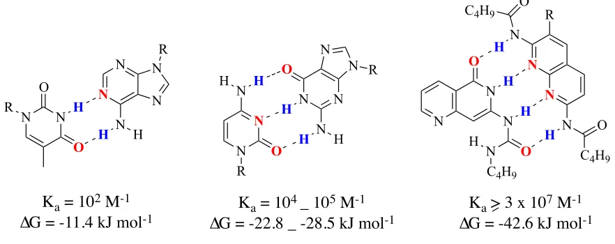
bonds and no repulsive secondary interactions.
N N N N R N H H O H N N N H H O R N N N N R N H H N N O O R H
Ka = 102 M-1 ∆G = -11.4 kJ mol-1
Ka = 104 _ 105 M-1 ∆G = -22.8 _ -28.5 kJ mol-1
N
N N
O C4H9 R
N
H
C4H9 O N
N N
N H
C4H9 O O
H
H H
Figure 2: Secondary interactions based on the sequences of donors and acceptors in
triply hydrogen bonded complexes.
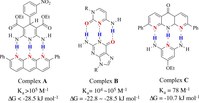
For example, complex A has a Ka value >105 M-1 in CDCl3 (Figure 3).7 In
comparison, the AAD-DDA complex has two attractive secondary hydrogen bonds and
two repulsive secondary interactions and because of that it has a lower Ka value than
complex B (Ka= 104-105 M-1). 6 Finally, the ADA-DAD complex with four repulsive
secondary interactions has the lowest Ka value for triply hydrogen bonded complexes
(complex C with Ka = 78 M-1).1
Figure 3: Ka values of triply hydrogen-bonded complexes in CDCl3.
N N N
Ph Ph N N H N H O OEt O OEt H NO2 N N N N N N O N H H O R R N H H H
N N N
N Ph Ph O H N H H N H H OEt
H H H
Complex A Complex B Complex C
Ka >105 M-1
∆G < -28.5 kJ mol-1
Ka= 104 ~105 M-1
∆G = -22.8 ~ -28.5 kJ mol-1
Ka = 78 M-1
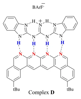
In 2011, Leigh and coworkers combined the effect of both hydrogen bond number
and the secondary interactions to synthesize complex D, which contains an AAAA–
DDDD quadruple hydrogen-bond complex that has an extremely high Ka value in
non-polar solvents (Figure 4).8
Figure 4: an AAAA–DDDD quadruple hydrogen-bond complex.
N N N N
tBu tBu
N
N N N
N N
N
H H
BArF
-H H H H
Complex D
Ka > 3 x 10¹² M–1, ∆G = - 71.1 kJ mol-1in CH2Cl2
Ka= 1.5 x 10⁶ M–1, ∆G = - 35.2 kJ mol-1 in CH3CN
1.1.2.3 Acidity and basicity of the donor and acceptor
The individual or primary hydrogen bond strength is affected by the acidity of the donor
and basicity of the acceptor. Applying that to a multiple hydrogen bonding complex, we
can determine the strength and stability based on the expecting the pKa values and
altering it by changing the functional groups on the complex.1 In 1966, Yoshimasa and
coworkers investigated the effect of substituents of the hydrogen bonding of adenine and
uracil derivatives on the association constants of their complexes (Figure 5). Their study
indicated the role substituents might play to enhance or decrease the strength of the
adenine-uracil association.
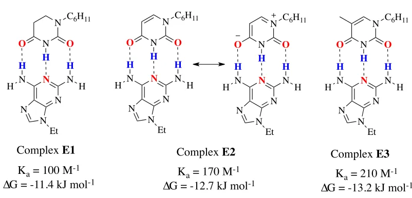
Figure 5: The effect of substituents on the association constant.
First, the loss of aromaticity because of the hydrogenation in 5,6-dihydrouracil is
responsible for the increase of the Ka value in complex E2 than E1. The resonance in
N
N C6H11
O O H N N N H H N H H N N Et N
N C6H11
O O H N N N H H N H H N N Et N
N C6H11
O O H N N N H H N H H N N Et
Ka = 100 M-1
∆G = -11.4 kJ mol-1
Ka = 170 M-1
∆G = -12.7 kJ mol-1 Ka = 210 M
-1
∆G = -13.2 kJ mol-1
Complex E1 Complex E2 Complex E3
N
N C6H11
complex E2 gives the 4-carbonyl oxygen atom more electron density and more basicity
as a result. In addition, the pKa of the amide group increased from 9.4 in uracil to 11 or
larger in E2. Secondly, adding a methyl group to uracil in complex E3 increased the
association complex. This increase is due to the role of the methyl group as an electron
donor which increased further the basicity of the 4-carbonyl oxygen and increased the
pKa value of the amide group from 9.4 to 9.9.9
Moreover, having appropriate charge in the complex will increase stability.1
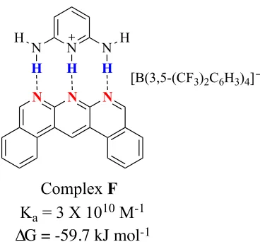
We can see the huge increase in strength that a complex gains from the Ka value of complex
F, (Ka = 3 X 10 10
M-1
in CH2Cl2) because the positive charge in the cationic DDD
enhances the hydrogen bond donor character and gives electrostatic stabilization to the complex.10
Figure 6: the effect of charge on the association constant.
N N N
N
H
N
H
H N
H
H
[B(3,5-(CF3)2C6H3)4]
Complex F Ka = 3 X 10¹⁰ M-1
1.1.2.4 Solvents
Another important factor that affects the strength and stability of hydrogen bonded complexes is the solvent. Generally, there is an inverse relationship between solvent polarity and hydrogen bonding strength. Less polar solvents such as hydrocarbons increase the strength while protic or polar solvents such as water or DMSO decrease it.1
Therefore most hydrogen bonded supramolecular complexes are characterized in
chloroform or toluene, but unfortunately the solubilities of some donors or acceptors are poor in these solvents and this can cause complications. In 2007, Hunter and his
coworkers studied the effects of different solvents on the association constants for
formation of a 1:1 complex between tri-n-butylphosphine oxide and perfluoro-tert-butyl
alcohol at 295 K. The results in the table below illustrates the higher association constants
in CCl4 and cyclohexane over DMSO and other polar solvents due to lack of competition
with the hydrogen bonds of the complex.11
Solvents
Association constants (K
ain
M
-1)
DMSO 6.8 x10⁻¹
Acetone 6.5 x10⁻¹
Acetonitrile 1.6 x10²
Tetrahydrofuran 2.4 x10²
CHCl3 2.7 x10³
CCl4 7.6 x10⁴
In another study, Rehm and Schmuck reported many solutions to increase the strength of self-assembly in polar solvents based on previous research and their own research because most hydrogen bond assemblies are not stable enough to be useful in polar protic solvents on their own. 2
First, using strong metal–ligand interactions in addition to hydrogen bonds gives the hydrogen bond-mediated assembly stability in water because a coordination bond between the metal and the ligand can be as stable as a covalent bond. On the other hand, this technique can have disadvantages such as toxicity and high cost. Ion-pair formation is the second solution because the electrostatic
interaction is more stable than hydrogen bonding but not as much as the coordinate bond in metal–ligand interaction and this charge interaction beside hydrogen bonding is very important for the stability of a self- assembly in polar protic solvents. Finally, adding functional groups that have an additional electron-donor or acceptor to the monomer contributes with the other methods to form a stable and strong self-assembly in polar protic solvents.2
However, these solutions may still not work in polar protic solvents if we employ only individual complexes. Multivalency of many of the same groups on the surface of a nanoparticle will also contribute to the stability of an aggregate by increasing the number of recognition events.
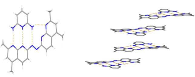
and formed a 1:1 complex with four primary hydrogen bonds (Figure 7, right).
Figure 7: The intended 2:1 donor: acceptor complex (left) and the resulting 1:1 complex (right) observed in the solid state.
The crystal was obtained from acetonitrile and isopropyl ether, and there are four primary hydrogen bonds. In addition, there are π-π stacking interactions between the planar complexes (Figure 8).12
Based on the previously reported complex F, and because of the extra hydrogen bond this complex should have Ka >> 3 X 10
10
M-1
in chlorinated solvents.
Figure 8: The x-ray crystal structure of the 1:1 complex.12 Primary hydrogen bonds (left)
and π-π stacking interactions (right ) are evident in the structure.
N N N
N N N
N N N H H H H H N N N H H H H
H N N N N
N N N N H H H H H N B Ph Ph Ph Ph
B Ph Ph Ph Ph B
1.2 Magnetic Resonance Imaging (MRI)
1.2.1 MRI and Tumour imaging
In another important field, scientists continually attempt to find the best techniques for clinical diagnosis in order to help them differentiate between normal and diseased tissue without having to perform surgery to elucidate a patient’s illness. Since the invention of Magnetic Resonance Imaging (MRI), many contrast agents have been used to change the relaxation times of protons in water molecules in order to better distinguish between normal and diseased tissue. MRI has been used for cancer imaging for many years, 13
as cancer is one of the leading causes of death. While most previously developed contrast agents distinguish tumors from normal tissue on the basis of their leaky vasculature, there is a potential for tumor imaging based on the fact that differences in pH exist between normal tissues and the blood stream which are around 7-7.4 and between tumors which are more acidic where the pH can reach 6 in the extracellular core of the tumor.14
1.2.2. MRI Principles
The principles of MRI are similar to those of Nuclear Magnetic Resonance (NMR) spectroscopy and depend on the density of protons in the targeted area and the degree of polarization of the nuclear spin states.15 When placed in a strong magnetic field (B
0),
proton nuclei, which have a non-zero magnetic moment, will orient either anti-parallel (high energy state) with the main magnetic field (B0) or parallel (low energy state) with a
slight majority reaching a thermal equilibrium, and a precessional motion about B0 at
jobs in MRI. First, excitement of the proton nuclei occurs in response to a radiofrequency (RF) pulse from this coil, which creates a new magnetic field (B1). The RF pulse affects
the orientation of the proton nuclei, causing tipping of the overall net magnetization toward the transverse plane, which is perpendicular to the main magnetic field (B0). This
tipping is due to transition of some spins from the low energy state to high energy state after absorbing energy from the RF. Also, the spins become phase coherent in the transverse plane. In order to begin detecting MR signals, the RF pulse will be turned off allowing nuclei to return into their original states (before performing a radiofrequency (RF) pulse). These MR signals that are caused from the oscillation of the protons translate into electrical current in the same radiofrequency (RF) coil creating the MR signal.16
There are two relaxation mechanisms depending on how the nuclei return to their original states. T1 and T2 are relaxation times corresponding to longitudinal and
transverse relaxation mechanisms. T1 is defined by the time that a radiofrequency excited
proton in (B1) magnetic field takes to return to the main magnetic field (B0) or to the
longitudinal axis after turning off the RF pulse. T2 corresponds to the decay of the proton
around the transverse axis (B0) after losing phase coherence.
15 The differences in water
concentration and environment between healthy and diseased tissues give inherent differences in T1 and T2 relaxation times. For example, since the water concentration is
higher in cancerous tissue than the normal tissue, the T2 values for them are different,
0.043 seconds for normal and 0.056 seconds for cancerous tissue, and they exhibit different T1 values as well.
1.2.3. Paramagnetic Contrast Agents
The using of paramagnetic contrast agents has rendered MRI one of the most important medical imaging techniques currently developed because of the advantages of these paramagnetic contrast agents comparing to those for other imaging modalities. For example, they do not emit ionizing radiation. They are not visualized directly but rather affect the relaxation of water and fat protons in their surrounding areas.17
There are two main types of paramagnetic contrast agents that are clinically used based on their effect on T1 or T2 relaxation times. First, gadolinium contrast agents affect T1 relaxation times
more strongly and are called positive contrast agents because they lead to increases in the MRI signal intensity under most pulse sequences. Second, superparamagnetic iron oxide nanoparticles alter T2 relaxation times more strongly and they are called negative contrast
agents. They lead to decreases in the MRI signal intensity, often called signal voids in their vicinity.
1.2.4 Superparamagnetic iron oxide nanoparticles (SPIO) as contrast agents
The emergence of superparamagnetic iron oxide nanoparticles was very important to avoid some disadvantages such as short retention time in vivo, toxicity and
biocompatibility of other contrast agents like gadolinium.13
Another advantage of iron oxide nanopaticles is their sensitivity and they present inhomogenities in the magnetic field causing a decrease in T2 relaxation times of healthy tissue. This decrease generates a
big difference in T2 relaxation time between normal and diseased tissues and, as a result,
the differences in the signal between them.16,19
In order to allow these nanoparticles to work in cells, iron oxide nanoparticles are coated by water soluble polymers such as dextran to render them soluble in aqueous or biological media and also to make a colloidal suspension more stable.20
Iron oxide nanoparticles were functionalized with azide groups and further derivatized using click chemistry.19
This method facilitates the conjugation of the nanoparticles to any active targeting group for many applications in MRI.
1.5 Targeted iron oxide nanoparticles
One of the important uses of the iron oxide nanoparticles is targeting specific
areas in the body by taking advantage of in vivo macrophages or attaching specific
targeting groups to the surface of the nanoparticle. First of all, iron oxide nanoparticles
can be used to image the liver and spleen due the natural uptake of the nanoparticles by
macrophages, which work as non-specific defenses against any foreign material.21
Despite the benefit of macrophages in imaging, it makes it difficult to image any other
nanoparticles is a highly desirable goal. The size of the nanoparticles should be less than
100 nm to increase the prolonged circulation in the blood stream and avoid non-specific
uptake by macrophages. There are many examples of attaching peptides, antibodies and
small molecules to the surface of nanoparticles. As an example of attaching peptides, in
2005, glioma brain tumors were detectable by both MRI and fluorescence imaging. This
detection was enabled by attaching chlorotoxin and Cy5.5 to coated iron oxide
nanoparticles with covalently bound bifunctional poly(ethylene glycol) (PEG).
Chlorotoxin peptide has a high affinity for the membrane-bound matrix
metalloproteinase-2 endopeptidases that are found in gliomas, medulloblastomas, the
neuroectodermal origin tumors.22 Secondly, in the area of using antibodies, having a
prostate-specific membrane antigen mostly on the neovasculature of solid tumors and on
the surface of prostate cells helped to develop biotinylated anti-PSMA iron oxide
nanoparticles that improve the magnetic resonance detection of prostate cancer cells.23
Finally, folic acid was conjugated to superparamagnetic iron oxide nanoparticles coated
by covalently bound bifunctional PEG. It is known that the folate receptor is
overexpressed on the surfaces of many human tumor cells such as ovarian, lung, breast,
endometrial, renal, and colon cancers. This conjugation of folic acid on the nanoparticles
results in a significant uptake by target cells in comparison with nanoparticles coated with
PEG or dextran alone.24 Thus, in order to target specific areas for in vivo imaging,
N N
O N
N N
N N
H
H H
H H
O O O
Primary hydrogen bond
Attractive secondary interaction
Click to substrate/ nanoparticles
N
1.3 The Proposal
This project combines the two described important fields above. It involves the synthesis of a pH-sensitive hydrogen bonded complex and its attachment to iron oxide
nanoparticles by click chemistry in order to increase the sensitivity of these nanoparticles for tumor imaging. This increased sensitivity will be achieved because the hydrogen bonding moieties will be designed to assemble selectively at the mildly acidic pH of tumor tissue. This bonding is anticipated to induce clustering of the nanoparticles. It has been previously demonstrated that nanoparticle clusters exhibit significantly higher relaxivity than individual nanoparticles.25
Therefore, the ability to detect the tumor will be enhanced by this selective clustering in tumor tissue. Based on the requirement for
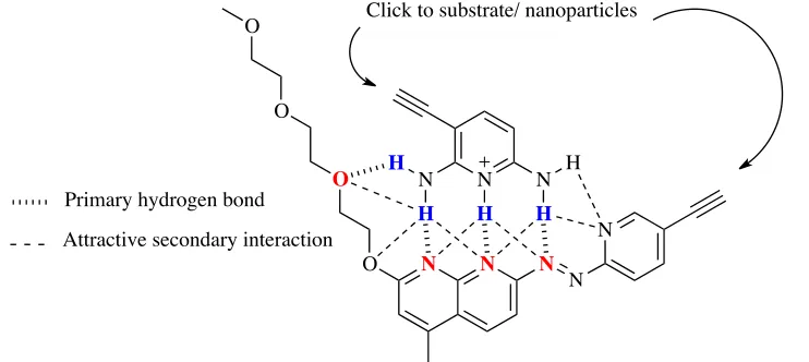
hydrogen bonding induced by mildly acidic pH and previous reports on hydrogen bonding moieties,10,12 the monomers were chosen (Figure 10). The hydrogen bonding
monomers are 3-ethynylpyridine-2,6-diamine (donor molecule) and
(E)-7-((5- ethynylpyridin-2-yl)diazenyl)-2-(2-(2-(2-methoxyethoxy)ethoxy)ethoxy)-4-methyl-1,8-naphthyridine (acceptor molecule).
The pKa value of the donor molecule was measured by weak acid-strong base titration
and it was found to be 6.5.
This value means that under pH 6.5 there will be more protonated molecule, 2,6-diamino-3-ethynylpyridin-1-ium than 3-ethynylpyridine-2,6-diamine. The formation of this cation and having more of it within this region should enhance assembly by hydrogen bonding and π-π stacking. The design of the pH-sensitive hydrogen bonded complex takes into account some of the factors that can help to make it a strong and stable complex and that will work in polar protic solvents such as water. First, the sequences of donors and acceptors is an AAAA-DDDD arrangement, so it increases the number of attractive secondary electrostatic interactions between donor and acceptor atoms and eliminates any repulsive interaction between them. Second, using a hydrophilic group (triethylene glycol) in the acceptor molecule improves its water solubility. Also, adding functional groups that have electron-acceptors (oxygen atoms) to the acceptor molecule increases the number of the hydrogen bonds in the complex in order to form a stable self-assembly in polar protic solvents.
N
H2N NH2
H N
H2N NH2
1.3.1 The design
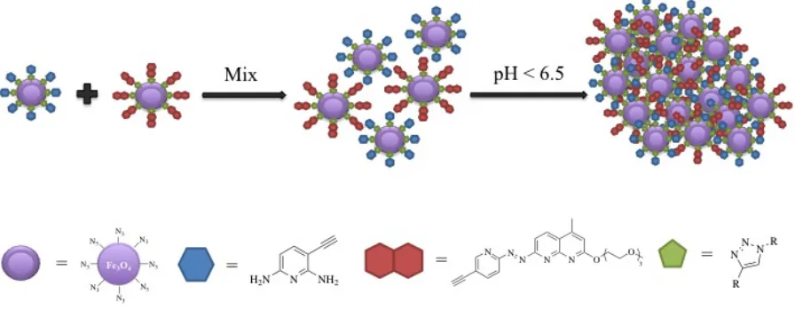
The design of this project has two main lines of inquiry. First, the donor and the acceptor molecules are going to be attached to the iron oxide nanoparticles by click chemistry between the alkyne group in the hydrogen bonding molecules and the azide group in the nanoparticles, separately and then mix them together (Figure 11) and their behavior will be observed with respect to aggregation in response to pH changes.
Figure 11: clicking donor and acceptor molecules to SPIO nanoparticles. Second, the donor and the acceptor molecules are going to be attached to bifunctional monomers by click chemistry, between the alkyne group in the hydrogen bonding
Figure 12: clicking donor and acceptor molecules to a bifinctional monomer.
2.1. Synthesis
2.1.1 Synthesis of Donor Molecule.
Scheme 2: Synthesis of the donor molecule.
2,6-diamino-3-ethynylpyridin-1-ium 4 was chosen as the DDD molecule and contains an
alkyne functional group for eventual click chemistry to a covalent scaffold. The synthesis
of 4 began with the iodination of commercially available, 2,6-diaminopyridine (Scheme
2). This reaction was performed based on a previously reported synthesis 26 and was
monitored while adding iodine dropwise over 4 hours to avoid the formation of
3,5-diiodopyridine-2,6-diamine, since NH2 groups in positions 2 and 6 on the pyridine ring
activate both carbons 3 and 5 for electrophilic aromatic substitution. A saturated aqueous
solution of sodium thiosulfate was used in the workup to remove extra unreacted iodine,
and the mixture was filtered through a celite pad to remove potassium iodide. A 57%
yield of the monoiodo-pyridine-2,6-diamine was obtained and this yield was the best
compared to other methods undertaken to obtain 1 and much easier. Sonogashira
N
H2N NH2 H2N N NH2
Si
N
H2N NH2 N N N
H K2CO3, I2
N
H2N NH2 I THF, overnight,
R.T.
PdCl2(PPh3)2, CuI TEA, TMSA overnight, R.T.
Methanol, 2h, R.T.
1(57%) 2 (> 99%)
3 (55%)
H H
H H
pH below 6.5 K2CO3
coupling was then used to convert 1 to 2 as a first step to attach the alkyne group.27 The
steps to produce 2 and 3 were performed very carefully using oven-dried glassware,
deoxygenated solvents and a nitrogen atmosphere. Saturated aqueous sodium bicarbonate
was used in the workup of the first step to neutralize the trace acid in the products and it
gave a quantitative yield of 2. Removal of the trimethylsilyl (TMS) protecting group was
performed using 2 equivalents of K2CO3 in methanol,28 and gave a 55 % yield after
2.1.2 Synthesis of Acceptor molecule.
Scheme 3: Synthesis of 6.
The synthesis of the acceptor subunit 14 began with the formation of 5 from a
condensation between 2,6- diaminopyridine and ethyl acetoacetate in acidic conditions
and a yield of 46% was obtained (Scheme 3).29,30 Naphthyridine 5 work up was done by
simple dilution and neutralization in water to precipitate the product in pure form most of
the time. For further purification, recrystallization was performed on the recovered solid
using acetone:diethyl ether (1:1). The nucleophilic substitution to form triethylene glycol
derivative 6 was performed by adding 1-iodo-3,6,9-trioxodecane 8 within half an hour of
adding base and the reaction was stirred overnight.
N
H2N NH2 O
O O H
3PO4, reflux, 3 h, 110 °C
N N OH H2N
O I
5 (46%)
6 (63%)
60% NaH, DMSO
N N O
H2N O 3
3
Scheme 4: Synthesis of 8.
Iodo-3,6,9-trioxodecane 8 was synthesized to be a derivative of tri(ethylene glycol)
bearing a good leaving group for the above described synthesis of 6 (Scheme 4). Both
molecules 7 and 8 were prepared according to literature procedures.31,32
Scheme 5: Synthesis of 12.
The synthesis of 5-iodo-2-nitrosopyridine, 9, was initiated by iodination of
aminopyridine (Scheme 5). 2 equivalents of iodine were added all at once to
2-aminopyridine and the reaction was stirred overnight in DMF at room temperature.33 A
O O
O
HO O O
O O O O O I NaI Acetone, reflux, overnight NaOH, TsCl
THF, H2O, overnight, R.T.
7 (93%)
8 (80%)
Ts
N
DMS, NCS, 0 °C, 6 h
NaOCH3, CH3OH, H2O
Overnight, R.T.
N
m-CPBA, DCM, 0 °C, 3 h
DMS, 1 h, R.T.
10 (> 99%)
PdCl2(PPh3)2, CuI
TEA, TMSA overnight, R.T. N Si N Si
11 (58%)
I
N
I2
DMF, Overnight, R.T.
9 (30%)
12 (75%)
saturated aqueous solution of sodium thiosulphate was added to remove the unreacted
iodine and to neutralize the reaction mixture. This reaction was followed by a
Sonogashira coupling reaction to afford 9 in a quantitative yield.34 The sulfimine
derivative 10 was synthesized by a previously reported methodwith a modification of the
reaction time to obtain higher yield. Flash column chromatography was performed using
EtOAc as eluent to yield 58% of 11. Oxidation using 1.3 equivalent of m
-chloroperbenzoic acid (m-CPBA) at 0 °C was performed to give 12 in 75% yield after
addition of dimethylsulfide to remove the extra unreacted m-CPBA.35,36 Workup of the
reaction required evaporation at 20-30 °C to avoid producing a dimer. It has been shown
in a previous study that 2-nitrosopyridine molecules exist in organic solvents as a mixture of the monomer and azodioxy dimer in equilibrium at room temperature.37
Scheme 6: Synthesis of 13.
After the synthesis of 12 and 6, the azo product
(E)-2-(2-(2-(2-
methoxyethoxy)ethoxy)ethoxy)-4-methyl-7-((5-((trimethylsilyl)ethynyl)pyridin-2-yl)diazenyl)-1,8-naphthyridine was synthesized from them using 1.3 equivalent of
iodosobenzene diacetate (Scheme 6).38 Purification by column chromatography using 4%
methanol in DCM, eluent gave 32% of 13.
N H2N N N O
PhI(AcO)2
DCM, 30 °C, Overnight O
3 N N N O
O
3
N N Si
Si
12 6 13 (32%)
Scheme 7: Synthesis of the acceptor molecule, 14.
Finally, 1 M tetra-n-butylammonium fluoride (TBAF) in THF was used to remove the
trimethylsilyl (TMS) protecting group in 13 under dry conditions and in just 10 minutes,
providing an 80% yield of the acceptor (Scheme 7).39
N N O
N O 3
N N
Si
THF, 10 mins, R.T. 1 M TBAF
N N O
N O 3
N N
2.1.3 Synthesis of Dextran Coated Superparamagnetic Iron Oxide Nanoparticles
The synthesis of water soluble SPIO nanoparticles was performed using a previously
reported method.40 The first step involved the decomposition of FeCl2 and FeCl3 in the
presence of ammonium hydroxide and dextran to produce water soluble dextran coated
nanoparticles. As Weissleder and coworkers reported,6 a cross-linked dextran shell with
amine groups ( Scheme 8) on the surface was synthesized, by using the previous
synthesized coated nanoparticles and treating them first with epichlorohydrin and then
aqueous ammonium hydroxide.
Scheme 8: Synthesis of dextran-coated Fe3O4 nanoparticles.
Dynamic light scattering (DLS) was used to measure the sizes of the particles (Figure
13). The Z-average size was found to be 66 nm for the dextran-coated Fe3O4
nanoparticles 15 and 77 nm for the cross-linked amine functionalized nanoparticles 16.
FeCl3.6H2O
FeCl2.4H2O
Dextran 70 K
7.5% NH4OH
O Cl
5M NaOH
30% NH4OH
NH2 H2N
H2N
H2N
NH2
NH2
NH2
NH2
Fe3O4 Fe3O4
15
Figure 13: Size distrubution of nanoparticles 15 (gray) and 16 (dark purple).
Figure 24: UV-Vis absorbance of nanoparticles 15 (gray)and 16 (dark purple).
0 5 10 15 20
0.1 1 10 100 1000 10000
V ol u me (%) Diameter (nm) Nanoparticles 16 Nanoparticles 15 0 0.5 1 1.5 2 2.5 3 3.5
300 350 400 450 500
UV-Vis absorption was also performed following nanoparticle degradation with
hydrochloric acid and hydrogen peroxide on both nanoparticles 15 and 16 to determine
the concentration of iron.41 The calculation of concentration was based on the UV-Vis
absorption that was found to be 3.01 at 365 nm for nanoparticles 15 corresponding to a
concentration of 10.9 mg/mL and 1.80 at 365 nm for 16 corresponding to a concentration
of 6.49 mg/mL.
2.1.4 Synthesis of the linker.
Scheme 9: Synthesis of the linker.
The synthesis of the linker for attachment of the hydrogen bonding moieties to the
nanoparticles began with a monotosylation of tri(ethylene glycol) by reaction with 0.5
equivalents of triethylamine and 0.25 equivalents of 4-toluenesulfonyl chloride, affording
88% of 17 (Scheme 9).42 Crude 17 was carried forward to the synthesis
2-(2-(2-azidoethoxy)ethoxy)ethanol, 18, without any purification.43 After reaction, flash column
HO O O OH HO O O O Ts
O
O N3
O O O N3
HO
O O
O
O N3 O
O N O
O 0.5 eq TEA, 0.25 eq TsCl
DCM, 1 h, R.T.
2 eq NaN3
DMF, Overnight R.T.
Br
OH O
, 2 eq 60% NaH
dry THF, refux 24 h.
N O
O OH
DCC, CH3CN HO
18 (85%)
19 (62%)
17 (88%)
chromatography was performed on the residue using 1:1 ethyl acetate:hexane as eluent,
affording 85% of 18. The carboxylic acid derivative 19 was synthesized by using sodium
hydride (dispersion) as a base and alkylating the resulting oxo-anion with bromoacetate.44
The final step to make the succinate ester linker, 20 was performed according to a literature procedure and afforded a similar yield.19
2.1.5 Conjugating donor and acceptor to nanoparticles
Scheme 10: Synthesis of the functionalized nanoparticles.
The next step in order to install the azide groups on the nanoparticles, was to react linker
20 with the cross-linked nanoparticles 16 (Scheme 10). This reaction was obtained by
simply stirring 2.5 µmol of linker 20 per milligram of iron at a pH of 7.4 overnight in the
dark.19 The pH was reached by performing a predialysis against 25 mM pH 7.4 citrate
O O O N3
O
O N O
O
H2O/DMSO
overnight in the dark
Or
CuSO4, sodium ascorbate in H2O
Or 20 21 22 23 N3 N3 N3 N3 N3 N3 N3 N3 Fe3O4
NH2 H2N
H2N
H2N NH2
NH2 NH2
NH2 Fe3O4
N H2N NH2
= N N N O O3
N N
= NNN
R R = = N3 N3 N3 N3 N3 N3 N3 N3
buffer. The UV-visible absorbance was measured again following nanoparticle
degradation with hydrochloric acid and hydrogen peroxide after performing dialysis of
nanoparticles, 16 and for 21. The absorption was found to be 1.34 at 365 nm
corresponding to a concentration of 4.82 mg/mL for 16 and0.508 at 365 nm
corresponding to a concentration of 1.83 mg/ml for 21 (Figure 15). The Z-average size of
particles 21 was found to be 90 nm (Figure 16). In addition, the presence of the azide
groups on the nanoparticle surface was verified using infrared (IR) spectroscopy by the
presence of the distinctive azide stretch at 2100 cm-1 (Figure 17).
Figure 15: UV-Vis absorbance of nanoparticles 16 in pH 7.4 aqueous buffer (dark
purple) and 21 (light purple).
0 0.2 0.4 0.6 0.8 1 1.2 1.4
300 350 400 450 500
A
b
sor
b
an
ce
Wavelength (nm)
Nanoparticles 21
Figure 16: Size distrubution of nanoparticles 21.
Figure 17: Infrared spectra illustrating changes on nanoparticle surface.
Finally, attachment of the donor and acceptor moieties to the surfaces of the
nanoparticles was accomplished using a click reaction between their alkyne group and the
0 5 10 15 20 25
0.1 1 10 100 1000
V
ol
u
me
(%)
Diameter (nm)
Nanoparticles 21
0.9 0.92 0.94 0.96 0.98 1
1300 1800 2300 2800 3300 3800
Wavenumbers (cm-1)
Nanoparticles 21
azide functions on the nanoparticles (Scheme 10). The click reaction was performed
using CuSO4 and sodium ascorbate and dissolving the donor and acceptor molecules in
DMF with the nanoparticles. The overnight reaction was followed by dialysis against 10
mM ethylenediaminetetraacetic acid (EDTA) to remove residual copper salts, followed
by pure water.19 The disappearance of the peak corresponding to the azide stretch was
verified using infrared (IR) on the nanoparticles confirming completion of the reaction
(Figure 17). The Z-average sizes of the particles were found to be 210 nm for 22 and 110
nm for 23 using DLS (Figure 18).
Figure 18: Size distribution of nanoparticles 22 (red) and 23 (blue).
0 5 10 15
0.1 1 10 100 1000 10000
V
ol
u
me
(%)
Diameter (nm)
18). 2.1.6 Synthesis of Donor and Acceptor monomers
Scheme 11: Synthesis of the PEG derivative.
Synthesis of the PEG derivative 25 was necessary in order to do the click
chemistry described above (Scheme 11). Synthesis of 24 was first performed by reacting
hexa(ethylene glycol) with 6.6 equivalents of KOH and 2.5 equivalents of TsCl.45
Recrystallization was performed to remove the mono tosylated derivatives. Compound 25
was synthesized according to a literature procedure.46
Donor and acceptor monomers were synthesized by click reactions (Scheme 12).
The synthesis of the donor monomer 26 was performed by reaction between 2 equivalents
of 3-ethynylpyridine-2,6-diamine 3 and 1 equivalent of 25, while the acceptor monomer
27 was synthesized by reaction between 2 equivalents of
(E)-7-((5-ethynylpyridin-2-yl)diazenyl)-2-(2-(2-(2-methoxyethoxy)ethoxy)ethoxy)-4-methyl-1,8-naphthyridine, 14
and 1 equivalent of 25. Both click reactions were performed in the present of 0.65
equivalents of CuI and 6.5 equivalents of 1,8-diazabicycloundec-7-ene (DBU).47
Different workups were used for the two reactions due to the insolubility of the donor
OH O
H Ts O O Ts
N3 O
NaN3
DMF, 2 days 110 °C
KOH, water
TsCl, THF 36 h, R.T.
N3
6 6
5
24 (90%)
monomer 26 in most organic solvents. A saturated solution of EDTA was required as a
chelating agent to scavenge the metal ions in the workup.
Scheme 12: Synthesis of the donor, 26 and Acceptor, 27 monomers.
N O
N N
N NH2
H2N
N O
N N
N N H2N NH2
N N O
N O3
N
N 0.65 eq CuI, 6.5 eq DBU
Toluene, 2h, 70 °C, N2
N3
O N3 5
5 N
N N
N H2N
NH2
5 N N N N N N N N O O N N N N O O 3 3 Or Or = =
3 14 25
26 (33%)
2.2. pKa Titrations
2.2.1.pKa of the Donor Molecule.
Before starting the synthesis of 3, a weak acid-strong base titration was performed to
determine the pKa value for the nitrogen atom in the 2,6-diaminopyridine ring. This
titration was performed by adding 1 mL of 0.1 M NaOH to 0.1 M of
2,6-diaminopyridin-1-ium chloride per minute and measuring the pH using a glass pH electrode at each time
point. Diaminopyridin-1-ium chloride was synthesized by the protonation of
2,6-diaminopyridine with HCl (gas) that was produced by adding H2SO4 dropwise to NaCl
(Scheme 13). Figure 19 shows the change in the pH while adding NaOH to
2,6-diaminopyridin-1- ium chloride and from the figure, the pKa can be determined which is
likely very similar to the pKa for 3.
Scheme 13: synthesizing 2,6-diaminopyridin-1-ium chloride.
N
H2N NH2
H N
H2N NH2
Cl HCl (gas)
Figure 19: Weak acid-strong base titration of 2,6-diaminopyridinium chloride.
2.2.2. pKa of the Donor Monomer.
A weak acid-strong base titration was performed on 26 to determine the effect of the
triazole group on the pKa of the donor. This titration was performed by adding 1 mL of 5
x 10-4 M NaOH to 5 x 10⁻⁴ M of HCl salt of 26 per minute and measuring the pH using a
glass pH electrode at each time point. The HCl salt of 26 was synthesized by dissolving
26 (0.1 g) in 0.1 M HCl (2 mL) and adding it dropwise to a saturated solution of NaCl (3
mL). Figure 20 demonstrates that the effect of adding a triazole group to the donor
2.3. The Effect of Changing pH on the Aggregation of the Nanoparticles
The effect of changing pH on the aggregation of the nanoparticles was studied by
measuring the Z-average size of 0.2 mg/mL nanoparticle solution (0.1 mg/mL of 22 + 0.1
mg/mL of 23) at different pHs. The solution’s pH was altered by adding 1 x 10⁻³ M HCl
and 1 x 10⁻³ M NaOH and the pH was measured with a pH meter. This test was also
performed on both nanoparticles 22 and 23 alone at the same iron concentration.
Figure 21: Z-average size changing with pH.
Figure 21 shows that an increase in the Z-average size of the nanoparticle mixture started
at pH = 6 which is the pKa of the donor molecule and reached the maximum size at 5.
The decrease in the size after that point may be due the start of protonation of the
naphthyridine rings in the acceptor molecule. This protonation of the acceptor is
responsible for the Z-average size increase between 4 > pH > 3 in nanoparticle 22.
Although having these changes, they were not significant as we expected. 0
50 100 150 200 250 300 350
3 4 5 6 7 8
Z-ave
rage
s
iz
e
pH
2.4. X-ray crystallography
An X-ray crystal structure was obtained for the acceptor 14 that explained the unexpected
results of the aggregation tests (Figure 22).
Figure 22: X-ray crystal structure of 14.
Iodosobenzene diacetate was used to join the pyridine and naphthyridine components
together but it was unclear whether an azo or azoxy bonds was created. MS was
performed on the sample and showed an azo bond but it was not until this crystal
structure was obtained that it was revealed that actually an azoxy bond was synthesized
instead of the oxygen (O1), a structural difference that was very difficult to elucidate by
simple NMR spectroscopy. This improper placement blocks a hydrogen bonding site and
disrupts the intended complexation with the donor. Based on the X-ray crystal structure
of the acceptor molecule, the following revised scheme explains what happened in each
step.
Scheme 14: synthesis of the acceptor
N
PhI(AcO)2
DCM, 30 °C, Overnight
THF, 10 mins, R.T. 1 M TBAF Si
12 N O N
H2N NH2 O
O O
H3PO4, reflux, 3 h, 110 °C
N NH O
H2N
O I
5
6 60% NaH, DMSO
N N O
H2N 3
8
O
3
6
N N O
H2N
O
3
13
N N O
N O 3 N N Si O 14
N N O
2.5. 1H NMR Titration
An 1H NMR titration was performed between 2,6-diaminopyridinium tetrafluoroborate
(donor) and (E)-2,4-dimethyl-7-(pyridin-2-yldiazenyl)-1,8-naphthyridine (PAN)
(acceptor). This complex (figure 23) is structurally similar to the desired system so the
association constant will provide insight into the strength of the proposed complex. It was
performed in DMSO, which is a very competitive solvent, and we obtained a Ka value of
194 M-1.
Figure 23: Titration curve for complex G.
N
N N
N
N N N H
H N
H
H H BF4
Primary hydrogen bond Attractive secondary interaction
Complex G
This thesis explored the synthesis of donor and acceptor hydrogen bonding
moieties for conjugation to SPIO. The aim was to develop a system that would undergo
aggregation in the mildly acidic environment of tumors, in order to provide enhanced
contrast. Though there were a few previous examples of controlled clustering of such
nanoparticles in the presence of stimuli, clustering induced by very small pH changes had
not previously been demonstrated. The donor molecule was successfully synthesized in
three simple steps including a Sonogashira coupling and deprotection of the TMS group.
The acceptor molecule synthesis involved the preparation of an amine derivative and a
nitroso derivative. Then, both derivatives were oxidized to obtain the azo group and
followed by deprotection of the TMS group. SPIO were synthesized and then
cross-linked after coating them with dextran. A linker was synthesized to obtain azide group on
the surface of the SPIO for further click reaction with the donor and acceptor molecules.
Donor and acceptor molecules were added to azide functionalized SPIO via click
chemistry. After conjugation to the nanoparticle surface, the results of clustering mixed
nanoparticles within the accessible tumor pH range were not significant. The presence of
the TEG group on one nitrogen atom in the naphthyridine ring and oxygen on the azo
group unexpectedly was revealed by x-ray crystallography during the final stages of this
thesis. This made the acceptor molecule too weak to form a strong complex, explaining
the lack of SPIO clustering. However, the idea of this project may be worth carrying
forward based on the strength of a similar complex that showed good binding strength in
DMSO.
Although synthesizing non-symmetrical azo heterocyclic molecules has proven to
shows a successful route without achieving an azoxy as an intermediate and adopting this
procedure is highly recommended for completion of this project. The PAN molecule
(Scheme 15) was successfully synthesized by a Ph.D. candidate Jeffrey Pleizier in the
Wisner group and the rest of the scheme is a suggested synthesis for an alternative
acceptor molecule.48,49,50,51
Scheme 15: Suggested future work.
N N
H2N
1.5 eq. NaNO2
H2SO4(aq)
N N
HO POCl Cl N N
3
62% 88%
2-hydrazinopyridine
nPrOH, 80 - 100 oC
79%
N HN
N H
N N
1.2 eq. PhI(OAc)2
DCM:DMSO
1:1 N N N
N N
PAN, 91%
2 eq. NBS, BPO, CCl4
70 oC, 12 h N N
N N N Br Br O O
60% NaH, DMSO
3 H N N N N N O Br O 3
60% NaH, DMSO
1 eq.
HO N N
4.1. General:
All chemicals were of reagent quality and used as obtained from commercial sources
unless otherwise noted. Dry solvents were obtained from an Innovative Technology
SPS-400-5 Solvent Purification System. Flasks were oven dried and nitrogen-purged prior to
use. Reactions were carried out under an inert atmosphere (dry nirogen) unless otherwise
indicated. Flash chromatography was performed using Silicycle pharma grade silica
(40-63µm). Dialysis was performed against water using a Spectra/Por regenerated cellulose
membrane (12-14,000 MW cutoff). Ultrafiltration was carried out using a 300 KDa
molecular weight cutoff (MWCO) membrane of regenerated cellulose purchased from
Amicon. The 1H and 13C NMR data were obtained on 400 or 600 MHz spectrometers. All
spectra were obtained in deuterated chloroform and/or DMSO-d6. The spectra were
referenced to residual chloroform (at δ 7.25 ppm for 1H spectra and the center peak of the
triplet at δ 77.0 (t) for 13C spectra) and DMSO-d6 (at δ 2.49 ppm for 1H spectra and the
center peak of the multiplet at δ 39.50 (m) for 13C spectra). When peak multiplicities are
given, the following abbreviations are used: s, singlet; d, doublet; t, triplet; q, quartet; m,
multiplet; EI (electron ionization) mass spectra were obtained on Finnigan MAT 8200
4.2 Experimental Section
4.2.1 Synthesis of Donor Molecule
3-Iodo-pyridine-2,6-diamine (1).
A suspension of 2,6-diaminopyridine (10.01 g, 0.091 mol) and
potassium carbonate (12.65 g, 0.091 mol) in THF (100 mL) was
stirred for 1 hour. A solution of iodine (23.3 g, 0.091 mol) in THF
(50 mL) was added dropwise over 4 hours to the suspension. The reaction mixture was
stirred overnight at room temperature. After the reaction time, the resulting residue was
filtered through a celite pad and the filtrate was washed with water and a saturated
aqueous solution of sodium thiosulphate to remove the extra-unreacted iodine. The
organic layer was dried over potassium carbonate and evaporated under reduced pressure
affording a gray solid of the title compound 1 (12.24 g, 57 %). 1H NMR (CDCl3, 400
MHz): δ 4.22 (s, 2H), 4.68 (s, 2H), 5.72 (d, 1H, J=8.2), 7.50 (d, 1H, J=8.2). 13C NMR
(400 MHz, CDCl3): δ 61, 100.5, 147.9, 156.3, 157.6. HRMS calcd for C5H6IN3 [M]+:
234.9606, found: 234.9607.
3-(Trimethylsilyethynyl) pyridine-2,6-diamine (2).
3-Iodo-pyridine-2,6-diamine (1, 12.0 g, 51.0 mmol),
PdCl2(PPh3)2 ( 0.35 g, 0.51 mmol, 1.0 mol %), and CuI ( 0.19 g,
0.99 mmol, 2.0 mol %) were charged in an oven-dried,
nitrogen-purged 250 mL round bottom flask under N2 atmosphere. Triethylamine (100 mL) which
was deoxgenated by being bubbled with N2 gas for 30 mintues was added via syringe
followed by the addition of TMSA (5.99 g, 61.01 mmol). The reaction stirred overnight
N
H2N NH2 I
N
under N2 at room temperature. After reaction time, solvent was removed under reduced
pressure. Saturated aqueous sodium bicarbonate (50 mL) was added to the residue and
chloroform (100 mL). The mixture was further extracted with chloroform (3 x 20 mL).
The combined organic layers were dried over potassium carbonate and evaporated under
reduced pressure to obtain dark brown solid of the titled compound 2 (10.4 g, 100%). 1H
NMR (CDCl3, 400 MHz): δ 0.23 (s, 9H), 4.36 (s, 2H), 4.82 (s, 2H), 5.81 (d, 1H, J=8.2),
7.31 (d, 1H, J=8.2). 13C NMR (400 MHz, CDCl3): δ 0.13, 97.7, 98, 101.7 141.9, 157.4,
159.2. HRMS calcd for C10H15N3Si [M]+: 205.1035, found: 205.1037.
3-Ethynylpyridine-2,6-diamine (3).
To 3-(trimethylsilyethynyl) pyridine-2,6-diamine (2, 10.4 g, 50.65
mmol) and potassium carbonate ( 13.99 g, 101.3 mmol), methanol
(100 mL) which was deoxgenated by being bubbled with N2 gas for
30 mintues was added under N2. The reaction solution stirred for 2 hours at room
temperature. After reaction, the solution was evaporated under reduced pressure and the
remaining residue was dissolved in chloroform and diluted with water. Extraction was
performed with chloroform (3 x 50 mL). The combined organic layers were dried over
potassium carbonate and evaporated under reduced pressure to afford (3.6 g, 55%) of the
donor molecule after recrystallization using chloroform: hexane. 1H NMR (CDCl3, 400
MHz): δ 3.31 (s, 1H), 4.36 (s, 2H), 4.81 (s, 2H), 5.84 (d, 1H, J=8.2), 7.34 (d, 1H, J=8.2).
13C NMR (400 MHz, CDCl
3): δ 80.5, 81.1, 97.8, 142.3, 157.6, 159.5. HRMS calcd for
[M]+ calc’d for C7H7N3: 133.0640, found: 133.0643
N
4.2.2 Synthesis of Acceptor Molecule.
7-Amino-2-hydroxy-4-methyl-1,8-naphthyridine (5).
2,6-Diaminopyridine (32.74 g, 300.01 mol) and ethyl
acetoacetate (39.05 g, 300.01 mol) were heated in phosphoric
acid (130 mL) for 3 hour at 110 °C. After cooling to room
temperature, the solution was poured into distilled water and neutralized by adding
ammonium hydroxide until pH: 7 and filtered affording a beige solid (24.03 g, 45.6 %) of
the titled product 5. 1H NMR (DMSO, 400 MHz):
δ 2.27 (s, 3H), 5.99 (s, 1H), 6.33 (d,
1H, J=8.6), 6.66 (s, 2H), 7.69 (d, 1H, J=8.6). HRMS calcd for C9H9N3O [M]+: 175.0746,
found: 175.0748.
Functionalized Naphthyridine (6).
To 7-amino-2-hydroxy-4-methyl-1,8-naphthyridine (5, 1.02 g, 5.8
mmol ) in DMSO (50 mL), 60 % sodium hydride (0.23 g, 5.8
mmol) was added to the solution carefully under N2. After 30
minutes, 1-iodo-3,6,9-trioxodecane (8, 1.58 g, 5.8 mmol) was
added via syringe and reaction stirred overnight under N2 at room temperature. After
reaction time, the solution was poured into distilled water and extracted many times with
dichloromethane. The organic layers were combined and washed with distilled water and
dried over potassium carbonate. The solvent was removed by rotary evaporation
affording a brown oil (1.17 g, 62.9 %) of the titled product 6. 1H NMR (CDCl3, 400
MHz): δ 2.31 (s, 3H), 3.36 (s, 3H), 3.51-3.53 (m, 2H), 3.60-3.63 (m, 4H), 3.70-3.72 (m,
2H), 3.75 (t, 2H, J=6.6), 4.63 (t, 2H, J=6.6) 4.97 (s, 2H), 6.29 (s, 1H), 6.36 (d, 1H,
N NH O
H2N
N N O
H2N