Human Identification with Face Detection Using Vector
Segmentation
Kandimalla Narmadha & P.S.S. Chakravarthy.
1M.tech-Scholar, Dept of ECE, Narasaraopeta Engineering College, Narsaraopet, A.P, India 2Associate Professor, Dept of ECE, Narasaraopeta Engineering College, Narsaraopet, A.P, India
ABSTRACT: We propose a new way to solve a very general blind inverse problem of multiple simultaneous degradations, such as blur, resolution reduction, noise, and contrast changes, without explicitly estimating the degradation. The proposed system is depends on combining semantic non-rigid patches, problem-specific high-quality prior data, and non-rigid registration tools. We exhibit how a significant quality enhancement can be achieved, both visually and quantitatively, in the facial images. The method is determined on the problem of cellular photography quality enhancement of dark facial images for different identities, expressions, and poses, and it is compared with the state-of-the-art denoising, deblurring, super-resolution, and color-correction methods.
Key words: Prior-based image quality enhancement, similarity measures, non-rigid registration, denoising, deblurring, super-resolution.
I. INTRODUCTION
In the past decades, handling common image flaws has gradually improved with the use of more sophisticated image priors and models. Early methods used pixel-based statistics, such as smoothness, piecewise
smoothness, total-variation, pixel
correlation, or wavelet decomposition for image reconstruction. In recent years, nonparametric patch-based methods, such as Nonlocal Means and BM3D, exploited local and nonlocal self-similarities. Other patch-
based, training-based methods were using Markov Random Fields and dictionary learning.
Today’s main state-of-the-art methods are based on square patches with little if any semantic context. In recent years, using generic image priors has started to reach an optimality bound; for example, for super-resolution and denoising. For facial images, facial priors were then used to break this limit; For example, face hallucination, or image compression using K-SVD. We propose an alternative concept of using large non-rigid patches with high semantic value.
also assume that no matches of high quality (HQ) and low quality (LQ) data are available for learning. As there is no degradation model, one also cannot generate faithfully LQ images by degrading HQ images (e.g. adding noise to a clean image). Experimental results are demonstrated on the problem of dark cellular image enhancement.
II.EXISTED SYSTEM
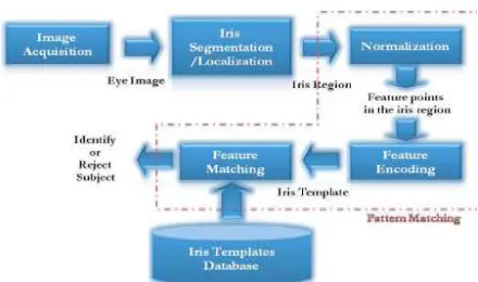
The existed system is to be composed of a number of sub-systems according to each stage of iris recognition. These stages are: • Image acquisition: Image acquisition means capturing image of eye.
• Segmentation: locating the iris region in an eye image is known as segmentation
• Normalization: Normalization means
creating a dimensionally consistent
representation of the iris region
• Feature encoding: Feature encoding creates a template containing only the most discriminating features of the iris.
The input to the system will be an eye image, and the output will be an iris template, which will provide a mathematical representation of the iris region.
A. Image Acquisition: The iris image is
rich in iris texture as the feature extraction stage is based on the image quality. The following attentions have been taken care at the time of grabbing the image.
• High resolution and good sharpness: It is necessary for the detection of outer and inner circle boundaries accurately.
• Good lighting condition: The system of diffused light is utilized for preventing spotlight effect.
B. Segmentation: The initial stage of iris
recognition is to isolate the actual iris region in a digital eye image. The iris region can be approximated by two circles, one for the iris/sclera boundary and another for the
iris/pupil boundary. The success of
segmentation depends on the quality of eye images. The center of pupil can be utilized for detecting the outer radius of iris patterns. The iris inner and outer boundaries are located by finding the edge image by utilizing the Canny edge detector.
Fig 1. Iris Detection System
The algorithm runs in 5 separate steps: 1. Smoothing: Filtering and blurring of the image is for removing noise such that pixels creating indifferent spots can be reduced. 2. Finding gradients: At the points/pixels where color pattern falls in the similar threshold region are grouped together. The edges should be marked where the gradients of the image has large magnitudes.
matching the closets shape is taken out for only local maxima and then should be marked as edges.
4. Double thresholding: Potential edges are regulated by the thresholding.
5. Edge tracking by hysteresis: Final edges are determined by suppressing all edges that are not connected to a very certain (strong) edge.
C. Image Normalization: After the iris
region is segmented the next stage is to normalize this part for enabling generation of the iris code and their comparisons. Since variations in the eye such as optical size of the iris, position of pupil in the iris, and the iris orientation change person to person, it is required to normalize the iris image. Hence, the representation is common to all with similar dimensions. Normalization process is unwrapping the iris and transforming it into its polar equivalent. It is done by utilizing Daugman’s Rubber sheet model. The centre of the pupil is assessed as the reference point and a remapping formula is utilized for converting the points on the Cartesian scale to the polar scale. Feature extraction is done to detect the iris.
III.PROPOSED SYSTEM
The objective of proposed system was to design and implement face recognition in MATLAB which will detect human faces in an image similar to the training images. A wide spectrum of techniques have been utilized which including color analysis, template matching, neural networks, support vector machines (SVM), maximal rejection classification and model based detection. It
is difficult for implement the algorithms that work for all illuminations, sizes and
geometries, face colors, and image
backgrounds. As a result, face detection remains as an art as science. Our method utilizes rejection based classification.
The face detector contains a set of weak classifiers which rejects the non-face regions. The non-skin color regions are rejected by utilizing color segmentation. A set of morphological operations are applied for filtering the clutter. The remaining connected regions are divided based on their geometry and the number of holes. Template matching is utilized for detecting zero or more faces in each connected region. A block diagram of the proposed system is shown in Figure 1.
Fig 2. Block diagram of proposed system
a. Skin Color Segmentation: The skin
color segmentation is to reject the non-skin color regions from the input image. Segmentation is based on the color of the human face across all races which agrees closely in its chrominance value and varies mainly in its luminance value. We select the RGB (Red, Green, Blue) color space for segmentation. Since, it decouples the
chrominance information from the
The faces in each training image were extracted by utilizing the ground truth data and a histogram was plotted for their color components.
The histogram reveals that the RGB color components for faces are clustered. This
information was utilized to define
appropriate thresholds for RGB space that correspond to faces. The threshold values were embedded into the color segmentation. During the execution of the detector, segmentation is performed as follows: 1. The input image is subsampled for improving the computational efficiency 2. The resulting image is converted to RGB color space
3. All pixels that fall outside the RGB thresholds are rejected (marked black).
b. Morphological Processing: The skin
color segmentation is for rejecting non-skin colors from the input image. However, the resulting image has a bit of noise and clutter. A series of morphological operations are performed to clean up the image. The goal is to end up with a mask image that can be applied to the input image to yield skin color regions without noise and clutter.
A description of each step is as follows: 1. Since morphological operations work on intensity images, the color segmented image is transformed into a gray scale image. 2. Intensity thresholding is performed for breaking dark regions into many smaller
regions. They can be cleaned by
morphological opening. The threshold is set low enough. So that it doesn’t chip away parts of a face but only create holes in it.
3. Morphological opening is performed by removing very small objects from the image while preserving the shape and size of larger objects in the image. The definition of a morphological opening of an image is the erosion which is followed by dilation utilizing the same structuring element for both operations.
4. Hole filling is done for keeping the faces as single connected regions in anticipation of a second much larger morphological opening. Otherwise, the mask image will consists of many cavities and holes in the faces.
5. Morphological opening is performed for removing small to medium objects that are safely below the size of a face.
c. Template Matching: The basic idea of
template matching is for convolve the image with another image (template) which is
representative of faces. Finding an
appropriate template is a challenge. Since, ideally the template (or group of templates) should match any given face irrespective of the size and exact features.
Viola – Jones Technique: The Viola–
Jones object detection is the initial object detection for providing competitive object detection rates in real-time. It is proposed in 2001 by Paul Violaand Michael Jones. It can be trained for detecting a variety of object classes and it was motivated by the problem of face detection.
within rectangular areas. As such, they bear some resemblance to Haar basis functions, which have been utilized previously in the image-based object detection. However, since the features are utilized by Viola and Jones all rely on more than one rectangular area, they are generally more complex. The figure at right illustrates the four different types of features are utilized in the framework. The value of any given feature is always simply the sum of the pixels within clear rectangles subtracted from the sum of the pixels within shaded rectangles. As is to be expected, rectangular features of this sort are rather primitive when compared
to alternatives like steerable filters.
Although they are sensitive to vertical and horizontal features, their feedback is considerably coarser.
However, with the utilization of an image representation known as the integral image, rectangular features can be evaluated in
constanttime that gives them a considerable
speed advantage over their more
sophisticated relatives. Each rectangular area in a feature is always adjacent to at least one other rectangle.
IV. RESULTS
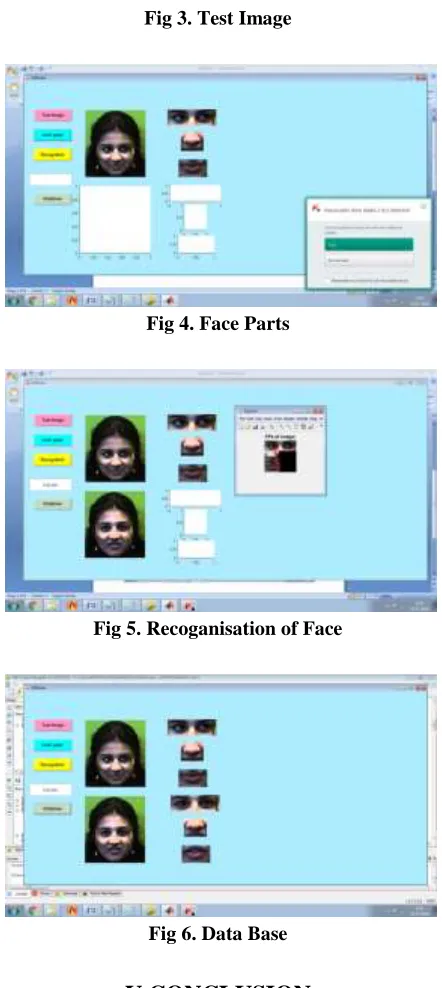
Fig 3. Test Image
Fig 4. Face Parts
Fig 5. Recoganisation of Face
Fig 6. Data Base
V.CONCLUSION
We proposed the work to overcome classical image processing limits by combining semantic patches and registration methods
for visual image enhancement. We
photography devices, our model assumes
that high-quality personal priors are
available, but that we are blind to the degradation model and its parameters. A low-to-moderate degradation may include an unknown mix of noise, nonlinear post-processing artifacts, certain motion blur, resolution reduction and color-change. The blind model assumption allows a very general correction mechanism which is not device and scenario dependent. To solve this we utilize non-rigid semantic patches and a registration algorithm, which is robust to noise and blur, and can infer a high quality solution based on the priors.
VI.REFERENCES
[1] B. K. Horn and B. G. Schunck, “Determining optical flow,” in 1981 Technical symposium east. International Society for Optics and Photonics, 1981, pp. 319–331.
[2] D. Geman and G. Reynolds,
“Constrained restoration and the recovery of discontinuities,” IEEE Transactions on Pattern Analysis & Machine Intelligence, no. 3, pp. 367–383, 1992.
[3] L. I. Rudin, S. Osher, and E. Fatemi, “Nonlinear total variation based noise removal algorithms,” Physica D: Nonlinear Phenomena, vol. 60, no. 1, pp. 259–268, 1992.
[4] J. Huang and D. Mumford, “Statistics of natural images and models,” in Computer Vision and Pattern Recognition, 1999. IEEE Computer Society Conference on., vol. 1. IEEE, 1999.
[5] E. P. Simoncelli, “Bayesian denoising of visual images in the wavelet domain,” in
Bayesian inference in wavelet-based
models. Springer, 1999, pp. 291–308.
[6] A. Buades, B. Coll, and J.-M. Morel, “A review of image denoising algorithms, with a new one,” Multiscale Modeling & Simulation, vol. 4, no. 2, pp. 490–530, 2005. [7] K. Dabov, A. Foi, V. Katkovnik, and K. Egiazarian, “Color image denoising via sparse 3d collaborative filtering with
grouping constraint in
luminance-chrominance space,” in Image Processing, 2007. ICIP 2007. IEEE International Conference on, vol. 1. IEEE, 2007, pp. I– 313.
KANDIMALLA NARMADHA
completed her B.Tech in AM Reddy
Engineering College. She is pursuing M.Tech in Narasaraopet Engineering
College, Narasaraopeta. Her
Specialisation is digital electronics and communication systems.
P.S.S. CHAKRAVARTHY completed
his B. Tech in N.B.K.R.I.S.I (S.V. University) Vidyanagar, Nellore Dt and M. Tech in JNTU College of Engineering, Kakinada.At present he
is working as professor in
Narasaraopet Engineering College,
Narasaraopeta. He has 18 years of